https://doi.org/10.1177/2048872619885346
European Heart Journal: Acute Cardiovascular Care 2020, Vol. 9(1) 76 –89
© The European Society of Cardiology 2020 Article reuse guidelines:
sagepub.com/journals-permissions DOI: 10.1177/2048872619885346 journals.sagepub.com/home/acc
Introduction
This paper provides an update on the European Society of Cardiology (ESC) task force report on the management of chest pain.1 A consensus statement on the pre-hospital man-agement of patients with chest pain and/or dyspnoea of car-diac origin has previously been published.2
Patients presenting with chest pain to the emergency department (ED) constitute a diagnostic and logistic challenge as the majority have symptoms related to non-cardiac and often benign disorders that do not need emergency treatment or hospitalisation. The introduction of high-sensitivity cardiac troponin (hs-cTn) assays allowing the detection and quantifi-cation of minute degrees of myocardial injury has led to the development of new decision algorithms that enable a more accurate and rapid rule-in and rule-out of acute coronary syn-dromes (ACSs).3 Implementation of these new decision algo-rithms may speed up the triage of chest pain patients in the ED and prevent the unnecessary hospitalisation of patients with non-critical disorders. The progress of computed tomog-raphy angiogtomog-raphy (CTA) has made it possible to examine
Diagnosis and risk stratification of
chest pain patients in the emergency
department: focus on acute coronary
syndromes. A position paper of the
Acute Cardiovascular Care Association
Janina Stepinska
1, Maddelena Lettino
2, Ingo Ahrens
3, Hector Bueno
4,
Luis Garcia-Castrillo
5, Abdo Khoury
6, Patrizio Lancellotti
7,
Christian Mueller
8, Thomas Muenzel
9, Anna Oleksiak
1, Roberta Petrino
10,
Maria Rubini Guimenez
11, Doron Zahger
12and Christiaan JM Vrints
13Document reviewers: Sigrun Halvorsen (chair), Elia de Maria, Gregory YH Lip,
Roberta Rossini, Marc Claeys, Kurt Huber*
Abstract
This paper provides an update on the European Society of Cardiology task force report on the management of chest pain. Its main purpose is to provide an update on the decision algorithms and diagnostic pathways to be used in the emergency department for the assessment and triage of patients with chest pain symptoms suggestive of acute coronary syndromes.
Keywords
Chest pain, biomarkers, troponin, acute myocardial infarction
Date received: 9 October 2019; accepted: 30 September 2019
1 Department of Intensive Cardiac Therapy, Institute of Cardiology, Poland 2 Cardiovascular Department, Humanitas Research Hospital, Italy 3 Department of Cardiology and Medical Intensive Care, Augustinerinnen
Hospital, Germany
4 Cardiology Department, Hospital Universitario 12 de Octubre and
Instituto de Investigación Sanitaria Hospital 12 de Octubre (imas12), Madrid, Spain and Centro Nacional de Investigaciones Cardiovasculares (CNIC), Spain
5 Emergency Department, Marques de Valdecilla Hospital, Spain 6 Department of Emergency Medicine and Critical Care Clinical
Investigation Center, University Hospital of Besançon, France
7 Department of Cardiology, University of Liège Hospital, Belgium 8 Cardiovascular Research Institute, University Hospital of Basel, Switzerland 9 Universitätsmedizin Mainz, Zentrum für Kardiologie, Germany 10 Emergency Department, S Andrea Hospital, Italy
11 Cardiovascular Research Institute, University Hospital of Basel,
Switzerland
12 Department of Cardiology, Soroka University Medical Center, Israel 13 Department of Cardiology, Antwerp University Hospital Belgium
Corresponding author:
Christiaan JM Vrints. Department of cardiology, Antwerp University Hospital, Belgium.
Email: christiaan.vrints@uantwerpen.be
non-invasively the coronary arteries in patients with sus-pected unstable angina, and this imaging method has become the first-line test in patients with suspected acute aortic dis-eases or high-risk pulmonary embolism (PE).3
Since the publication of the latest ESC guidelines on the diagnosis and management of ACSs4,5 the new decision algorithms for the rapid rule-in and rule-out of ACSs have been further validated. The main purpose of the present paper is therefore to provide an update on the decision algo-rithms and diagnostic pathways to be used in the ED for the assessment and triage of patients with chest pain symptoms suggestive of ACS.
Definition and epidemiology
Definition
Acute chest pain is the perception of non-traumatic pain or other thoracic discomfort occurring within the preceding 24 hours, localised anteriorly, between the base of the nose and the umbilicus and, posteriorly, between the occiput and the 12th vertebra. Descriptors include a range of features, including stabbing or burning, and also as pressure, tight-ness or heartburn.
Acute chest pain can be caused by an extensive variety of disorders (Table 1) ranging from life-threatening syn-dromes such as ACSs, acute aortic diseases and PE to con-ditions that are relatively harmless.4–7 It must be emphasised that there are important gender differences in epidemiol-ogy, pathophysiolepidemiol-ogy, clinical presentation, response to therapy and clinical outcome of acute chest pain syn-dromes.8 ACSs in women are more often caused by micro-vascular disease, and the Takotsubo syndrome is almost 10 times more frequently observed in women than in men.9 An underdiagnosed cause of ACSs that predominantly occurs in women is spontaneous coronary artery dissection.10
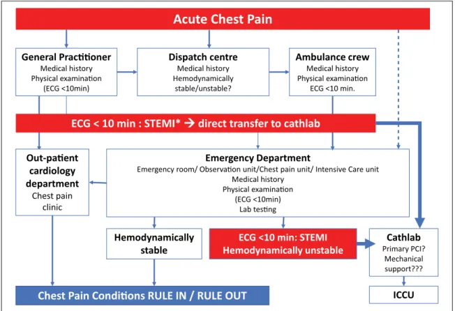
Formulating an accurate differential diagnosis within a very short timeframe and differentiating between an acute chest pain of cardiac or aortic origin and non-cardiac chest pain is a major challenge for all those involved in triage, clinical assessment and treatment of patients presenting with acute chest pain, such as general practitioners, emer-gency medical services (EMS) personnel, emeremer-gency phy-sicians, nurses and cardiologists (Figure 1).
Epidemiology
Acute chest pain is one of the most common reasons to attend the ED, accounting for approximately 10% of non-injury-related visits.12–17 The incidence of chest pain-related visits to the ED is 8–19 per 1000 person-years,12,16,17 being higher in urban than in rural hospitals, with a mean age of 52–61 years, and with 49–57% of men.12,13,15–17 In current practice, about half of the patients presenting with chest pain can be discharged without further hospitalisation from the ED.15,16 The great majority of these patients (83%) are
discharged with a non-cardiac cause of the chest pain (unspecified chest pain in 48% and other non-cardiac causes in 35%).16 Of the patients who are admitted to hos-pital, on average only 25% (range 12.2–59.1%) have a final diagnosis of an ACS.15–17 Another 25% of patients will be discharged with the diagnosis of angina pectoris (3.5–6.6%) or with another non-ischaemic cardiac problem (10–19%). In the remaining half of the admitted patients the final diag-nosis will be unspecified chest pain (26%) or a non-cardiac cause (27%).16 Acute vascular emergencies and PE account only for a very small minority (2–3%) of the patients.17
Approximately 2% of chest pain patients with an ACS are erroneously discharged from the ED, which is associ-ated with a twofold increase in 30-day morbidity and mor-tality.18 Indeed, the absence of typical chest pain does not exclude an ACS; in patients presenting to the ED without diagnosis-specific symptoms, acute myocardial infarction (MI) was the final diagnosis in 1.6%.15
Clinical assessment of acute chest
pain
Emergency physicians face a major challenge to identify rapidly and accurately the small group of patients who require hospitalisation for acute management and the larger group with more benign conditions who can be safely dis-charged from the ED.
If the patients arrive by way of EMS, it is vital that pre-hospital presentation, findings from the EMS ECG and any treatments provided are formally communicated in a struc-tured handover to ED clinicians.
The triage of chest pain patients in the ED is based on careful history-taking, physical examination, recording and interpretation of a 12-lead ECG within 10 minutes of arrival and measurement of cardiac biomarkers.5
The first priority is to identify the patients who need urgent transfer to the catheterisation laboratory. Immediate percutaneous coronary intervention (PCI) (<2 hours) is recommended in ST-segment elevation myocardial infarction (STEMI) patients4 and in some non-ST elevation ACS (NSTE-ACS) patients with at least one of the very-high-risk criteria (haemodynamic instability or cardiogenic shock, recurrent or ongoing chest pain refractory to medical treatment, life-threaten-ing arrhythmias or cardiac arrest, mechanical complica-tions of MI, acute heart failure with refractory angina or ST deviation, recurrent dynamic ST or T-wave changes, particularly with intermittent ST-elevation).5 However, in case of a haemodynamically unstable patient, initial haemodynamic stabilization (e.g. cardiogenic shock management, drugs, intubation, mechanical ventilation) may be necessary before the invasive procedure.
Although clinical judgement based on the results of the above-mentioned basic clinical tools allows the identifi-cation of a small high-risk group with clinical features
(Table 2) highly suggestive of a NSTE-ACS mandating an early invasive coronary strategy, the findings will remain inconclusive in many low-risk patients. In order to avoid inadvertent early discharge from the ED these patients must be observed and monitored for a prolonged period in
which an accelerated diagnostic protocol is performed, comprising serial ECG recordings and cardiac injury bio-marker measurements obtained over 1–3 hours to diag-nose accurately (rule-in) or exclude (rule-out) an ACS. Most often this is carried out in an observation unit in the
Table 1. Causes of chest pain.
Primary cardiovascular Primary non-cardiovascular
Acute coronary syndromes
ST-segment elevation myocardial infarction (STEMI) Non-ST elevation ACS (NSTE-ACS)
Non-ST-segment elevation myocardial infarction Unstable angina
Acute pericarditis, pericardial effusion Acute myocarditis
Severe hypertensive crisis
Stress cardiomyopathy (Takotsubo syndrome) Tachyarrhytmias
Hypertrophic cardiomyopathy, aortic stenosis Severe acute heart failure
Acute aortic syndrome (dissection, haematoma) Pulmonary embolism, pulmonary infarction Cardiac contusion
Oesophageal spasm, oesophagitis, gastroesophageal reflux (GER) Peptic ulcer disease, cholecystitis, pancreatitis
Pneumonia, bronchitis, acute asthma Pleuritis, pleural effusion, pneumothorax
Pulmonary embolism, severe pulmonary hypertension Thoracic trauma
Costochondritis, rib fracture
Cervical/thoracic vertebral or discal damage Herpes zoster
Psychogenic
Reproduced with modifications from the 2018 Acute Cardiovascular Care Association (ACCA) toolkit.11
Figure 1. Clinical pathways of acute chest pain.
*If timely (<120 minutes from first medical contact) primary percutaneous coronary intervention (PCI) cannot be performed after ST-segment elevation myocardial infarction (STEMI) diagnosis, fibrinolytic therapy is recommended within 12 hours of symptom onset in patients without contraindications.
ED or in a dedicated chest pain unit (CPU), within or close to the ED.19 A negative accelerated diagnostic evaluation allows early discharge, whereas patients who are triaged towards rule-in are usually admitted to a monitored unit and require early coronary angiography.
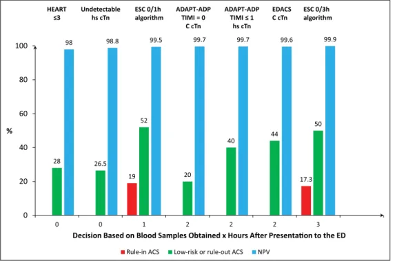
Clinical risk stratification tools may help clinicians to integrate symptoms, ECG findings and biomarkers in the risk stratification of chest pain patients. These tools have been incorporated in accelerated diagnostic pathways that facilitate fast triage and safe early discharge of low-risk chest pain patients.20,21 The diagnostic pathways based on the ADAPT (2-hour Accelerated Diagnostic protocol to Assess Patients with chest pain symptoms using contempo-rary Troponins as the only biomarker),22 EDACS (ED Assessment of Chest Pain Score)23–25 and HEART26 scores have the strongest scientific evidence supporting their use. Although these scores perform equally well, this taskforce recommends the use of the HEART score as it most closely follows the clinical reasoning process in the diagnosis of acute chest pain.
The HEART score (Figure 2) was specifically devel-oped for unselected patients with chest pain presenting at the ED.26 The HEART score differs from other risk stratifi-cation tools as it also includes the clinical suspicion by the physician and the presence of multiple coronary risk factors in its calculation. Moreover, as it is mainly based on simple clinical parameters it can be easily calculated at the bed-side. The HEART score represents the patients’ risk of developing a major adverse cardiac event (MACE) within 6 weeks after initial presentation. The HEART score has been tested and validated in numerous studies performed in Europe,27–29 the United States30,31 and Asia Pacific.26,32
Overall a HEART score of 3 or less allowed us to identify 35–46% low-risk patients with a very high sensitivity and negative predictive value.33 Patients with a HEART score of 7 or greater are a very high risk subgroup with over 50% of MACE within 6 weeks,27 who therefore should be imme-diately admitted to an intensive cardiac care unit. The HEART score was initially validated for use in conjunction with contemporary cardiac troponin (cTn) assays. Use of the HEART score based on the results of hs-cTn assays should therefore be done with caution as the troponin-related points may vary with the assay used. However, in two retrospective cohort studies in which hs-cTn assays were used, application of the HEART score resulted in the identification of 31.6–37.2% of low-risk patients with a sensitivity of 93.7–100% and a negative predictive value of 98.3–100%, results very similar to those observed in earlier validation studies.28,34
The utility of clinical risk stratification tools has been reduced since the advent of diagnostic pathways using hs-cTn assays that allow a very accurate and fast diagnosis of acute MI.
The role of nurses
Nurse-led early triage of patients with acute chest pain has been reported to increase timely ECG recording, medicine use in high-risk patients and appropriate admission to the coronary care unit.35 In rural hospitals, emergency nurse practitioners have been shown to have high adherence to clinical guidelines, a high level of diagnostic accuracy, reduce chest pain patients’ waiting times and length of stay without compromising safety.36 The diagnostic accuracy of ED nurses has been shown in one study to be similar to that of ED physicians, with a nurse-led, accelerated discharge protocol having a 1.1% rate of MACE at 30 days.37 Although these studies need further confirmation, it appears that the active participation of well-trained nurses can be very helpful in the triage of chest pain patients.
Diagnostic testing
Electrocardiogram
The 12-lead ECG within 10 minutes of EMS arrival or patient presentation (first medical contact defined as the time of first assessment by a healthcare professional who is able to obtain and interpret a 12-lead ECG) is pivotal in the decision-making algorithm for the management of patients presenting with acute chest pain. Only a small number of patients presenting with acute chest pain to the ED show a typical STEMI ECG pattern. The majority have either a completely normal ECG (40–60%) or atypical non-ischae-mic ECG changes.38–42 In a recent study of patients present-ing with acute chest pain almost 60% had a normal admission ECG, with a very low (5.0% (3.9–6.3%)) infarction rate.42
Table 2. High-risk criteria for chest pain suggestive of ACS.
Symptoms Prolonged ongoing chest pain (≥20 minutes) History Prior PCI in the last 6 months
Prior CABG Clinical
findings Pulmonary oedema most likely due to ischaemiaHypotension Tachycardia
New mitral regurgitation murmur Acute heart failure Killip class >1 New systolic murmur at ERB’s point
ECG Dynamic ST changes >0.5 mm during chest pain New or presumably new left or right bundle branch block
Sustained ventricular tachycardia High degree atrioventricular block Biomarkers Elevated cardiac troponins Score GRACE risk score ≥140
HEART score ≥7
ACS: acute coronary syndrome; PCI: percutaneous coronary intervention; CABG: coronary artery bypass graft surgery; ECG: electrocardiogram; GRACE: Global Registry of Acute Coronary Events; HEART: History, ECG, Age, Risk factors and Troponin.
In these patients, although the incidence of acute MI is low, it is not zero, and therefore a normal admission ECG should not be used alone to rule out an ACS. If the ECG is normal or non-diagnostic, the ECG should be repeated after a 10-minute interval, especially if chest pain recurs. A 12-lead ECG is sufficient in most patients. However, patients with an occluded circumflex coronary, acute occlusion of a vein graft, or left main disease, may present without ST-segment elevation.43 The use of additional posterior chest wall leads (V7–V9) should be considered if there is high clinical suspi-cion of posterior MI.4 If the 12-lead ECG shows an acute inferior MI then recording of additional right precordial leads (V3R and V4R) should be considered to identify con-comitant right ventricular infarction.4
Cardiac troponin testing
Clinical assessment combined with an ECG is not suffi-cient to diagnose or exclude,44 additional measurement of cTn T or I with serial measurements is crucial.5,43,45,46
CTn T and I are structural proteins unique to the heart. The cTn complex is immobilised on the thin filament of the contractile apparatus and plays a critical role in the regula-tion of excitaregula-tion–contracregula-tion coupling in the heart. In MI, cTn I and cTn T are released from necrotic myocardium both as intact proteins and degradation products.47
hs-cTn assays allow the precise quantification of cardio-myocyte injury around the 99th percentile, substantially increasing the accuracy of MI diagnosis based on the blood sample obtained at presentation.48–53 Although hs-cTn assays quantify the amount of cardiomyocyte damage, they
need to be interpreted as quantitative variables and not in a binary (negative/positive) fashion; the higher the cTn level, the higher is the likelihood of the presence of MI.
The latest ESC non ST-segment elevation ACS guide-lines give a IA recommendation for the use of hs-cTn as the standard biomarker for clinical practice, and a IB recom-mendation for the application of two main novel algorithms with cardiac biomarkers for the management of patients with acute chest pain in the ED.5 In patients presenting with suspected non ST-segment elevation ACS it is recom-mended to use the 0/3 hour ESC algorithm (Figure 3). As an alternative, the 0/1 hour ESC algorithm is recommended when hs-cTn assays (hs-cTn T: Elecsys; hs-cTn I: Architect, Centaur) with a validated algorithm are available (Figure 4).
0/3 Hour ESC algorithm. MI is ruled out if the
concentra-tions of hs-cTn remain in the normal range (below the respective 99th percentile) in the blood sample drawn at presentation and 3 hours later, and if the patient fulfils two additional requirements: to be pain-free and to be at low risk of in-hospital mortality, as quantified by a GRACE score44 below 140.5 In patients presenting more than 6 hours after the onset of chest pain, in whom chest pain onset can be reliably quantified, one single blood sample at presentation is considered sufficient. Patients are ruled in if they have a clearly elevated hs-cTn blood concentration at presentation, or if the 3-hour sample shows a relevant change. This approach has been recommended by the ESC guidelines since 2011, and is the standard of care in many institutions worldwide (Figure 3). Its use regarding the rule-out of MI seems to be safe for all hs-cTn assays.54 The
Figure 2. Calculation of the HEART score and incidence of 6-week major adverse cardiac events (MACE) according to the
exact performance for rule-in cannot be quantified, as no precise definitions of its rule-in cut-off levels are given.
0/1 Hour ESC algorithm. The concept of the 0/1 hour ESC
algorithm is based exclusively on information provided by hs-cTn blood concentrations. The decision points derived and validated for each assay are assay-specific (Figure 4).5,55–60 The 0/1 hour ESC algorithm obviates the need for the formal use of clinical scores and allows the safe rule-out of MI even in patients with mild, non-specific ECG abnormalities.
Undetectable level of cTn. An undetectable level of
hs-cTn (below the level of detection of the assay) at presen-tation in combination with an ECG without ischaemic changes may rule out an acute MI with sufficient sensi-tivity in patients presenting 1–3 hours after the onset of chest pain.61–64 Although the use of this criterion may allow us to rule out acute MI in one-third of the patients, it should be applied with caution in patients aged 65 years and less due to an increased risk of a false negative result.65
Time of blood sampling, turnaround time and time to decision. The 0/1 hour ESC algorithm refers to the
time-points at which blood is sampled. The turnaround time is the time period from sampling to reporting results back to the treating clinician. This is about 1 hour in a well-run hospital using an automated platform in a central laboratory. Adding the local turnaround time to the time of blood sampling determines the earliest timepoint for clinical decision-making based on hs-cTn concentrations. The clinical and economic
benefits of the 0/1 hour ESC algorithm versus, for example, the 0/3 hour algorithm are therefore independent of the local turnaround time.
Central laboratory versus point of care. Point-of-care devices
usually require 10–20 minutes for the measurement itself, providing results back to clinicians about 40 minutes earlier than the central laboratory. However, they have a number of potential disadvantages:
To date no hs-cTn assay on a point-of-care device has been appropriately validated to justify routine clinical use. However, several point-of-care hs-cTn I assays are in the late stages of development/valida-tion.66 Large diagnostic multicentre studies will be required to determine their performance versus the well validated hs-cTn T/I assays run on automated platforms.
Current point-of-care assays are either conventional or sensitive assays. Therefore, the second sample should be taken at 3–6 hours depending on the assay specifics.
Point-of-care devices have lower precision (less well standardised), and a higher error rate compared with central laboratory assays.
Other reasons for elevated hs-cTn levels. cTn T and I are
organ but not disease-specific markers and thus many factors other than acute myocardial ischaemia may cause hs-cTn elevation. In the absence of myocardial isch-aemia, elevated cTn levels are often interpreted as ‘false positive’ hs-cTn results. However, in the majority of cases this term is incorrect as they mostly reflect
Figure 3. The European Society of Cardiology 0/3 hour rule-out and rule-in algorithm of non-ST-segment elevation acute
previously undetected or underestimated cardiac disease, including valvular heart disease, heart failure, hyperten-sive heart disease and chronic coronary artery disease.67,68 Many cardiac disorders and non-cardiac disorders with cardiac involvement may lead to substantial amounts of cardiomyocyte injury and thereby hs-cTn elevation, even in the absence of macrovascular coronary artery disease. Elevated hs-cTn levels are associated with a worse prog-nosis. Dynamic changes of hs-cTn during serial sampling help to distinguish ischaemic from non-ischaemic causes of chest pain and minor degrees of troponin elevation (Table 3).
The diagnostic algorithms using hs-cTn assays improve substantially the efficiency of the triage of chest pain patients in the ED. The increased sensitivity of hs-cTn assays abridges the troponin blind period early after the onset of MI and allows marked shortening of the time interval to the second blood sample needed to
demonstrate a significant rise of the biomarker. The 0/1 hour ESC algorithm is as effective as the 0/3 hour ESC in ruling in and ruling out acute MI with a very high nega-tive predicnega-tive value.69 Moreover, it allows us to detect – 1 hour earlier – a greater number of patients eligible for early discharge than the previously widely used acceler-ated diagnostic pathways using contemporary cTn assays (ADAPT-ADP) that included the additional calculation of a clinical low-risk score (Figure 5). Institutional stand-ard operational procedures for the diagnosis of acute chest pain based on the 0/1 hour ESC algorithm will therefore not only increase patients’ safety but also mark-edly shorten the duration of stay in the ED, which may lead to important cost savings. A prospective interna-tional multicentre trial recently confirmed the applicabil-ity, efficacy and safety of the routine clinical use of the ESC 0/1 hour algorithm for the management of patients presenting with acute chest pain to the ED.70
Figure 4. The European Society of Cardiology 0/1 hour rule-out and rule-in algorithm using high-sensitivity cardiac troponin
(hs-cTn) assays in patients presenting with suspected NSTEMI to the emergency department. *If chest pain onset is greater than 3 hours. For three additional hs-cTn I assays a 0/1 hour algorithm has been derived and validated and the respective manuscripts are currently in peer review.
Echocardiography
Echocardiography should be routinely available in the ED or CPU, and performed by a trained staff member.72 It is not required where a non-cardiac diagnosis is obvious or in whom the probability of an acute cardiovascular cause is considered very low. Transthoracic echocardiography (TTE) is indicated in patients with acute chest pain and: (a) a diagnosis, or a high clinical suspicion of an acute coro-nary syndrome; (b) haemodynamic instability; (c) acute heart failure; (d) suspicion of acute aortic syndromes, myo-carditis, or pericarditis; (e) underlying cardiac disease such as aortic valve stenosis, hypertrophic cardiomyopathy. Although it is reasonable for individual risk stratification during the hospital stay, echocardiography is not recom-mended in haemodynamically stable, normotensive patients with suspected PE.6 Transoesophageal echocardiography (TOE) may be indicated when TTE is non-diagnostic. In the case of suspected aortic dissection TOE is more sensi-tive than TTE.7 Echocardiographic signs suggestive of myocardial ischaemia or necrosis include: (a) segmental wall motion abnormalities; (b) impaired myocardial perfu-sion detected by contrast echocardiography; (c) reduced regional function using strain and strain rate imaging.72
Chest X-ray
The chest X-ray is often performed in the evaluation of patients attending the ED. In one large study a quarter of such patients showed significant findings: cardiomegaly, pneumonia and pulmonary oedema.73 When there is a high clinical suspicion of acute life-threatening conditions other than ACS (pericardial effusion, acute aortic dissection, PE,
pneumothorax or pneumonia) chest X-ray is indicated and should be available, preferably within 30 minutes.
Computed tomography
Coronary computed tomography angiography (CCTA) has been proposed as a rapid and accurate diagnostic technique to rule out obstructive coronary artery disease given its very high negative predictive value.74,75 Three multicentre studies have evaluated the feasibility, safety and diagnostic accuracy of early CCTA compared to usual care in the tri-age of chest pain patients in the ED.76–78 A meta-analysis showed that a diagnostic strategy using early CCTA is as safe as usual care of chest pain patients in the ED and results in a significant reduction of cost and length of hos-pital stay.79
However, in a recent multicentre study that compared early CCTA with standard optimal care diagnostic proto-cols based on the use of hs-cTn assays, early CCTA failed to identify more patients with significant coronary artery disease requiring coronary revascularisation, shorten hos-pital stay, or allow for more direct discharge from the ED.80 A selective use of CCTA may be considered in the 20% of chest pain patients in whom the diagnosis of NSTE-ACS cannot be reliably ruled out or ruled in by the ECG and hs-cTn diagnostic algorithms.5,81
CTA of the aorta plays a central role in the diagnosis, risk stratification and management of acute aortic syn-dromes. In most patients with suspected acute aortic dissec-tion, CTA is the preferred initial imaging modality.7
Pulmonary CTA allows the detection of PE and ade-quate visualisation of the pulmonary arteries down to at least the segmental level. Pulmonary CTA is the
Table 3. Conditions other than MI associated with cTn elevations.
Tachyarrythmias Heart failure
Hypertensive emergencies
Critical illness (e.g. shock, sepsis, burns) Myocarditis
Takotsubocardiomyopathy
Structural heart disease (e.g. aortic stenosis) Aortic dissection
Pulmonary embolism, pulmonary hypertension Renal dysfunction and associated cardiac disease Coronary spasm
Acute neurological event (e.g. stroke or subarachnoid haemorrhage, meningitis)
Cardiac contusion or cardiac procedure (CABG, PCI, ablation, pacing, cardioversion, endomyocardial biopsy) Hypo and hyperthyroidism
Infiltrative diseases (e.g. amyloidosis, haemochromatosis, sarcoidosis, scleroderma) Myocardial drug toxicity or poisoning
Extreme endurance efforts Rhabdomyolysis
second-line test in patients with suspected non-high-risk PE and an elevated D-dimer level, whereas it is the first-line test in patients with suspected high-risk PE, i.e. pre-senting with shock or hypotension or patients with a high clinical probability of PE.
The high accuracy of CTA in the diagnosis of PE and acute aortic dissection and the utility of CCTA in exclud-ing coronary artery disease have led to the development of a triple rule-out scan protocol allowing the simultaneous assessment of all three causes of acute chest pain with a single scan.82 Even with modern scanners, which offer a wider coverage and a greater temporal resolution, this necessitates a longer scanning time and an increased con-trast volume. In a recent registry a triple rule-out protocol was associated with a slightly higher yield of PE and acute aortic dissection than CCTA, specifically in patients pre-senting in the ED.83
Ultrasonography (other than
echocardiography)
Ultrasonography can help in the management of acute chest pain, in particular when evaluating possible non-car-diac causes. Lung ultrasonography is useful to detect pleu-ral effusion or pneumothorax. The chest X-ray may miss the diagnosis when the volume of fluid or air is small, while ultrasonography has a higher sensitivity and speci-ficity (>90%). Typical findings in pleural effusion are the
‘quad and sinusoid signs’, while the ‘seashore sign’ (lung sliding) and ‘stratosphere (barcode) sign’ suggest pneumo-thorax.84 Lung ultrasound is also important to detect B-lines or ‘comets’, which indicate the amount of extravas-cular lung water and correlate with acute heart failure that can be associated with acute ischaemic chest pain. When gastrointestinal causes of chest pain are suspected (e.g. cholecystitis, biliary colic, pancreatitis) abdominal ultra-sonography is appropriate.
Further predischarge testing
Exercise ECG or non-invasive stress testing has been rec-ommended in low-risk patients as the final confirmatory test before safe discharge from the ED.85,86 Routine use of pre-discharge ischaemia testing may, however, lead to a longer length of stay in the ED or observation units, more downstream invasive angiography and revascularisation procedures, more radiation exposure and greater costs without any improvement in clinical outcome.87–90 Use of the HEART score may aid in identifying the patients in whom predischarge testing should be considered.30,31 In the HEART pathway implementation trial, patients with a HEART score of 3 or less in whom an ACS was excluded by serial troponin testing could be safely discharged with-out further testing.91 Based on these studies it is proposed to limit pre-discharge exercise testing and cardiac imaging to patients with a HEART score greater than 3 (Figure 6).
Figure 5. Comparison of the performance in ruling in and ruling out acute myocardial infarction (MI) or identifying low-risk chest
pain patients of various risk stratification scores and/or diagnostic pathways either using contemporary (C-cTn) or high sensitivity cardiac troponin assays (hs-cTn).20,71 The 0/1 hour European Society of Cardiology algorithm is very effective in ruling in and ruling
Conclusion
The diagnosis and management of patients presenting with chest pain to the ED can be considerably improved by the implementation of new diagnostic clinical algorithms based on the use of hs-cTn assays in conjunction with meticulous assessment of clinical symptoms and signs, the ECG and other bedside examination techniques. The new diagnostic algo-rithms allow a more effective and rapid triage of chest pain patients, as an acute MI can be diagnosed (rule-in) or excluded (rule-out) in three of four patients based on the measurement of hs-cTn on admission and one hour later. Up to halve of the patients are eligible for early discharge if differential diagno-ses are excluded. Although the new hs-cTn-based diagnostic algorithms obviate the use of clinical risk stratification scores, it is proposed to add a HEART score of 3 or less as an addi-tional criterion for the decision on early discharge as this score predicts a very low MACE risk during early follow-up. The small group of patients in whom the diagnosis is unclear should remain in the ED, CPU or observation units until fur-ther ECG monitoring, repeated hs-cTn measurement, bedside
echocardiography and additional CCTA or ischaemia testing allows us to clarify the diagnosis (Figure 6).
The new hs-cTn-based diagnostic algorithms allow a more effective and rapid triage of chest pain patients allow-ing early discharge and preventallow-ing ED crowdallow-ing. It is time to implement them universally!
Consensus statements
A careful evaluation of symptoms and a prognostic risk stratification should be made in all patients presenting with chest pain, in order to initiate specific therapy when indi-cated and reduce avoidable admissions and inappropriate discharges.
- At the first medical contact, the clinician should look for signs of haemodynamic instability and manifesta-tions of life-threatening condimanifesta-tions.
- 12-Lead ECG recording and interpretation is indi-cated as soon as possible within 10 minutes after the first medical contact.
Figure 6. Management of chest pain within the emergency department. TTE: transthoracic echocardiography.
- Additional 12-lead ECG recordings should be per-formed in the case of recurrent symptoms or diagnos-tic uncertainty.
- Additional ECG leads (V3R, V4R, V7–V9) are rec-ommended if ongoing ischaemia is suspected when standard leads are inconclusive.
- A normal admission ECG should not be used as a single test to exclude ACS.
- Routine blood sampling for serum markers is indicated as soon as possible after admission to the ED. - It is recommended to measure cardiac troponins,
preferably with high-sensitivity assays and obtain the results within 60 minutes.
A rapid rule-out and rule-in protocol at 0 hours and 1 hour is recommended if a hs-cTn test with a validated 0/1 hour algorithm is available. Additional testing after 3–6 hours is indicated if the first two troponin measurements are not conclusive and the clinical condition is still sugges-tive of ACS.
The HEART score should be used to risk stratify patients with chest pain in the ED.
TTE is indicated in patients with acute chest pain and: increased cardiac necrosis biomarkers and
non-diag-nostic ECG
haemodynamic instability acute heart failure
suspicion of acute aortic syndromes, myocarditis, or pericarditis
underlying cardiac disease (aortic valve stenosis, hypertrophic cardiomyopathy. . .)
In acute chest pain patients with inconclusive results after an accelerated diagnostic chest pain protocol using hs-cTn assays CCTA should be considered to exclude the pres-ence of coronary artery disease.
In suspected acute aortic dissection, CTA is the preferred initial imaging modality.
CCTA is the second-line test in patients with suspected non-high-risk PE and an elevated D-dimer level, and the first-line test in patients with suspected high-risk PE pre-senting with shock or hypotension or patients with a high clinical probability of PE.
Lung ultrasound is useful to detect pleural effusion or pneumothorax. It can also detect B-lines that indicate lung congestion in ACS complicated by acute heart failure.
Patients in whom ACS is ruled out, with a HEART score of 3 or less and in whom other diagnoses of chest pain are excluded may safely be discharged without further in-hos-pital testing.
Conflict of interest
The authors declare that there is no conflict of interest.
Funding
The authors received no financial support for the research, author-ship, and/or publication of this article.
*Document reviewers
Sigrun Halvorsen (Acute Cardiovascular Care Association Review Coordinator) - Department of Cardiology, Oslo University Hospital Ulleval, and University of Oslo, Oslo, Norway; Elia de Maria, Cardiology Unit, Ramazzini Hospital, Carpi (Modena), Italia; Gregory Y. H. Lip, Liverpool Centre for Cardiovascular Science, University of Liverpool and Liverpool Heart & Chest Hospital, Liverpool, United Kingdom; and Aalborg Thrombosis Research Unit, Department of Clinical Medicine, Aalborg University, Aalborg, Denmark. Roberta Rossini, Cardiology Ospedale S. Croce e Carle, Cuneo, ITALY. Marc Claeys, Department of cardiology, Antwerp University Hospital, Antwerp, Belgium. Kurt Huber, Wilhelminenhospital and Sigmund Freud University, Vienna, Austria.
References
1. Erhardt L, Herlitz J, Bossaert L, et al. Task force on the man-agement of chest pain. Eur Heart J 2002; 23: 1153–1176. 2. Beygui F, Castren M, Brunetti ND, et al. Pre-hospital
man-agement of patients with chest pain and/or dyspnoea of car-diac origin. A position paper of the Acute Cardiovascular Care Association (ACCA) of the ESC. Eur Heart J Acute
Cardiovasc Care 2015; 2048872615604119.
3. Achenbach S, Marwan M, Ropers D, et al. Coronary com-puted tomography angiography with a consistent dose below 1 mSv using prospectively electrocardiogram-triggered high-pitch spiral acquisition. Eur Heart J 2010; 31: 340–346. 4. Ibanez B, James S, Agewall S, et al. 2017 ESC Guidelines
for the management of acute myocardial infarction in patients presenting with ST-segment elevation: the Task Force for the Management of Acute Myocardial Infarction in Patients Presenting with ST-segment Elevation of the European Society of Cardiology (ESC). Eur Heart J 2018; 39: 119–177.
5. Roffi M, Patrono C, Collet J-P, et al. 2015 ESC Guidelines for the management of acute coronary syndromes in patients presenting without persistent ST-segment elevation: the Task Force for the Management of Acute Coronary Syndromes in Patients Presenting without Persistent ST-Segment Elevation of the European Society of Cardiology (ESC). Eur Heart J 2016; 37: 267–315.
6. Konstantinides SV, Torbicki A, Agnelli G, et al. 2014 ESC Guidelines on the diagnosis and management of acute pulmonary embolism: the Task Force for the Diagnosis and Management of Acute Pulmonary Embolism of the European Society of Cardiology (ESC). Endorsed by the European Respiratory Society (ERS). Eur Heart J 2014; 35: 3033–3073.
7. Erbel R, Aboyans V, Boileau C, et al. 2014 ESC Guidelines on the diagnosis and treatment of aortic diseases. Document covering acute and chronic aortic diseases of the thoracic and abdominal aorta of the adult: the Task Force for the Diagnosis and Treatment of Aortic Diseases of the European Society of Cardiology (ESC). Eur Heart J 2014; 35: 2873–2926.
8. Regitz-Zagrosek V, Oertelt-Prigione S, Prescott E, et al. Gender in cardiovascular diseases: impact on clinical manifestations, management, and outcomes. Eur Heart J 2016; 37: 24–34.
9. Templin C, Ghadri JR, Diekmann J, et al. Clinical features and outcomes of Takotsubo (stress) cardiomyopathy. N Engl
J Med 2015; 373: 929–938.
10. Adlam D, Alfonso F, Maas A, et al.; European Society of Cardiology, Acute Cardiovascular Care Association, SCAD Study Group. A position paper on spontaneous coronary artery dissection. Eur Heart J 2018; 39: 3353–3368. 11. Bueno H and Vranckx P. The Acute Cardiovascular Care
Association clinical decision-making toolkit. 2018, https:// www.escardio.org/Education/Practice-Tools/Clinical-Decision-Making-Toolkit#pdf (accessed 23 October 2019)
12. Goodacre S, Cross E, Arnold J, et al. The health care burden of acute chest pain. Heart 2005; 91: 229–230.
13. Martínez-Sellés M, Bueno H, Sacristán A, et al. Chest pain in the emergency department: incidence, clinical characteris-tics, and risk stratification. Revista Española de Cardiología
(English edn) 2008; 61: 953–959.
14. Bhuiya F, Pitts S and McCaig L. Emergency department vis-its for chest pain and abdominal pain: United States, 1999– 2008. NCHS Data Brief 2010; 43: 1–8.
15. Möckel M, Searle J, Muller R, et al. Chief complaints in medical emergencies: do they relate to underlying disease and outcome? The Charité Emergency Medicine Study (CHARITEM). Eur J Emerg Med 2013; 20: 103–108. 16. Ekelund U, Akbarzadeh M, Khoshnood A, et al. Likelihood
of acute coronary syndrome in emergency department chest pain patients varies with time of presentation. BMC Research
Notes 2012; 5: 420.
17. Bjørnsen LP, Naess-Pleym LE, Dale J, et al. Description of chest pain patients in a Norwegian emergency department.
Scand Cardiovasc J 2019; 53: 28–34.
18. Pope JH, Aufderheide TP, Ruthazer R, et al. Missed diagno-ses of acute cardiac ischemia in the emergency department.
N Engl J Med 2000; 342: 1163–1170.
19. Claeys MJ, Ahrens I, Sinnaeve P, et al. The organiza-tion of chest pain units: posiorganiza-tion statement of the Acute Cardiovascular Care Association. Eur Heart J Acute
Cardiovasc Care 2017; 6: 203–211.
20. Hollander JE, Than M and Mueller C. State-of-the-art evalua-tion of emergency department patients presenting with potential acute coronary syndromes. Circulation 2016; 134: 547–564. 21. Chang AM, Fischman DL and Hollander JE. Evaluation of
chest pain and acute coronary syndromes. Cardiol Clin 2018; 36: 1–12.
22. Than M, Cullen L, Aldous S, et al. 2-Hour accelerated diag-nostic protocol to assess patients with chest pain symptoms using contemporary troponins as the only biomarker: the ADAPT Trial. J Am Coll Cardiol 2012; 59: 2091–2098. 23. Than M, Flaws D, Sanders S, et al. Development and
valida-tion of the Emergency Department Assessment of Chest pain Score and 2 h accelerated diagnostic protocol. Emerg Med
Australas 2014; 26: 34–44.
24. Flaws D, Than M, Scheuermeyer FX, et al. External vali-dation of the emergency department assessment of chest pain score accelerated diagnostic pathway (EDACS-ADP).
Emerg Med J 2016; 33: 618–625.
25. Than MP, Pickering JW, Aldous SJ, et al. Effectiveness of EDACS versus ADAPT accelerated diagnostic pathways for chest pain: a pragmatic randomized controlled trial embed-ded within practice. Ann Emerg Med 2016; 68: 93–102.e1. 26. Six AJ, Backus BE and Kelder JC. Chest pain in the
emer-gency room: value of the HEART score. Neth Heart J 2008; 16: 191–196.
27. Backus BE, Six AJ, Kelder JC, et al. A prospective validation of the HEART score for chest pain patients at the emergency department. Int J Cardiol 2013; 168: 2153–2158.
28. Santi L, Farina G, Gramenzi A, et al. The HEART score with high-sensitive troponin T at presentation: ruling out patients with chest pain in the emergency room. Intern Emerg Med 2017; 12: 357–364.
29. Carlton EW, Khattab A and Greaves K. Identifying patients suit-able for discharge after a single-presentation high-sensitivity tro-ponin result: a comparison of five established risk scores and two high-sensitivity assays. Ann Emerg Med 2015; 66: 635–645.e1. 30. Mahler SA, Hiestand BC, Goff DCJ, et al. Can the HEART
score safely reduce stress testing and cardiac imaging in patients at low risk for major adverse cardiac events? Crit
Pathw Cardiol 2011; 10: 128–133.
31. Mahler SA, Riley RF, Hiestand BC, et al. The HEART Pathway randomized trial: identifying emergency depart-ment patients with acute chest pain for early discharge. Circ
Cardiovasc Qual Outcomes 2015; 8: 195–203.
32. de Hoog VC, Lim SH, Bank IE, et al. HEART score perfor-mance in Asian and Caucasian patients presenting to the emer-gency department with suspected acute coronary syndrome.
Eur Heart J Acute Cardiovasc Care 2018, 7: 591–560.
33. Van Den Berg P and Body R. The HEART score for early rule out of acute coronary syndromes in the emergency depart-ment: a systematic review and meta-analysis. Eur Heart J
Acute Cardiovasc Care 2018; 7: 111–119.
34. Carlton EW, Cullen L, Than M, et al. A novel diagnostic pro-tocol to identify patients suitable for discharge after a single high-sensitivity troponin. Heart 2015; 101: 1041–1046. 35. O’Neill L, Smith K, Currie P, et al. Nurse-led Early Triage
(NET) study of chest pain patients: a long term evalua-tion study of a service development aimed at improving the management of patients with non-ST-elevation acute coronary syndromes. Eur J Cardiovasc Nursing 2014; 13: 253–260.
36. Roche TE, Gardner G and Jack L. The effectiveness of emergency nurse practitioner service in the management of patients presenting to rural hospitals with chest pain: a multisite prospective longitudinal nested cohort study. BMC
Health Serv Res 2017; 17: 445.
37. Carlton EW, Khattab A and Greaves K. Beyond triage: the diagnostic accuracy of emergency department nursing staff risk assessment in patients with suspected acute coronary syndromes. Emerg Med J 2016; 33: 99–104.
38. Lee TH and Goldman L. Evaluation of the patient with acute chest pain. N Engl J Med 2000; 342: 1187–1195.
39. Forest RS, Shofer FS, Sease KL, et al. Assessment of the standardized reporting guidelines ECG classification system: the presenting ECG predicts 30-day outcomes. Ann Emerg
Med 2004; 44: 206–212.
40. Kontos MC, Roberts BD, Tatum JL, et al. Mortality based on the presenting electrocardiogram in patients with
myocar-dial infarction in the troponin era. Am J Emerg Med 2009; 27: 146–152.
41. Kontos MC, Diercks DB and Kirk JD. Emergency depart-ment and office-based evaluation of patients with chest pain.
Mayo Clin Proc 2010; 85: 284–299.
42. Knowlman T, Greenslade JH, Parsonage W, et al. The asso-ciation of electrocardiographic abnormalities and acute coro-nary syndrome in emergency patients with chest pain. Acad
Emerg Med 2017; 24: 344–352.
43. Thygesen K, Alpert JS, Jaffe AS, et al. Fourth universal defi-nition of myocardial infarction (2018). Eur Heart J 2019; 40: 237–269.
44. Fox KAA, Dabbous OH, Goldberg RJ, et al. Prediction of risk of death and myocardial infarction in the six months after pres-entation with acute coronary syndrome: prospective multina-tional observamultina-tional study (GRACE). BMJ 2006; 333: 1091. 45. Thygesen K, Alpert JS, White HD, et al.; on behalf of the
Joint ESC/ACCF/AHA/WHF Task Force for the Redefinition of Myocardial Infarction. Universal definition of myocardial infarction. Eur Heart J 2007; 28: 2525–2538.
46. Thygesen K, Alpert JS, Jaffe AS, et al. Third universal definition of myocardial infarction. Eur Heart J 2012; 33: 2551–2567.
47. Mair J, Lindahl B, Hammarsten O, et al. How is cardiac troponin released from injured myocardium? Eur Heart J
Acute Cardiovasc Care 2018; 7: 553–560.
48. Reichlin T, Hochholzer W, Bassetti S, et al. Early diagno-sis of myocardial infarction with sensitive cardiac troponin assays. N Engl J Med 2009; 361: 858–867.
49. Twerenbold R, Boeddinghaus J, Nestelberger T, et al. Clinical use of high-sensitivity cardiac troponin in patients with sus-pected myocardial infarction. J Am Coll Cardiol 2017; 70: 996–1012.
50. Mueller C. Biomarkers and acute coronary syndromes: an update. Eur Heart J 2014; 35: 552–556.
51. Möckel M, Giannitsis E, Mueller C, et al. Rule-in of acute myocardial infarction: focus on troponin. Eur Heart J Acute
Cardiovasc Care 2017; 6: 212–217.
52. Mueller C, Giannitsis E, Möckel M, et al. Rapid rule out of acute myocardial infarction: novel biomarker-based strate-gies. Eur Heart J: Acute Cardiovasc Care 2017; 6: 218–222. 53. Keller T, Zeller T, Peetz D, et al. Sensitive troponin I assay in
early diagnosis of acute myocardial infarction. N Engl J Med 2009; 361: 868–877.
54. Wildi K, Nelles B, Twerenbold R, et al. Safety and efficacy of the 0 h/3 h protocol for rapid rule out of myocardial infarc-tion. Am Heart J 2016; 181: 16–25.
55. Rubini Gimenez M, Twerenbold R, Jaeger C, et al. One-hour rule-in and rule-out of acute myocardial infarction using high-sensitivity cardiac troponin I. Am J Med 2015; 128: 861–870.e4. 56. Reichlin T, Twerenbold R, Wildi K, et al. Prospective valida-tion of a 1-hour algorithm to rule-out and rule-in acute myo-cardial infarction using a high-sensitivity cardiac troponin T assay. CMAJ 2015; 187: E243–E252.
57. Reichlin T, Schindler C, Drexler B, et al. One-hour rule-out and rule-in of acute myocardial infarction using high-sensitivity cardiac troponin T. Arch Intern Med 2012; 172: 1211–1218.
58. Mueller C, Giannitsis E, Christ M, et al. Multicenter evalua-tion of a 0-hour/1-hour algorithm in the diagnosis of
myocar-dial infarction with high-sensitivity cardiac troponin T. Ann
Emerg Med 2016; 68: 76–87.e4.
59. Twerenbold R, Neumann JT, Sörensen NA, et al. Prospective validation of the 0/1-h algorithm for early diagnosis of myo-cardial infarction. J Am Coll Cardiol 2018; 72: 620–632. 60. Boeddinghaus J, Twerenbold R, Nestelberger T, et al. Clinical
validation of a novel high-sensitivity cardiac troponin I assay for early diagnosis of acute myocardial infarction. Clin Chem 2018; 64: 1347–1360.
61. Rubini Giménez M, Hoeller R, Reichlin T, et al. Rapid rule out of acute myocardial infarction using undetectable levels of high-sensitivity cardiac troponin. Int J Cardiol 2013; 168: 3896–3901.
62. Thelin J, Melander O and Öhlin B. Early rule-out of acute coronary syndrome using undetectable levels of high sensi-tivity troponin T. Eur Heart J Acute Cardiovasc Care 2015; 4: 403–409.
63. Body R, Mueller C, Giannitsis E, et al. The use of very low concentrations of high-sensitivity troponin T to rule out acute myocardial infarction using a single blood test. Acad Emerg
Med 2016; 23: 1004–1013.
64. Mokhtari A, Lindahl B, Smith JG, et al. Diagnostic accuracy of high-sensitivity cardiac troponin T at presentation com-bined with history and ECG for ruling out major adverse car-diac events. Ann Emerg Med 2016; 68: 649–658.e3. 65. Ljung L, Reichard C, Hagerman P, et al. Sensitivity of
unde-tectable level of high-sensitivity troponin T at presentation in a large non-ST-segment elevation myocardial infarction cohort of early presenters. Int J Cardiol 2019; 284: 6–11. 66. Pickering JW, Young JM, George PM, et al. Validity of a
novel point-of-care troponin assay for single-test rule-out of acute myocardial infarction. JAMA Cardiol 2018; 1(3): 1108–1111.
67. Mair J, Lindahl B, Müller C, et al. What to do when you ques-tion cardiac troponin values. Eur Heart J Acute Cardiovasc
Care 2018; 7: 577–586.
68. Yan AT, Yan RT, Kennelly BM, et al. Relationship of ST ele-vation in lead aVR with angiographic findings and outcome in non-ST elevation acute coronary syndromes. Am Heart J 2007; 154: 71–78.
69. Badertscher P, Boeddinghaus J, Twerenbold R, et al. Direct comparison of the 0/1h and 0/3h algorithms for early rule-out of acute myocardial infarction. Circulation 2018; 137: 2536–2538.
70. Twerenbold R, Costabel JP, Nestelberger T, et al. Outcome of applying the ESC 0/1-hour algorithm in patients with sus-pected myocardial infarction. J Am Coll Cardiol 2019; 74: 483–494.
71. Boeddinghaus J, Nestelberger T, Twerenbold R, et al. Direct comparison of 4 very early rule-out strategies for acute myo-cardial infarction using high-sensitivity cardiac troponin I.
Circulation 2017; 135: 1597–1611.
72. Lancellotti P, Price S, Edvardsen T, et al. The use of echocar-diography in acute cardiovascular care: recommendations of the European Association of Cardiovascular Imaging and the Acute Cardiovascular Care Association. Eur Heart J Acute
Cardiovasc Care 2015; 4: 3–5.
73. Buenger RE. Five thousand acute care/emergency depart-ment chest radiographs: comparison of requisitions with radiographic findings. J Emerg Med 1988; 6: 197–202.
74. Budoff MJ, Dowe D, Jollis JG, et al. Diagnostic performance of 64-multidetector row coronary computed tomographic angiography for evaluation of coronary artery stenosis in indi-viduals without known coronary artery disease: results from the prospective multicenter ACCURACY (Assessment by Coronary Computed Tomographic Angiography of Individuals Undergoing Invasive Coronary Angiography) Trial. J Am Coll
Cardiol 2008; 52: 1724–1732.
75. Meijboom WB, Meijs MFL, Schuijf JD, et al. Diagnostic accuracy of 64-slice computed tomography coronary angi-ography: a prospective, multicenter, multivendor study. J Am
Coll Cardiol 2008; 52: 2135–2144.
76. Goldstein JA, Chinnaiyan KM, Abidov A, et al. The CT-STAT (Coronary Computed Tomographic Angiography for Systematic Triage of Acute Chest Pain Patients to Treatment) Trial. J Am Coll Cardiol 2011; 58: 1414–1422. 77. Hoffmann U, Truong QA, Schoenfeld DA, et al. Coronary
CT angiography versus standard evaluation in acute chest pain. N Engl J Med 2012; 367: 299–308.
78. Litt HI, Gatsonis C, Snyder B, et al. CT angiography for safe discharge of patients with possible acute coronary syn-dromes. N Engl J Med 2012; 366: 1393–1403.
79. Hulten E, Pickett C, Bittencourt MS, et al. Outcomes after coronary computed tomography angiography in the emergency department: a systematic review and meta-analysis of rand-omized, controlled trials. J Am Coll Cardiol 2013; 61: 880–892. 80. Dedic A, Lubbers MM, Schaap J, et al. Coronary CT angiogra-phy for suspected ACS in the era of high-sensitivity troponins. Randomized Multicenter Study. J Am Coll Cardiol 2016; 67: 16–26.
81. Ferencik M, Hoffmann U, Bamberg F, et al. Highly sensitive troponin and coronary computed tomography angiography in the evaluation of suspected acute coronary syndrome in the emergency department. Eur Heart J 2016; 37: 2397–2405. 82. Gallagher MJ and Raff GL. Use of multislice CT for the
evaluation of emergency room patients with chest pain:
the so-called “Triple rule-out”. Catheter Cardiovasc Interv 2008; 71: 92–99.
83. Burris AC, Boura JA, Raff GL, et al. Triple rule out ver-sus coronary CT angiography in patients with acute chest pain. J Am Coll Cardiol Imag 2015; 8: 817–825.
84. Ricci F, Aquilani R, Radico F, et al. Role and importance of ultrasound lung comets in acute cardiac care. Eur Heart J
Acute Cardiovasc Care 2015; 4: 103–112.
85. Amsterdam EA, Kirk JD, Bluemke DA, et al. Testing of low-risk patients presenting to the emergency department with chest pain: a scientific statement from the American Heart Association. Circulation 2010; 122: 1756–1776.
86. Amsterdam EA, Wenger NK, Brindis RG, et al. 2014 AHA/ ACC Guideline for the management of patients with non-ST-elevation acute coronary syndromes. J Am Coll Cardiol 2014; 64: e139–e228.
87. Safavi KC, Li S-X, Dharmarajan K, et al. Hospital variation in the use of noninvasive cardiac imaging and its associa-tion with downstream testing, intervenassocia-tions, and outcomes.
JAMA Intern Med 2014; 174: 546–553.
88. Foy AJ, Liu G, Davidson WR Jr, et al. Comparative effective-ness of diagnostic testing strategies in emergency department patients with chest pain: an analysis of downstream testing, interventions, and outcomes. JAMA Intern Med 2015; 175: 428–436.
89. Reinhardt SW, Lin C-J, Novak E, et al. Noninvasive car-diac testing vs clinical evaluation alone in acute chest pain: a secondary analysis of the ROMICAT-II Randomized Clinical Trial. JAMA Intern Med 2018; 178: 212–219. 90. Howell SJ, Prasad P, Vipparla NS, et al. Usefulness of
predis-charge cardiac testing in low risk women and men for safe, rapid discharge from a chest pain unit. Am J Cardiol 2019; 123: 1772–1775.
91. Mahler SA, Lenoir KM, Wells BJ, et al. Safely identifying emergency department patients with acute chest pain for early discharge. Circulation 2018; 138: 2456–2468.