Dual Function of ERRa in Breast Cancer and Bone
Metastasis Formation: Implication of VEGF and
Osteoprotegerin
Anaïs Fradet1,2, Hel!ene Sorel1,2, Lamia Bouazza1,2, Delphine Goehrig1,2, Baptiste D"epalle1,2,
Akeila Bellahc!ene4, Vincent Castronovo4, H"el!ene Follet1,2, Fran¸coise Descotes3, Jane E. Aubin5,
Philippe Cl"ezardin1,2, and Edith Bonnelye1,2
Abstract
Bone metastasis is a complication occurring in up to 70% of advanced breast cancer patients. The estrogen receptor-related receptor alpha (ERRa) has been implicated in breast cancer and bone development, prompting us to examine whether ERRa may function in promoting the osteolytic growth of breast cancer cells in bone. In a mouse xenograft model of metastatic human breast cancer, overexpression of wild-type ERRa reduced metastasis, whereas overexpression of a dominant negative mutant promoted metastasis. Osteoclasts were directly affected and ERRa upregulated the osteoclastogenesis inhibitor, osteoprotegerin (OPG), providing a direct mechanistic basis for understanding how ERRa reduced breast cancer cell growth in bone. In contrast, ERRa overexpression increased breast cancer cell growth in the mammary gland. ERRa-overexpressing primary tumors were highly vascularized, consistent with an observed upregulation of angiogenic growth factor, the VEGF. In support of these findings, we documented that elevated expression of ERRa mRNA in breast carcinomas was associated with high expression of OPG and VEGF and with disease progression. In conclusion, our results show that ERRa plays a dual role in breast cancer progression in promoting the local growth of tumor cells, but decreasing metastatic growth of osteolytic lesions in bone. Cancer Res; 71(17); 5728–38.!2011 AACR.
Introduction
Bone metastasis is a frequent complication of cancer, occurring in up to 70% of patients with advanced breast cancer. Bone metastasis is not a direct cause of death but is associated with significant morbidity (1). For cancer cells to grow in bone, malignant cells recruit and activate osteoclasts (OC; bone resorbing cells) to resorb the bone matrix. Indeed, osteolytic breast cancer metastasis are characterized by an increase in OC number and activity at the bone metastatic site, where excessive bone destruction provides a permissive microenvironment for breast cancer cells to proliferate and expand (2, 3). Unfortunately, current treatments for bone metastasis that rely on antiresorptive agents are only pallia-tive, raising the need for a better understanding of the
molecular mechanisms involved in this pathology so as to design potential alternative therapies (3, 4).
Nuclear steroid receptors are transcription factors that comprise both ligand-dependent molecules such as estrogen receptors (ER) and a large number of so-called orphan recep-tors, for which no ligand has yet been determined (5). Three orphan receptors, estrogen receptor-related receptor alpha (ERRa), ERRb, and ERRg, share structural similarities with ERa and ERb (5), but they do not bind estrogen (6, 7). Sequence alignment of ERRa and the ERs reveals a high similarity (68%) in the 66 amino acids of the DNA binding domain, but only a moderate similarity (36%) in the ligand-binding domain, which may explain the fact that ERRa recognizes the same DNA binding elements as ERs but does not bind estrogen (8). Although ERRa activity is decreased by the synthetic molecule XCT790, no natural ligand has yet been found (9–11).
ERRa is known to regulate fatty acid oxidation and the adaptive bioenergetic response (12, 13). It is widely expressed in normal tissues, but several RNA expression studies show its presence in a range of cancerous cells including breast, prostate, endometrial, colorectal, and ovarian tumor tissues (14–20). ERRa was markedly increased in neoplastic versus normal tissues, and ERRa-positive tumors were associated with more invasive disease and higher risk of recurrences (14, 15). On the contrary, ERa and ERb were significantly lower in neoplastic versus normal tissue and were associated with better prognosis (14, 17, 18). ERRa is also highly expressed in skeletal tissues (21, 22) and has been reported to regulate
Authors' Affiliations:1Inserm, UMR1033;2University of Lyon;3Centre
hospitalier Lyon Sud, Lyon, France;4University of Li!ege, Li!ege, Belgique;
and5University of Toronto, Toronto, Canada
Note: Supplementary data for this article are available at Cancer Research Online (http://cancerres.aacrjournals.org/).
Corresponding Author: Edith Bonnelye, INSERM, UMR1033, UFR de M"edecine Lyon-Est, Rue G Paradin 69372 Lyon cedex 08, France. Phone: 33-4-787-857-38; Fax: 33-4-787-787-72; E-mail:
edith.bonnelye@inserm.fr
doi: 10.1158/0008-5472.CAN-11-1431
osteoblast, OC differentiation, and bone formation in vitro (21, 22, 23, 24) and in vivo (25–27). Consistent with these observa-tions, osteopontin (OPN) has been reported to be a direct target gene of ERRa in osteoblastic cell lines (28–30). The role of ERRa in bone metastasis formation is currently unknown. In the light of these findings, we asked here whether ERRa is involved in breast cancer bone metastasis formation and progression, and whether modulating its activity abrogates bone destruction.
Materials and Methods Ethics statement
BALB/c and NMRI mice were purchased from Charles River laboratories. All procedures involving animals, including hous-ing and care, the method by which they were killed, and experimental protocols, were conducted in accordance with a code of practice established by the local ethical committee (CREEA: comite Regionale d’Ethique pour l’Exp"erimentation Animale). Studies involving human primary breast tumors were carried out according to the principles embodied in the Declara-tion of Helsinki. Patients were included anonymously in this study. All human experiments were approved by the Experi-mental Review Board from the Laennec School of Medicine. Breast cancer tissue specimens
The autopsy files of the Department of Pathology (Pr. J. Boniver, Centre Hospitalier Universitaire of Li!ege, Belgium) were searched for diagnosis of disseminated breast cancer with histologically proven bone metastasis during the period 1991 to 1998. Slides were retrieved, and clinical history was obtained. Two breast cancer patients who died with dissemi-nated disease, including bone metastasis, were selected for immunohistochemistry. Soft tissue metastasis (TM) was fixed with formalin, dehydrated, and paraffin embedded.
Breast cancer cohort of patients
In the cohort, patients (n ¼ 251) were selected according to the following criteria: primary breast tumor without inflam-matory features and no previous treatment (31). Breast cancer tissue biopsies were obtained by surgery, selected by the pathologist, and immediately stored in liquid nitrogen until processing. The biopsies were pulverized using a MikroDis-membrator (B.Braun Biotech International), and total RNA was extracted using TRI Reagent (Sigma). RNA quality was verified using an Agilent Bioanalyser 2100 (Agilent Technologies). Real-time reverse transcriptase PCR (RT-PCR) was carried out. Cell lines and transfection
MDA-BO2-FRT (BO2) cells and stably transfected clonal derivatives were cultured in complete Dulbecco's modified Eagle's medium (Invitrogen), 10% FBS (Perbio), and 1% peni-cillin/streptomycin (Invitrogen) at 37"C in a 5% CO2incubator.
Characteristics of MDA-MB-231/BO2-FRT (BO2) breast cancer cells were previously described (32). To avoid potential effects of different insertion sites, a pcDNA5/FRT vector (Invitrogen) was used to obtain the stable BO2-ERRaWT, BO2-ERRaDAF2, and BO2 (CT) cell lines. Human ERRa cDNA [wild type (WT) and
DAF2-AD] was obtained from mRNA extracted from BO2-FRT cells, by using RT-PCR with specific primers [(NM_004451.3): ERRa upstream (177bp): GGG AAG CTT AGC GCC ATG TCC AGC CAG; ERRa downstream (WT; 177-1461 bp): GGG GGA TCC CCA CCC CTT GCC TCA GTC C; ERRa downstream (DAF2-AD): GGG GGA TCC TCA TGT CTG GCG GAG GAG (177-1350 bp; helix11-12 deletion (32 amino acids)]. Amplimers were sequenced for verification. The pcDNA5/FRT/ERRa-WT and pcDNA5/FRT/ERRa-DAF2-AD constructs were cotrans-fected with the plasmid POG44 (Invitrogen) conferring the specific integration into the FRT site present in the BO2 cells. For clonal selection, cells were cultured for 4 weeks in the presence of hygromycin (20 mg/mL; Invitrogen). Conditioned medium from all clones and from BO2 treated with the inverse agonist XCT-790 at 5.10#7mol/L (Sigma) was obtained after 48
hours ina-MEM supplemented with 0.5% of serum, then filter sterilized and proteins quantified to use equal concentration of proteins for each condition (25mg).
Animal studies
Tumor fat pad experiments were carried out using BO2-ERRaWT-1, BO2-ERRaDAF2 (pool of AF2-1, -2, and -3 clones), and BO2 (CT1/2) cell lines (106cells in 50mL of PBS) injected
into the fat pad of the fourth mammary gland of female 4-week-old NMRI nude mice (Charles River). Tumor progression was followed by bioluminescence (NightOwl, Berthold), then tumor size and weight were determined after sacrifice at 66 days.
Bone metastasis experiments using the same pool of clones were carried out in 4-week-old BALB/c nude mice as pre-viously described (33). Cells were suspended at a density of 5 $ 105 in 100 mL of PBS and inoculated intravenously into
animals. Radiographs (LifeRay HM Plus, Ferrania) of animals were taken at 35 days after inoculation using X-ray (MX-20; Faxitron X-ray Corporation). Animals were sacrificed; hind limbs were collected for histology and histomorphometrics analyses. Tibiae were scanned using microcomputed tomo-graphy (Skyscan1076, Skyscan) with an 8.8 voxel size, and three-dimensional (3D) reconstructions were carried out with a dedicated visualization software (Amira 5.2, Visage Imaging Inc.). The area of osteolytic lesions was measured using the computerized image analysis system MorphoExpert (Explor-anova). The extent of bone destruction for each animal was expressed in square millimeter.
Bone histomorphometry and histology
Hind limbs from animals were fixed and embedded in paraffin. Five millimeter sections were stained with Goldner's Trichrome and processed for histomorphometric analyses to calculate the bone volume/tissue volume (BV/TV) ratio and the tumor burden/tissue volume (TB/TV) ratio. The in situ detection of OC was carried out on sections of bone tissue with metastasis using the tartarte-resistant acid phosphatase (TRAP) activity Kit assay (Sigma). The resorption surface (Oc. S/BS) was calculated as the ratio of TRAP-positive trabecular bone surface (Oc.S) to the total bone surface (BS) using the computerized image analysis system MorphoExpert (Ex-ploranova).
Osteoclastogenesis assay
Bone marrow cells from 6-week-old OF1 male mice were cultured for 7 days in differentiation medium:a-MEM med-ium containing 10% fetal calf serum (Invitrogen), 20 ng/mL of macrophage colony-stimulating factors (M-CSF; R&D Sys-tems), and 200 ng/mL of soluble recombinant receptor acti-vator of nuclear factorkB ligand (RANKL; ref. 34). Cells were continuously (day 1–7) exposed to conditioned medium extracted (25 mg proteins for each conditions) from BO2 clones. After 7 days, mature multinucleated OC were stained for TRAP activity (Sigma-Aldrich) and counted as OC when containing 3 or more nuclei.
Immunofluorescence
BO2 cultures were fixed in culture wells with 3.7% paraf-ormaldehyde (Sigma) in PBS for 10 minutes and permeabi-lized with 0.2% Triton X-100 in PBS. Immunodetection was carried out using a goat polyclonal antibody against human ERRa (Santa Cruz, Tebu) and the secondary antibody (fluor-escein isothiocyanate-conjugated donkey anti-goat; Rock-land, Tebu-bio). The distribution of F-actin was visualized using phalloidin (Molecular Probes; ref. 14). Cells were observed using an LMS510 laser scanning confocal micro-scope (Zeiss) with a 63$ (numerical aperture 1.4) Plan Neo Fluor objective.
Immunoblotting
Cell proteins were extracted, separated in 4% to 12% SDS-PAGE (Invitrogen), then transferred to nitrocellulose mem-branes (Millipore) using a semidry system. For ERRa and a-tubulin detection, the same goat polyclonal antibody ERRa (Santa Cruz) and a mouse polyclonal antibody against human a-tubulin (Sigma-Aldrich) were used. Membrane was incu-bated with secondary antibody horseradish peroxide (HRP)-conjugated donkey anti-goat (Santa Cruz) and anti-mouse (Amersham), respectively. An ECL kit (PerkinElmer) was used for detection.
Immunocytochemistry
Five micrometer sections were subjected to immunohisto-chemistry using the same goat polyclonal antibody ERRa (Santa Cruz) and a rabbit polyclonal antibody against human osteoprotegerin (OPG; Abbiotec). Sections were incubated with secondary antibody HRP-conjugated donkey anti-goat and anti-rabbit, respectively (Amersham; dilution 1/300) for 1 hour. After washing, the sections were revealed by 3,30
-dia-minobenzidine (Dako). Real-time RT-PCR
Total RNA was extracted with Trizol reagent (Sigma) from cancer cells and OCs. Real-time RT-PCR was carried out on a
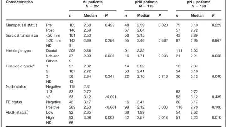
Table 1. Clinical and biological characteristics and ERR
a
mRNA expression in breast cancer patientsCharacteristics All patients
N ¼ 251 pN0 patientsN ¼ 115 pNþ patientsN ¼ 136
n Median P n Median P n Median P
Menopausal status Pre 105 2.68 0.425 48 2.59 0.020 79 3.10 0.229
Post 146 2.59 67 2.04 57 2.72
Surgical tumor size <20 mm 101 2.53 58 2.15 43 2.89
&20 mm 142 2.69 0.256 55 2.46 0.662 87 2.95 0.967
ND 8
Histologic type Ductal 205 2.68 91 2.32 114 3.03
Lobular 37 2.09 0.026 16 1.71 0.208 21 2.21 0.058 Others 9 Histologic gradea 1 27 2.32 14 2.22 13 2.37 2 107 2.72 53 2.41 54 3.18 3 58 2.84 0.341 22 2.16 0.718 36 3.12 0.040 ND 13
Node status Negative 115 2.31
1–3 83 2.72 83 2.72
>3 53 3.12 <0.001 53 3.12 0.439
RE status Negative 42 3.17 16 3.47 26 3.17
Positive 209 2.53 <0.001 99 2.12 0.003 110 2.78 0.106
VEGF statusb Low 92 2.35 38 1.99 54 2.62
High 93 3.08 0.002 42 2.57 0,018 51 3.23 0.010
ND 66
NOTE: P values correspond to Mann–Whitney test or Kruskall–Wallis test (histologic grade and node status). aHistologic grade defined only in ductal carcinomas.
Roche Lightcycler Module (Roche) with specific primers (see Supplementary Table S2). Real-time RT-PCR was carried out by using SYBR Green (Qiagen) on the LightCycler system (Roche) according to the manufacturer's instructions. Ampli-mers were all normalized to corresponding L32 values. Data analysis was carried out using the comparative Ctmethod.
Real-time RT-PCR on breast cancer tissue biopsy mRNA was carried out using primers specific for human L32 (101 bp): 50-CAAGGAGCTGGAAGTGCTGC-30, 50
-CAGCTCTTTCCAC-GATGGCT-30; TATA-box binding protein (TBP; 138 bp) 50
-TGGTGTGCACAGGAGCAAG-30, 50
-TTCACATCACAGCT-CCCCAC-30; ERRa (101 bp): 50
-ACCGAGAGATTGTGGT-CACCA-30, 50-CATCCACACGCTCTGCAGTACT-30 and OPG
(Supplementary Table S2) and SYBR green (Invitrogen) in 96-well plates on a Mastercycler EP system (Realplex2, Eppen-dorf) according to the manufacturer's instructions with an initial step for 10 minutes at 95"C followed by 40 cycles of
20 seconds at 95"C, 15 seconds at T
m(L32: 62"C, TBP: 67"C,
ERRa: 59"C), and 10 seconds at 72"C. ERRa and OPG
expres-sion were normalized with the average of the genes expresexpres-sion encoding the ribosomal protein L32 and the TBP.
Cell invasion assay
Invasion assays were carried out using Bio-Coat migration chambers (Becton Dickinson) with 8mm filters coated with Matrigel as described previously (35). BO2 cells (5 $ 104) were
plated in the upper chambers and the chemoattractant (10% FBS) in the lower chambers. After 24 hours at 37"C in 5% CO
2
incubator, cells that had migrated through the filters were fixed and stained. Cells were counted (200$ magnification). All experiments were run in triplicate, and invasion was expressed in cells/square millimeter.
OPG ELISA
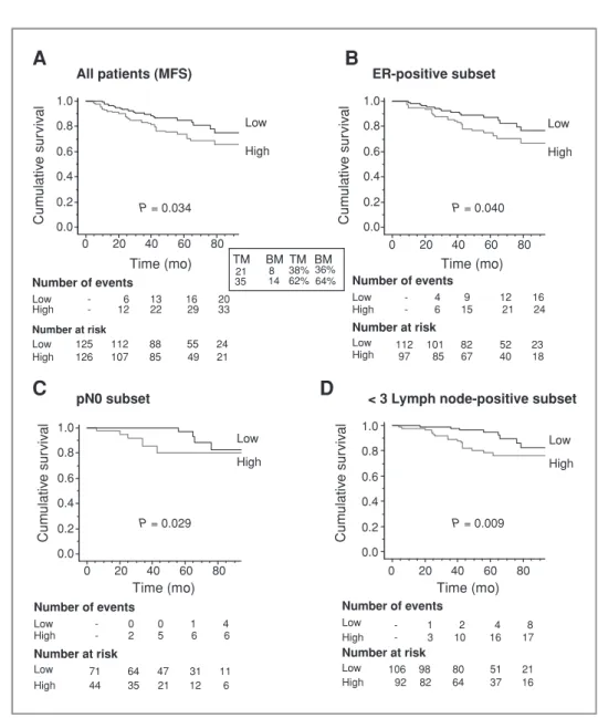
Conditioned medium obtained from CT(1/2), BO2-ERRa-WT-1, and BO2-FRT-ERRaDAF2 (pool of AF2-1, -2 Figure 1.ERRa is a bad
prognostic marker. Kaplan–Meier curves show correlation between high expression of ERRa, categorized with median value, and MFS in patients in (A) the whole population (N ¼ 251), (B) the ER-positive subset (N ¼ 209), (C and D) the pN0 subset patients (N ¼ 115) and the <3 lymph node-positive subset (N ¼ 198). Low: '50% quartile; high: &50%.
C
Cum ulativ e sur viv al Cum ulativ e sur viv al Cum ulativ e sur viv al Cum ulativ e sur viv alA
Low High 0.0 0.2 0.4 0.6 0.8 1.0 0.0 0.2 0.4 0.6 0.8 1.0 0 20 40 60 80 Number of events Low High Number at risk Low High - 6 13 16 20 - 12 22 29 33 125 112 88 55 24 126 107 85 49 21 All patients (MFS) P = 0.034 Time (mo) ER-positive subset Time (mo)B
Low High 0 20 40 60 80 Number of events Low High Number at risk Low High - 4 9 12 16 - 6 15 21 24 112 101 82 52 23 97 85 67 40 18 P = 0.040 pN0 subset Time (mo) Low High P = 0.029 0.0 0.2 0.4 0.6 0.8 1.0 0.0 0.2 0.4 0.6 0.8 1.0 0 20 40 60 80 Time (mo) 0 20 40 60 80 Number of events Low High Number at risk Low High - 0 0 1 4 - 2 5 6 6 71 64 47 31 11 44 35 21 12 6< 3 Lymph node-positive subset
D
Low High Number of events Low High Number at risk Low High - 1 2 4 8 - 3 10 16 17 106 98 80 51 21 92 82 64 37 16 P = 0.009 BM TM BM TM 21 35 814 38% 36%62%64%and -3 clones) were diluted following the manufacturer's instructions, and OPG concentration was evaluated using the ELISA Kit (RayBiotech).
Statistical analysis
Data were analyzed statistically by one-way ANOVA fol-lowed by post hoc t-tests to assess the differences between groups for in vitro and in vivo studies. Concerning the cohort, the median follow-up at the time of analysis was 54 months. The criterion for statistical analyses was the metastasis free survival (MFS), that is, the delay between the time of primary surgery and the first event: nodal or distant metastasis or death. Analysis of the distribution of ERRa expression in relation to the usual prognostic parameters was carried out using the Mann–Whitney or Kruskall–Wallis test. Survival probabilities were estimated using Kaplan–Meier estimators and were compared using the log-rank test. Univariate ana-lysis was carried out using the Cox proportional hazard model. Results of P< 0.05 were considered significant.
Results and Discussion
ERRa mRNA and protein expression in human primary breast tumors and bone metastasis
We analyzed ERRa mRNA expression by real-time RT-PCR in a cohort of 251 breast tumor biopsies (Supplementary
Table S1; ref. 31). As reported previously by others (14, 15, 17, 18), a statistically significant association was detected in all patients analyzed between ERRa expression and histologic type, node status, and ERs (radioligand method; P ¼ 0.026, P< 0.001, P < 0.001; Table 1). The Kaplan–Meier curve was constructed after segmentation into 2 groups on the basis of the median value for ERRa expression (Fig. 1A–D). It was observed that high levels of ERRa mRNA expression were related to a decrease in MFS (N ¼ 251, P ¼ 0.034; Fig. 1A). Sixty-two percent of patients (35/56) with high ERRa expres-sion levels exhibited liver, lung, bone, and soft tissue metas-tasis compared with 38% of patients (21/56) having low ERRa levels (Fig. 1A, see frame). This paralleled the frequencies seen in patients (n ¼ 22) who had developed "only" bone metastasis (BM), that is, 64% (high ERRa) and 36% (low ERRa; Fig. 1A) suggesting that ERRa is an overall bad prognostic factor that is not a determinant of metastasis location of breast cancer cells. Moreover, high ERRa expression correlated with a higher risk of recurrence at an early stage of the disease in the ER-positive group (N ¼ 209), the pN0 subset, and in the pN < 3 lymph-node-positive subset (P ¼ 0.04; P ¼ 0.029, and P ¼ 0.009; log-rank test), when compared with low ERRa (Fig. 1B– D) suggesting that ERRa may be a very useful early prognostic marker in breast cancer. Finally, as previously described (15), ERRa protein was present in situ and in invasive breast carcinoma cells (Supplementary Fig. S1B and C, respectively)
0 0.5 1 1.5 2 2.5 3 3.5 4
A
B
T T GP GP ERRα/actin CT- ERRαC
0 2 4 6 8 10 12 14 ERRα/L32E
0 1 2 3 4 5 6 VEGF/L32 OPN/L32 ERRα α-Tubulin *** *** *** *** *** * ** * *** * WT AF-2 AF2-3 AF2-2 AF2-1 WT-1 CT-2 CT-1 AF2-3 AF2-2 AF2-1 WT-1 AF2-3 AF2-2 AF2-1 WT-1 AF2-3 AF2-2 AF2-1 WT-1 CT-1/2 C E AF2-AD C E WTAF-2 Relativ e nor maliz ed expression (f old change) Relativ e nor maliz ed expression (f old change) Relativ e nor maliz ed expression (f old change) CT-1/2 CT-1/2D
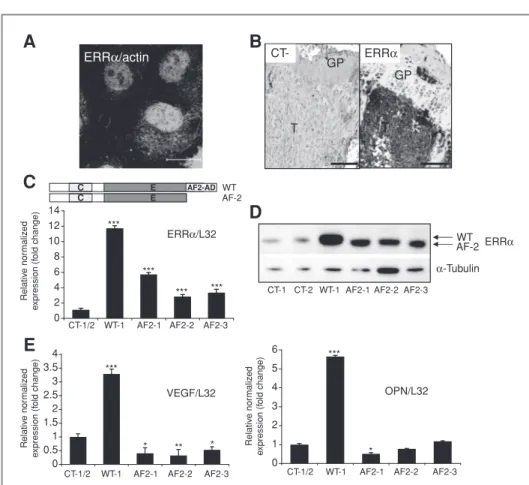
Figure 2.Modulation of ERRa in BO2 breast cancer cell line. A, ERRa protein expression (nucleus and cytoplasm) in BO2 cells by immunofluorescence and confocal microscopy and (B) in vivo by immunohistochemistry in bone metastasis present 30 days after intravenous injection of BO2 cells. C, isolation after stable transfection of three independent BO2-ERRaDAF2 clones (ERRa dominant-negative form), one clone BO2-ERRaWT and two controls (CT-1 and CT-2) BO2-CT (empty vector). ERRa expression was assessed by real-time PCR on triplicate samples and normalized against that of the ribosomal protein gene L32 (ANOVA, P< 0.0001) and (D) by Western blotting. (E) VEGF and OPN expression was increased in BO2-ERRaWT and decreased or not regulated in BO2-ERRaDAF2 (ANOVA, P< 0.0001 for VEGF and OPN in WT-1 orDAF2 versus CT). (A) bar ¼ 20 mm and (B) bar ¼ 200 mm. T, tumor; GP, growth plate.
but not in normal breast epithelial cells (Supplementary Fig. S1A). ERRa was also clearly present in breast cancer cells that metastasized to bone (Supplementary Fig. S1D see T). As previously reported by us (21), ERRa was also detected in osteocytes embedded in the bone matrix.
ERRa expression in breast cancer cells reduces their ability to induce osteolytic lesions in vivo
To assess whether ERRa is involved in bone metastasis formation, we used MDA-BO2-FRT (BO2) cells, a subpopula-tion of the human MDA-231 breast cancer cell line, that was
selected for the high efficiency with which it metastasizes to bone (32). ERRa protein was seen in the nucleus and cyto-plasm of BO2 cells in vitro (Fig. 2A) and in situ in bone metastasis from legs of animals, 30 days after intravenous tumor cell inoculation (Fig. 2B).
To establish a functional role for ERRa in bone metastasis development, we next transfected B02 cells with a full-length (WT) ERRa or a truncated version of ERRa lacking the coactivator binding domain AF2, ERRaDAF2, which acts as a dominant-negative form (22, 23, 36; Fig. 2C). Constructs of human ERRaWT and ERRaDAF2 were stably transfected into the genomic FRT site present in the BO2 cells. Three inde-pendent BO2-ERRaDAF2 (1, 2, 3), one BO2-ERRaWT, and two BO2-CT (empty vector) clones were obtained, named AF2-1, AF2-2, AF2-3, WT-1, CT-1, and CT-2, respectively. As judged by real-time PCR, total ERRa mRNA expression was increased when compared with CT-1/2 clones (Fig. 2C). Western blotting detected a band of approximately 50 kD for ERRa protein in CT1-2 and WT-1 that was increased in WT-1 and AF2-1, AF2-2, and AF2-3 cells. The presence of a band with a slightly lower molecular weight in AF2-1, AF2-2, and AF2-3 cells corre-sponded well with the expected size for truncation of the AF2 domain (42 amino acids; Fig. 2D). mRNA expression levels of the ERRa target genes VEGF and OPN were statistically significantly increased in WT-1 cells compared with CT-1/2 cells (Fig. 2E). By contrast, VEGF and OPN mRNA levels remained reduced or unchanged in AF2 clones (Fig. 2E), confirming the increased activity and the dominant-negative functions of the WT and truncated ERRaDAF2 constructs, respectively.
To assess the involvement of ERRa in bone metastasis formation, CT (pool of CT-1 and -2 clones), WT-1, and AF2 (pool of AF2-1, -2 and -3 clones) cells were inoculated intra-venously into female BALB/c nude mice. Thirty-five days after tumor cell injection, radiographic analysis revealed that ani-mals bearing WT-1 tumors had osteolytic lesions that were 40% smaller than those of mice bearing CT tumors (Fig. 3A,B, and J). By contrast, there was a 3-fold increase in the extent of osteolytic lesions in animals bearing AF2 tumors, when com-pared with control (Fig. 3A, C, and J). The inhibitory effect of ERRa on cancer-induced bone destruction was confirmed using 3D micro-CT reconstruction (Fig. 3D–F), histology (Fig. 3G–I), and histomorphometric analyses of tibiae (BV/ TV; skeletal tumor burden, TB/STV; Fig. 3J). Taken together, our results indicated that overexpression of ERRa in breast cancer cells reduced the formation of osteolytic lesions. Regulation of OC formation by ERRa#expressing BO2 cells
Given these data, we next asked whether modulation of ERRa in breast cancer cells could alter OCs, the bone resorbing cells. TRAP staining of tibial sections of meta-static legs from animals bearing WT-1 and AF2 tumors showed a 43% decrease and a 143% increase of TRAP-positive OC surface (Oc.S/BS) at the bone/tumor cell inter-face, respectively, when compared with CT tumors (Fig. 4A and 3J; Supplementary Fig. S2). Consistent with these in vivo data, the treatment of primary mouse bone marrow cell
AF2 WT-1 CT AF2 WT-1 CT AF2 WT-1 CT T T T
C
B
A
F
E
D
I
H
G
CT WT BO2-FRT Clones Radiography 10.9 ± 1.9 (mm2/animal) (n = 9) 11.4 ± 1.1 (n’ = 13) 35.9 ± 4.2 (n’ = 13) 49.3 ± 3.4 (n’ = 14) 7.1 ± 3.4* (n = 8) 15.1 ± 0.9* (n’ = 13) 21 ± 4.8* (n’ = 13) 21 ± 5.6** (n’ = 13) 30.7 ± 7.2*** (n = 9) 6.4 ± 0.4*** (n’ = 14) 71 ± 3.2*** (n’ = 14) 70 ± 6*** (n’ = 16) Bone volume (BV/TV, %) Tumor burden (TB/STV, %) Osteolyse (Oc.S/BS, %)J
∆AF ERRαFigure 3.Overexpression of ERRa inhibits development of bone metastasis. A–C, BO2-ERRaWT-1, BO2-ERRaDAF2 (pool), or BO2-CT (pool) cells were inoculated into BALB/c nude mice; 35 days postinoculation, radiography revealed smaller osteolytic lesions in mice injected with BO2-ERRaWT-1 cells and much larger lesions in mice injected with BO2-ERRaDAF2 cells compared with mice injected with CT cells (see white arrows). D–F, 3D micro-CT reconstructions of tibiae and (G–J) histology after Goldner's Trichrome staining confirmed the radiography results. J, quantification of bone destruction. T, tumor.
cultures with RANKL and M-CSF together with the condi-tioned medium of WT-1 cells inhibited the formation of TRAP-positive multinucleated OCs compared with that observed with the conditioned medium of CT cells (Fig. 4B and C). By contrast, the conditioned medium from AF2 cells stimulated OC formation (Fig. 4B and C). In addition, the conditioned medium from parental BO2 cells treated with the inverse agonist XCT-790, which blocks ERRa activity, increased OC formation compared with control (dimethyl sulfoxide; Fig. 4D), confirming our osteo-clastogenesis data obtained with the conditioned medium of AF2 cells.
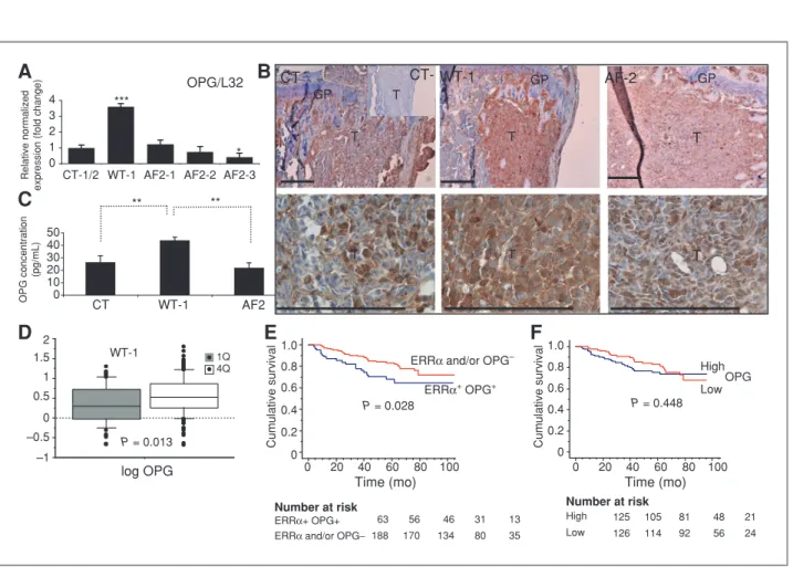
ERRa regulates OPG expression in breast cancer cells We showed that B02 breast cancer cells overexpressing wild-type ERRa markedly inhibited osteolysis in vivo (Fig. 3J) and reduced OC formation in vitro (Fig. 4). We quantified several markers involved in osteoblasts and OC differentia-tion, and we found that the OPG, a soluble decoy receptor for RANKL that inhibits osteoclastogenesis, was regulated by ERRa (Fig. 5A; ref. 37). By immunohistochemistry, we show that OPG expression was higher in skeletal WT-1 tumors compared with that observed in AF2 and CT tumors (Fig. 5B). In addition, as judged by ELISA, WT-1 cells secreted higher amounts of OPG compared with CT-1/2 and AF2 cells (pool of AF2-1, -2 and -3 clones; Fig. 5C). OPG mRNA expres-sion was also quantified by real-time RT-PCR in the cohort of 251 patients. OPG levels were statistically significantly higher
in ERRa-positive tumors compared with ERRa-negative tumors (Fig. 5D; P ¼ 0.013). Moreover, there was a positive correlation between high mRNA expression levels of both ERRa and OPG (ERRaþ/OPGþ) and a decrease in relapse-free
survival (P ¼ 0.028, log-rank test; Fig. 5E). All together, the significant correlation between high ERRa and OPG in patients and the regulation of OPG by ERRa in BO2 cells provide a mechanistic basis for the reduction of osteo-clastogenesis in vitro and in vivo. Interestingly, OPG in our preclinical data suggest that, alone it had no prognostic value in breast carcinomas (Fig. 5F) whereas in association with high ERRa mRNA levels, a correlation with a poor clinical outcome in patients was found (Fig. 5E). OPG is not only an osteoclastogenesis inhibitor, but also a survival factor for human breast cancer cells (38, 39). It also promotes angiogen-esis (40), and its overexpression in human MCF-7 breast cancer cells enhances tumor growth following orthotopic inoculation in animals (41). ERRa has been implicated in tumor progression, and the positive association between high ERRa/OPG mRNA levels and increased risk of recurrences in patients (Fig. 5E) suggested that ERRa could play a role on primary tumor expansion.
ERRa stimulates tumor growth and angiogenesis in vivo To address this hypothesis, orthotopic tumors were induced with CT (pool of CT-1 and -2 clones), WT-1, or AF2 (pool of AF2-1, -2 and -3 clones) cells upon inoculation within the mammary fat pad of NMRI nude female mice.
0 200 400 600 800 1,000 1,200 1,400 1,600 CT WT-1 AF-2 NT
A
C
B
D
Number of OCs/ w ells AF2-3 AF-2-2 AF2-1 WT-1 CT-2 CT-1 NT ** *** *** ** 0 100 200 300 400 500 Number of OCs/w ells XCT790 (5 × 10⁻⁷mol/L) CT BO2 ** 0 20 40 60 80 OCs surf ace/bone surf ace (%) ** *** AF-2 WT-1 CTFigure 4.ERRa expression in BO2 cells regulates OCs formation. A, TRAP staining of OCs in sections of tibiae taken from mice injected with BO2-ERRaWT-1, BO2-ERRaDAF2 (pool), or BO2-CT (pool) cells shows decreased and increased surface of OCs in BO2-ERRaWT-1 and BO2-ERRaDAF2, respectively compared with CT (ANOVA, P< 0.0001). B and C, primary mouse bone marrow cells were cultured in the presence of RANKL and M-CSF and treated or not with medium conditioned by BO2-ERRaWT-1, BO2-ERRaDAF2, or BO2-CT cells. Fewer OCs formed in cultures treated with BO2-ERRaWT-1 conditioned medium, whereas more formed in cultures treated with BO2-ERRaDAF2 conditioned medium, compared with CT (refs. 1, 2) conditioned medium (ANOVA, P< 0.0001). D, conditioned medium obtained from parental BO2 cells treated with the ERRa inverse agonist XCT-790 increased OCs formation, mimicking the results obtained with BO2-ERRaDAF2 conditioned medium (ANOVA, P < 0.001). Bar, 100 mm.
Bioluminescence analysis from day 5 to day 66 revealed a dramatically greater tumor progression in WT-1 tumor-bearing animals compared with that observed with CT and AF2 tumor-bearing animals a modest increase in AF2 tumor burden was also observed at day 62 and 66 (Fig. 6A and B). Tumor weight/size at day 66 (Fig. 6C and D) correlated well with bioluminescence quantification (Fig. 6B and C). Interestingly, WT-1 tumors were highly vascularized compared with CT and AF2 tumors (Fig. 6E), an observation correlating with higher VEGF mRNA levels observed in WT-1 versus AF2 or CT tumors (Fig. 6E). Moreover, if these results are in agreement with previous data describing VEGF as a target gene for ERRa in breast cancer (42), we show for the first time a positive association between high levels of ERRa and VEGF in breast tumors from patients (P ¼ 0.002; Table 1). Interestingly, OPG expression that can be stimulated by VEGF in endothelial cells, is also known to be a positive regulator of microvessel formation in vivo (43) and therefore can participate to the neovascu-larization observed in WT-1 tumors. We also observed that
ERRa promoted BO2 breast cancer cell invasion in vitro (Supplementary Fig. S3A) but has a slightly effect on pro-liferation (data not shown). Consistent with this we found matrix metalloproteinases MMP1 and MMP13 regulated by ERRa (Supplementary Fig. S3B). These results were in agreement with previous findings showing that the silen-cing of ERRa dramatically reduced the in vitro migratory capacity of breast cancer cell lines (44). Taken together, these results strongly suggested that ERRa promoted tumor growth, mainly through the stimulation of angiogenesis and invasion. Based on our results on high ERRa/OPG/VEGF in our preclinical study, we propose that OPG worked in concert with VEGF to stimulate tumor angiogenesis which, in turn, promoted the growth of BO2-ERRaWT cells. Con-versely, in bone metastasis although the angiogenic factor VEGF was overproduced in BO2-ERRaWT cells, tumor-derived VEGF had probably a low impact on progression of osteolytic lesions. Indeed, recent studies have shown that hypoxia was nonessential for bone metastasis while pro-moting angiogenesis in lung metastasis and primary tumor
0 10 20 30 40 50
A
B
CT WT-1 AF-2 T GP GP GP T T CT-TD
log OPG 4Q 1Q 2 1.5 1 0.5 0 –0.5 –1 P = 0.013 WT-1 OPG concentration (pg/mL) ** **E
0 20 40 60 80 100 0 20 40 60 80 100Cumulative survival Cumulative survival
0 0.2 0.4 0.6 0.8 1.0 0 0.2 0.4 0.6 0.8 1.0 Number at risk ERRα+ OPG+ ERRα and/or OPG−
63 56 46 31 13 188 170 134 80 35
P = 0.028
ERRα and/or OPG–
ERRα+ OPG+
Time (mo) Time (mo)
F
Number at risk High Low 125 105 81 48 21 126 114 92 56 24 P = 0.448 High LowOPG 0 1 2 3 4 5 4 3 2 1 OPG/L32 * Relative normalizedexpression (fold change)
*** AF2-3 AF2-2 AF2-1 WT-1 CT-1/2
C
T T T CT WT-1 AF2Figure 5.Correlation of ERRa and OPG in BO2 cells and breast cancer patients. A, real-time PCR carried out on RNA extracted from BO2 clones showed increased expression of OPG by ERRa (ANOVA, P < 0.0001). B, staining for OPG is higher in bone metastasis induced by BO2-ERRaWT cells compared with BO2-CT and BO2-ERRaDAF2 cells; tissues collected 35 days postcell inoculation. C, ELISA quantification confirmed the increased secretion of OPG by BO2-ERRaWT compared with BO2-CT (pool) and BO2-ERRaDAF2 (pool) cells (ANOVA, P ¼ 0.0064; P < 0,01 CT versus WT-1 and WT-1 versus AF-2). D and E, a significant correlation was also found between levels of ERRa mRNA and median values of OPG mRNA in the cohort (ERRa 1st quartile and median OPG ¼ 2.03; ERRa 2nd–4th quartile and median OPG ¼ 3.45). Kaplan–Meier curves show that ERRaþ/OPGþexpression was associated with
growth (45). Therefore, modulating angiogenesis through VEGF and the proangiogenic role of OPG may have no impact on angiogenesis in bone, as bone is already extre-mely vascularized (46), but have dramatic impact on vas-cularization and progression of primary breast tumors or metastasis to nonbone sites. These data provide novel insights into how ERRa can be a bad prognostic factor in the primary tumor (angiogenesis via VEGF and OPG) but a favorable biomarker in the very special case of bone metastasis (inhibition of OC formation through OPG).
In conclusion, our results show for the first time that ERRa plays a dual role, promoting the progression and invasion of primary tumors, but decreasing osteolytic lesions in bone. In addition, our data show that OPG is modulated by ERRa that
probably contributes to the overall negative clinical outcome which is associated with the expression of ERRa in human breast carcinomas.
Disclosure of Potential Conflicts of Interest
No potential conflicts of interest were disclosed.
Acknowledgments
We are very grateful to C. Lionnet and C. Chamot from PLATIM (IFR 128 Lyon Biosciences) for their help with imaging experiments and to Blandine Deux, Vincent Gonin, and Pascale Heneaux for their technical help. The authors also thank the CeCIL platform (Facult"e de M"edecine Laennec, Lyon, France) for technical assistance.
B
A
D
E
CT WT-1 AF2 Day 66 WT-1 AF2 CT WT-1 AF2 CT 0 0.5 1 1.5 *** T um or w ei gh t ( g) CT WT-1 AF2 0 1 2 3 4 WT-1 AF2 VEGF/L32 *** * Relative normalizedexpression (fold change) CT
CT WT ∆AF Tumorigenesis (fat pad-day 66) BLU (photons/sec 106) Volume (mm3/tumor) Weight (g/tumor) 1.7 ± 0.35*** (n = 10) 932 ± 152*** (n = 10) 0.98 ± 0.15*** (n = 10) 0.1 ± 0.3 (n = 10) 104.4 ± 36 (n = 10) 0.16 ± 0.03 (n = 10) 0.34 ± 0.1 (n = 10) 244 ± 67 (n = 10) 0.32 ± 0.1 (n = 10) BO2-FRT Clones ERRα
C
2 2.5 CT WT-1 *** 0.5 1 1.5 BLU (photons/sec10 6) AF2 ** ** * * * 0 70 60 50 40 30 20 10Days Figure 6.Stimulation of tumor
progression and angiogenesis by ERRa in vivo. A–C, BO2-ERRaWT, BO2-ERRaDAF2 (pool), or BO2-CT (pool) cells were inoculated into the fat pad of NMRI nude mice. Tumor progression was followed by bioluminescence from day 5 to 66. Greater tumor expansion was observed in mice with BO2-ERRaWT-1 compared with BO2-ERRaDAF2 (pool) or BO2-CT (pool) cells. C and D, weight and volume of tumors dissected at endpoint (E) and VEGF expression normalized against L32 (ANOVA, P< 0.001) within tumors (pool of n ¼ 3 for each condition) correlated with greater tumor vascularization.
Grant Support
This work was supported by the CNRS (EB), Inserm, the University of Lyon, "Ligue Regionale contre le Cancer" (Is!ere; E. Bonnelye). A. Fradet is supported by the Ligue Nationale contre le Cancer, B. Depalle by region Rhône Alpes, and A. Bellahc!ene from the National Fund for Scientific Research, Belgium.
The costs of publication of this article were defrayed in part by the payment of page charges. This article must therefore be hereby marked advertisement in accordance with 18 U.S.C. Section 1734 solely to indicate this fact.
Received April 29, 2011; revised June 29, 2011; accepted June 29, 2011; published OnlineFirst July 6, 2011.
References
1. Roodman GD. Mechanisms of bone metastasis. N Engl J Med 2004;350:1655–64.
2. Kozlow W, Guise TA. Breast cancer metastasis to bone: mechanisms of osteolysis and implications for therapy. J Mammary Gland Biol Neoplasia 2005;10:169–80.
3. Clezardin P, Teti A. Bone metastasis: pathogenesis and therapeutic implications. Clin Exp Metastasis 2007;24:599–608.
4. Clezardin P. Bisphosphonates’ antitumor activity: An unravelled side of a multifaceted drug class. Bone Epub2010 Jul 22.
5. Benoit G, Cooney A, Giguere V, Ingraham H, Lazar M, Muscat G, et al. International Union of Pharmacology. LXVI. Orphan nuclear receptors. Pharmacol Rev 2006;58:798–836.
6. Kallen J, Schlaeppi JM, Bitsch F, Filipuzzi I, Schilb A, Riou V, et al. Evidence for ligand-independent transcriptional activation of the human estrogen-related receptor alpha (ERRalpha): crystal structure of ERRalpha ligand binding domain in complex with peroxisome proliferator-activated receptor coactivator-1alpha. J Biol Chem 2004;279:49330–7.
7. Greschik H, Wurtz JM, Sanglier S, Bourguet W, van Dorsselaer A, Moras D, et al. Structural and functional evidence for ligand-indepen-dent transcriptional activation by the estrogen-related receptor 3. Mol Cell 2002;9:303–13.
8. Giguere V, Yang N, Segui P, Evans RM. Identification of a new class of steroid hormone receptors. Nature 1988;331:91–4.
9. Busch BB SWJ, Martin R, Ordentlich P, Zhou S, Sapp DW, Horlick RA, et al. Identification of a selective inverse agonist for the orphan nuclear receptor estrogen-related receptor alpha. J Med Chem 2004;47: 5593–6.
10. Willy PJ, Murray IR, Qian J, Busch BB, Stevens WC, Martin R, et al. Regulation of PPARgamma coactivator 1alpha (PGC-1alpha) signal-ing by an estrogen-related receptor alpha (ERRalpha) ligand. Proc Natl Acad Sci U S A 2004;101:8912–7.
11. Lui K TT, Mori T, Zhou D, Chen S. MCF-7aro/ERE, a novel cell line for rapid screening of aromatase inhibitors, ERalpha ligands and ERRal-pha ligands. Biochem Pharmacol 2008;76:208–15.
12. Luo J, Sladek R, Carrier J, Bader JA, Richard D, Giguere V. Reduced fat mass in mice lacking orphan nuclear receptor estrogen-related receptor alpha. Mol Cell Biol 2003;23:7947–56.
13. Huss JM, Imahashi K, Dufour CR, Weinheimer CJ, Courtois M, Kovacs A, et al. The nuclear receptor ERRalpha is required for the bioenergetic and functional adaptation to cardiac pressure overload. Cell Metab 2007;6:5–37.
14. Ariazi EA, Clark GM, Mertz JE. Estrogen-related receptor alpha and estrogen-related receptor gamma associate with unfavorable and favorable biomarkers, respectively, in human breast cancer. Cancer Res 2002;62:6510–8.
15. Suzuki T, Miki Y, Moriya T, Shimada N, Ishida T, Hirakawa H, et al. Estrogen-related receptor alpha in human breast carcinoma as a potent prognostic factor. Cancer Res 2004;64:4670–6.
16. Cheung CP, Yu S, Wong KB, Chan LW, Lai FM, Wang X, et al. Expression and functional study of estrogen receptor-related recep-tors in human prostatic cells and tissues. J Clin Endocrinol Metab 2005;90:1830–44.
17. Gao M, Sun P, Wang J, Zhao D, Wei L. Expression of estrogen receptor-related receptor isoforms and clinical significance in endometrial adenocarcinoma. Int J Gynecol Cancer 2006;16: 827–33.
18. Cavallini A, Notarnicola M, Giannini R, Montemurro S, Lorusso D, Visconti A, et al. Oestrogen receptor-related receptor alpha (ERRal-pha) and oestrogen receptors (ERalpha and ERbeta) exhibit different
gene expression in human colorectal tumour progression. Eur J Cancer 2005;41:1487–94.
19. Sun P, Sehouli J, Denkert C, Mustea A, K€onsgen D, Koch I, et al. Expression of estrogen receptor-related receptors, a subfamily of orphan nuclear receptors, as new tumor biomarkers in ovarian cancer cells. J Mol Med 2005;83:457–67.
20. Stein RA, McDonnell DP. Estrogen-related receptor {alpha} as a therapeutic target in cancer. Endocr Relat Cancer 2006;13 Suppl 1: S25–32.
21. Bonnelye E, Merdad L, Kung V, Aubin JE. The orphan nuclear estro-gen receptor–related receptor (ERR) is expressed throughout osteo-blast differentiation and regulates bone formation in vitro. J Cell Biol 2001;153:971–83.
22. Bonnelye E, Zirngibl RA, Jurdic P, Aubin JE. The orphan nuclear estrogen receptor-related receptor-{alpha} regulates cartilage forma-tion in vitro: implicaforma-tion of Sox9. Endocrinology 2007;148:1195–205. 23. Bonnelye E, Saltel F, Chabadel A, Zirngibl RA, Aubin JE, Jurdic P. Involvement of the orphan nuclear estrogen receptor-related receptor ERRalpha in osteoclast adhesion and transmigration. J Mol Endocri-nol 2010;45:365–77.
24. Rajalin AM PH, Aarnisalo P. ERRalpha regulates osteoblastic and adipogenic differentiation of mouse bone marrow mesenchymal stem cells. Biochem Biophys Res Commun 2010;396:477–82.
25. Delhon I, Gutzwiller S, Morvan F, Rangwala S, Wyder L, Evans G, et al. Absence of estrogen receptor related alpha increases osteoblastic differentiation and cancellous bone mineral density. Endocrinology 2009;150:4463–72.
26. Teyssier CGM, Rabier B, Monfoulet L, Dine J, Macari C, Espallergues J, et al. Absence of ERRalpha in female mice confers resistance to bone loss induced by age or estrogen-deficiency. PLoS One 2009;4: e7942.
27. Wei W WX, Yang M, Smith LC, Dechow PC, Sonoda J, Evans RM, et al. PGC1beta mediates PPARgamma activation of osteoclastogen-esis androsiglitazone-induced bone loss. Cell Metab 2010;11:503–16. 28. Bonnelye E, Vanacker JM, Dittmar T, Begue A, Desbiens X, Denhardt DT, et al. The ERR-1 orphan receptor is a transcriptional activator expressed during bone development. Mol Endocrinol 1997;11:905– 16.
29. Vanacker JM, Delmarre C, Guo X, Laudet V. Activation of the osteo-pontin promoter by the orphan nuclear receptor estrogen receptor related alpha. Cell Growth Differ 1998;9:1007–14.
30. Zirngibl RA CJ, Aubin JE. Estrogen receptor-related receptor alpha (ERRalpha) regulates osteopontin expression through a non-canoni-cal ERRalpha response element in a cell context-dependent manner. J Mol Endocrinol 2008;40:61–73.
31. Berthier ASS, Sasco AJ, Bobin JY, De Laroche G, Datchary J, Saez S, et al. High expression of gabarapl1 is associated with a better out-come for patients with lymph node-positive breast cancer. Br J Cancer 2010;102:1024–31.
32. Peyruchaud O, Winding B, Pecheur I, Serre CM, Delmas P, Clezardin P. Early detection of bone metastases in a murine model using fluorescent human breast cancer cells: application to the use of the bisphosphonate zoledronic acid in the treatment of osteolytic lesions. J Bone Miner Res 2001;16:2027–34.
33. Le Gall CBA, Bonnelye E, Gasser JA, Castronovo V, Green J, Zim-mermann J, et al. A cathepsin K inhibitor reduces breast cancer induced osteolysis and skeletal tumor burden. Cancer Res 2007; 67:9894–902.
34. David MWE, Descotes F, Jansen S, Deux B, Ribeiro J, Serre CM, et al. Cancer cell expression of autotaxin controls bone metastasis
formation in mouse through lysophosphatidic acid-dependent activa-tion of osteoclasts. PLoS One 2010;5:e9741.
35. Boissier SFM, Peyruchaud O, Magnetto S, Ebetino FH, Colombel M, Delmas P, et al. Bisphosphonates inhibit breast and prostate carci-noma cell invasion, an early event in the formation of bone metas-tases. Cancer Res 2000;60:2949–54.
36. Vanacker JM, Pettersson K, Gustafsson JA, Laudet V. Transcriptional targets shared by estrogen receptor- related receptors (ERRs) and estrogen receptor (ER) alpha, but not by ERbeta. Embo J 1999;18: 4270–9.
37. Boyle WJ, Simonet WS, Lacey DL. Osteoclast differentiation and activation. Nature 2003;423:337–42.
38. Rachner TDBP, Rauner M, Goettsch C, Singh SK, Schoppet M, Hofbauer LC. Osteoprotegerin production by breast cancer cells is suppressed by dexamethasone and confers resistance against TRAIL-induced apoptosis. J Cell Biochem 2009;108:106–16. 39. Malyankar UMSM, Suchland KL, Yun TJ, Clark EA, Giachelli CM.
Osteoprotegerin is an alpha vbeta 3-induced, NF-kappa B-dependent survival factor for endothelial cells. J Biol Chem 2000;275:20959–62. 40. McGonigle JSGC, Scatena M. Osteoprotegerin and RANKL differen-tially regulate angiogenesis and endothelial cell function. Angiogen-esis 2009;12:35–46.
41. Fisher JLT-MR, Elliott J, Hards DK, Sims NA, Slavin J, Martin TJ, et al. Osteoprotegerin overexpression by breast cancer cells enhances orthotopic and osseous tumor growth and contrasts with that deliv-ered therapeutically. Cancer Res 2006;66:3620–8.
42. Stein R, Gaillard S, McDonnell D. Estrogen-related receptor alpha induces the expression of vascular endothelial growth factor in breast cancer cells. J Steroid Biochem Mol Biol 2009;114: 106–12.
43. Benslimane-Ahmim Z, Heymann D, Dizier B, Lokajczyk A, Brion R, Laurendeau I, et al. Osteoprotegerin, a new actor in vasculogenesis, stimulates endothelial colony-forming cells properties. J Thromb Haemost 2011;9:834–43.
44. Dwyer MAJJ, Wade HE, Eaton ML, Kunder RS, Kazmin D, Chang CY, et al. WNT11 expression is induced by estrogen-related receptor alpha and beta-catenin and acts in an autocrine manner to increase cancer cell migration. Cancer Res 2010;22:9298–308.
45. Lu XYC, Yuan M, Wei Y, Hu G, Kang Y. In vivo dynamics and distinct functions of hypoxia in primary tumor growth and organotropic metastasis of breast cancer. Cancer Res 2010;70:3905–14. 46. Fei JPF, Malaval L, Vico L, Lafage-Proust MH. Imaging and
quanti-tative assessment of long bone vascularization in the adult rat using microcomputed tomography. Anat Rec (Hoboken) 2010;293:215–24.