PEGylated quaternized copolymer/DNA complexes for gene delivery
Benoît Vromana, Isabel Ferreiraa, Christine Jérômeb, Robert Jérômeb, Véronique Préata
a Université Catholique de Louvain, Unité de Pharmacie Galénique, Avenue E. Mounier 73.20, 1200 Brussels, Belgium
b
Center for Education and Research on Macromolecules (CERM), University of Liège, Sart-Tilman, B6, 4000 Liège, Belgium
Abstract
The aim of this study was to improve the colloidal stability, decrease unspecific interactions with cells and blood components of a novel gene delivery system composed of ε-caprolactone and quaternized ε-caprolactone. For this purpose, diblock 50/50 copolymer was used to generate complexes with DNA by either the solvent evaporation technique and by dialysis. The size, surface charge and degree of interaction of the plasmid-loaded formulations were measured. Then, polyplexes were combined with a poly(CL)-b-PEG copolymer to create a hydrophilic corona on the surface of the complexes. The cytotoxicity, transfection efficiency and cellular uptake of polyplexes and their association with PEG were evaluated on HeLa cells. The dialysis method did not allow to reduce the size of complexes as compared to the solvent evaporation method. The zeta potential of polyplexes became positive from a charge ratio of 4. The degree of interaction of copolymer with plasmid DNA was very high. Cytotoxicity and transfection efficiency were found to be comparable to polyethylenimine 50 kDa. Association of polyplexes with poly(CL)-b-PEG copolymer led to a small increase in particle size and a sharp decrease of charge surface. Cytotoxicity, transfection efficiency and cellular uptake were significantly reduced relative to unshielded copolymer/DNA complexes. The PEGylated formulations may be an attractive approach for an in vivo application.
Keywords: Gene therapy; Polyplexes; DNA; Polyethylene glycol; Transfection
1. INTRODUCTION
Gene therapy is a promising approach for the treatment of a wide range of diseases. Among non-viral gene delivery systems, "polyplexes" seem to be attractive. The polyplexes form spontaneously as a result of electrostatic interactions between the positively charged groups of the polycation and the negatively charged phosphate groups of the DNA. This results in DNA condensation, protection from the nuclease digestion and more efficient delivery of plasmid into the cells (De Smedt et al., 2000). Unmodified cationic polymers lead to complexes with DNA displaying a relatively high cationic surface charge. The positive charge of the
polymer/DNA complex facilitates cellular uptake of the complex by electrostatic interaction with negatively charged cell membranes (Lee and Kim, 2005) but represents a problem when polyplexes are introduced in the blood circulation (Kichler, 2004). Indeed, strong positive charge on the polymer/DNA complexes induces unspecific interactions with the extracellular matrix and cell surfaces (Davis, 2002). Interactions of positively charged polyplexes with blood components such as erythrocytes and plasma proteins results in their dissociation and aggregation, leading to rapid clearance by the reticuloendothelial system (Merdan et al., 2003). Polyplexes dissociation might lead to liberated DNA, which displays unfavorable pharmacokinetics, poor cellular uptake and consequently very poor transfection activity (De Wolf et al., 2005).
A frequently applied strategy to stabilize copolymer/DNA complexes and neutralize positive charges is to shield the outer surface of the complexes with hydrophilic and uncharged polymers (Funhoff et al., 2005). Association of polyplexes with PEG is advantageous: (i) it reduces the cytotoxicity of polymer/DNA complexes; (ii) PEG shields excess positive charges of polymer/DNA complexes, resulting in the reduction of unspecific interactions between polyplexes and blood components and cells; (iii) PEG can be used as a spacer between a targeting lig-and lig-and the polymeric carrier, which facilitates the access of the liglig-and to its receptor (Lee lig-and Kim, 2005). In a previous study, copolymers composed of ε-caprolactone monomers (CL) and γ-bromo-ε-caprolactone monomers quaternized by reaction with pyridine (Py+CL) were used to generate DNA nanoparticulate systems.
Copolymers with diblock or random structure, each of them available with two monomer ratios (50 Py+CL/50 CL and 80 Py+CL/20 CL) were investigated. Cytotoxicity and transfection efficiency were found to be comparable to polyethylenimine 50 kDa, a conventional polymer used in gene therapy (Vroman et al., 2007). However, the positive charges of the polyplexes might lead in vivo (i) to unspecific interactions with blood components and cells and (ii) to unstability (floculation/aggregation) at high ionic strength. Poly(CL-b-Py+CL) 50/50 diblock copolymer was chosen for the associations studies with poly(CL)-b-PEG copolymer according to three parameters: (i) the diblock structure is more adapted for the association with PEG copolymer than a random structure, (ii) it was shown that the particle size of diblock 50/50 copolymer incubated in serum was smaller than this for other copolymers tested (data not shown) and (iii) poly(CL-b-Py+CL) 50/50 copolymer was found to be less cytotoxic than poly(CL-b-Py+CL) 20/80 copolymer (Vroman et al., 2007). The plasmid-loaded formulations have been characterized by their size, surface charge and degree of interaction.
The main aim of this present study was to improve the colloidal stability and in vivo biodistribution of the novel gene delivery systems based on ε-caprolactone and quaternized ε-caprolactone (Detrembleur et al., 2000a,b; Vroman et al., 2007) by reducing the size and/or introducing a hydrophilic corona. Firstly, diblock 50/50 copolymer was used to generate complexes with DNA by either the solvent evaporation technique (Vroman et al., 2007) and by dialysis (Gaucher et al., 2005) By using the dialysis technique, it was expected to formulate polyplexes with a reduced size compared to the solvent evaporation technique. In the second part of the work, polyplexes were combined with a poly(CL)-b-PEG copolymer to create a hydrophilic corona on the surface of the complexes. In vitro transfection efficiency, cytotoxicity and cellular uptake of polyplexes and their
association with PEG were evaluated on HeLa cell line. Shielded polyplexes were formed by mixing preformed poly(CL)-b-PEG copolymer with plasmid DNA and polycation during the formation of particles. This method is an easy and versatile preparation procedure, by which the size and the extent of PEG shielding can be controlled by modifying the proportions of the components within the mixture used for polyplex preparation. Moreover, other functionalities, like targeting ligands, can be easily introduced in the outer layer of polyplexes by using preformed functionalized PEG copolymer for the preparation of particles (Garinot et al., 2007).
2. MATERIALS AND METHODS 2.1. Materials
Plasmid pGL2CMVLuc (6.233 kb) encoding luciferase gene reporter and under the control of a human CMV promoter was obtained from Gibco® BRL (Carlsbad, CA, USA).
7-Oxabicyclo[2.2.1]heptane(Sigma-Aldrich,St.Louis,MO, USA), 48% (aqueous) HBr (Sigma-Aldrich, St. Louis, MO, USA), pyridinium chlorochromate (Sigma-Aldrich, Bornem, Belgium) and 75% m-chloroperoxybenzoic acid (Sigma-Aldrich, Bornem, Belgium) were used as received. Br-CL was synthesized starting from
7-oxabicyclo[2.2.1]heptane as reported elsewhere (Detrembleur et al., 2000a) and dried by repeated (three times) azeotropic distillation of toluene just before polymerization. ε-CL (Sigma-Aldrich, Bornem, Belgium) was dried over calcium hydride for 48 h at room temperature and distilled under reduced pressure just before use. Toluene (ChemLab, Somme-Leuze, Belgium) was dried by refluxing over calcium hydride and distilled under nitrogen atmosphere. Aluminium isopropoxide [Al(OiPr)3] (Sigma-Aldrich, St. Louis, MO, USA) was twice sublimated and then dissolved in toluene under nitrogen. Triethylaluminium (Fluka) was diluted in dry toluene and the solution concentration was determined by volumetric titration (Vangeyte and Jérôme, 2004). Methylene chloride and pyridine were dried by reflux over calcium hydride for at least 48 h before distillation. Ethylene oxide (EO) (Messer), triethylene glycol monoethyl ether (Fluka) and potassium hydroxyde (Fluka) were used as received. HeLa (human cervic carcinoma) cells were purchased from ATCC (American Type Culture Collection, Manassas, VA, USA).
Ampicillin, 3-(4,5-dimethylthiazol-2-yl)-2,5-diphenyl tetra-zolium bromide (MTT) and PEI 50 kDa polymer were acquired from Sigma-Aldrich (St. Louis, MO, USA). PEI 50 kDa was used as 10 mM aqueous stock solution [9mg of the 50% (w/v) commercial solution diluted in 10 ml of water]. The solution was neutralized with HC1 and filtered (Millipore, 0.2 µm). Dul-becco's modified Eagle's medium (DMEM), fetal bovine serum (FBS), trypsin-EDTA and penicillin-streptomycin mixtures were from Gibco® BRL (Carlsbad, CA, USA). Spectra/Porl dialysis membranes (MWCO = 6-8000) were acquired from Spectrum Laboratories (Breda, The Netherlands). YOYO-1 iodide was purchased from Molecular Probes (Leiden, The Netherlands). The Endofree plasmid kit was obtained from Qiagen (Hilden, Germany), the Picogreen® assay kit from Molecular Probes (Leiden, The Netherlands), the Micro BCA Protein Assay Kit from Pierce (Rockford, USA) and the Luciferase Assay System from Promega (Madison, USA). Ultra-pure water was used throughout and all other chemicals
were of analytical grade.
2.2. Preparation of plasmid DNA
Plasmid pGL2CMVLuc was propagated in Escherichia coli, extracted by the alkali lysis technique and purified using an Endofree plasmid kit according to the manufacturer's instructions. The quantity and quality of the purified plasmid DNA was assessed by optical density at 260 and 280 nm and by gel electrophoresis. Plasmid DNA was resuspended in water and stored at -20 °C until use.
2.3. Synthesis and characterization of copolymers
Synthesis of diblock 50/50 copolymer of ε-caprolactone and quaternized ε-caprolactone was performed by ring-opening polymerization of CL and BrCL initiated by Al(OiPr)3 followed by quaternization with pyridine (Detrembleur et al., 2000a,b; Vroman et al., 2007). Poly(CL)-b-PEG copolymer was also synthesized by ring-opening polymerization using triethyla-luminium as catalyst as previously described (Vangeyte and Jérôme, 2004). 1H NMR (250 MHz) spectra were recorded in CDCl3 with a Brüker AM 250 apparatus at 25 °C. Size-exclusion chromatography (SEC) was carried out in THF with a Waters chromatograph equipped with a Waters 410 differential refrac-tometer and 5 µm Waters columns of increasing pore size (102, 103, 105 and 106Å). Polystyrene standards were used for calibration.
2.4. Formulation of copolymer/DNA complexes
Polyplexes were formulated at various charge ratios N/P copolymer/DNA using the solvent evaporation technique (Soppimath et al., 2001; Hans and Lowman, 2002; Vroman et al., 2007) as well as by dialysis (Gaucher et al., 2005). Charge ratio N/P copolymer/DNA was expressed as the ratio between the number of pyridinium groups on copolymer and the number of phosphate groups on plasmid DNA. For biophysical studies (size, zeta potential and degree of interaction), complexes were prepared at a plasmid concentration of 50 µg/ml in water, NaCl 20 mM or NaCl 150 mM. For in vitro applications (cytotoxicity, transfection efficiency and cellular uptake), polyplexes were formulated at a plasmid concentration of 10 µg/ml in culture medium. 2.4.1. Solvent evaporation technique
Firstly, diblock copolymer was dissolved in dichloromethane. Then, the copolymer solution was emulsified in the plasmid DNA solution by homogenization at 8000 rpm for 1 min using a high-speed homogenizer Diax 900 (Heidolph Instruments, Germany). Finally, the organic solvent was eliminated by stirring of the emulsion at room temperature for 2 h (Vroman et al., 2007). For PEGylation of polyplexes, copolymer/DNA complexes were associated with a poly(CL)-b-PEG copolymer at proportions of 1/9, 1/3 and 1/1 poly(CL)-b-PEG/poly(CL-b-Py+CL) copolymers during the formation of nanoparticles.
2.4.2. Dialysis technique
Poly(CL-b-Py+CL) copolymer was dissolved in dimethyl-formamide. DNA solution was added at once to the copolymer solution and the mixture was stirred at room temperature for 15 h. Then the solution was poured in a Spectra/Porl dialysis membrane (MWCO 6000-8000) and dialyzed against water for 24 h to remove
dimethylformamide.
2.5. Size and zeta potential of copolymer/DNA complexes
The mean hydrodynamic diameter and polydispersity of the complexes were determined by photon correlation spectroscopy using a Malvern NanoZS (Malvern Instruments, UK). Measurements were performed at a temperature of 25 °C and at a detection angle of 90°. The instrument was routinely calibrated using standard reference latex particles (Malvern Instruments, UK). The particle size of each sample was expressed as the mean hydrodynamic particle diameter ± standard deviation of three measurements.
Surface charge of copolymer/DNA complexes was measured at 25 °C with a Malvern NanoZS after calibration with a polystyrene dispersion (Malvern Instruments, UK) of known zeta potential. Given values were the average of at least five runs.
2.6. Degree of association copolymer/DNA
The degree of interaction between plasmid DNA and copolymer was evaluated by measuring the amount of plasmid DNA that was not associated with particles and therefore remained in the supernatant upon
centrifugation of the particle suspension (Perez et al., 2001; Vroman et al., 2007). Copolymer/DNA particles were spun down at 15,000 rpm for 60 min. The supernatant was mixed with Picogreen® reagent and the fluorescence was measured using a Packard Fluorocount Microplate Fluorometer (Packard BioScience
Company, Meriden, USA) with an excitation wavelength of 485 nm and an emission wavelength of 530 nm. The amount of plasmid DNA was read from a calibration curve setup with standard DNA solutions in the 25 ng/ml to 1 µg/ml concentration range (Molecular Probes, Leiden, The Netherlands) (Vroman et al., 2007).
2.7. Cell culture
HeLa cells (human cervix epithelial carcinoma cells) were cultured in Dulbecco's modified Eagle medium (DMEM) supplemented with 10% (v/v) fetal bovine serum and 100 IU/ml of penicillin G sodium and 100 µg/ml of streptomycin sulfate. Cells were maintained at 37 °C in a humidified 5% CO2 atmosphere. Cells were sub-cultured using trypsin/EDTA when almost confluent.
2.8. Transfection efficiency protocol
For transfection studies, HeLa cells were seeded in 96-well plates at an initial cell density of 104 cells/well in 100 µl of culture medium supplemented with 10% FBS. After 24 h, the medium was replaced with 100 µl of serum-free medium containing unshielded polyplexes and PEGylated copolymer/DNA (1 µg) complexes prepared at N/P charge ratios ranging from 1 to 4. Following 2 h of incubation with the formulations, 100 µl of culture medium with 20% of serum was added in each well. After 4 h treatment with polyplexes, the transfection mixtures were removed and the cells overlaid with fresh culture medium containing 10% FBS. Transfected cells were further incubated for 44 h to allow protein expression. Polyethyleneimine 50 kDa/DNA complexes formulated at a N/P charge ratio of 9, according to Boussif et al. (1995), were used as a positive control of transfection. All transfection experiments were carried out in two identical 96-well plates. One plate was tested for luciferase activity; the other plate was tested for cell viability using a MTT colorimetric assay. Measurements were performed in triplicate for the determination of luciferase activity. Six determinations were done for evaluation of cell viability. Two independent experiments were carried out (Vroman et al., 2007).
2.9. Detection of luciferase activity
Luciferase gene expression was determined 48 h after transfection by using a commercial luciferase assay kit according to the manufacturer's instructions. Briefly, after removing culture medium, cells were washed twice with PBS and permeabilized with 200 µl of lysis buffer (Promega). For measurement of samples, 100 µl of luciferase substrate was automatically injected into 20 µl of cell lysate with a Glomax microplate luminometer (Turner Biosystems, Sunnyvale, USA). Light emitted was measured over 10 s with 2 s delay. Protein
concentration was determined by using a BCA protein assay kit. Thirty microliters of the remaining cell lysate was transferred in individual wells of a 96-well microplate and diluted with 120 µl of PBS pH 7.4. BCA reagent (150 µl) was added, the mixtures allowed to incubate for 2 h at 37 °C and the absorbance measured at 570 nm with a BioRad microplate reader. A calibration curve with bovine serum albumin standards was used to evaluate the amount of protein present. Transfection activity was expressed as relative light units (RLUs) per milligram cell protein (Vroman et al., 2007).
2.10. Evaluation of cell viability
Evaluation of cytotoxicity of polyplexes was performed 48 h after transfection. The metabolic activity of HeLa cells in each well was measured by using the MTT colorimetric assay (Mosmann, 1983). At the end of the transfection experiment, culture medium was replaced with 100 µl of sterile filtered MTT (0.5mg/ml) stock solution in culture medium and cells were incubated for 1 h at 37 °C. MTT containing medium was removed and 100 µl of DMSO was added to dissolve the for-mazan crystals. Absorbance was measured at 570 nm using a BioRad microplate reader. Untreated cells were taken as control with 100% viability and cells without addition of MTT were used as blank to calibrate the spectrophotometer to zero absorbance. Triton X-100 1% was used as positive control of cytotoxicity. The cell viability (%) was calculated according to the following equation:
where OD570nm(sample) represents the measurement from the wells treated with polyplexes and OD570 nm(control) from the wells treated with culture medium only (Vroman et al., 2007).
2.11. Cellular uptake
Plasmid DNA was labelled with the cell-impermeable fluorescent dye YOYO-1 iodide (Molecular Probes) with a ratio of 1 dye molecule to 300 bp followed by 1 h incubation in the dark at room temperature. Twelve-well plates were seeded with 105 cells per well and the cells were incubated at 37 °C for 24 h. After 24 h, the medium was aspirated off from the wells and gently replaced with unshielded and PEGylated polyplexes prepared in serum-free culture medium at charge ratios N/P ranging from 1 to 4. At certain time intervals (30, 60, 120 and 240 min, respectively) the complexes were aspirated off from the wells. The cells were extensively washed with ice-cold PBS. They were then harvested by trypsinization and centrifuged at 1200 rpm for 5 min. Finally, they were recovered in 0.5 ml of ice-cold PBS and stored on ice until analysis was performed. The percentage of fluorescent cells in the population and the level of cell-associated fluorescence were measured by flow cytometry. Flow cytometry analysis was performed using a FACScan flow cytometer (Bec-ton Dickinson, Le Pont-De-Claix, France) with an argon laser at wavelength of 488 nm. The filters used for emission were a 530/30-nm band-pass for YOYO-1 iodide, which was excited by the 488-nm laser line. Data were analyzed using the Cel-lQuest (Becton Dickinson) software and were expressed as the percentage of cells containing the labelled DNA and the mean fluorescence associated with cells. The individual fluorescence of 10,000 cells were collected for each sample (Deshpande et al., 2004).
2.12. Statistical analysis
Statistical analysis was performed using Student's t-test. To compare the mean and standard deviations of more than two groups, a Tukey's procedure was used. A difference between means was considered significant if the p value was less than or equal to 0.05. Software JMP 5.1 was used to evaluate the statistical significance of the data.
3. RESULTS AND DISCUSSION
The aim of this study was to improve the colloidal stability, decrease unspecific interactions with cells and prolong plasma half-life time of poly(CL-b-Py+CL) 50/50 diblock polyplexes. For this purpose, poly(CL-b-Py+CL) 50/50 diblock copolymer was used to generate complexes with DNA and was combined with a poly(CL)-b-PEG copolymer to create a hydrophilic corona on the surface of the polyplexes.
3.1. Characterization of copolymers
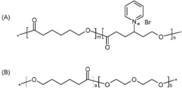
The structure and properties of copolymers used in the study are presented in Fig. 1 and Table 1, respectively. Poly(CL-b-Py+CL) 50/50 diblock copolymer had an average molecular weight of ~12,500Da and polydispersity of ~1.25. The quaternization yield was 75%. The experimental molar composition of the copolymer is close to the expected values, 50 mol% CL. The molecular weight of poly(CL)-b-PEG copolymer was in the range of 19,500 Da with a PEG chain of 4600 Da and with a polydispersity index of ~ 1.15.
Table 1.Physicochemical characteristics of the copolymers used in the study
Copolymer Molecular weighta,b Polydispersitya,c Degree of polymerizationd mol% of cationic unitsd
Poly(CL-b-Py+CL)50/50 12,300 1.25 69 49
Poly(CL)-b-PEG 14,900 CL/4600 PEG 1.15 100 (CL)/105 (PEG) -
a Apparent molecular weight and polydispersity were determined by size exclusion chromatography (polystyrene calibration). b M
n of the PCL block in poly(CL-b-PEG) copolymer was calculated from the intensity of the signal at 2.31 ppm for PCL and from Mn of PEG (4600).
c
Polydispersity=Mw/Mn. d Determined by proton NMR.
3.2. Influence of the method of preparation
First, non-PEGylated polyplexes were formulated at various N/P charge ratios using the solvent evaporation technique as well as dialysis to decrease the size of the particles. The replacement of the solvent evaporation method by the dialysis method did not allow a decrease of the size of polyplexes.
The particle sizes of the copolymer/DNA complexes (Fig. 2A) show a tendency to decrease with an increase of charge ratio. In agreement with previous studies (Jeong et al., 2001; Jeong and Park, 2002; Vroman et al., 2007), large aggregates were formed at a charge ratio of 2 for polyplexes prepared by the solvent evaporation technique. At this charge ratio, the surface charge of the copolymer/DNA complexes was near the neutrality, as confirmed by zeta potential data (Fig. 2B). At charge ratios of 4 and 10, particle sizes of approximately 200 nm were produced for the two methods of preparation of polyplexes.
At low charge ratios, zeta potential of copolymer/DNA complexes were negative (Fig. 2B). With increasing charge ratio, zeta potentials of complexes rapidly increased up to charge ratio of 4 whereas they slowly increased from charge ratios 4 to 10 (39.5 ± 1.5 and 26 ± 7.2 mV for polyplexes prepared either by the solvent evaporation technique and by dialysis, respectively). Complete shielding of negative charges of plasmid DNA occurred at a charge ratio of 4 both for solvent evaporation technique and dialysis method.
To demonstrate the interaction between copolymer and plasmid DNA, a Picogreen® assay was performed. As shown in Fig. 2C, the interaction between copolymer and plasmid DNA progressively increased according to the charge ratio and reached a plateau at around a charge ratio of 2 for the two methods of preparation of
copolymer/DNA complexes. At low charge ratios, plasmid DNA was still accessible for Picogreen® reagent explaining the negative zeta potential values found for the particles at these charge ratios. From a charge ratio of 2, plasmid DNA is completely bound to the polymer and protected inside the particles.
The results show that the method of preparation of polyplexes has no influence on the size and zeta potential of the DNA complexes and on the condensation capacity of poly(CL-b-Py+CL) 50/50 diblock copolymer. Since similar trends were observed for the two methods of preparation of polyplexes, the solvent evaporation technique, which was already used in our previous work (Vroman et al., 2007), was selected for the association studies with poly(CL)-b-PEG copolymer.
Fig. 2. Comparison of the solvent evaporation technique and of the dialysis method. (A) Particle size of 50/50
diblock copolymer/DNA complexes with various N/P charge ratios. Values are the average of three
measurements (±standard deviation). (B) Zeta potential values of polyplexes. (C) Degree of association between 50/50 diblock copolymer and plasmid DNA, determined by the Picogreen® assay (n = 4).
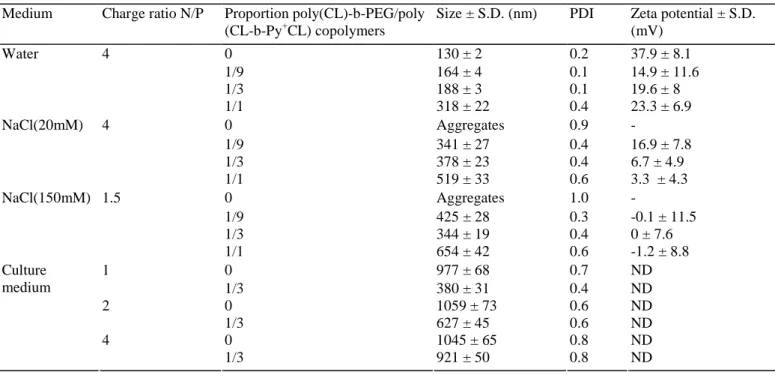
Table 2. Mean particle size and zeta potential of unshielded and PEGylated polyplexes (n = 3)
Medium Charge ratio N/P Proportion poly(CL)-b-PEG/poly (CL-b-Py+CL) copolymers
Size ± S.D. (nm) PDI Zeta potential ± S.D. (mV) 0 130 ± 2 0.2 37.9 ± 8.1 1/9 164 ± 4 0.1 14.9 ± 11.6 1/3 188 ± 3 0.1 19.6 ± 8 Water 4 1/1 318 ± 22 0.4 23.3 ± 6.9 0 Aggregates 0.9 - 1/9 341 ± 27 0.4 16.9 ± 7.8 1/3 378 ± 23 0.4 6.7 ± 4.9 NaCl(20mM) 4 1/1 519 ± 33 0.6 3.3 ± 4.3 0 Aggregates 1.0 - 1/9 425 ± 28 0.3 -0.1 ± 11.5 1/3 344 ± 19 0.4 0 ± 7.6 NaCl(150mM) 1.5 1/1 654 ± 42 0.6 -1.2 ± 8.8 0 977 ± 68 0.7 ND 1 1/3 380 ± 31 0.4 ND 0 1059 ± 73 0.6 ND 2 1/3 627 ± 45 0.6 ND 0 1045 ± 65 0.8 ND Culture medium 4 1/3 921 ± 50 0.8 ND
PDI: polydispersity index, is a measure of the distribution of the size of the particles; ND: not determined.
3.3. PEGylation of polyplexes
To increase the colloidal stability and decrease unspecific interactions, polyplexes were PEGylated by the addition of poly(CL)-b-PEG copolymer in the formulation. The amount of PEG copolymer introduced in the DNA complexes was optimized keeping the following factors into consideration: (i) reduction of the surface charge of polyplexes; (ii) limited increase of the particle size and (iii) stabilization of copolymer/DNA complexes in physiological medium. At first, PEGylated copolymer/DNA complexes were formulated at a charge ratio N/P of 4, at which well-defined and positively charged non-PEGylated polyplexes were observed (Fig. 2B). Poly(CL)-b-PEG copolymer was combined with poly(CL-b-Py+CL) polyplexes in water at proportions of 1/9, 1/3 and 1/1 poly(CL)-b-PEG/poly(CL-b-Py+CL) copolymers. Particle size and zeta potential values of the various PEG associations are shown in Table 2. The most efficient association seemed to be this with a
proportion of 1/9 poly(CL)-b-PEG/poly(CL-b-Py+CL) copolymers. Indeed, the surface charge decreased by approximately 22 mV while a slight increase in particle size was observed from 130 to 164nm. The increment in size can be attributed to the presence of PEG chains on the surface of complexes (Lee et al., 2001). Moreover, to study the effect of PEGylation on particle size of polyplexes in physiological medium, positively charged polyplexes were formulated in NaCl 20 mM and neutral polyplexes in NaCl 150mM and combined with PEG copolymer at previously mentioned proportions. In NaCl 20 mM, the particle sizes of the associations with proportions of 1/9 and 1/3 poly(CL)-b-PEG/poly(CL-b-Py+CL) copolymers were similar but a lower surface charge was obtained for the association with a proportion of 1/3 poly(CL)-b-PEG/poly(CL-b-Py+CL) copolymers. The particle size in NaCl 150 mM of the association with a proportion of 1/3
poly(CL)-b-PEG/poly(CL-b-Py+CL) copolymers was lower than the particle size of polyplexes combined with a proportion of 1/9 poly(CL)-b-PEG/poly(CL-b-Py+CL) copolymers. Based on the results of the association studies, a proportion of 1/3 poly(CL)-b-PEG/poly(CL-b-Py+CL) copolymers was selected for cytotoxicity, transfection and cellular uptake studies.
3.4. Cytotoxicity, transfection efficiency and cellular uptake
Transfection efficiency of unshielded and PEGylated polyplexes prepared by the solvent evaporation technique was evaluated on HeLa cells and compared to polyethylen-imine 50kDa and naked DNA. Among polycationic carriers, polyethylenimine (PEI) has been extensively exploited as an effective gene delivery vehicle (Nimesh et al., 2006). Polyethylenimine/DNA complexes were formulated at a charge ratio N/P of 9 at which an optimal transfection efficiency was previously observed (Boussif et al., 1995).
As shown in Fig. 3, transfection efficiency of poly(CL-b-Py+CL)/DNA complexes was high at charge ratios of 1 and 2. However, at N/P charge ratio of 4, an increase in cell death (Fig. 4) dramatically decreased the protein expression. Poly(CL-b-Py+CL)/DNA complexes formulated at a charge ratio N/P of 2 were equally efficient at transfecting cells in vitro as PEI 50kDa prepared at a charge ratio N/P of 9 (p>0.05). The transfection activity of PEGylated polyplexes was significantly reduced relative to unshielded polyplexes for all N/P charge ratios (p < 0.0001 at N/P charge ratios of 1 and 2).
Because transfection efficiency of a polymeric carrier is influenced by the cytotoxicity of the cationic polymers (Fischer et al., 1999; Jeong et al., 2001), the cytotoxicity of the plasmid-loaded formulations used in transfection experiments with HeLa cell line were investigated (Fig. 4). It was found that PEGylated polyplexes had a substantially lower cytotoxicity than unshielded polyplexes (p< 0.0001 for all N/P charge ratios tested). PEGylated polyplexes exhibited good cell viability (over 85%) whereas diblock 50/50 copolymer/DNA complexes were quite toxic for HeLa cells.
As transfection can be promoted by the formation of aggregates in the transfection medium (De Wolf et al., 2005), size studies were performed in culture medium (Table 2). Size studies performed in the culture medium used for the transfection studies showed that polyplexes aggregated. The higher transfection efficacy exhibited by the large aggregated particles could result from the conjunction of several features: (i) they sediment onto the cell surface more rapidly than the small complexes thereby intensifying the contact with the cells which
stimulates cellular internalization, (ii) since they contain a large proportion of free cationic polymers in addition to those complexed with DNA, they destabilize the membrane favoring their entry into cells, and (iii) their endosomolytic activity is far higher than that of the small particles (Zaric et al., 2004).
Fig. 3. Transfection efficiency of unshielded and PEGylated polyplexes in HeLa cell line. Transfection was
performed at proportions of 1/3 poly(CL)-b-PEG copolymer/poly(CL-b-Py+CL) copolymer. The quantity of plasmid DNA was maintained constant at 1 µg for all groups. The luciferase expression in relative light units (RLU) is normalized to mg of protein. Data represent the mean ± standard deviation of three measurements (***p < 0.0001).
Fig. 4. Cytotoxicity of the various transfection mixtures to HeLa cells. Transfection was performed at
proportions of 1/3 poly(CL)-b-PEG copolymer/poly(CL-b-Py+CL) copolymer. The quantity of plasmid DNA was maintained constant at 1 µg for all groups. Results were compared with the cytotoxicity of PEI 50 kDa
polyplexes formulated at a N/P charge ratio of 9 and with naked DNA (n = 6) (***p< 0.0001).
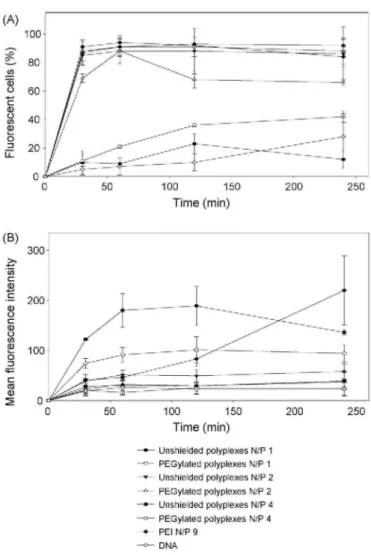
The reduced transfection activity and lower cytotoxicity of PEGylated polyplexes relative to unshielded polyplexes could be due to the formation of complexes with improved colloidal stability and reduced size (as demonstrated in Table 2) and to a reduced cellular interaction and internalization (Kursa et al., 2003; Merdan et al., 2003; Deshpande et al., 2004). Hence, the cellular uptake of unshielded and PEGylated polyplexes was evaluated at different time intervals using flow cytometry. The percentage of cells that showed an increase in fluorescence compared to the untreated control cells and the level of cell-associated fluorescence are shown in Fig. 5. At charge ratios N/P of 1 and 2, a decrease in mean cell-associated fluorescence was observed for PEGylated polyplexes compared to unshielded polyplexes (p<0.05), while the percentage of cell population showing associated fluorescence was relatively high. At charge ratio N/P of 4, the level of cellular uptake was similar to the level of internalization of naked DNA. The percentage of cells showing associated fluorescence for unshielded polyplexes formulated at a charge ratio N/P of 1 was comparable to PEI 50 kDa, although the level of fluorescence varied at the various incubation times for the two types of polymers. For poly(CL-b-Py+CL) 50/50 diblockpolyplexes, cell-associated fluorescence was relatively high at the earliest detection times and declined thereafter. In contrast, the amount of DNA in the cells gradually increased for PEI 50 kDa polyplexes. Flow cytometry results show that the introduction of PEG reduced the cellular uptake of DNA which could at least partially explain their low transfection efficiency and cytotoxicity. However, the low cellular association and low transfection efficiency obtained using PEGylated polyplexes could be beneficial to deliver DNA to specific target cells by conjugating a targeting ligand while preventing unspecific cellular interactions and transfection (Lam et al., 2004).
Fig. 5. FACS analysis of unshielded and PEGylated polyplexes. (A) Percentage of cells exhibiting associated
fluorescence (YOYO-1 labelled DNA) at different time intervals (n = 3). (B) Mean fluorescence associated with cells at different time intervals (n = 3). N/P corresponds to the charge ratio polymer/DNA.
4. CONCLUSION
To increase colloidal stability and decrease unspecific interactions with cells and blood components, poly(CL-b-Py+CL)/DNA complexes were PEGylated by adding poly(CL)-b-PEG4600 in the formulation. PEGylation induced a slight increase in particle size and an efficient shielding of surface charge of copolymer/DNA complexes. Cytotoxicity, transfection activity and cellular uptake of PEGylated polyplexes were significantly reduced relative to unshielded copolymer/DNA complexes. The PEGylated formulations may be an attractive approach for an in vivo application where non-specific interactions with blood components needs to be minimized. The conjugation of a targeting ligand to the PEG copolymer used for the formulation of shielded particles could lead to further improvements of vector efficiency. The application of this new polymer to animal studies in vivo is in progress.
Acknowledgements
The authors would like to thank Fonds de la Recherche Scientifique Médicale (FRSM) for financial support and Michaël Mazza for the synthesis of poly(CL-b-Py+CL) copolymer.
References
Boussif, O., Lezoualch, R, Zanta, M.A., Mergny, M.D., Scherman, D., Demeneix, B., Behr, J.P., 1995. A versatile vector for gene and oligonucleotide transfer into cells in culture and in vivo: polyethylenimine. Proc. Natl. Acad. Sci. U.S.A. 92, 7297-7301.
Davis, M.E., 2002. Non-viral gene delivery systems. Curr. Opin. Biotechnol. 13, 128-131.
De Smedt, S.C., Demeester, J., Hennink, W.E., 2000. Cationic polymer based gene delivery systems. Pharm. Res. 17, 113-126.
De Wolf, H.K., Luten, J., Snel, C.J., Oussoren, C, Hennink, W.E., Storm, G., 2005. In vivo tumor transfection mediated by polyplexes based on biodegradable poly(DMAEA)-phosphazene. J. Contr. Release 109, 275-287. Deshpande, M.C., Davies, M.C., Garnett, M.C., Williams, P.M., Armitage, D., Bailey, L., Vamvakaki, M., Armes, S.R, Stolnik, S., 2004. The effect of poly(ethylene glycol) molecular architecture on cellular interaction and uptake of DNA complexes. J. Contr. Release 97, 143-156.
Detrembleur, C, Mazza, M., Halleux, O., Lecomte, Ph., Mecerreyes, D., Hedrick, J.L., Jérôme, R., 2000a. Ring-opening polymerization of γ-bromo-ε-caprolactone: a novel route to functionalized aliphatic polyesters. Macromolecules 33, 14-18.
Detrembleur, C, Mazza, M., Lou, X., Halleux, O., Lecomte, Ph., Mecerreyes, D., Hedrick, J.L., Jérôme, R., 2000b. New functional aliphatic polyesters by chemical modification of copolymers of ε-caprolactone with γ-(2-bromo-2-methylpropionate)-ε-caprolactone, γ-bromo-ε-caprolactone and a mixture of β and
-y-ene-ε-caprolactone. Macromolecules 33, 7751-7760.
Fischer, D., Bieber, T., Li, Y, Elsasser, H.P., Kissel, T., 1999. A novel non-viral vector for DNA delivery based on low molecular weight, branched polyethylenimine: effect of molecular weight on transfection efficiency and cytotoxicity. Pharm. Res. 16, 1273-1279.
Funhoff, A.M., Monge, S., Teeuwen, R., Koning, G.A., Schuurmans-Nieuwenbroek, N.M., Crommelin, D.J., Haddleton, D.M., Hennink, W.E., van Nostrum, C.E, 2005. PEG shielded polymeric double-layered micelles for gene delivery. J. Contr. Release 102, 711-724.
Garinot, M., Fievez, V., Pourcelle, V., Stoffelbach, F, des Rieux, A., Theate, I., Freichels, H., Jérôme, C, Marchand-Brynaert, J., Schneider, Y.-J., Préat, V., 2007. PEGylated PLGA-based nanoparticles targeting M cells for oral vaccination. J. Contr. Release 120, 195-204.
Gaucher, G., Dufresne, M.-H., Sant, V.P, Kang, N., Maysinger, D., Leroux, J.-C, 2005. Block copolymer micelles: preparation, characterization and application in drug delivery. J. Contr. Release 109, 169-188. Hans, M.L., Lowman, A.M., 2002. Biodegradable nanoparticles for drug delivery and targeting. Curr. Opin. Solid State Mater. Sci. 6, 319-327.
Jeong, J.H., Song, S.H., Lim, D.W., Lee, H., Park, T.G., 2001. DNA transfection using linear poly(ethylenimine) prepared by controlled acid hydrolysis of poly(2-ethyl-2-oxazoline). J. Contr. Release 73, 391-399.
Jeong, J.H., Park, T.G., 2002. Poly(L-lysine)-g-poly(D,L-lactic-co-glycolic acid) micelles for low cytotoxic biodegradable gene delivery carriers. J. Contr. Release 82, 159-166.
Kichler, A., 2004. Gene transfer with modified polyethylenimines. J. Gene Med. 6, S3-S10.
Kursa, M., Walker, G.F, Roessler, V., Ogris, M., Roedl, W., Kircheis, R., Wagner, E., 2003. Novel shielded transferrin-polyethylene glycol—polyethylenimine/DNA complexes for systemic tumor-targeted gene transfer. Bioconjug. Chem. 14, 222-231.
Lam, J.K.W., Ma, Y, Armes, S.P, Lewis, A.L., Baldwin, T., Stolnik, S., 2004. Phosphorylcholine-polycation diblock copolymers as synthetic vectors for gene delivery. J. Contr. Release 100, 293-312.
Lee, H., Jeong, J.H., Park, T.G., 2001. A new gene delivery formulation of polyethylenimine/DNA complexes coated with PEG conjugated fusogenic peptide. J. Contr. Release 76, 183-192.
Lee, M., Kim, S.W., 2005. Polyethylene glycol-conjugated copolymers forplas-mid DNA delivery. Pharm. Res. 22, 1-10.
Merdan, T., Callahan, J., Petersen, H., Kunath, K., Bakowsky, U., Kopeckova, P., Kissel, T., Kopecek, J., 2003. Pegylated polyethylenimine-Fab' antibody fragment conjugates for targeted gene delivery to human ovarian carcinoma cells. Bioconjug. Chem. 14, 989-996.
Mosmann, T., 1983. Rapid colorimetric assay for cellular growth and survival: application to proliferation and cytotoxicity assays. J. Immunol. Methods 65, 55-63.
Nimesh, S., Goyal, A., Pawar, V., Jayaraman, S., Kumar, P., Chandra, R., Singh, Y, Gupta, K.C., 2006.
Polyethylenimine nanoparticles as efficient transfect-ing agents for mammalian cells. J. Contr. Release 110, 457-468.
Perez, C, Sanchez, A., Putnam, D., Ting, D., Langer, R., Alonso, M.J., 2001. Poly(lactic acid)-poly(ethylene glycol) nanoparticles as new carriers for the delivery of plasmid DNA. J. Contr. Release 75, 211-224. Soppimath, K.S., Aminabhavi, T.M., Kulkarni, A.R., Rudzinski, W.E., 2001. Biodegradable polymeric nanoparticles as drug delivery devices. J. Contr. Release 70, 1-20.
Vangeyte, P., Jérôme, R., 2004. Amphiphilic block copolymers of high-molecular-weight poly(ethylene oxide) and either ε-caprolactone or γ-methyl-ε-caprolactone: synthesis and characterization. J. Polym. Sci. Part A: Polym. Chem. 42, 1132-1142.
Vroman, B., Mazza, M., Fernandez, M.R., Jérôme, R., Préat, V., 2007. Copolymers of ε-caprolactone and quaternized ε-caprolactone as gene carriers. J. Contr. Release 118, 136-144.
Zaric, V., Weltin, D., Erbacher, P., Remy, J.-S., Behr, J.-P, Stephan, D., 2004. Effective polyethylenimine-mediated gene transfer into human endothelial cells. J. Gene Med. 6, 176-184.