Primary multiplication site of the vaccinia-rabies glycoprotein
recombinant virus administered to foxes by the oral route
I. Thomas, 1. B. Brochier, 1 B. Languet, 3 J. Blancou, 4 D. Peharpre, 2 M . P. Kieny, 5 P. Desmettre, 3
G. Chappuis 3 and P . - P . Pastoret I
t Department of Virology and Immunology, Faculty of Veterinary Medicine, University of Libge, 45 rue des Vbt~rinaires,
B-1070 Brussels, 2Service de la Rage, Institut Pasteur du Brabant, 28 rue du Remorqueur, B-1040 Brussels, Belgium,
3Rh6ne_Mbrieux, Laboratoire IFFA, 254 rue Marcel M~rieux, F-69007 Lyon, "*Centre National d'Etudes Vbtbrinaires et
Alimentaires, Centre National d'Etudes sur la Rage et la Pathologie des Animaux Sauvages, Ministbre de l'Agriculture,
B.P. No. 9, F-54220 Malzbville and 5 Transgbne S.A., 11 rue de Molsheim, F-67082 Strasbourg, France
The primary multiplication site of VVTGgRAB, a
recombinant vaccinia virus (VV) expressing the rabies
virus G glycoprotein, was studied in comparison with
that of the parental VV Copenhagen strain, after oral
administration to foxes. Foxes were fed with 10 s
TCID5o of either VVTGgRAB or VV and were
sacrificed 12, 24, 48 or 96 h after inoculation. Both
viruses were detected by viral isolation in the tonsils
during the first 48 h after inoculation at titres between
102 and 104.3 TCIDs0/ml. Indirect immunofluor-
escence confirmed the presence of the virus in tonsils of
some of the foxes. The polymerase chain reaction
allowed the detection of VVTGgRAB in the tonsils of
both of two foxes tested after 24 h, three of three foxes
after 48 h, in the buccal mucosa of one of two foxes
tested after 24 h and two of three foxes after 48 h and in
the soft palate of one of two foxes tested after 24 h and
one of three foxes after 48 h. VV was detected in the
tonsils of one fox tested after 48 h, in the buccal
mucosa of another fox tested after 24 h, and in the first
fox after 48 h by the same reaction. Foxes were
inoculated with virus isolated from fox tonsils 24 h
after oral administration (with or without cell culture
amplification) to perform back passages. No virus
could be isolated in either case after this passage. The
innocuity of VVTGgRAB was also demonstrated when
foxes were inoculated with passaged virus.
Introduction
Sylvan rabies remains a disease of major importance in
many parts of the world. The red fox
(Vulpes vulpes)
is
currently the major vector of the disease in western
Europe whereas striped skunks
(Mephitis mephitis)
and
raccoons
(Procyon lotor)
are the main vectors of the
disease in North America. For the last 25 years, most of
the research on the control of sylvan rabies has been
focused on the development of wildlife vaccination by
the oral route. Vaccination campaigns carried out in
several European countries with an attenuated strain of
the rabies virus have shown the feasibility of the method
(Steck
et al.,
1982; Schneider & Cox, 1983; Pastoret
et
al.,
1987; Brochier
et al.,
1988a). However the use of
these conventional rabies vaccines remains controversial
because they are still pathogenic for laboratory and wild
rodents (Wandeler
et al.,
1972; Schneider & Cox, 1983;
Leblois & Flamand, 1988) and are heat-sensitive.
Further technical developments are needed. A recom-
binant virus (VVTGgRAB-26D3 187XP strain) derived
from vaccinia virus (VV; Copenhagen strain) expressing
the immunizing rabies virus glycoprotein has been
developed by genetic engineering (Kieny
et al.,
1984). In
this strain the cDNA corresponding to the glycoprotein
of the ERA strain of rabies virus has been incorporated
under the control of the 7.5K promoter into the
thymidine kinase (TK) gene of VV. This virus
(VVTGgRAB) was demonstrated to be effective for the
oral immunization of foxes, raccoons and skunks,
eliciting high titres of rabies virus-neutralizing antibo-
dies and conferring long-term protection against rabies
(Blancou
et al.,
1986; Rupprecht
et al.,
1986, 1988;
Tolson
et al.,
1987, 1988; Brochier
et al.,
1988b). Oral
administration of VVTGgRAB was shown to be
perfectly safe for several domestic (Blancou
et al.,
1989;
Soria Baltazar
et al.,
1987), laboratory (Wiktor
et al.,
1988) and wildlife species (Brochier
et al.,
1989;
Rupprecht
et al.,
1986, 1988; Tolson
et al.,
1987, 1988).
Taking into account all the available experimental data
38
L Thomas and others
concerning the efficacy and safety of this recombinant, a
preliminary restricted field trial of fox vaccination
against rabies was carried out in 1987 in Belgium
(Pastoret
et al.,
1988). Nothing was known about the
multiplication site of this vaccine when administered
orally in foxes, nor about the fate of the parental VV
strain administered by the same route. Experiments were
therefore designed to determine the multiplication site in
foxes of the recombinant virus as compared with that of
the parental strain of the virus, by virus isolation,
titration and indirect immunofluorescence. The poly-
merase chain reaction (PCR; Saiki
et al.,
1985) was also
used to detect specific viral D N A in several fox organs.
In this paper we also investigate the recovery of the virus
from foxes after one passage with and without cell
culture amplification.
M e t h o d s
Animals. Twenty-six foxes aged from 4 to 8 months, captured in a rabiesffree area in France, were used in this study. They were weighed, their sex was determined and they were divided into several experimental groups.
Viruses. A live vaccinia (Copenhagen strain)--rabies glycoprotein (ERA strain) recombinant virus (VVTGgRAB-26D3 187XP strain) (Kieny et al., 1984) and the parental VV (Copenhagen strain) propagated in Veto cells were used for the inoculation at a titre of 108 TCIDso per ml.
Experimental protocol. In the first experiment nine animals were divided into two experimental groups (A and B). In group A, seven foxes were inoculated with 1 ml of recombinant virus. In group B, two foxes received 1 ml of the VV Copenhagen strain. One control animal was given 1 ml of phosphate-buffered saline (PBS). VVTGgRAB, VV and PBS were administered directly into the oral cavity. Foxes were sacrificed at various times after vaccination by intra-cardiac injection of T61 (Hoechst Veterin~ir), after sedation with Hypnorm (0.1 ml/kg) (Janssen Pharmaceutica). The protocol of experiment 1 is shown in Table 1.
In the second experiment, seven foxes were divided into two experimental groups (A and B). On day 0, the five foxes of group A were inoculated with 1 ml of VVTGgRAB and the two foxes of group B were inoculated with 1 ml of VV. One control fox was given 1 ml of PBS. The protocol of Experiment 2 is detailed in Table 2. Post-mortem examination was performed on all animals after euthanasia.
In the third experiment, four foxes were inoculated with VVTGgRAB isolated from the tonsils of foxes during the first experiment. The titre of the inoculum was 104.3 TCIDso/ml. Two of them were sacrificed after 24 h and the remaining two were observed over 28 days, after which they were sacrificed and immediately necropsied. The same experiment was repeated with four other foxes inoculated with the same virus, previously amplified on Vero cell culture, at a titre of 1085 TCIDso/mL
Sampling. Blood samples were collected from tbe jugular vein of foxes at the time of death, and faeces were removed at necropsy. The following organs were collected from each animal: brain, buccal mucosa, tonsils, spleen, parotid glands, maxillary glands, soft palate, retropharyngeal lymph nodes, submaxillary lymph nodes and mesen- teric lymph nodes. Samples of approximately 1 cm 3 were taken in triplicate, placed in individual Petri dishes and maintained at - 70 °C or placed in liquid nitrogen until used.
Virus isolation and titration. Samples of each organ were placed in 5 ml of Eagle's MEM containing gentamicin and ground before being centrifuged for 15 rain at 1500 r.p.m. The supernatant (0.5 ml) was used for serial dilutions. Virus dilutions were inoculated into micro- wells simultaneously with a Veto cell suspension. After 5 days incubation, positive wells were counted, and virus titres were evaluated using Kfirber's methods (K/irber, 1931).
Detection of viral antigens by indirect immunofluorescence. Samples of each tissue were mounted on tissue holders and cut with a microtome at a thickness of 3 to 5 ~tm. A minimum of 15 sections were cut for each sample and transferred to microscope slides covered with water containing albumin. Sections were fixed in acetone at - 20 °C for 2 h. Half the slides were stained using a rabbit anti-VV serum (dilution 1:20) (Rh6ne-M6rieux) and a fluorescein isothiocyanate (FITC)- conjugated goat anti-rabbit IgG (dilution 1:40) (Flow Laboratories). The other half were stained using an anti-rabies glycoprotein monoclonai antibody (dilution 1 : 1000) kindly supplied by Dr M. Lafon (Pasteur Institute, Paris, France) and an FITC-conjugated rabbit anti- mouse IgG (dilution 1:60) (Cappel Laboratories). Examination was performed using an epifluorescence microscope (Reichert, model 'Polyvar').
Polymerase chain reaction
Preparation of samples for PCR. DNA was prepared by deproteini- zation of tissue samples by overnight incubation with 2 ~ 2- mercaptoethanoi, 10 mM-Tris-HCl pH 8, 100 mM-NaCI, 10 mM- EDTA, pH 8, 0"5% SDS, and proteinase K (0-2mg/ml) digestion followed by three phenol-chloroform extractions as described pre- viously (Maniatis et al., 1982). D N A from each organ was then mixed with 50 t~l of TE buffer (10 mM-Tris-HC1 pH 7-5, 1 mM-EDTA) and DNA concentrations were estimated by measuring the absorbance ratio at 260/280 nm.
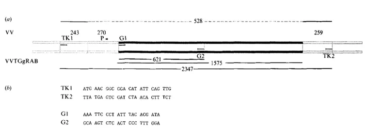
Synthesis and purification of oligonueleotides. Two pairs of oligonucleotide primers chosen following sequence analysis of the VV TK gene (Weir & Moss, 1983) and the rabies virus glycoprotein gene (Anilionis et al., 1981) were synthesized on an automatic D N A synthesizer and purified by PAGE (Fig. 1). The first set (TK1 and TK2), with 24 bp of the TK gene of VV flanking the insert for the rabies virus glycoprotein, allowed the amplification of a 528 bp sequence of VV DNA and a 2347 bp sequence of VVTGgRAB (Fig. 2). The other set (G 1 and G2), with 21 bp of the rabies virus glycoprotein gene was used for the amplification of a 621 bp sequence of VVTGgRAB DNA.
DNA sequence amplification. Ten ~tg of D N A was amplified through two series of 35 PCR cycles. The reaction mixture consisted of l0 [.tl of a 10-fold concentrated reaction buffer containing 500 mM-KCI, 100 mM-Tris-HCl pH 8, 15 mM-MgCl2, 0.1% gelatin, 200 ~tM of each of the four deoxynucleoside triphosphates (dATP, dCTP, dTTP, dGTP), 0.4 ~tl of Taq polymerase (0-5 units/ml), 1 p.g of each primer, water and DNA, to a total volume of 100 ~tl. The reaction mixture was covered with 50 p.l of mineral oil, to prevent evaporation. The amplification reaction was performed in a D N A thermal cycler (Perkin-Elmer/Ce- tus). The following conditions were found to be optimal: samples were first heated to 93 °C for 2-5 min, then submitted to 35 PCR cycles of 1 min at 93 °C to denature the DNA, 2 min at 55 °C to allow annealing of the primers, and 5 to l0 rain at 72 °C to allow primer extension. A final extension step was then performed for 10 min at 72 °C. Ten ~tl samples were then submitted to a second identical round of 35 PCR cycles.
Detection of amplified products. The amplified products were detected by direct gel analysis (for controls) and by dot blot hybridization assay using labelled oligonucleotide probes (for controls and samples). For direct analysis, 10 ~tl of the reaction mixture was subjected to electrophoresis on a 1 to 1-5~ agarose gel and the D N A
was observed by u.v. fluorescence after staining with ethidium
bromide. Mr markers were included in each gel. For dot blot analysis,
30 Izl of the final product was adjusted to 0-4 M-NaOH, 0.25 mM-EDTA
in a 200 btl volume, boiled for 2 min at 95 °C, cooled on ice, and
immediately applied to a nylon filter membrane (GeneScreen Plus) by
vacuum filtration using a Manifold I (Schleicher & Schiill). Wells were
rinsed with 20 x SSPE (1 x SSPE is 0-18 M-NaC1, 10 mM-Na2HPO4-
NaH2PO4, 1 mM-EDTA, pH 7-4), and the filters were then dried at
room temperature.
Oligonucleotide probes were labelled at their 5' end by phosphoryla-
tion with 3zPqabelled ATP (sp. act. > 5000 Ci/mmol; Amersham) and
T4 polynucleotide kinase (Amersham). The oligomers were then
purified on a 15% polyacrylamide/7 M-urea gel, which allows their
separation from their precursors. The nylon membrane was prehybri-
dized in a solution of 5 x SSPE, 5 x Denhardt's solution, 0.5% SDS
for 1 h at the hybridization temperature (TK1, 29 °C; TK2, 33 °C)
Approximately 100 ng of probe (2 x l06 c.p.m./ml) was then added and
hybridization was allowed for 1 night at the same temperature. The
filters were then rinsed twice in 2xSSPE, 0.1%SDS at room
temperature, and once for 10 min in 5 x SSPE containing 0-1% SDS at
Tm - 1 °C. Finally, the filters were subjected to autoradiography at
-70 °C (Fuji XR) for 4 h or 18 h.
Results
Virus isolation (experiments 1 and 3)
As shown in Table 1, small amounts of virus (102 to 104.3
T C I D s o / m l ) were recovered from the tonsils of five of
the seven foxes inoculated with V V T G g R A B , during the
first 48 h only. In group B, VV was detected in the tonsils
of the animal sacrificed after 24 h. Virus was not
detected ( < 10 ~'5 TCID50/ml) in any other organ, serum
or faeces of the vaccinated foxes, nor in any organ of the
control animal. V V T G g R A B could not be isolated from
foxes inoculated with viral stock recovered after one fox
passage nor in foxes inoculated with the same viral stock
amplified in cell culture.
Indirect immunofluorescence (experiment 1)
As shown in Table 1, the indirect immunofluorescence
test carried out with anti-VV rabbit polyclonal serum
confirmed the presence o f the virus in the tonsils o f four
of the five foxes from which V V T G g R A B had been
isolated and in the fox from which VV was isolated. The
indirect immunofluorescence test carried out with the
anti-rabies glycoprotein monoclonal antibody confirmed
the presence of virus in the tonsils of three of the five
foxes from which the virus was re-isolated. I m m u n o -
fluorescence was however too diffuse to allow precise
localization of the virus in the tonsil.
PCR results (experiment 2)
Determination of the sensitivity of the method
Negative controls consisted of D N A f r o m an uninfected
fox. Positive controls were p r e p a r e d with several
dilutions of V V T G g R A B or VV in normal fox D N A ; the
dilutions ranged f r o m approximately one infectious
particle per cell to one infectious particle per 106 cells.
T K 1 and T K 2 oligonucleotides were used for the
detection of VV and several combinations of oligo-
nucleotide primers ( T K 1 - T K 2 , G 1 - G 2 , T K 1 - G 2 , G 1 -
TK2), (Fig.
1) were used for the detection of
V V T G g R A B .
For each virus 10 ~1 of the amplified product of the
10 -2 dilution (corresponding to 1 infectious particle/100
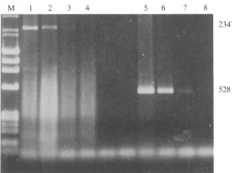
cells) was detected by direct gel analysis (Fig. 2). F o r
each virus 30 ~tl of the reaction mixture submitted to
two series of 35 P C R cycles were detected by dot blot
hybridization, up to and including the 10 -6 dilution (1
infectious particle/million cells) (Fig. 3). N o detectable
amplification was observed in different samples of
normal fox genomic D N A .
Detection of VVTGgRAB in DNA of vaccinated foxes
As shown in Table 2, using T K 1 - T K 2 as p r i m e r s and G1
as the probe, V V T G g R A B was detected in the tonsils of
two out of two inoculated foxes after 24 h and three of
three foxes after 48 h, in the buccal mucosa of one of two
foxes after 24 h and two of three foxes after 48 h and in
the soft palate of one of two foxes after 24 h and one of
three foxes after 48 h. N o virus was detected from any
other organ nor from any of the organs of the control fox.
Detection of VV in DNA of vaccinated foxes
After verification of the specificity of the amplified
product in controls, we used T K I and T K 2 as primers
and T K 1 as probe. VV was detected in the tonsils of the
inoculated fox tested after 48 h, and in the buccal mucosa
of two other foxes, one tested after 24 h and the other
after 48 h. Virus was not detected in any other organ.
Discussion
M a n y experiments have been carried out to demonstrate
the efficacy and safety of the v a c c i n i a - r a b i e s glycopro-
tein r e c o m b i n a n t virus for vaccination against rabies.
This vaccine could offer a suitable alternative to the use
of attenuated strains of rabies virus used until now and
has the advantage of good thermostability under field
conditions. T h e parental C o p e n h a g e n strain of VV was
associated with a limited n u m b e r of h u m a n encephalitis
complications in immunosuppressed people (Lane
et al.,
1969). H o w e v e r previous experiments h a v e demon-
strated that the introduction of a foreign gene into the
T K gene of VV greatly reduces the virulence of this virus,
even of neurovirulent strains (Buller
et al.,
1985).
Furthermore no lesion was ever observed in immunosup-
4 0
I. Thomas and others
(a) VV 243 270 T K I P . G I :::::::::::::::::::::::::::::::::::::::::::::::::::::::::::::::::::::::::::::::::::::::::::::::: I VVTGgRAB . . . 528 . . . 259 G2 621 1575 2347 [ 2= ::z z:z = ~.~¢.-'. zz.~a~:..'~.z 7JT;='_=.';..'7.,z..'=: z~ TK2(b) T K I A T G AAC GGC GGA CAT A T T CAG TTG
T K 2 T T A T G A GTC GAT CTA A C A CTT TCT
G1 A A A TTC CCT A T T TAC A C G A T A
G 2 GCA ACT CTC ACT CCC TTT G G A
Fig. 1. (a) The c D N A of the rabies glycoprotein gene under the control of a promoter (P) is inserted within the T K gene of the VV genome. Primers TK1 and TK2 are located on the TK gene and primers G1 and G2 are located on the glycoprotein gene. Numbers indicate the size (bp) of the different segments. (b) Sequence of synthetic oligonucleotide primers and complementary oligonucleotide probes.
T a b l e 1.
Detection by viral isolation and indirect immunofluorescence of VVTGgRAB and VV in tonsils of foxes vaccinated by
the oral route
Treatment
V V T G g R A B VV PBS
(Group A) (Group B) (Control)
Fox no. 1 2 3 4 6 7 9 5 8 10
Time of organ collection (h) 12 12 24 24 48 48 96 24 Virus titre* - 102 104 102g 1043 102.8 - 104.3 Detection of VV antigens by - - + + + + - +
indirect immunofluorescence$
Detection of rabies glycoprotein by - -- + - + + - -- indirect immunofluorescence
48 96
* Virus titres are expressed as TCIDs0/ml. t ( + ) , Viral antigen detected; ( - ) , not detected.
T a b l e 2.
Detection by PCR of VVTGgRAB or VV in orally vaccinated foxes
TreatmentV V T G g R A B VV PBS
(Group A) (Group B) (Control)
Fox no. 11 12 13 14 15 16 t7 18
Time of organ collection (h) 24 24 48 48 48 24 48 24
Tonsils + * + + + + - t + Buccal mucosa + - + - + + + Soft palate + - + . . . . Brain . . . . Spleen . . . . Parotid glands . . . . Maxillary glands . . . . Liver . . . .
Retropharyngeal lymph nodes . . . . Submaxillary lymph nodes . . . . Mesenteric lymph nodes . . . .
Blood . . . .
Faeces . . . .
* ( + ) , Viral D N A detected. t ( - ) , Viral D N A not detected.
528
Fig. 2. Electrophoretic gel analysis of PCR products of different
dilutions of VVTGgRAB (lane 1, 2, 3, 4) or VV (lane 5, 6, 7, 8) in
normal fox DNA. TK1 and TK2 were used as primers. Lane M, size
markers (bp)
(HindlII-digested 2 DNA, HaelII-digested q~X174 RF
DNA); lanes 1 and 5, 1 infectious particle/cell; lanes 2 and 6, 10 -1
infectious particle/cell; lanes 3 and 7, 10 -2 infectious particle/cell;
lanes 4 and 8, 10 -3 infectious particle/cell.
1 2 3 4 5 6 7
Fig. 3. Dot blot autoradiographofPCR products of different dilutions
of VVTGgRAB in normal fox DNA. Two series of 35 cycles were
performed using TK1 and G2 as primers and G1 as probe. Lane 1,
negative control; lane 2, 10 -1 infectious particle/cell; lane 3, 10 -2
infectious particle/cell; lane 4, 10 -3 infectious particle/cell; lane 5, 10 -4
infectious particle/cell; lane 6, 10 -2 infectious particle/cell; lane 7, 10 -6
infectious particle/cell.
pressed nude mice inoculated with the V V T G g R A B by
different routes (P. Desmettre, unpublished results).
Nevertheless in preparation for a large scale release of
the recombinant it is necessary to make a thorough study
of the pathogenesis of this vaccine and to improve
methods for detecting the virus.
Using different techniques (virus isolation, indirect
immunofluorescence and the very sensitive P C R
method), virus ( V V T G g R A B or VV) was detected
during the first 48 h following vaccination by the oral
route, but only in the tonsils, buccal mucosa and soft
palate. Similar results have been obtained by others in
raccoons (tonsils, parotid or submandibular lymph
nodes, buccal mucosa) using virus isolation (Rupprecht
et aL, 1988). The virus titres were very low in all cases,
suggesting that if there is any viral multiplication, it must
take place locally and at a very low level.
To ascertain our P C R results, the sensitivity of the
method was analysed. The limit of detection of the test in
our experimental conditions was one infectious particle
per 106 cells. On the other hand, as the organ samples
contained 10 p.g of D N A , the D N A content of about
1.5 x 10⁶ cells, any negative result would indicate the
absence of virus particles in the sample.
Experiments carried out with V V T G g R A B adminis-
tered into foxes' stomachs and to skunks intraduodenally
(Tolson
et al., 1987, 1988) showed that a reduced rate of
seroconversion and lower rabies virus-neutralizing anti-
body titres were obtained by those routes. Furthermore,
results of other experiments demonstrate that tonsillec-
tomy of the foxes does not affect the protection conferred
by vaccination with V V T G g R A B (I. Thomas
et al.,
unpublished results). All these data suggest that orally
administered recombinant virus multiplies at a low level
within the oral cavity, in the tonsils and also in other
tissues. Viraemia was never observed on days 0, 2, 3, 4, 5,
6, 7, 8 and 14 in foxes inoculated with V V T G g R A B or
VV (I. Thomas
et al., unpublished results). N o virus
could be detected in salivary glands (parotid glands and
maxillary glands) of foxes; the risk of transmission by
saliva from one animal to another is therefore very low.
This explains why transmission of the virus from
vaccinated animals to unvaccinated controls was shown
not to occur in foxes (one case) (Blancou
et al., 1986;
Tolson
et al., 1988; Brochier et al., 1988c), in cats and
dogs (Blancou
et al., 1986) and in ferrets (Brochier et al.,
1989). However in raccoons, some unvaccinated animals
held in contact with vaccinated ones had rabies virus-
neutralizing antibodies and resisted challenge (Rup-
precht
et al., 1988).
In our experiments no difference was observed in the
replication sites of the recombinant virus as compared to
the parental strain of VV, demonstrating that recombi-
nation does not modify the tropism of VV. N o n e of these
viruses were detected in the brain, suggesting that the
recombinant has no predilection for nerve cells. These
results were consistent with those of other studies
reporting the absence of detectable cytological abnorma-
lities in cerebrospinal fluid from raccoons orally vaccina-
ted with the recombinant virus (Hanlon
et al., 1989).
It is very important also to verify that the virus will not
adapt to the fox and will not acquire some pathogenicity.
In our experiments, virus was not detected in foxes
inoculated with virus isolated after one passage, nor in
others inoculated with the same virus after cell culture
amplification. However, further experiments should be
carried out with a higher number of animals to avoid any
misinterpretation due to individual variations. In any
case, neither lesions nor clinical signs were detected in
animals vaccinated with these viruses during a 28-day
observation period.
42
I. Thomas and others
The fact that the recombinant virus multiplies only in
restricted sites minimizes the eventual risk of recombina-
tion with other orthopoxviruses, bearing in mind that
there is no known reservoir of VV in the wild.
All these results represent additional arguments in
favour of the use of the recombinant virus in the field.
However even if risks appear to be very unlikely it is
important to be cautious. In this respect, the PCR
technique will be a useful tool for safety controls in field
trials because of its very high sensitivity and specificity.
Even in samples with a very low viral titre, the presence
of the virus was detected. This technique should
therefore permit the detection of the recombinant virus
in any animal collected in a vaccinated area, suspected of
carrying a pox lesion.
The authors wish to thank M. Georges and G. Vassart for the helpful suggestions and for their hospitality and M. Lafon for donating the anti-rabies virus glycoprotein monoclonal antibody. This work was supported by a grant from the Commission of the European Communities (BAP no. 0368) and by the Ministry of R6gion wallonne for the Environment (Convention 88/31279).
References
ANILIONm, A., WUN~R, W. H. & CtmTIS, P. (1981). Structure of the glycoprotein gene of rabies virus. Nature, London 294, 275-278. BLANCOU, J., KIENY, M. P., LATHE, R., LECOCQ, J. P., PASTORET, P.-P.,
SOULEBOT, J. P. & DESMETTRE, P. (1986). Oral vaccination of the fox against rabies using a live recombinant vaccinia virus. Nature, London 322, 373-375.
BLANCOU, J., ARTOIS, M., BROCHIER, B., THOMAS, I., PASTORET, P.-P., DESMETrRE, P., LANGUET, B. & KIENY, M. P. (1989). Innocuit6 et efficacit6 du virus recombinant vaccine-rage administr6 par vole orale chez le renard, le chien et le chat Annales de Recherches
V$t$rinaires 20, 195-204.
BROCHIER, B., THOMAS, I., IOKEM, A., GINTER, A., KALPEP, S, J., PAQUOT, A., COSTY, F. & PASTORET, P.-P. (1988a). A field trial in Belgium to control fox rabies by oral immunization. Veterinary Record 123, 618-621.
BROCHIER, B., LANGUET, B., BLANCOU, J., KIENY, i . P., LECOCQ, J. P., COSTY, F., DESMETI'RE, P. & PASTORET, P.-P. (1988b). Use of a recombinant vaccinia-rabies virus for oral vaccination of fox cubs (Vulpes vulpes, L.) against rabies. Veterinary Microbiology 18, 103- 108.
BROCHIER, B., BLANCOU, J., THOMAS, I., LANGUET, B., ARTOLS, M., KIENY, M. P., LECOCQ, J. P., COSTY, F., DESMETrRE, P. & PASTORET, P.-P. (1989). Use of recombinant vaccinia-rabies glycoprotein virus for oral vaccination of wildlife against rabies: innocuity to several non-target bait consuming species. Journal of Wildlife Disease (in press).
BULLER, R. M. L., SMITH, G. L., CREMER, K., NOTIONS, A. L. & Moss, B. (1985). Decreased virulence of recombinant vaccinia virus expression vectors is associated with a thymidine kinase-negative phenotype. Nature, London 31"/, 813.
HANLON, C. A., ZIEMERS, E. L., HAMIR, A. N. & RUPPRECHT, C. E. (1989). Cerebrospinal fluid analysis of rabies and vaccinia-rabies glycoprotein recombinant in orally vaccinated raccoons (Procyon lotor). American Journal of Veterinary Research 50, 364-367. K~BER, G. (1931). Beitrag zur kollectiven Behandlung Pharmakolo-
gischer Reiherversuche. Archive ff*r experimentelle Pathologie und Pharmakologie 162, 480--483.
KIENY, M. P., LATHE, R., DRILLIEN, R., SPEHNER, D., SKORY, S., SCHMITT, D., WIKTOR, T., KOPROWSKI, H. & LECOCQ, J. P. (1984).
Expression of rabies virus glycoprotein from a recombinant vaccinia virus. Nature, London 312, 163-166.
LANE, J. M., RUBEN, F. L., NEFF, J. M. & MmLARD, J. D. (1969). Complications of smallpox vaccination, 1968: results of ten statewide surveys. Journal of Infectious Diseases 122, 303-309. LEBLOIS, H. & FLAMAND, A. (1988). Studies on pathogenicity in mice of
rabies virus strains used for oral vaccination of foxes in Europe. In Vaccination to Control Rabies in Foxes, pp. 101-104. Edited by P.-P. Pastoret, B. Brochier, I. Thomas & J. Blancou. Brussels: Office for Official Publications of the European Communities.
MANIATIS, T., FRITSCH, E. F. & SAMBROOK, J. (1982). Molecular Cloning: A Laboratory Manual. New York: Cold Spring Harbor Laboratory.
PASTORET, P.-P., FRISCH, R., BLANCOU, J., BROCHIER, B., WOLFF, E., ROBOLY, O. & SCHNEIDER, L. G. (1987). Campagne internationale de vaccination antirabique du renard par vole orale men6e au grand- duch~ de Luxembourg, en Belgique et en France. Annales de M$decine Vbt~rinaire 131, 441-447.
PASTORET, P.-P., BROCI-IIER, B., LANGUET, B., THOMAS, I., PAQUOT, A., BAUDUIN, B., KIENY, M. P., LECOCQ, J. P., DE BRUYN, J., COSTY, F., ANTOINE, H. & DESMETTRE, P. (1988). First field trim of fox vaccination against rabies using a vaccinia-rabies recombinant virus. Veterinary Record 123, 481-483.
RUPPRECHT, C. E., WIKTOR, T. J., JOHNSTON, D. H., HAMIR, A. N., DIE'rzscItOLD, B., WUNNER, W. H., GLICKMAN, L. T. & KOPROWSKI, H. (1986). Oral immunization and protection of raccoons (Procyon lotor) with a vaccinia-rabies glycoprotein recombinant virus vaccine. Proceedings of the National Academy of Sciences, U.S.A. 83, 7947- 7950.
RUPPRECHT, C. E., HAMIR, A. N., JOHNSTON, D. H. & KOFROWSKI, H. (1988). Efficacy of a vaccinia-rabies glycoprotein recombinant virus vaccine in raccoons (Procyon lotor). Research Towards Rabies Prevention, Reviews of Infectious Disease 10, $803-$808.
SAIKI, R. K., SCHARF, S., FALOONA, F., MULLIS, K. B., HORN, G. T., ERLICH, H. A. & ARNl-mIra, N. (1985). Enzymatic amplification of fl- globin genomic sequence and restriction site analysis for diagnoses of sickle cell anemia. Science 230, 1350-1354.
SCHNEIDER, L. G. • COX, J. H. (1983). Ein Feldversuch zur oralen immunisierung von Ffichsen gegen die Tollwut in der Bundesrepub- Ilk Deutschland. I. Unsch~dlichkeit, Wirksamkeit und Stabilit/it der Vakzine SAD B19. Tieri~rztliche Umschau 38, 315-324.
SORIA BALTAZAR, R., BLANCOU, J. & ARTOLS, M. (1987). R~sultats de l'administration par vole orale au mouton de deux vaccins contenant un virus de la rage modifi6 (SAD B19) ou un recombinant du virus de la vaccine et de la rage (187XP). Annales de M~decine V~t~rinaire 131, 481-486.
STECK, F., WANDELER, A. I., BICHSEL, P., CArL S. & SCHNEIDER, L. (1982). Oral immunization of foxes against rabies. A field study. Zentralblatt J~r Veterini~rmedizin 29, 372-396.
TOt,SON, N. D., CHARLTON, K. M., STEWART, R. B., CAMPBELL, J. B. & WIKTOR, T. J. 0987). Immune response in skunks to a vaccinia virus recombinant expressing the rabies virus glycoprotein. Canadian Journal of Veterinary Research 51, 363-366.
TOLSON, N. D., CHARLTON, K. M., CASEY, G. A., KNOWLES, M. K., RUPPRECHT, C. E., LAWSON, K. F. & CAMPBELL, J. B. (1988). Immunization of foxes against rabies with a vaccinia recombinant virus expressing the rabies glycoprotein. Archives of Virology 102, 297-301.
WANDELER, A., WACHENDORFER, G., FORSTER, U., KREKEL, H., MULLER, J. & STECK, F. (1972). Rabies in wild carnivores in Central Europe. II. Virological and serological examinations. ZentralblattJ~r
Veterini~rmedizin B21, 757-764.
WEre, J. P. & Moss, B. (t983). Nuclootide sequence of vaccinia virus TK gene. Journal of Virology 46, 530-537.
WmTOR, T. J., KIEI~Y, M. P. & LATHE, R. (1988). New generation of rabies vaccine. Vaccinia rabies glycoprotein recombinant virus. In Applied Virology Research. Volwne !: New Vaccines and Chemo- therapy, pp. 69-89. Edited by E. Kurstak, R. G. Marusyk, F. A. Murphy & M. H. V. Van Regenmortel. New York: Plenum Press.