Université de Montréal
Characterization of antimicrobial resistance in Aeromonas and
Vibrio isolated in Canada from fish and seafood
par
F. Carl Uhland
Département de pathologie et microbiologie
Faculté de médecine vétérinaire
Mémoire présenté à la Faculté de médecine vétérinaire
en vue de l’obtention du grade de
Maître ès Sciences (M.Sc.)
en sciences vétérinaires
option microbiologie
June 2011
Université de Montréal
Faculté de médecine vétérinaire
Ce mémoire intitulé
Characterization of antimicrobial resistance in Aeromonas and Vibrio
isolated in Canada from fish and seafood
présenté par
F.Carl Uhland
a été évalué par un jury composé des personnes suivantes
Philippe Fravalo, président-rapporteur
Marie Archambault, directrice de recherche
Josée Harel, codirectrice
Patrick Boerlin, codirecteur
Ann Letellier, membre du jury
Summary
Multiple studies have examined antimicrobial susceptibility in bacteria from aquacultured products microorganisms and their environment. However, no information is available concerning antimicrobial resistance in bacterial flora of fish and seafood available at the retail level in Canada. This is particularly true for the common aquatic commensals, Aeromonas and Vibrio, for which some species are known zoonotic pathogens. In the course of this study, the antimicrobial susceptibility among Aeromonas spp. and Vibrio spp. from domestic and imported fish and seafood was characterized. Aeromonas and Vibrio spp. isolates cultured from finfish and shrimp samples were evaluated for antimicrobial susceptibility by broth microdilution and/or disk diffusion techniques. Antimicrobial classes examined in detail included: tetracyclines (TET), folate pathway inhibitors (sulfadimethoxine-trimethoprim, SXT), florfenicol (FLO), and the quinolones (nalidixic acid / enrofloxacin, NA/ENO). Epidemiological cut-off values (ECV’s) for Aeromonas/Vibrio were established using normalized resistance interpretation (NRI) of disk diffusion data. Isolates were further examined by PCR and microarray for genes associated with their antimicrobial resistance. Of 201 Aeromonas and 185 Vibrio isolates, those classified as resistant were as follows, respectively: TET (n=24 and 10), FLO (n=1 and 0), SXT (n=2 and 8), NA (n=7 and 5) and ENO (n=5 and 0). Various combinations of tet(A), tet(B), tet(E), floR, sul1, sul2 and intI1 genes were detected with tet(E), intI1, sul2 and tet(B) being the most common. Vibrio and Aeromonas species isolated from retail fish and seafood sources can harbor a variety of resistance determinants, although their occurrence is not high. The risk represented by these resistances remains to be evaluated in view of the potential for bacterial infection and their role as a reservoir for antimicrobial resistance.
Key words: Aeromonas, Vibrio, antimicrobial resistance, normalised resistance interpretation (NRI), microarray, PCR, resistance genes
Résumé
Plusieurs études ont examiné la sensibilité aux antimicrobiens chez les bactéries d’organismes provenant de produits issus de l’aquaculture ou de leur environnement. Aucune information n’est cependant disponible concernant la résistance aux antimicrobiens dans les bactéries de la flore de poissons ou de fruits de mer vendus au détail au Canada. C’est particulièrement vrai en ce qui a trait aux bactéries des genres Aeromonas et Vibrio, dont certaines espèces sont des agents pathogènes zoonotiques connus. Au cours de cette étude, la sensibilité aux antimicrobiens d’isolats d’Aeromonas spp. et de Vibrio spp. provenant de poissons et de crevettes domestiques et importés a été mesurée à l’aide de techniques de micro dilution en bouillon et/ou de diffusion sur disque. Les classes d’antimicrobiens examinés comprenaient les tétracyclines (TET), les inhibiteurs de la voie des folates (sulfadiméthoxine-triméthoprime, SXT), le florfenicol (FLO), et les quinolones (acide nalidixique / enrofloxacine, NA/ENO). Des valeurs seuils épidémiologiques pour Aeromonas et Vibrio ont été établies en utilisant la méthode d’interprétation normalisée des données de résistance provenant de diffusion sur disque. La recherche de gènes de résistance associés au profil de résistance des isolats a été effectuée en utilisant des PCR s et des puces ADN. Le nombre d’isolats résistants aux divers antimicrobiens parmi les 201 isolats d’Aeromonas et les 185 isolats de Vibrio étaient respectivement les suivants: TET (n=24 et 10), FLO (n=1 et 0), SXT (n=2 et 8), NA (n=7 et 5) et ENO (n= 5 et 0). Diverses associations de gènes tet(A), tet(B), tet(E), floR, sul1, sul2, et intI1 ont été détectées, les gènes tet(E), intI1, sul2 et tet(B) étant les plus communs. Les espèces d’Aeromonas et de Vibrio isolées de poissons au détail et de fruits de mer peuvent héberger une variété de gènes de résistance, bien que peu fréquemment. Le risque que représente ces gènes de résistance reste à évaluer en considérant le potentiel infectieux des bactéries, l’utilisation des ces agents antimicrobiens pour le traitement des maladies en aquaculture et en médecine humaine et leur rôle en tant que réservoir de la résistance antimicrobienne.
Mots-clefs: Aeromonas, Vibrio, résistance aux antimicrobiens, normalised resistance interpretation (NRI), puce d’ADN, PCR, gènes de résistance
Table of contents
Identification of the jury ___________________________________________________ ii
Summary ___________________________________________________________ iii Résumé ____________________________________________________________iv Table of contents __________________________________________________________ viii List of Figures __________________________________________________________ ix List of abbreviations _______________________________________________________ x Acknowledgements _______________________________________________________ xii Introduction ____________________________________________________________ 1 Review of the literature ____________________________________________________ 3 Vibrio and Aeromonas: pathogens of humans and animals _____________________ 3 Importance of antimicrobial resistance ______________________________________ 4 Mechanisms of antimicrobial resistance _____________________________________ 6
Intrinsic resistance____________________________________________________________________ 6 Enzymatic degradation/inactivation ______________________________________________________ 6 Reduced accumulation (efflux pumps) ____________________________________________________ 7 Target modification and protection ______________________________________________________ 7
Resistance acquisition ___________________________________________________ 8 Mutation ___________________________________________________________________________ 8 Transformation ______________________________________________________________________ 8 Transduction ________________________________________________________________________ 9 Conjugation ________________________________________________________________________ 10 Transposition _______________________________________________________________________ 11 Integrative elements _________________________________________________________________ 12 Integrating conjugative elements (ICE’s) or Genomic islands (GEIs) __________________________ 13
Antimicrobial resistance in Aeromonas and Vibrio ___________________________ 14
β-lactams __________________________________________________________________________ 14 Tetracyclines _______________________________________________________________________ 18 Phenicols __________________________________________________________________________ 23 Sulfonamides and diaminopyrimidines __________________________________________________ 26
Aminoglycosides and aminocyclitols ____________________________________________________ 30 Quinolones ________________________________________________________________________ 34 Macrolides, lincosamides, streptogramins, ketolides and oxazolidinones (MLSKO antimicrobials) __ 37 Rifamycins ________________________________________________________________________ 38
Scientific article #1 _______________________________________________________ 40 General discussion _______________________________________________________ 61 Identification of Aeromonas and Vibrio spp. ________________________________ 61 Susceptibility testing of Aeromonas and Vibrio isolates ________________________ 64 Identification of resistance genes in Aeromonas and Vibrio ____________________ 66 Conclusion ___________________________________________________________ 69
References ___________________________________________________________ 70
Annex 1: β-lactamase classification ____________________________________ xiii
Annex 2: MIC’s of mobile β-lactamases reported in Aeromonas and Vibrioa (in
μg/ml) _____________________________________________________xiv
Annex 3: Tetracycline resistance genes identified in Aeromonas and Vibrio _____ xv
Annex 4: MIC’s associated with the tetracycline resistance genes of ___________ xv
Aeromonas spp. and Vibrioa spp. ________________________________ xv
Annex 5: Aeromonas MIC’s for naladixic acid, oxalinic acid and
ciprofloxacin/enrofloxacin, and associated QRDR mutations ________ xvii
Annex 6: Vibrio MIC’s for naladixic acid, oxalinic acid and
ciprofloxacin/enrofloxacin and associated QRDR mutations _______ xviii
Annex 7: Identification Scheme for Aeromonas and Vibrio _________________ xix
Annex 8: Sensitivity and specificity for Vitek2® identification of Aeromonas to the
genus and species level using an rpoB gold standard _______________ xix Annex 9: Sensitivity and specificity for Vitek2® identification of Vibrio to the genus
and species level using an rpoB gold standard _____________________ xx
Annex 10: Prevalence of resistance phenotypes in Aeromonas and Vibrio _______ xx
microarray and simple PCR ___________________________________ xxi
Annex 12: Antimicrobial resistance genes identified by microarray ____________ xxii
List of Tables
Review of the Literature
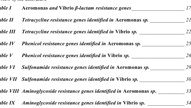
Table I Aeromonas and Vibrio β-lactam resistance genes ____________________ 17
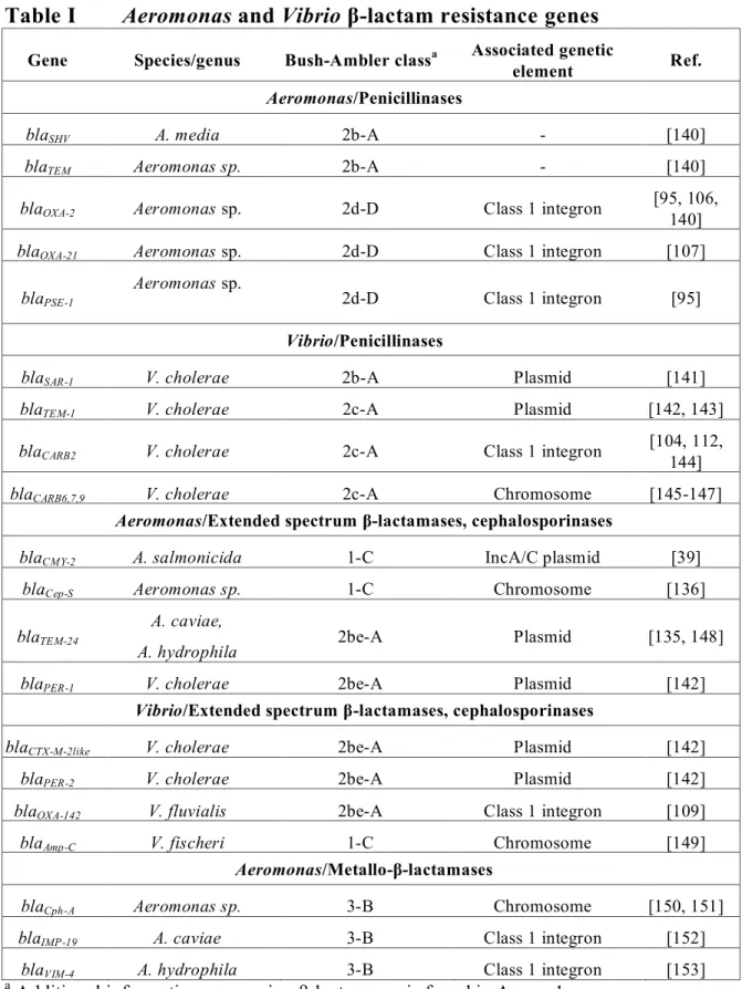
Table II Tetracycline resistance genes identified in Aeromonas sp. _____________ 21
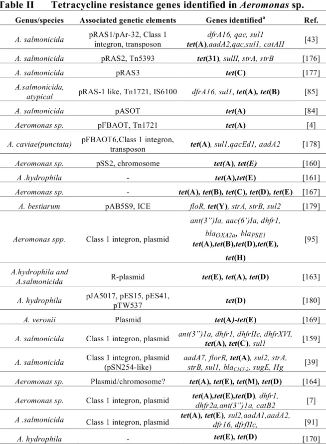
Table III Tetracycline resistance genes identified in Vibrio sp. _________________ 22
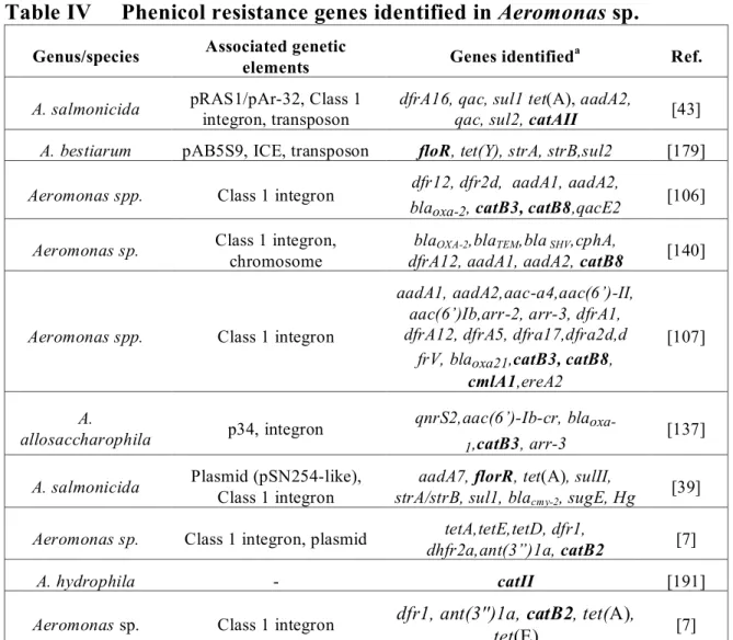
Table IV Phenicol resistance genes identified in Aeromonas sp. ________________ 25
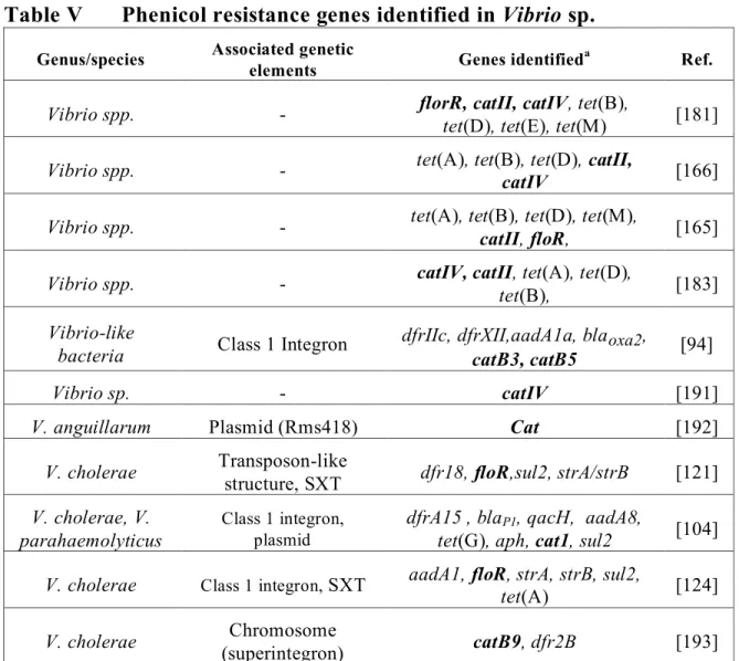
Table V Phenicol resistance genes identified in Vibrio sp. ____________________ 26
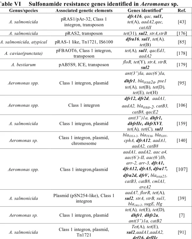
Table VI Sulfonamide resistance genes identified in Aeromonas sp. _____________ 29
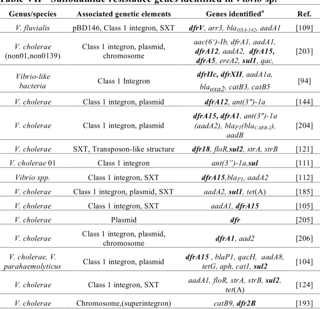
Table VII Sulfonamide resistance genes identified in Vibrio sp. _________________ 30 Table VIII Aminoglycoside resistance genes identified in Aeromonas sp. __________ 33
Table IX Aminoglycoside resistance genes identified in Vibrio sp. ______________ 34
Scientific Article #1
Table I: NRI-ECVs calculated for all Vibrio sp. and theV. parahaemolyticus
subpopulation _________________________________________________ 56
Scientific Article #2
Table I: Primers used for simple PCR analysis of resistance genes __________ xxxviii
Table II: Sensitivity and specificity for Vitek2® identification of Aeromonas to the genus and species level using an rpoB gold standard _______________ xxxix Table III: Differences in antimicrobial resistance gene detection noted between
microarray and simple PCR ______________________________________ xl Table IV: Antimicrobial resistance genes identified by microarray _______________ xli
TableV: Isolates with resistance phenotypes of four antimicrobial families and
List of Figures
Scientific Article #1Figure 1: Histogram of inhibition zone diameters of Vibrio sp. population __________ 54
Figure 2: Comparison of MIC and disc diffusion zones for Vibrio sp. isolates ________ 55
Figure 3: Comparison of the zone diameter distributions of Nalidixic acid____________ 56
(NA) and Enrofloxacin (ENO). ____________________________________________ 56
Scientific Article #2
Figure 1: Histogram of inhibition zone diameters of the Aeromonas sp. population
List of abbreviations
AAC : N-acetyltransferases ABC : ATP binding cassettes ANT : O-nucleotidyltransferase APH : O-phosphotransferases AMR : Antimicrobial resistance APW : Alcaline peptone water
ATCC : American type culture collection BP : Base pair
Clav : Clavulanic acid
CAT : Chloramphenicol acetyl transferase
CIPARS : Canadian Integrated Program for Antimicrobial Resistance Surveillance CLSI : Clinical and Laboratory Standards Institute
DHFR : Dihydrofolate reductase
DHPS : Dihydropteroic acid synthase enzyme DNA : Deoxyribonucleic acid
EBP: Epidemiologic breakpoint ECV : Epidemiological cut-off value EDTA : Ethylenediamine-tetra-acetic acid EPI : Efflux pump inhibitors
ENO : Enrofloxacin FFC : Florfenicol
ESBL : Extended spectrum β-lactamase GEI : Genomic islands
HGT : Horizontal gene transfer ICE : Integrating conjugative element IS : Insertion sequence element KBP : Kilobase pair
NA : Nalidixic acid
NRI : Normalised resistance interpretation
MATE : Multidrug and toxic compound extrusion MBL : Metallo-β-lactamase
MDF : Multi-drug resistant
MFS : Major faciliator superfamily MIC : Minimum inhibitory concentration
MLSKO antimicrobials : Macrolides, lincosamides, streptogramins, ketolides and oxazolidinones
mRNA : Messenger ribonucleic acid
NADPH : Nicotinamide adenine dinucleotide phosphate ORF : Open reading frame
PBP : Penicillin binding protein PCR : Polymerase chain reaction PMF : Protein motive force
QRDR : Quinolone resistance determining region RFLP : Restriction fragment length polymorphism RNA : Ribonucleic acid
rRNA : Ribosomal ribonucleic acid RND : Resistance nodulation cell division RPP : Ribosomal protection protein SMR : Small multidrug resistance SXT : Sulfadimethoxine-trimethoprim
SXT constin : Conjugative, self-transmissible, and integrating element TET : Tetracycline
TSI : Triple sugar iron agar
0129 : 2,4- diamino-6,7-di-isopropyl- pteridine phosphate XAT : Xenobiotic chloramphenicol acetyl transferase
Acknowledgements
Returning to school after being in the workforce for a number of years is a daunting undertaking, at least it was for myself. I could not have succeeded without the help of many people and friends around me. I sincerely wish to thank:
My wife Lucie, who without hesitation encouraged me to reorient my career and pursue a project that has interested me for quite some time. I must also recognize her patience and that of my children, Ann-Rose, Rébecca and Nicolas who lived with a distracted and sometimes absent husband and father, and who supported me throughout this adventure. Dr. Marie Archambault for accepting me as one of her students, for her enthusiasm for the project which I had proposed as well as her patience and guideance during the different phases of my studies. I must also thank Drs. Josée Harel and Patrick Boerlin, who accepted to accompany me as co-directors, and whose questions and constructive criticism helped develop my critical thinking and forced me to better myself.
Dr. Richard Reid-Smith and the Public Health Agency of Canada, who accepted to support me and allow me to pursue my interests.
Drs. Peter Smith and Göran Kronvall who piqued and justified my interest in AMR in aquaculture, and convinced me to think about things “outside of the box”.
Dr. Luc Masson, BRI for providing a modified version of the E. coli maxivirulence 1 microarray slide.
Jérôme del Castillo who generously invited me into his laboratory, providing me the infrastructure, comraderie and advice necessary to complete my thesis.
The numerous colleagues and technicians who were implicated near or far in this project, as without them, success would not have been possible; Nicol Janecko, Philippe Garneau, Andrea Desruisseau, Brent Avery, Gabhan Chalmers, Jessica Breton, Jessica Breznik, Annie Sinotte, Cindy Love-Tremblay, Audrey Charlebois, Michaël Ferland-Beaudry and Geneviève Pelletier-Jacques.
Introduction
Aquaculture is the fastest growing animal production industry in the world. It is predicted that in the year 2020, worldwide demand will surpass the wild fisheries supply by 15-20% [1]. This would indicate that more fish will be produced in the aquaculture setting if supply is to meet the demand. Not only will the demand for seafood surpass the wild fisheries capacity, but the annual personal consumption of seafood is also expected to increase from 16 kg today to 31 kg in 2030 [2]. This would predict an increased human exposure to fish, seafood and their bacterial flora during the production, marketing and consumption activities.
Aquaculture was in its infancy in Canada in the early 1980’s and now salmon growing operations on the East and West coasts of Canada compare favourably with other modern agriculture industries. Disease control is maintained with strict biosecurity measures as well as preventative measures such as vaccination, site fallowing/rotation and in certain cases, the judicious use of antimicrobials. Although antimicrobial exposure in the aquaculture setting is generally considered low as compared to other types of animal production, with an increase in consumption of aquacultured products as opposed to wild caught, there is a concomitant relative increase in the exposure to fish, seafood and their bacterial flora which possibly have had exposure to antimicrobials in an aquaculture setting. There are equally important differences in the regulations governing antimicrobial usage in agriculture (including aquaculture) depending upon the country examined. The availability of antimicrobials for use in aquaculture is extremely limited in North America where only three classes are available; tetracyclines, potentiated sulfa’s and phenicols. Although there are regulatory mechanisms to obtain antimicrobials without homologation, this is the exception rather than the rule. In other areas of the world such as in Asia, it is believed that antimicrobial usage is widespread and poorly regulated. Antimicrobial therapy in aquatic production is primarily administered orally whereas treatment by injection is reserved for highly valuable individuals such as broodstock. Therefore, in addition to the bacterial population causing disease, the bacterial flora on the fish and in the surrounding environment are equally exposed to the antimicrobials which evade consumption or their active metabolites secreted by the diseased organism.
Resistance to antimicrobials in bacteria derived from aquacultured animals and/or the aquatic environment have been reported in numerous publications in the scientific literature [3]. In addition, there have been studies that indicate transmission of resistance
determinant between terrestrial and aquatic environments does take place [4, 5]. Multiple laboratory studies have also demonstrated that resistance determinants can be transferred between aquatic bacteria which are low pathogen risks to humans to Enterobacteriaceae for example [6-8]. If this exchange occurs, at what frequency it occurs, and how it occurs in the environment or in association with seafood has yet to be elucidated.
It could reasonably be assumed that there is a certain level of risk of AMR exposure from aquatic bacteria pathogenic to humans, and from the transfer of resistance determinants from aquatic bacteria found on seafood and in the environment to bacteria pathogenic for humans, but the level of risk is unknown. The first step towards clarifying this question is examining and quantifying the presence of resistance elements in aquatic bacteria.
Aeromonas and Vibrio are among the most common bacterial genera found on fish, shellfish and the aquatic environment. They are recognized pathogens of aquatic animals, causing economically important aquaculture diseases, as well as zoonotic pathogens capable of causing severe disease in humans. As such, there exists the possibility of the exposure of these genera to antimicrobials, and perhaps the development of resistance. In addition, they are easily cultivated on usual bacteriological media which is important when categorizing bacteria via standardized phenotypic AMR susceptibility testing methods. In this study, the prevalence of Aeromonas and Vibrio in retail seafood will be examined, as well as the prevalence of AMR and the genetic basis for the observed AMR phenotypes.
Review of the literature
Vibrio and Aeromonas: pathogens of humans and animals
Aeromonas and Vibrio species are Gram-negative, mobile, facultative anaerobic bacteria that are present in aquatic systems worldwide. Until 1986, Aeromonas and Vibrio species were found in the family Vibrionaceae. Following analysis of molecular genetic evidence by Colwell et al. in 1986, Aeromonas species were subsequently transfered to a new family, the Aeromonadaceae [9]. There are currently 22 and 83 named species in the Aeromonas and Vibrio genera, respectively. Members of both genera are recognized as human and animal pathogens, and certain are zoonotic in nature. For details of the different species of this genera the following resources should be consulted [10, 11].
Vibrio species are predominantly halophilic and are therefore found more frequently in marine systems although certain species are reported in brackish and freshwater systems. They are among the most common bacteria isolated from marine molluscs and seafood [12, 13]. The Vibrio species anguillarum, ordalli, salmonicida, alginolyticus, and vulnificus are common pathogens of cultured marine fish causing septicaemia or focal chronic disease, the last two being zoonotic agents [14-17]. Vibrio vulnificus, Vibrio mimicus, Vibrio parahaemolyticus, Vibrio cholerae, are the species most often associated with disease in humans, and often in relation with consumption of raw or undercooked seafood, or in the case of V. cholerae, with fecal contamination of foodstuffs including water. The symptom most commonly encountered with Vibrio infections is gastroenteric upset, however in certain cases systemic disease may result, especially with V. vulnificus [18].
Aeromonas salmonicida as well as Aeromonas hydrophila and other motile Aeromonas species are also frequently found in fish, shellfish and other seafoods [19, 20]. This genus is associated with severe acute septicemic and chronic disease in aquacultured animal species including salmonids and non-salmonids such as carp and frogs. Aeromonas in humans can cause serious disease including extra-intestinal infections such as bacteraemia, meningitis, pulmonary and wound infections although food poisoning and associated gastroenteritis is probably the most common sequel to exposure [16, 21, 22].
Importance of antimicrobial resistance
Antimicrobial resistance (AMR) can be defined as the ability of microorganisms to resist the effect of an antibiotic or antimicrobial agent. This inefficacy of antimicrobials can be associated with intrinsic bacterial resistance, a mutation of the bacterial genome or by the acquisition of genetic material or a combination of these factors which will be discussed further on.
The presence of antimicrobial resistance in a zoonotic species of either genera causing severe systemic disease may decrease the chances of successful therapy [23-27]. Although the spectre of outright therapeutic failure and mortality is the most feared outcome of antimicrobial resistance development, the impact of antimicrobial resistance on humans and animals is difficult to evaluate. In human populations, increased levels of antimicrobial resistance have been associated with higher morbidity, mortality and increased hospitalization rates in the literature [28, 29]. This in turn may be explained by antimicrobial treatment failure or increased virulence of resistant bacterial strains, although the presence of pre-existing disease conditions and inadequate or delayed therapy may equally contibute [28, 30, 31]. Not all researchers agree however. Cosgrove (2006), Suneshine (2007) and Maragakis (2008) demonstrated an association between AMR and increases in mortality, morbidity and increased treatment costs whereas, conversely, Devasia et al. (2005) found no differences in treatment outcomes comparing patients infected with multi-drug-resistant ampC (MDR-AmpC) and pansusceptible Salmonella Newport [32]. The relationship between disease outcome and AMR is not clear cut. In addition to the health concerns, AMR represents a financial burden including direct costs such as hospitalisation for community acquired disease, increased hospital stay duration, prolonged therapy or changes to more costly medications, and repeat consulations [33]. Estimates of the monetary cost of AMR in the United States have been pegged from 1.3 to 5 billion dollars in the 1990’s [33, 34].
Antimicrobials are an important part of disease control in most animal production systems and are vital for cost effective production in treatment of episodic disease. They are used for infectious disease control/treatment, prevention of disease in high risk situations and for growth promotion [35]. Aquaculture differs in that the use of antimicrobials for growth promotion is not a current production practice. The use of antimicrobials in animal production, including aquaculture, has been fingered as an
important source of both resistant pathogenic and commensal bacteria [36, 37]. In aquaculture, there are multiple contributors to the development of resistance in aquatic bacteria which are likely similar to other agriculture production systems. Important factors include poor husbandry, lack of an accurate diagnosis followed by indiscriminate antimicrobial usage, repeated use of the same antibiotic, undiagnosed underlying disease processes, and inappropriate record keeping, [3].
The most direct impact of non-susceptible bacterial pathogens in aquatic production is treatment failure. The availability of a limited number of antimicrobials approved for aquatic species; florfenicol (Aquaflor®), potentiated sulfas (Romet-30® and Tribrissen®) and tetracyclines (Terramycin-Aqua®), at least in North America, exacerbates this situtation. Strains of Aeromonas salmonicida resistant to all of the aforementionned medications have been reported in fresh water aquaculture in Canada [38, 39]. The costs incurred by the presence of AMR in aquatic animal production are difficult to estimate although attempts have been made to model costs of disease in other species [40, 41]. In addition to the financial burden directly incurred through loss of stock via inefficacious treatments, the impact of medication costs, increased manhours needed for treatment activities (medication preparation and administration, removal of dead animals etc.), and fees charged by health professionals are also to be considered.
Likely the most hotly debated issue of importance concerning AMR in agriculture/aquaculture is the impact on humans where the presence of antimicrobial resistance in a zoonotic species causing severe systemic disease may decrease the chances of successful therapy [23-27]. The danger that AMR development in animal production presents to humans is twofold. The first is the direct transmission of resistant human pathogens (eg. E. coli, Aeromonas sp. etc.) from animal to human, and the second is the contamination/infection by resistant commensal bacteria during manipulation or consumption somewhere along the food chain with a subsequent resistance gene transmission to human pathogens. Recent surveillance data generated by the Canadian Integrated Program for Antimicrobial Resistance Surveillance ( CIPARS ) program has furthered evidence of antimicrobial use and transmission of resistant bacteria by demonstrating trend associations between cephalosporin usage and resistant Salmonella in retail chicken [42]. Multiple laboratory studies have demonstrated that resistance determinants can be transferred between aquatic bacteria, which are low pathogen risks to humans (ex. Aeromonas salmonicida to Enterobacteriaceae) [6, 7, 43]. If this exchange
occurs, at what frequency it occurs, and how it occurs in the environment or in association with seafood has yet to be elucidated. Studies by Rhodes (2000) and Furushita (2003) make the argument that transmission of resistance determinants between terrestrial and aquatic environments does take place [4, 5]. Further, Rhodes (2000) suggests that aquatic and terrestrial environments should be considered as one interactive unit [4]. If this is true, aquatic bacteria susceptibility will not only be affected by selective antimicrobial pressure on farm but from the availability of resistance determinants acquired from aquatic and terrestrial sources.
Mechanisms of antimicrobial resistance
To evade the effects of antimicrobials, bacteria have developed multiple strategies to neutralize their effects including avoidance, target modification and protection, inactivation, and active elimination of the offending molecules from the bacterial cytoplasm.
Intrinsic resistance
Bacteria which exhibit intrinsic or innate resistance would include those which lack or restrict access to targets for the antimicrobial in question. For example, anaerobic bacteria are insensitive to aminoglycosides because of they’re inability to successfully carry out oxygen dependent antimicrobial transport across the cytopolasmic membrane and into the bacterial cell [44]. Additionally, certain resistance determinants are permanent fixtures in the bacterial genome such as described in species of Aeromonas with chromosomally located β-lactamases [45].
Enzymatic degradation/inactivation
Modification or destruction of antimicrobials to render them inactive is a strategy used by many bacteria for several classes of antimicrobial drugs including the aminoglycosides, macrolides, β-lactams and phenicols. Acetyltransferases for example are inactivating enzymes which are common to aminoglyocosides and phenicol resistance. Transfer of an acetyl group from an acetyl co-enzyme A donor to the antibiotic affects amino acid interaction and inhibits binding at strategic sites in the ribosome [46, 47]. Another example is the three constitutive chromosomal β-lactamases groups which can be present in some Aeromonas species including a penicillinase/carbapenemase, a cephalosporinase and an oxacillinase [45, 48]. They have different substrate preferences,
but their mode of action is similar, where enzymatic hydrolysis of amide bond in the β-lactam ring is responsible for inactivation [49].
Reduced accumulation (efflux pumps)
Antimicrobial avoidance by decreasing cytoplasmic concentrations of offending chemicals is common among many bacterial species. Bacterial efflux pumps serve as efficient gate keepers allowing entry of ions and nutrients, permitting communication between bacteria and their environment and limiting accumulation of unwanted metabolites or other toxic products [50]. In fact, it is quite likely that the affected antimicrobials are not the intended pump substrate. The first antimicrobial resistance efflux pumps were discovered on plasmids coding for resistance to tetracycline in E. coli, and have now been identified in many bacterial genera [51, 52]. Some efflux pumps may be substrate specific as is seen in bacteria which are producers of antimicrobial compounds such as the actinomycetes [53]. They may equally act upon many different substrates such as the multidrug resistance pump (MDR) AheABC of A. hydrophila described by Henrould (2008), and more than one efflux pump may be present in the same bacterium [54, 55]. Five major efflux pump classes have been identified including ATP binding cassettes (ABC), resistance nodulation cell division (RND), major faciliator superfamily (MFS), small multidrug resistance (SMR) and multidrug and toxic compound extrusion (MATE ) [50, 56]. The ABC efflux pumps use ATP as the energy source to drive antimicrobial export, whereas MFS, SMR and RND efflux systems use the proton motive force (PMF) of the transmembrane electrochemical proton gradient to effectuate this action. MATE export pumps in contrast are considered H+ or Na+ coupled drug transporters [57]. Although differing in action, it remains that all functionally are considered to be capable of transporting a wide variety of substances including antimicrobials [56]. MFS, RND and SMR efflux pumps have been identified in Aeromonas species, whereas RND, MFS, SMR and MATE classes have been described in Vibrio sp. [50, 58].
Target modification and protection
Bacteria can evade antimicrobial action by modifiying or shielding antimicrobial targets to render them refractory to antimicrobial effects. This can arise from a mutational event or from horizontal gene transfer. Perhaps one of the best characterized is the resistance to sulfonamides via their interaction with folic acid metatoblism. Folic acid is an
important intermediate in many vital metabolic pathways in bacteria including the production of DNA and NADPH among others. Sulfonamides act as structural analogs of p-amino benzoic acid which competes for the dihydropteroic acid synthase enzyme (DHPS), thereby inhibiting the production of folic acid [59]. The acquisition of genes coding for DHPS enzymes with greater affinity for p-amino benzoic acid than sulfonamides renders the bacteria resistant [59]. This type of resistance has been reported in both Aeromonas and Vibrio genera.
Resistance acquisition
Acquired resistance refers to a modification or acquisition of genetic material which confers antimicrobial resistance to a bacterium. This may be associated with mutation of the bacterial genome conferring resistance or transfer and incorporation of resistance determinants via exchange of naked DNA, bacteriophage infection or mobile genetic structures such as plasmids, transposons and integrative conjugative elements (ICE’s) [60]. Collectively, these non-mutational mechanisms are referred to as horizontal gene transfer or HGT. Further, the maintenance of acquired genes is generally facilitated by environmental selective pressure where survival of the bacteria with the genetic modifications is favoured, as with chronic antibacterial use in hospitals or animal production for example.
Mutation
Spontaneous mutation of the bacterial genome occurs during normal bacterial growth due to copy errors in DNA replication. Mutation rates are variable, dependent upon the bacteria under consideration but are generally found to be 10-10 – 10-9 per base pair replicated [61]. Those bacteria harbouring a mutation which is beneficial to survival in a given environment are favoured. Therefore in a situation where a bacterium is exposed to an “antimicrobial environment”, those with an adaptive resistant mutation will survive, followed by clonal expansion of the bacterium within the bacterial population. Resistance to quinolones (ex. naladixic acid) is an example where point mutations principally in the gyrase (gyrA) or topoisomerase IV (parC) genes confer increased resistance to 1st and 2nd generation quinolones [48, 62]
Transformation
resulting in a different genotype [63]. Experimental or in-vitro transformation is used to artificially introduce DNA into bacteria following competence induction by chemical, physical or enzymatic treatments. Natural transformation is a function encoded in the bacterial genome and implies the survival of naked DNA in the environment, localisation and internalisation by a competent host followed by incorporation into the host genome. This type of acquisition occurs in both Gram-positive and Gram-negative species although they differ principally because of the differences in cellular barriers [64]. Natural transformation of plasmid DNA has been reported for Vibrio sp. with frequencies ranging from 0.3 to 3.1 x 10⁻⁸ transformants/recipient in sediment microcosms [65]. AMR transfer by transformation has not been described in Vibrio or Aeromonas, but is a well known mechanism first recognized in the Gram-positive species Streptococcus pneumonia [66, 67]. More recently, Neilsen (2010), describes transfer of erm (B), mef (E), mef (I), tet (M) and catQ genes between streptococcal isolates [68]. Although it has been argued that this form of genetic exchange is less important than conjugative events as they relate to HGT and AMR, transformation does not require conjugative elements, physical contact, or even a live DNA donor [69]. Therefore, its importance in complex bacterial communities such as biofilms may be underestimated.
Transduction
Bacterial infection with viral particles or bacteriophages may also occasion the transfer of genetic material between bacteria resulting in the incorporation of novel genetic material into the bacterial genome [70]. Bacteriophages are ubiquitous microorganisms endowed with a simple structure including the viral coat or capsid, an injection apparatus and the genetic material, variably DNA or RNA being simple or double stranded [71]. The viral infection of bacteria may have two general results: bacterial death (lytic phages) or incorporation of viral DNA into the bacterial genome (lysogenic phages). Cellular targets of bacteriophages are not clearly understood, however it is generally considered that they are specific to receptor types and likely to bacterial species and are not a probable genetic transfer mechanism between distantly related bacterial genera. However, it has been shown recently that transfer of virulence determinants between different bacterial genera via bacteriophages can occur with as high a frequency as intrageneric exchange [72]. Bacteriophage DNA capacity may effectively limit the efficacity of resistant determinant transfer. Bacteriophages are known to be important for the transmission of virulence
factors such as the case with the CTX prophage coding for cholera toxin in Vibrio cholerae. Lan (2009) describes prophages in Vibrio parahemolyticus containing putative bacterial DNA. They suggest that this bacteriophage may be involved in horizontal genetic exchange and may contribute to bacterial genetic diversity in this species [70]. Large bacteriophage capacity is estimated to be approximately 200 kb wherein resistance determinants such as tet(A) (1250 bp) and cat (1348 bp) would fit comfortably [71, 73-75]. Additionally, bacteriophages in the aquatic environment are considered the most abundant biological entity (up tot 2.5x10⁸ per ml) and presumably an important element for genetic diversification [76, 77]. Bacteriophages have been shown to transfer various AMR determinants in Pseudomonas aeruginosa [78] . Although there are numerous references to bacteriophages reported in the literature concerning Aeromonas and Vibrio species, there is no specific mention of AMR containing viral particles [79, 80].
Conjugation
Conjugation refers to the exhange of DNA between bacteria via pili, and is often erroneously referred to as “sexual reproduction”. Requirements for successful conjugation include the intimate (direct) contact of a gene donor and recipient, the gene targeted for transfer and the presence of the “machinery” necessary for the transfer of the genetic material. The most commonly recognized conjugative mechanism is plasmid mediated genetic exchange, first described in E. coli by Tatum and Lederberg in 1947 [81]. Plasmids are autonomously replicating, extrachromosomal circular or linear double stranded DNA fragments, with sizes ranging form 300 bp to 2400 kpb [82]. Minimally, a conjugative plasmid must contain transfer genes (tra), which code for the pilus assembly and associated transfer related proteins as well as a replicative starting point or origin of transfer (oriT) which is distinct from oriR which is necessary for intrabacterial replication [83]. This is followed by a transfer of one DNA strand to the recipient cell and the synthesis of the complementary strands in the donor and recipient cells. The transmission of resistance determinants associated with plasmids has been reported by many authors and transfer frequency in experiments between A. salmonicida and E. coli in the laboratory have been found to range between 10-1 – 10-9 [7, 39, 84, 85]. Upon examining plasmid transfer in a natural microenvironment involving salmon contaminated with resistant A. salmonicida and E. coli on a cutting board, a transfer frequency of 3 x 10⁻⁶ to 8 x 10⁻³ was noted [6]. Broad host range plasmids such as the IncU class possess shorter rigid pili which
lend to greater transfer efficiency, as much as 2,000 to 300,000 times faster on solid surfaces [43, 86, 87]. This is an important distinction when considering consumer risk originating from aquatic animals or their environment. Coexistence of different plasmids within the same bacterium is possible, however, two plasmids which share the same or similar types of plasmid transfer genes (considered as being of the same incompatibility group) are inhibitory to each other [83]. Certain types of plasmid incompatibility groups may be more commonly associated with specific bacterial genera. The plasmids of the incompatibility groups U and C are the most common in the aermonads, whereas group C plasmids are found principally in Vibrio species [88-90]. Plasmids from these incompatibility groups have a wide host range and have been associated with phenotypic resistance in these species.
Not all plasmids are capable of conjugative transfer. Smaller plasmids which do not contain the necessary 35 kb of transfer genes may piggyback on other mobile plasmids if they simply contain the appropriate origin of replication (oriT) recognized by the transfer machinery of the co-residing plasmid. Most early studies examining the genetic causes of antimicrobial resistance phenotypes concentrated on the absence or presence of mobile genetic elements which could confer AMR to sensitive bacterial strains. Aeromonas and Vibrio species are known to harbour a plethora of plasmids, ranging in size from 11 – 200 kbp, coding for resistance to various antibiotics [8].
Transposition
DNA elements which can “hop” or transpose from one location to another in bacteria are called transposons. They consist minimally of an open reading frame coding for a transposase enzyme (which controls their movement among different DNA locations) bounded by inverted repeats. These most simple transposons are called insertion sequence elements (IS) and may be only 1000 bp in length. IS’s may “hop” close enough together on chromosomes or plasmids to mobilize the intervening DNA forming “composite transposons” [83]. This interaction of related transposons may mobilize larger DNA elements, including AMR genes, within the bacterium. Non-composite transposons may also contain AMR genes as a part of the minimal transposable unit. They may also have the capacity to integrate AMR genes or “cassettes” due to the presence of integrons in their structure (see below). Transposons in association with plasmids and integrons appear to be involved in long-range AMR distribution. The truncated or complete transposon Tn1721
containing tet(A) for example, has been found on pRAS1 or pRAS1-like plasmids in A. salmonicida from different regions including Scotland, Norway and Japan [4, 43, 91]. Integrative elements
Integrons are genetic elements capable of capturing and incorporating genetic material into their DNA structure. They are not capable of self replication or transmission, but are rendered “mobile” by other mobile genetic platforms such as transposons and plasmids. The integrons have been categorized into classes depending on the type of integrase that is incorporated in their structure (intI1, intI2, intI3 and intI4). Classes 1-4 have been identified in Aeromonas and Vibrio sp. Class 1 integrons contain minimally a gene coding for an integrase enzyme (intI), a promoter and a recombination site (attI) in the 5’ conserved segment of the integron [92]. The components intI and attI act on gene cassettes which are comprised of a promoterless gene, often coding for antimicrobial resistance, and a recombination site (attC) recognizing the complementary site (attI) of the integron [93]. Integrated genes become a part of the integron structure and many genes cassettes may become associated with the integron. PCR amplification of the variable region of the integron permits the identification of the residing cassettes. Class 1 integrons, thought to be degenerate transposons, are the most common in Gram-negative bacteria. They usually contain sul1 and qacE∆ genes, coding for resistance to sulfonamides and quaternary ammonium compounds respectively, and an open reading frame (orf) of unknown function in their conserved 3’ segment, downstream of the attcI site. The presence of sul1, along with qacE∆ is often used as markers for class 1 integrons though the presence of these genes or truncated relatives is variable [94, 95]. The functional capacity of the integron to accumulate resistance gene cassettes as well as the inclusion of resistant genes in their basic structure (ex. sul1 in class 1 integrons) can lead to variable AMR phenotypes often exhibiting multiple drug resistance (MDR) in addition to diaminopyramidine resistance. Class 2 integrons have a similar structure and are found within transposons of the Tn7 family, commonly code for trimethoprim resistance (dhf) [96, 97]. Class 2 integrons appear to be more limited in resistance cassette arrangements than Class 1, likely due to a non-functional integrase gene [96]. The recently described Class 3 integrons have a similar organization to Class 1 and Class 2 integrons including the capacity to carry resistance determinants [98]. Their presence was signalled in Aeromonas sp. from the african aquaculture environment [95]. Finally, Class 4 integrons have been
identified on the small chromosome of Vibrio cholerae, containing 100’s of genes of mostly unknown function [99, 100]. Although the origin of integrons is unclear, these “Super-integrons” identified in Vibrio and also in Pseudomonas species are thought to be involved in their presence [101, 102]. Tapping into this vast depot of genetic material in the super-integrons may partly explain the seeming ease with which bacteria adjust to new antimicrobial molecules [103]. The presence of integrons is commonly reported in aquatic bacteria and clinical Aeromonas and Vibrio isolates [95, 104-112]. Class 1 integron carriage has been reported as varying from 3.6 – 73% [94, 95, 106, 107, 113]. These studies indicate that not only are Aeromonas and Vibrio species present in diverse environments, both as disease causing agents and commensals, but also that the various integron types are equally present which may contribute to resistance determinant mobility. This supports the argument that antimicrobial pressure in the environment is important for the maintenance and spread of AMR determinants in the bacterial population.
Integrating conjugative elements (ICE’s) or Genomic islands (GIs)
In the early 1980’s transposons were identified which not only had the capacity to integrate into the bacterial genome but also a conjugative capacity. ICE’s tend to be large DNA structures due to the accompanying tra genes necessary for conjugative transfer however they are incapable of autonomous replication like plasmids [114]. Their DNA excision and integration functions (xis and int genes) resemble more phages than true transposons, hence their name [115]. Recently, it has been argued they should be included in a larger overarching family of syntenic blocks of transferred DNA called Genomic islands or GI’s [83, 115]. The interbacterial transfer mechanism however, is similar to plasmids, where following excision from the bacterial DNA, a circular DNA intermediate where an oriT sequence is formed. (Hinerfield and Senghas in Snyder)[116]. ICEs can code for a variety of functions including virulence factors, antimicrobial resistance, and various metabolic functions [115]. The first ICE’s, were identified in Streptococcus faecalis and Bacteroides fragilis in the early 1980’s and were followed by several others [117, 118]. In 1996 Waldor et al. identified the first ICE in the Vibrionaceae, named SXT, in Vibrio cholerae [119]. Beaber described the SXT sequence as a melting pot of composite genes including those from bacteriophages, plasmids and other diverse sources [120]. The SXT element or “constin” (conjugative, self-transmissible, and integrating) has been found to be capable of antimicrobial resistance mediation, often in association with
integrons and transposons, and may also play a role in mobilizing plasmids carrying resistance determinants [121-123]. The resistance genes floR, strA, strB, sul2, dfrA18, and dfrA1 have been associated with presence of V. cholerae SXT constin [124].
Antimicrobial resistance in Aeromonas and Vibrio
β-lactams
The β-lactam class of antibiotics includes the penicillins and the cephalosporins, carbapenems and monobactams, all characterized by a central β-lactam ring. This class of antibiotics are structural analogs of the terminal acyl-D-alanyl-D-alanine of the bacterial cell wall peptidoglycan subunit. Penicillin bindings proteins (PBP’s) preferentially bind the β-lactams leading to an inhibition of cell wall synthesis and cell death [125]. Resistance to β-lactams is derived from multiple mechanisms including antimicrobial efflux, mutation of PBP’s (reducing penicillin binding affinity), β-lactamase activity, overexpression of intrinsic β-lactamase activity and decreased permeability [49].
PBP’s and their mutant variants are normally associated with resistance to β-lactams in Gram-positive bacteria and have not been reported to be a mechanism of importance for Aeromonas, Vibrio spp. or other Gram-negative bacteria associated with the presence of a plethora of β-lactamases in these species. Decreased outer membrane permeability in Gram-negative bacteria can be associated with low level resistance to the β-lactams as well as other antimicrobials. Oliver (2002) noted that alterations in porin expression in E. coli were linked to differences in β-lactam susceptibility, and furthermore Nikaido (1987) demonstrated a synergism between β-lactamase presence and decreased membrane permeability resulting in increased AMR in E. coli [126, 127]. Similar results have been shown for A. salmonicida where mutants with changes in outer membrane proteins demonstrated higher levels of AMR, including the β-lactams [128-130].
β-lactamase production by Gram-negative bacteria is the most important element when considering innate (chromosomal) and acquired resistance to β-lactams. Several classification schemes have been proposed for these enzymes based on molecular or functional characteristics, those of Ambler (1980) and Bush (1989, 1995) among the most commonly cited in the published literature [131-133]. The Ambler scheme proposes four classes (A-D) based on amino acid sequences where classes A, C and D are serine proteases and class B is a metallo-β-lactamase. The Bush scheme classifies β-lactamases as
to their preferred substrate and inhibiting molecules resulting in a more complex separation of enzymes [131, 133, 134]. These classifications schemes are contrasted in Annex 1.0, with mention of enzymes and substrates particular to each class.
Several enzymes classified loosely as pencillinases have been identified in Aeromonas and Vibrio. Aeromonads are generally considered resistant to penicillins due to the commonly identified chromosomally located β-lactamase enzymes, CeP-S, AMP-S and CPH-S. Vibrio species are not known to possess similar chromosomal enzymes. The presence of chromosomal β-lactamases in Aeromonas can be quite variable, where a penicillinase, a cephalosporinase and/or a metallo-β-lactamase may be present in different combinations depending on the species and strain examined [45, 135]. Walsh (1997) evaluated the prevalence of the three chromosomal β-lactamase genes in different Aeromonas species and found the blaAmp-S gene presence varied from 25 - 45% depending
on the species, with A. veronii having the highest prevalence at 45% [136]. The blaCep-S
gene was almost uniformly present in the A. caviae, A. veronii and A. hydrophila strains examined and its presence confers resistance to 1st generation cephalosporins [133, 136].
Although the chromosomal β-lactamases are considered immobile, others have been identified on mobile genetic structures making them more important when considering HGT of resistance elements. They are commonly found present as gene cassettes in class 1 integrons, with either a plasmidic or chromosomal location. In most cases they were associated with resistance determinants for other antimicrobials within the same integron or plasmid [106, 137].
The extended spectrum β-lactamases (ESBL’s) have become extremely important when considering β-lactam therapy due to their large spectrum of activity, and their genetic mobility. ESBL’s are generally recognized as being capable of hydrolyzing penicillins, 1st, 2nd, and 3rd generation cephalosporins (with the exception of cephamycins and carbapenems) and monobactams [138]. To date three ESBL’s have been identified in both the Aeromonas and Vibrio genera (see Table I below).
The metallo-β-lactamases (MBL) to date have only been recognized in Aeromonas species. They are categorized as Bush class 3/Ambler class B and can hydrolyze most classes of β-lactams. [134]. Wild chromosomal MBL + strains may be differentiated form acquired MBL’s due to their susceptibility to carbapenem [139]. Acquired MBL’s are commonly found as gene cassettes within class1 integrons and as such may be associated with other AMR determinants. European MBL’s strains often exhibit a truncated gene
cassette fused with the gene aacA4, resulting in resistance cross-selection between aminoglycosides and β-lactam [139]. The following table denotes the various determinants responsible for β-lactam resistance described for Aeromonas and Vibrio sp. Additional information concerning the β-lactamase genes described above as well as available associated MIC data can be found in Annex 1 and 2.
Table I
Aeromonas and Vibrio β-lactam resistance genes
Gene Species/genus Bush-Ambler classa Associated geneticelement Ref.
Aeromonas/Penicillinases
blaSHV A. media 2b-A - [140]
blaTEM Aeromonas sp. 2b-A - [140]
blaOXA-2 Aeromonas sp. 2d-D Class 1 integron [95, 106, 140]
blaOXA-21 Aeromonas sp. 2d-D Class 1 integron [107]
blaPSE-1
Aeromonas sp.
2d-D Class 1 integron [95]
Vibrio/Penicillinases
blaSAR-1 V. cholerae 2b-A Plasmid [141]
blaTEM-1 V. cholerae 2c-A Plasmid [142, 143]
blaCARB2 V. cholerae 2c-A Class 1 integron [104, 112,
144]
blaCARB6,7,9 V. cholerae 2c-A Chromosome [145-147]
Aeromonas/Extended spectrum β-lactamases, cephalosporinases
blaCMY-2 A. salmonicida 1-C IncA/C plasmid [39]
blaCep-S Aeromonas sp. 1-C Chromosome [136]
blaTEM-24
A. caviae,
A. hydrophila 2be-A Plasmid [135, 148]
blaPER-1 V. cholerae 2be-A Plasmid [142]
Vibrio/Extended spectrum β-lactamases, cephalosporinases
blaCTX-M-2like V. cholerae 2be-A Plasmid [142]
blaPER-2 V. cholerae 2be-A Plasmid [142]
blaOXA-142 V. fluvialis 2be-A Class 1 integron [109]
blaAmp-C V. fischeri 1-C Chromosome [149]
Aeromonas/Metallo-β-lactamases
blaCph-A Aeromonas sp. 3-B Chromosome [150, 151]
blaIMP-19 A. caviae 3-B Class 1 integron [152]
blaVIM-4 A. hydrophila 3-B Class 1 integron [153]
Tetracyclines
Tetracyclines are bacteriostatic antimicrobial compounds derived from Streptomyces spp. Their effects are mediated by the interaction with the 30S subunit of the ribosome, thereby preventing association with aminoacyltRNA and thus protein synthesis [154]. Resistance to the tetracycline family was recognized in Aeromonas species as early as 1959 by Snieszko and later attributed to a transferable R factor by Aoki in 1971 [155]. At least 41 tetracycline resistance genes have been characterized to date. They are divided into four classes including efflux proteins (26), ribosomal protection proteins (11), enzymatic modifying proteins (3), and one with an unknown mechanism [156, 157].
The first category, the efflux proteins, which are members of the major facilitator superfamily (MFS), reduce intrabacterial tetracycline concentrations via an energy dependent protonic exchange [158]. The efflux proteins associated with tetracycline resistance in Gram-negative bacteria are doted with two components, a repressor and an efflux protein, where in the presence of tetracycline, the efflux protein coding gene is derepressed allowing transcription [154]. The tet efflux genes are not found as gene cassettes within class 1 integrons, however they are commonly found in other mobile genetic structures such as on mobile plasmids and within transposons [73, 84, 157, 159]. The tet(A) gene is an example of a tet gene which has been associated with full or truncated Tn1721 transposon in A. salmonicida and other Aeromonas species [43, 85]. Early studies localized certain tet efflux genes on nonmobile plasmids or chromosomes due to apparent low experimental transfer frequencies [160, 161]. Subsequently, other authors have described these genes as highly prevalent on transferable plasmids, perhaps due to different or improved experimental methods [161-164]. Due to the plasmidic location of tet genes co-resistance to other antimicrobials is often reported. Co-resistance to sulfonamides ± trimethoprim as well as streptomycin is common, likely due to presence of integrons on tet determinant containing plasmids [7, 85, 95]. The tet efflux genes are commonly found singly, but they may also cohabit in the same bacteria either chromosomally or on the same mobile genetic element with other tet genes or resistance determinants to other antibiotic classes [154]. Determinant combinations in Aeromonas spp. have been reported in various studies, including tet(A)-tet(E), tet(B)-(D), tet(A)-tet(C), and tet(E)-tet(D) and even tet(A), tet(B), tet(D)/tet(H), and tet(E) [95, 159-161, 163, 164]. The presence of more than one tet determinant has also been signalled in Vibrio spp. including tet(A)/tet(B),
tet(A)/tet(B)/tet(D), tet(D)/tet(E) combinations [165, 166]. Therefore, one or combinations of tet determinants may be responsible for observed phenotypes in these genera.
Seven efflux tet genes have been identified in Aeromonas species to date including tet(A), tet(B), tet(C), tet(D), tet(E), tet(Y), tet(31). The tet(A) and tet(E) determinants are often quoted as being the most common although few studies have examined the prevalence of all tet determinants within a bacterial population. There have also been differences ascribed to variations in molecular techniques such as multiplex and single PCR protocols [163]. Prevalence for tet(A) in tetracycline resistant strains varies between 3 and 88% [4, 7, 84, 85, 91, 95, 160, 161, 163, 167], whereas prevalence of tet(E) genes among resistant isolates has been reported anywhere from 42% to 90% [95, 159-161, 163, 168-170]. The tet genes B, C and D appear to be present in lower numbers, ranging from as little as 1% to 28% in resistant bacteria [169, 170]. The lack of identifiable tet determinants in the literature is common [5, 7, 160, 161, 170]. In a study by Schmidt (2001) for example, only 30% (66/216) resistant isolates could be assigned a known tet determinant [7].
Several efflux pumps have also been identified in Vibrio species including tet(A), tet(B), tet(C), tet(D), tet(E), tet(G), and tet(35). The determinants tet(D) and tet(B) seem to be identified with a greater frequency in marine environments. The prevalence of tet(B) in Vibrio was 43% and 100% in studies conducted by Furushita (2003) and Kim (2007) respectively [5, 171]. The tet35 gene was identified in a tetracycline resistant strain of V. harveyi isolated from a prawn. Unlike previous tetracycline export pumps identified, tet35 appears to have a primary physiological role in Na+/H+ transport rather than antimicrobial export, is chromosomally located and results in inferior MIC’s in transconjugants as compared to tet(A) for example [172].
The second class of tet resistance genes are the ribosomal protection proteins (RPP’s). These act by permitting continual protein synthesis in the presence of tetracyclines. Although traditionally considered as Gram-positive tetracycline resistance genes these are now being identified in other cases including Gram-negative aquatic bacteria [171]. The determinant tet(M) is the only RPP identified among Vibrio and Aeromonas species [157]. It can be found in co-residence with other tet determinants such as tet(B), tet(D) and/or tet(E) [164, 165, 173]. The tet(M) determinant was the most common among tetracycline resistant Aeromonas isolates in an Australian study, where 7 out of 10 (70%) harboured the gene [164]. In Korean Vibrio isolates, the tet(M) and tet(B)
duo were found in 23 of 24 isolates and both tet determinants were located within a Tn10 tranposon. Similarly, Kim (2004) examined the tet resistance genes from 151 tetracycline resistant marine bacteria originating from Japan and Korea. The majority of the tet(M) positive Vibrio isolates were found to be associated with a transposon of the Tn1545-Tn1916 family and interestingly the Gram-positive bacteria, Lactococcus gerviae examined in the same study, carried a similar gene/transposon combination. The association of tet(M) resistance elements with transposable elements may be responsible for this ever-enlarging host range [154].
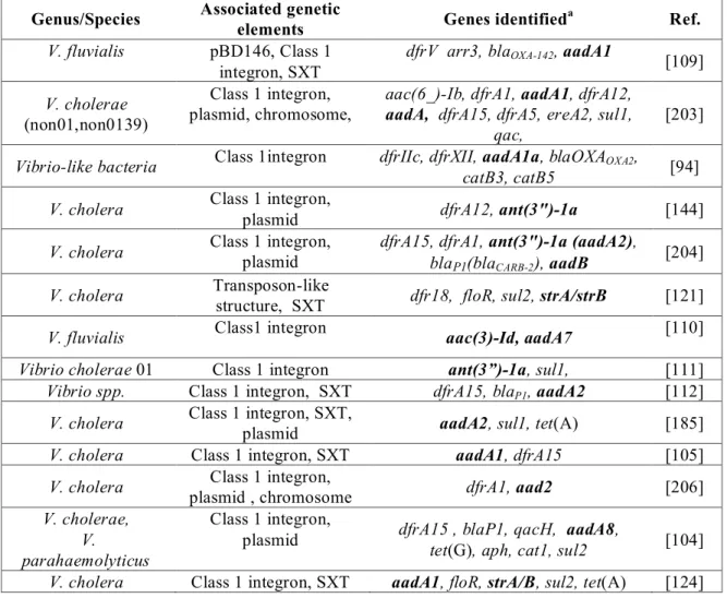
The last class of tet resistance elements are the enzymatic proteins which inhibit tetracycline activity by inactivation of the antimicrobial or by an acceleration of protein transcription bypassing the tet resistance mechanism and includes tet(34), tet(X) and tet(37) (refer to Annex 5) [174, 175]. They act via an NADPH-requiring oxidoreductase (tet(37) and tet(X)), or an enzyme similar to xanthine–guanine phosphoribosyl transferase from Vibrio cholerae (tet(34)). The tet(34) gene was identified in a Vibrio isolate grown from marine fish intestinal contents, and was associated with the relatively high MIC of 500μg/ml to oxytetracycline [174]. Additional information concerning the tetracycline resistance genes described above as well as available associated MIC data can be found in Tables II and III below and in Annex 3 and 4.
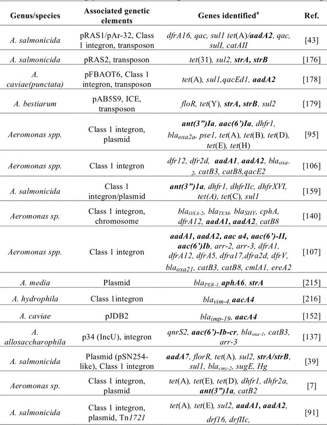
Table II
Tetracycline resistance genes identified in Aeromonas sp.
Genus/species Associated genetic elements Genes identifieda Ref.
A. salmonicida pRAS1/pAr-32, Class 1 integron, transposon tet(A),aadA2,qac,sul1, catAIIdfrA16, qac, sul1 [43]
A. salmonicida pRAS2, Tn5393 tet(31), sulII, strA, strB [176]
A. salmonicida pRAS3 tet(C) [177]
A.salmonicida,
atypical pRAS-1 like, Tn1721, IS6100 dfrA16, sul1, tet(A), tet(B) [85]
A. salmonicida pASOT tet(A) [84]
Aeromonas sp. pFBAOT, Tn1721 tet(A) [4]
A. caviae(punctata) pFBAOT6,Class 1 integron,
transposon tet(A), sul1,qacEd1, aadA2 [178]
Aeromonas sp. pSS2, chromosome tet(A), tet(E) [160]
A .hydrophila - tet(A),tet(E) [161]
Aeromonas sp. - tet(A), tet(B), tet(C), tet(D), tet(E) [167]
A. bestiarum pAB5S9, ICE floR, tet(Y), strA, strB, sul2 [179]
Aeromonas spp. Class 1 integron, plasmid
ant(3”)Ia, aac(6’)Ia, dhfr1, blaOXA2a
,
blaPSE1tet(A),tet(B),tet(D),tet(E), tet(H)
[95]
A.hydrophila and
A.salmonicida R-plasmid tet(E), tet(A), tet(D) [163]
A. hydrophila pJA5017, pES15, pES41,
pTW537 tet(D) [180]
A. veronii Plasmid tet(A)-tet(E) [169]
A. salmonicida Class 1 integron, plasmid ant(3”)1a, dhfr1, dhfrIIc, dhfrXVI, tet(A), tet(C), sul1 [159]
A. salmonicida Class 1 integron, plasmid
(pSN254-like)
aadA7, florR, tet(A), sul2, strA,
strB, sul1, blaCMY-2, sugE, Hg [39]
Aeromonas sp. Plasmid/chromosome? tet(A), tet(E), tet(M), tet(D) [164]
Aeromonas sp. Class 1 integron, plasmid tet(A),tet(E),tet(D), dhfr1,
dhfr2a,ant(3”)1a, catB2 [7]
A .salmonicida Class 1 integron, plasmid tet(A), tet(E), sul2,aadA1,aadA2,dfr16, dfrfIIc, [91]
A. hydrophila - tet(E), tet(D) [170]