Accurate Measurement of the Photon Flux Received Inside Two Continuous Flow Microphotoreactors by Actinometry
14
0
0
Texte intégral
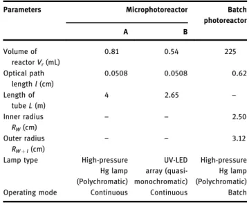
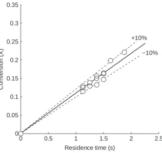
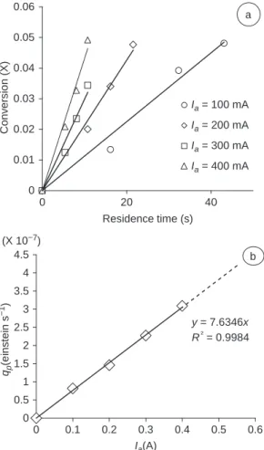
Figure
+5


Documents relatifs
