Reproducibility and repeatability of upper limb landmarks palpation for junior operators
Cédric Schwartz, Tatiana Fedrigo, Olivier Brüls, Serge Cescotto, Vincent Denoël, Jean-Louis Croisier, Bénédicte Forthomme Laboratory of Human Motion Analysis, University of Liège, Belgium ; email: [email protected]
SUMMARY
In human motion analysis, bone motions are usually expressed relatively to anatomical reference frames. The anatomical reference frames are constructed thanks to the localization of bony landmarks during a static phase prior to the acquisitions. These landmarks are identified by means of palpation. Accurate comparison between subjects and studies implies good reproducibility and repeatability of the palpation process. However, all investigators don’t have a long experience in palpation. In this paper, the reproducibility and repeatability of palpation for junior investigators were measured. Results show worse reproducibility and repeatability than what is usually expected. These errors have particularly an influence on the definition of the reference frames of the arm. This study therefore emphasizes on the need of a specific training of operators working in a motion lab.
INTRODUCTION
Optoelectronic systems are a common way to analyze 3D motions. The locations of the markers on the skin have, however, to be chosen carefully in order to limit as much as possible the effects of the soft tissue artifacts [1]. These locations are unfortunately different from easily defined bony landmarks. Therefore, the most common strategy to allow inter-subject / inter-studies comparison is to define a so-called anatomical reference frame. This reference frame is supposed to be fixed relatively to the local reference frame used to estimate the motion [2]. The palpation of landmarks is used to define the anatomical reference frame. The reproducibility (inter-investigators) and the repeatability (intra-investigators) are therefore major parameters for meaningful comparisons between subjects/studies. The repeatability of palpation has been shown to be fairly good [3]. However, the level of training has been shown to have an important influence on reproducibility and repeatability [4]. From our experience, people running experiments in motion labs are from various backgrounds (physicians, physiotherapists, engineers,…) and various skill levels (senior researchers, students,…). Therefore the results presented in the literature and coming from skilled investigators might not be representative from all the active population in the field. The goal of this paper is to establish the reproducibility and repeatability of palpation, which can be expected from junior investigators such as students and/or young operators.
METHODS
Two young researchers (one physiotherapist student and one biomedical engineer) realized the palpation of the bony landmarks recommended by the ISB [5] to construct the anatomical reference frames of the scapula, arm and forearm (Table 1). Each of the eleven landmarks was palpated ten times and the experiment was repeated on five male subjects (height: 1.79 m ± 0.01, weight: 73.8 kg ±12).
Table 1: List (and abbreviations) of the bony landmarks of the upper limb palpated during the study.
Abbreviation Complete name
IJ Suprasternal notch PX Xiphoid process
C7 Spinous process of the 7th cervical vertebra
T8 Spinous process of the 8th thoracic vertebra
AA Acromial angle TS Root of the scapular spine AI Inferior angle of the scapula EL Lateral epicondyle EM Medial epicondyle RS Radial styloid US Ulnar styloid
The position of the palpated points was expressed in each segment local reference frame. These local reference frames were constructed thanks to clusters of three markers placed on each segment (Figure 1). The reproducibility was obtained by computing the difference between the mean locations of the landmarks estimated by both investigators. The repeatability for each investigator was obtained from the standard deviation of the palpation measurements for each landmark.
The effect of these errors in terms of angles was also studied. For each segment, a mean anatomical reference frame was computed and compared to the ones constructed from each palpation sequence. No differences were made between palpations of both investigators. Regarding the arm anatomical frame, the glenohumeral head center was computed as a functional joint with the method described in Begon study [6].
In order to evaluate the accuracy of the couple acquisition system / pointer, we pointed a static point for 5 s. The standard deviation of the point position was equal to 0.65 mm.
Figure 1: Cluster of three markers on the arm. RESULTS AND DISCUSSION
Figure 2: Peak to peak value of the marker localization during the second experiment.
From Figure 2, we can see that both investigators don’t localize the bony landmarks at the same points. It is especially true for the scapula (difference up to 17.6 mm for AI). The main reason that explains this result is the larger amount of soft tissue between the bone and the skin for the scapula. As for reproducibility, Figure 2 also shows that repeatability is quite poor. When compared to values obtained by de Groot [3], the standard deviation is about two times larger (Table 2). For spinal landmarks, Billis [4] also reached the conclusion that students present poorest results Table 2: Norm of the standard deviation (mm) in the three axis direction in de Groot study [3] and in this study (mean of the two investigators).
in mm IJ PX C7 T8 AA TS AI EM EL de
Groot [3] 2.9 3.4 4.6 3.6 4.6 5.0 5.2 3.4 3.4 Present
study 3.7 5.0 7.6 7.9 8.5 7.0 9.2 8.7 7.2
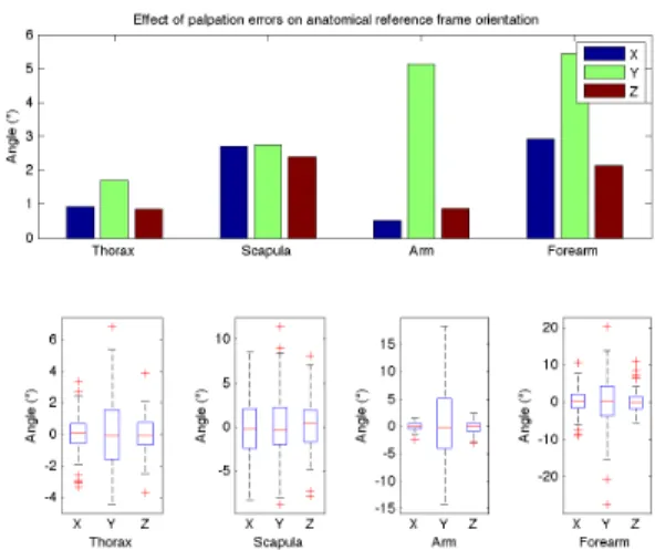
Figure 3 illustrates the impact of the errors in palpation on the orientation of the anatomical reference frame. Even if the variability of the palpation is greater for the scapula, Figure 3 indicates that palpation errors impact mostly the orientation of the reference frames of the arm and forearm (up to 5.44° for the forearm). This observation has already been documented in several papers including [5]. The reason
is the high sensitivity of EM / EL and RS / US on the axis definitions because these landmarks are very close to each other.
A closer look at the results shows high variability in the orientation of the reference frame. Indeed, even if the mean deviation remains under 6°, Figure 3 reveals that some palpations lead to much higher differences. The peak to peak value for the arm reaches 30°. This is directly linked to the lack of repeatability of the measures.
Figure 3: Top: Mean values of the absolute values of the angular deviations. Bottom: Distribution of the angular deviations (the central mark represents the median, the edges of the box the 25th and 75th percentiles – the whiskers shows
the most extreme values excepted outliers, which are represented by red crosses – if the data is normally distributed, outliers are values out of a 93.3 % coverage). CONCLUSIONS
This study focuses on the reproducibility and repeatability of the palpation by junior operators. The limited reproducibility and repeatability of the measures, especially in comparison to the literature, indicates that experience and training have an important influence on the quality of the palpation. Based on those findings, we would recommend that special attention is given to junior operators doing experiments in a motion lab. The best option would be to provide them with a special training. However, learning these skills might take some time. Therefore a more immediate but less satisfactory option would be to realize several times the calibration and to use the mean palpated points to construct the anatomical reference frames. This solution has the advantage to limit the intra-investigator variability.
REFERENCES
1. Bourne DA, et al, Annals of Biomedical Engineering.
DOI: 10.1007/s10439-010-0185-1, 2010.
2. Della Croce, et al, Gait and Posture. 21:226-237, 2005.
3. De Groot JH, Clinical Biomechanics. 12:461-472, 1997.
4. Billis EV, et al, Manual Therapy. 8:223-232, 2003.
5. Wu G, et al, Journal of Biomechanics.38:981-992, 2005.