DOI: 10.1212/01.wnl.0000327822.52212.c7
2008;71;1602-1608; originally published online Aug 20, 2008;
Neurology
Mundlos and W. B. Dobyns
Basel-Vanagaite, E. Leao-Teles, J. Vigneron, M. Foulon, M. Greally, J. Jaeken, S.
L. Van Maldergem, M. Yuksel-Apak, H. Kayserili, E. Seemanova, S. Giurgea, L.
laxa, Debré type
Cobblestone-like brain dysgenesis and altered glycosylation in congenital cutis
This information is current as of January 30, 2009
http://www.neurology.org/cgi/content/full/71/20/1602
located on the World Wide Web at:
The online version of this article, along with updated information and services, is
All rights reserved. Print ISSN: 0028-3878. Online ISSN: 1526-632X.
since 1951, it is now a weekly with 48 issues per year. Copyright © 2008 by AAN Enterprises, Inc.
® is the official journal of the American Academy of Neurology. Published continuously
Neurology
at UNIV MEDECINE LEIG 22827242 on January 30, 2009 www.neurology.org
Cobblestone-like brain dysgenesis and
altered glycosylation in congenital cutis
laxa, Debre´ type
L. Van Maldergem, MD
M. Yuksel-Apak, MD
H. Kayserili, MD
E. Seemanova, MD
S. Giurgea, MD
L. Basel-Vanagaite, MD
E. Leao-Teles, MD
J. Vigneron, MD
M. Foulon, MD
M. Greally, MD
J. Jaeken, MD
S. Mundlos, MD
W.B. Dobyns, MD
ABSTRACTObjective:
To delineate a new syndrome of brain dysgenesis and cutis laxa based on thedescrip-tion of 11 patients belonging to nine unrelated families recruited through an internadescrip-tional collab-oration effort.
Methods:
Careful clinical assessment of patients from birth to the age of 23 years with follow-upstudies ranging from 3 to 20 years. Biochemical studies of serum proteins glycosylation by iso-electric focusing and capillary zone electrophoresis were performed in 10 patients. Brain MRI studies using conventional methods were analyzed in eight patients.
Results:
An expanded clinical spectrum of a syndrome comprising facial dysmorphia (enlargedanterior fontanelles, downward slant of palpebral fissures, prominent root of the nose), a connec-tive tissue disorder (inguinal hernia, hip dislocation, high myopia), and neurologic impairment was defined. Early developmental delay was followed by onset of generalized seizures by the end of the first decade and a subsequent neurodegenerative course. A defect of N- or N- plus O-glycosylation of serum transferrins and ApoCIII was observed in 10 patients. An unusual cobblestone-like cortical malformation over the frontal and parietal regions was seen in eight patients and cerebellar abnormalities, including two patients with Dandy-Walker malformation, were observed in three patients.
Conclusions:
Our results suggest that autosomal recessive cutis laxa, Debre´ type, initiallyconsid-ered a dermatologic syndrome, is a multisystemic disorder with cobblestone-like brain dysgenesis manifesting as developmental delay and an epileptic neurodegenerative syndrome. It might repre-sent a metabolic cause of Dandy-Walker malformation. It is associated with a deficient N- and-O glycosylation of proteins and shares many similarities with muscle-eye-brain syndromes.
Neurology®2008;71:1602–1608
GLOSSARY
ARCL⫽ autosomal recessive congenital cutis laxa; CZE ⫽ capillary zone electrophoresis; DWM ⫽ Dandy-Walker malforma-tion; FCMD⫽ Fukuyama congenital muscular dystrophy; IEF ⫽ isoelectric focusing; MEB ⫽ muscle-eye-brain disease; WWS⫽ Walker-Warburg syndrome.
Autosomal recessive congenital cutis laxa (ARCL) represents a heterogeneous group of
disor-ders characterized by redundant skin present from birth. Although dominant inheritance has
been described (OMIM 123700), autosomal recessive inheritance is more common, with two
distinct subtypes described that differ at the ultrastructural level.
The pulmonary emphysema type of ARCL (ARCL type 1, OMIM 219100) is
character-ized by fragmentation of elastic fibers, which are present as unassembled primary
compo-nents. The Debre´ type (ARCL type 2, OMIM 219200) is characterized by developmental
Supplemental data at www.neurology.org
Address correspondence and reprint requests to Dr. L. Van Maldergem, Centre de Ge´ne´tique Humaine, CHU Sart-Tilman, Universite´ de Lie`ge, 4000 Lie`ge, Belgium
vmald@skypro.be
e-Pub ahead of print on August 20, 2008, at www.neurology.org.
From Centre de Ge´ne´tique Humaine (L.V.M.), CHU Sart-Tilman, Universite´ de Lie`ge, Belgium; Department of Medical Genetics (M.Y.-A., H.K.), Faculty of Medicine, University of Istanbul, Turkey; Department of Clinical Genetics (E.S.), Charles University, Prague, Czech Republic; Department of Neurology (S.G.), CHU Tivoli, La Louvie`re, Belgium; Department of Medical Genetics (L.B.-V.), Schneider Children’s Medical Center, Petah Tikva, Israel; Department of Pediatrics (E.L.-T.), San Joao Hospital, Porto, Portugal; Unite´ de Ge´ne´tique Me´dicale (J.V.), Service de Me´decine Ne´onatale, CHU Nancy, France; Department of Pediatrics (M.F.), CHU Charleroi, Belgium; College of Medicine and Medical Sciences (M.G.), Arabian Gulf University, Manama, Bahrain; Centre for Metabolic Diseases (J.J.), Katholieke Universiteit Leuven, Belgium; Institut fu¨r Medizinische Genetik (S.M.), Charite´, Campus Virchow, Berlin, Germany; and Departments of Human Genetics, Neurology, and Pediatrics (W.B.D.), The University of Chicago, IL.
delay, large anterior fontanelle, facial
dys-morphia, and a paucity of elastic fibers on
skin biopsy. ARCL is rare, with fewer than
40 patients with the pulmonary emphysema
type and fewer than 50 with the Debre´ type
reported (appendix e-1 on the Neurology
®Web site at www.neurology.org). A few other
patients have had variant or unspecified
phe-notypes (appendix e-1). In the pulmonary
em-physema subtype, homozygous mutations of
EGF-containing fibulin-like extracellular matrix
protein 2 (EFEMP2) and both homozygous and
heterozygous mutations of Fibulin-5 (FBLN5)
have been reported, the latter blurring the
dis-tinction between autosomal dominant and
re-cessive forms.
1-4Mutations in the elastin (ELN)
gene have been reported in six families with
au-tosomal dominant cutis laxa associated with
vari-able aortic aneurisms or pulmonary emphysema.
5-9Recently, a Dutch group described N- and
O-linked glycosylation defects in three patients
with autosomal recessive cutis laxa and
develop-mental delay.
10We recently established an international
collaboration to further delineate the
pheno-type and map the causative genes. Here we
describe an additional 11 patients from nine
families with Debre´ type ARCL and further
delineate the brain and neurologic features.
METHODSClinical reports. Patient 1. This girl was born to unrelated Belgian parents after a pregnancy complicated by
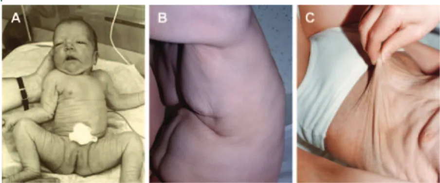
Figure 1 Photographs of seven patients at different ages show the evolution of the skin phenotype, shown here for patient 1 in A, 2 in B-I and B-II, 9 in C, 10 in D, 3 in E-I and E-II, 11 in F-I through F-II, and 4 in G-I through G-III
Photographs in infancy (A, B, E, G) show facial dysmorphia with downslanting palpebral fissures and prominent nasal root. Over the next several years, the facial features become more normal, but coarse hair (B-II and D) and hypotonia (F and G-II) are seen. By the second decade, a degenerative course is typically seen with severe hypotonia, drooling, and sometimes muscle wasting (F-III, G-III, and G-IV). Written authorization for publication of the photographs was obtained from the legal guardians of all patients.
Neurology 71 November 11, 2008 1603
at UNIV MEDECINE LEIG 22827242 on January 30, 2009 www.neurology.org
oligohydramnios. Her birthweight was 2,850 g and birth OFC 31.5 cm (–3 SD) at 37 weeks gestation. She had a dysmorphic appearance with downslanting palpebral fissures, prominent
or-bital ridges, and enlarged anterior fontanelle (7⫻ 9 cm), and
also generalized overfolding of her skin (figures 1A and 2A) con-sistent with Debre´ type ARCL. Skin biopsy demonstrated rar-efaction of the elastin network with fragmentation and rarity of elastin fibers.
She learned to sit at 1 year and walk late at 2.5 years. She entered mainstream school after 1 year of special education. At 6 years, eye examination detected a focal rupture of the Bruch membrane in her left eye. At 13 years, her weight, height (–2
standard deviations [SD]), and OFC (46.5 cm,⫺3.5 SD) were
all small, but she was doing well with no seizures. She had abun-dant, coarse hair. The cutis laxa became gradually less apparent on serial examinations. Brain MR imaging (figure 3, A through C) shows mildly reduced sulcation and thick cortex in the poste-rior frontal and possibly anteposte-rior parietal regions, short corpus callo-sum with absent rostrum and thick genu, subtle herniation of the inferior occipital lobe through the tentorial notch to present be-tween the splenium and superior vermis, and mild cerebellar vermis hypoplasia involving primarily the pyramis and uvula.
Patient 2. This girl was born at term to unrelated Belgian
parents with birthweight 3,300 g and OFC 33 cm (10th centile). Her neonatal history has been already described, including severe cutis laxa, unilateral choanal stenosis, Dandy-Walker
malforma-tion (DWM), and hydrocephalus.11Her developmental
mile-stones were delayed. She had marked fatigability, and onset of seizures at 7 years that subsequently proved difficult to control. Serial examinations showed cutis laxa that improved with age, and thick, abundant hair with an uncombable appearance (fig-ures 1B and 2B). Her IQ at 10 years was 45, but serial cognitive testing demonstrated a decline in function.
Her brain MRI (figure 3, D–F) demonstrated reduced sulcation with an irregular cortical surface and mildly thick 7–10 mm cortex over the posterior frontal, perisylvian, and parietal regions, thin white matter, moderately enlarged lateral ventricles, and thin corpus callosum. Lower images showed a short midbrain with small and bent tectum, elongated medulla, severe cerebellar vermis hypoplasia and upward rotation, moderate cerebellar hemisphere hypoplasia, and cystic dilation of the 4th ventricle resulting in a very large poste-rior fossa, typical for DWM.
Patients 3 to 11. The clinical histories for patients 3–11 are
available as supplemental material in appendix e-2, with
photo-graphs in figures 1 and 2, and brain imaging studies in figure 3 and figures e-1 and e-2.
Procedures. Isoelectric focusing (IEF) and capillary zone elec-trophoresis (CZE) of serum transferrins and IEF of serum apolipoprotein-CIII were performed according to published
methods.12-14
RESULTS
The details of these 11 patients are
sum-marized in the table. All 11 had cutis laxa consistent
with Debre´ type ARCL based on their clinical
exam-ination, particularly enlarged anterior fontanelles and
typical facial dysmorphia. Skin biopsies
demon-strated a sparse elastic network in 9 of 11 patients.
Birthweight and length were normal, in contrast to
patients in previous reports. Two patients had
genital choanal atresia or stenosis, and five had
con-genital hip dislocation. Erb’s palsy or clavicle fracture
was noted in four patients, and corneal dystrophy
and sensorineural hearing loss in one patient each.
All had mild to moderate developmental delay
and later mental retardation and hypotonia. All
teen-agers and adults had severe myopia and often
strabis-mus. All but one developed generalized seizures with
onset between 8 and 12 years that were often difficult
to treat. Several had lower cognitive function after
seizure onset, and several had a progressive course
including one girl (patient 11) with decline in
cogni-tive function and worsening spasticity and ataxia
de-spite having no seizures.
We reviewed brain MRI in nine patients (table).
The most common abnormality was a cortical
malfor-mation in eight patients consisting of broad and poorly
defined gyri separated by shallow sulci, and abnormally
thick 5–10 mm cortex that appeared smooth in some
areas and irregular in others, often with vertical streaks
within the cortex (figure 3 and figures e-1 and e-2).
However, no areas with well-defined microgyri were
seen. The cortical malformation appeared most severe
in the posterior frontal and anterior parietal lobes
in-cluding the perisylvian cortex, and extended to the
ante-rior frontal lobe and often the supeante-rior temporal lobe.
The white matter was normal in most patients,
al-though several had prominent perivascular spaces. The
corpus callosum appeared normal in all but one patient,
in whom it was abnormally short. Only three patients
had cerebellar involvement, including one with isolated
vermis hypoplasia and two with DWM consisting of
moderate to severe hypoplasia and upward rotation of
the vermis, cystic enlargement of the 4th ventricle, and
enlarged posterior fossa (figure 3). One girl (patient 11)
had generalized cerebral and cerebellar atrophy with no
malformations seen (figure e-1, P–R). Several of the
brain imaging studies were suboptimal.
IEF of serum sialotransferrins was performed in
all but patient 10 —the sib of patient 9 —and
Figure 2 In Debre´ type autosomal recessive congenital cutis laxa, the wrinkles or skin folds are narrow and shallow, especially in comparison to the skin folds in the pulmonary emphysema type
The skin abnormalities are always most prominent in newborns (A, patient 1), then become less marked but still evident at older ages (B, patient 2 at 8 years; C, patient 11 at 23 years).
showed an abnormal pattern consistent with
congen-ital disorder of glycosylation type 2 (CDG2) with an
increase of trisialo- and disialotransferrin (figure 4, B
and C). IEF of serum ApoCIII was analyzed in
pa-tients 6 and 7 and in their parents. Both affected
children showed a clearly lower disialo ApoCIII band
than their parents, pointing to hyposialylation
(fig-ure 4D). No serum was available from the parents of
the other patients, precluding firm conclusions about
their ApoCIII patterns.
DISCUSSION
Among these 11 patients with Debre´
type ARCL, we found a recognizable pattern of
ab-normalities involving facial appearance, connective
tissue structures especially the skin, and brain. The
most consistent craniofacial abnormalities consisted
of an abnormally large anterior fontanelle,
promi-nent supraorbital ridges and nasal root, telecanthus,
and downslanting palpebral fissures (figure 1). The
skin phenotype was characteristic of cutis laxa with
generalized overfolding and wrinkling, but no
hy-perelasticity as seen in Ehlers-Danlos syndrome. In
contrast to the pulmonary emphysema type of
ARCL, furrows in the skin are tightly spaced (figure
2). Most patients had abundant coarse hair, and
other features indicating a more generalized
connec-tive tissue dysplasia such as high myopia or dislocated
hips. Skin biopsies supported the diagnosis, showing
reduced number and fragmentation of elastin fibers
(data not shown). A trend toward improvement of
cutis laxa throughout childhood was seen.
All patients had moderately delayed
developmen-tal milestones and mendevelopmen-tal retardation with good
so-cial interactions, although speso-cial education was
required. Generalized seizures began between 6 and
12 years in all patients but one, and were often
in-tractable. The severe seizures probably contributed to
shortened survival, with death in childhood
occur-ring in two children reported in the literature and
four from our series, all by 17 years. Several patients,
especially patient 11 and her two affected sibs, had a
progressive course from early childhood
character-ized by dementia, spasticity, ataxia, and hearing loss
that led to death by 9 and 11 years in the two sibs
and a bedridden state in patient 11 by 16 years. This
distinct clinical course probably correlates with the
cerebral atrophy observed on brain imaging.
Brain imaging demonstrated a recurrent pattern
of dysgenesis consisting of a cortical malformation in
all but one patient, and variable microcephaly and
cerebellar hypoplasia (figure 3 and figures e-1 and
e-2). The cortical malformation involves the
poste-rior frontal, perisylvian, and parietal regions, and was
seen in all but one child. The abnormal cortex partly
resembles polymicrogyria, except that the cortical
ribbon appears smooth in some areas and irregular in
others, with some subtle vertical striations that are
never as prominent as in classic polymicrogyria. The
cortex is typically 7–10 mm thick, as in
polymicro-gyria. Two of the previously reported patients with
cutis laxa and CDG type 2 had a similar
malforma-tion.
10Cerebellar malformations were seen in three
of our patients, including CVH in one girl and
DWM in two others. DWM was previously reported
in one child with cutis laxa,
15but no images of the
cerebral cortex or IEF of serum transferrins were
pre-sented. Further, DWM and hydrocephalus without
the cortical malformation were reported in a boy
with severe congenital hypotonia, myopathy,
coagu-lopathy, and CDG type 2 and later associated with
Figure 3 Representative brain imaging studies in Debre´ type cutis laxa from patients 1 (A–C), 2 (D–F), and 3 (G–I)
Additional images are shown in figure e-1. In all three of these patients, axial and coronal images at the level of the lateral ventricles demonstrate an abnormally thick 6–10 mm cortex in the frontal and parietal regions on both sides with the frontal pole and posterior parietal regions less severely involved (white arrows in B, C, F). The cortical ribbon appears smooth in some images (I) and irregular in others (see figure e-1), which explains why the cortical malformation has been variably inter-preted as pachygyria or polymicrogyria in different patients. Midline sagittal and lower axial images show cerebellar vermis (v) hypoplasia in patients 1–3. This appears as mild hypoplasia of the poste-rior vermis in patient 1 (A), and as typical Dandy-Walker malformation with small and upwardly ro-tated vermis, cystic dilatation of the 4th ventricle, and enlarged posterior fossa in patients 2 (D, E) and 3 (G, H).
Neurology 71 November 11, 2008 1605
at UNIV MEDECINE LEIG 22827242 on January 30, 2009 www.neurology.org
Table Summary of clinical and brain imaging findings Patient no. 1 2 3 4 5 6 7 8 9 10 11 Ethnicity Belgian Belgian Turkish Italian Portuguese Bedouin Bedouin French Czech Czech Turkish Clinical features Birth OFC (SD) –3 –1.5 Mean –2.2 Mean –2 –4.5 –3 Mean –1 –4.5 Facial dysmorphia Yes Yes Yes Yes Yes Yes Yes Yes Yes Yes Yes Severity of MR Mild Severe Moderate Severe Severe Mild Moderate Moderate Mild Mild Severe Eye phenotype BMR HM HM/CD Normal Normal HM HM HM Hip dislocation ⫹ ⫹ ⫹ ⫹ ⫹ ⫹⫹⫹ Inguinal hernia ⫹⫹ ⫹ ⫹ ⫹ ⫹ Age at last evaluation (age at death), y 12 15 4 16 21 4 3 17 12 (6) 23 Age at onset of seizures, y None 7 None 10 10 None None 12 8 None None Glycosylation testing Transferrin IEF CDG2 CDG2 CDG2 CDG2 CDG2 CDG2 CDG2 CDG2 CDG2 In sib CDG2 ApoCIII-IEF — — — — — Abnormal Abnormal — — — — Brain imaging Microcephaly MIC — — MIC — MIC MIC — — — — Cortex COB COB COB COB COB COB COB — COB — — Other anomalies HCC HYD V-R — V-R V-R — ATR — ATR Cerebellum CVH DWM DWM — — — — — — — — OFC ⫽ occipito-frontal circumference; MR ⫽ mental retardation; BMR ⫽ Bruch membrane rupture; HM ⫽ high myopia; CD ⫽ corneal dystrophy; IEF ⫽ isoelectric focusing; CDG2 ⫽ IEF consistent with CDG type 2; ApoCIII ⫽ apolipoprotein-CIII; MIC ⫽ microcephaly at age of evaluation and not necessarily at birth; COB ⫽ cobblestone-like cortical malformation; HCC ⫽ hypoplasia (partial agenesis) of the corpus callosum; HYD ⫽ hydrocephalus; V-R ⫽ prominent perivascular (Virchow-Robin) spaces in central white matter; ATR ⫽ atrophy of both cerebrum and cerebellum; CVH ⫽ cerebellar vermis hypoplasia; DWM ⫽ Dandy-Walker malformation.
mutations in the
-1,4-galactosyltransferase-I or
B4GALT1 gene.
16,17The cortical malformation seen in eight of our
patients resembles the striking cobblestone cortical
malformation seen in Walker-Warburg syndrome
(WWS), muscle-eye-brain disease (MEB), and
Fukuyama congenital muscular dystrophy (FCMD).
In these syndromes, the gyral pattern varies from
smooth to irregular, and on autopsy the cortex is very
disorganized with neurons and glia mixed with
fibro-blasts and blood vessels in superficial regions.
18-20The malformation results from defects in the basal
lam-ina (pial limiting membrane) that allow lam-inappropriate
migration of neurons and glia into the subarachnoid
space to form extensive marginal glio-neuronal
hetero-topia.
21,22These syndromes are associated with
muta-tions in six genes that encode known or putative
O-linked glycosyltransferases: FCMD, FKRP, LARGE,
POMGnT1, POMT1, and POMT2.
23-28The encoded
proteins are involved in synthesis of alpha-mannosyl
side chains of alpha-dystroglycan, which bind to the
ex-tracellular matrix protein laminin in retina, brain, and
muscle.
29-31However, glycosylation of ApoCIII is
nor-mal in serum because ApoCIII is synthesized in the liver
and apparently not released into the circulation.
The cortical malformation seen in our patients
also resembles the malformation seen in two other
autosomal recessive syndromes. The first of these is
so-called bilateral frontal-parietal polymicrogyria
as-sociated with mutations of GPR56.
32-34Based on a
brain imaging appearance that closely resembles
MEB, one of the authors first reported this syndrome
as “cobblestone lissencephaly with normal eyes and
muscle” in 1996.
35Other authors described the same
malformation alternatively as pachygyria
36or
polymicrogyria.
33,34,37Interestingly, the GPR56
pro-tein is heavily glycosylated.
38The brain imaging
ap-pearance also resembles the CEDNIK syndrome
caused by mutations in SNAP29.
39We suggest using
the term frontal predominant or frontoparietal
cobblestone-like cortical malformation for these
dis-orders. We hypothesize that the brain malformations
observed in Debre´ type ARCL and these other
disor-ders share a common pathogenesis.
In further support of this analysis, mutations in the
a2 subunit of the V-type H
⫹-ATPase (ATP6V0A2)
were recently identified in all our patients except for
patient 2.
40Received October 3, 2007. Accepted in final form April 4, 2008.
REFERENCES
1. Loeys B, Van Maldergem L, Mortier G, et al.
Homozygos-ity for a missense mutation in fibulin-5 (FBLN5) results in a severe form of cutis laxa. Hum Mol Genet 2002;11: 2113–2118.
2. Markova D, Zou Y, Ringpfeil F, et al. Genetic
heterogene-ity of cutis laxa: a heterozygous tandem duplication within the fibulin-5 (FBLN5) gene. Am J Hum Genet 2003;72: 998–1004.
3. Elahi E, Kalhor R, Banihosseini SS, et al. Homozygous
missense mutation in fibulin 5 in an Iranian autosomal recessive pedigree and associated haplotype. J Invest Der-matol 2006;126:1506–1509.
4. Hucthagowder V, Sausgruber N, Kim KH, Angle B,
Mar-morstein LY, Urban Z. Fibulin-4: a novel gene for an au-tosomal recessive cutis laxa syndrome. Am J Hum Genet 2006;78:1075–1080.
5. Tassabehji M, Metcalfe K, Hurst J, et al. An elastin gene
mutation producing abnormal tropoelastin and abnormal elastic fibres in a patient with autosomal dominant cutis laxa. Hum Mol Genet 1998;7:1021–1028.
6. Zhang M-C, He L, Giro M, Yong SL, Tiller GE,
David-son JM. Cutis laxa arising from frameshift mutations in exon 30 of the elastin gene (ELN). J Biol Chem 1999;274: 981–986.
7. Rodriguez-Revenga L, Iranzo P, Badenas P, Puig S, Carrio
A, Mila M. A novel elastin gene mutation resulting in an autosomal dominant form of cutis laxa. Arch Dermatol 2004;140:1135–1139.
Figure 4 Separation of protein isoforms in autosomal recessive cutis laxa
Capillary zone electrophoresis (CZE) in a normal control (A) shows small peaks for disialo-(2) and trisialo- (3) transferrins, and a high peak for tetrasialotransferrins (4). CZE in patient 1 (B) demonstrates a pattern typical for type 2 congenital disorders of glycosylation with increased disialo- (peak at 5.057) and trisialo- (peak at 5.180) transferrins, and decreased tetrasialotransferrins (peak at 5.312). Isoelectric focusing (IEF) of serum transferrin pat-tern in patient 6 (P6), patient 7 (P7), and their parents (M, mother; F, father) demonstrate increased size of the peaks for disialo- and trisialotransferrins in the two affected sibs, whereas their parents’ profiles are normal (C). The numbers on the left and right indicate the sialotransferrin fractions. IEF of apolipoprotein CIII in the same family (D) shows decreased intensity of disialo fractions with respect to their parents’ normal profile.
Neurology 71 November 11, 2008 1607
at UNIV MEDECINE LEIG 22827242 on January 30, 2009 www.neurology.org
8. Urban Z, Gao J, Pope FM, Davis EC. Autosomal domi-nant cutis laxa with severe lung disease: synthesis and ma-trix deposition of mutant tropoelastin. J Invest Dermatol 2005;124:1193–1199.
9. Szabo Z, Crepeau MW, Mitchell AL, et al. Aortic
aneuris-mal disease and cutis laxa caused by defects in the elastin gene. J Med Genet 2006;43:255–258.
10. Morava E, Wopereis S, Coucke P, et al. Defective protein
glycosylation in patients with cutis laxa syndrome. Eur J Hum Genet 2005;13:414–421.
11. Biver A, De Rijcke S, Toppet V, Ledoux-Corbusier M,
Van Maldergem L. Congenital cutis laxa with ligamentous laxity and delayed development, Dandy-Walker malfor-mation and minor heart and osseous defects. Clin Genet 1994;45:318–322.
12. Jaeken J, Van Eijk HG, van der Heul C, Corbeel L,
Eeck-els R, Eggermont E. Sialic acid-deficient serum and cere-brospinal fluid transferring in a newly recognized genetic syndrome. Clin Chim Acta 1984;144:244–247.
13. Carchon HA, Chevigne R, Falmagne JB, Jaeken J.
Diag-nosis of congenital disorders of glycosylation by capillary zone electrophoresis. Clin Chem 2004;50:101–111.
14. Wopereis S, Gru¨newald S, Morava E, et al. Apolipoprotein
C-III isofocusing in the diagnosis of genetic defects in O-glycan biosynthesis. Clin Chem 2003;49:1839–1845.
15. Litzman J, Buckova H, Ventruba J, Holcikova A, Mikyska
P, Lokaj J. A concurrent occurrence of cutis laxa, Dandy-Walker syndrome and immunodeficiency in a girl. Acta Paediatr 2003;92:861–864.
16. Hansske B, Thiel C, Lubke T, et al. Deficiency of
UDP-galactose: N-acetylglucosamine beta-1,4-galactosyltransferase I causes the congenital disorder of glycosylation type IId. J Clin Invest 2002;109:725–733.
17. Peters V, Penzien JM, Reiter G, et al. Congenital disorder
of glycosylation IId (CDG-IId)—a new entity: clinical presentation with Dandy-Walker malformation and my-opathy. Neuropediatrics 2002;33:27–32.
18. Williams RS, Swisher CN, Jennings M, Ambler M,
Cavi-ness VS Jr. Cerebro-ocular dysgenesis(Walker-Warburg syndrome): neuropathologic and etiologic study. Neurol-ogy 1984;34:1531–1541.
19. Dobyns WB, Kirkpatrick JB, Hittner HM, Roberts RM,
Kretzer FL. Syndromes with lissencephaly II: Walker-Warburg and cerebro-oculo-muscular syndromes and a new syndrome with type II lissencephaly. Am J Med Genet 1985;22:157–195.
20. Haltia M, Leivo I, Somer H, et al. Muscle-eye-brain
disease: a neuropathological study. Ann Neurol 1997; 41:73–80.
21. Miller G, Ladda RL, Towfighi J. Cerebro-ocular
dysplasia-muscular dystrophy (Walker-Warburg) syndrome: find-ings in a 20-week-old fetus. Acta Neuropathol 1991;82: 234–238.
22. Takada K, Nakamura H, Suzumori K, Ishikawa T,
Sug-iyama N. Cortical dysplasia in a 23-week fetus with Fukuyama congenital muscular dystrophy (FCMD): a case analysis. Acta Neuropathol 1987;74:300–306.
23. Kobayashi K, Nakahori Y, Miyake M, et al. An ancient
retrotransposal insertion causes Fukuyama-type congenital muscular dystrophy. Nature 1998;394:388–389.
24. Beltran-Valero de Bernabe D, Voit T, Longman C, et al.
Mutations in the FKRP gene can cause muscle-eye-brain disease and Walker-Warburg syndrome. J Med Genet 2004;41:e61.
25. Longman C, Brockington M, Torelli S, et al. Mutations in
the human LARGE gene cause MDC1D, a novel form of congenital muscular dystrophy with severe mental retarda-tion and abnormal glycosylaretarda-tion of alpha-dystroglycan. Hum Mol Genet 2003;12:2853–2861.
26. Yoshida A, Kobayashi K, Manya H, et al. Muscular
dystro-phy and neuronal migration disorder caused by mutations in a glycosyltransferase, POMGnT1. Dev Cell 2001;1: 717–724.
27. Beltran-Valero de Bernabe D, Currier S, Steinbrecher
A, et al. Mutations in the O-mannosyltransferase gene POMT1 give rise to the severe neuronal migration dis-order Walker-Warburg syndrome. Am J Hum Genet 2003;71:1033–1043.
28. van Reeuwijk J, Janssen M, van den Elzen C, et al.
POMT2 mutations cause alpha-dystroglycan hypoglycosy-lation and Walker-Warburg syndrome. J Med Genet 2005;42:907–912.
29. Chiba A, Matsumura K, Yamada H, et al. Structures of
sialylated O-linked oligosaccharides of bovine periph-eral nerve alpha-dystroglycan: the role of a novel O-mannosyl-type oligosaccharide in the binding of alpha-dystroglycan with laminin. J Biol Chem 1997; 272:2156–2162.
30. Henry MD, Satz JS, Brakebusch C, et al. Distinct roles for
dystroglycan, beta1 integrin and perlecan in cell surface laminin organization. J Cell Sci 2001;114:1137–1144.
31. Olson EC, Walsh CA. Smooth, rough and upside-down
neocortical development. Curr Opin Genet Dev 2002;12: 320–327.
32. Piao X, Hill RS, Bodell A, et al. G protein-coupled
receptor-dependent development of human frontal cortex. Science 2004;303:2033–2036.
33. Chang BS, Piao X, Bodell A, et al. Bilateral frontal
polymi-crogyria: clinical and radiological features in ten families with linkage to chromosome 16. Ann Neurol 2003;56: 596–606.
34. Piao X, Chang BS, Bodell A, et al. Genotype-phenotype
analysis of human frontoparietal polymicrogyria syn-dromes. Ann Neurol 2005;58:680–687.
35. Dobyns WB, Patton MA, Stratton RF, Mastrobattista JM,
Blanton SH, Northup H. Cobblestone lissencephaly with normal eye and muscle. Neuropediatrics 1996;27:70–75.
36. Straussberg R, Gross S, Amir J, Gadoth N. A new
autoso-mal recessive syndrome of pachygyria. Clin Genet 1996; 50:498–501.
37. Piao X, Basel-Vanagaite L, Straussberg R, et al. An
autoso-mal recessive form of bilateral frontoparietal polymicro-gyria maps to chromosome 16q12.2-q21. Am J Hum Genet 2002;70:1028–1033.
38. Jin Z,Tietjen I, Bu L, et al. Disease-associated mutations
affect GPR56 protein trafficking and cell surface expres-sion. Hum Mol Genet 2007;16:1972–1985.
39. Sprecher E, Ishida-Yamamoto A, Mizrahi-Koren M, et al.
A mutation in SNAP29, a SNARE protein involved in intracellular trafficking causes a new neurocutaneous syn-drome characterized by cerebral dysgenesis, neuropathy, ichthyosis, and palmoplantar keratoderma. Am J Hum Genet 2005;77:242–251.
40. Kornak U, Reynders E, Dimopoulou A, et al., ARCL
De-bre´ Study Group. Impaired glycosylation and cutis laxa caused by mutations in the vesicular H⫹-ATPase subunit ATP6V0A2. Nat Genet 2008;40:32–34.
DOI: 10.1212/01.wnl.0000327822.52212.c7
2008;71;1602-1608; originally published online Aug 20, 2008;
Neurology
Mundlos and W. B. Dobyns
Basel-Vanagaite, E. Leao-Teles, J. Vigneron, M. Foulon, M. Greally, J. Jaeken, S.
L. Van Maldergem, M. Yuksel-Apak, H. Kayserili, E. Seemanova, S. Giurgea, L.
laxa, Debré type
Cobblestone-like brain dysgenesis and altered glycosylation in congenital cutis
This information is current as of January 30, 2009
& Services
Updated Information
http://www.neurology.org/cgi/content/full/71/20/1602
including high-resolution figures, can be found at:
Supplementary Material
12.c7/DC1
http://www.neurology.org/cgi/content/full/01.wnl.0000327822.522
Supplementary material can be found at:
Subspecialty Collections
orders
http://www.neurology.org/cgi/collection/other_neurocutaneous_dis
neurocutaneous disorders
Other
http://www.neurology.org/cgi/collection/cortical_dysplasia
Cortical dysplasia
http://www.neurology.org/cgi/collection/developmental_disorders
Developmental disorders
ed
http://www.neurology.org/cgi/collection/metabolic_disease_inherit
(inherited)
Metabolic disease
http://www.neurology.org/cgi/collection/mri
MRI
following collection(s):
This article, along with others on similar topics, appears in the
Permissions & Licensing
http://www.neurology.org/misc/Permissions.shtml
or in its entirety can be found online at:
Information about reproducing this article in parts (figures, tables)
Reprints
http://www.neurology.org/misc/reprints.shtml
Information about ordering reprints can be found online:
at UNIV MEDECINE LEIG 22827242 on January 30, 2009 www.neurology.org


![Angular analysis of the B[superscript 0] → K[superscript *0]e[superscript +]e[superscript −] decay in the low-q[superscript 2] region](data:image/gif;base64,R0lGODlhAQABAIAAAP///wAAACH5BAEAAAAALAAAAAABAAEAAAICRAEAOw==)