RESEARCH
Clinical-scale expansion of mesenchymal
stromal cells: a large banking experience
Chantal Lechanteur
1*, Alexandra Briquet
1, Olivier Giet
1, Olivier Delloye
1, Etienne Baudoux
1and Yves Beguin
1,2Abstract
Background: Mesenchymal stromal cells (MSC) are largely investigated in clinical trials aiming to control inappro-priate immune reactions (GVHD, Crohn’s disease, solid organ transplantation). As the percentage of MSC precursors in bone marrow is very low, these must be expanded in vitro to obtain therapeutic cell doses. We describe here the constitution of an allogeneic human third-party MSC bank from screened healthy volunteer donors in compliance with quality specifications and ISCT-release criteria and report follow-up of different aspects of this activity since 2007. Methods: 68 clinical-grade large-scale MSC cultures were completed and analyzed. The whole process was
described, including volunteer donor screening, bone marrow collection, mononuclear cell isolation and expansion over 4 weeks, harvesting, cryopreservation, release, administration and quality controls of the cells (including microbi-ology, phenotype, and potency assays).
Results: From 59 validated donors, 68 cultures were completed (mean of final yields: 886 × 106 cells/culture) and
a total of 464 MSC aliquots have been produced and stored in liquid nitrogen (mean of 132.8 × 10⁶ cells/bag). Each
MSC batch underwent extensive testing to verify its conformity with EBMT and ISCT release criteria and was individu-ally validated. As of June 1 2015, 314 bags have been released and infused to patients included in 6 different clinical protocols. All thawed MSC units satisfied to release criteria and no infusion-related toxicity was reported.
Conclusion: In conclusion, despite low passage cultures, we have been able to create an allogeneic “off-the-shelf” MSC bank with a large number of frozen aliquots and report here an efficient clinical-grade MSC banking activity in place for more than 7 years. Our challenge now is to produce MSC in compliance with good manufacturing practices (GMP) as, in the meantime, MSC have become considered as advanced therapy medicinal products (ATMP). Another significant challenge remains the development of relevant potency assay.
Keywords: Banking, Clinical-grade, GMP, MSC, Mesenchymal stem cells
© 2016 The Author(s). This article is distributed under the terms of the Creative Commons Attribution 4.0 International License (http://creativecommons.org/licenses/by/4.0/), which permits unrestricted use, distribution, and reproduction in any medium, provided you give appropriate credit to the original author(s) and the source, provide a link to the Creative Commons license, and indicate if changes were made. The Creative Commons Public Domain Dedication waiver (http://creativecommons.org/ publicdomain/zero/1.0/) applies to the data made available in this article, unless otherwise stated.
Background
Mesenchymal stromal cells (MSC) were identified more than three decades ago by Friedenstein et al. as the stro-mal cells of the marrow microenvironment that support
hematopoiesis [1]. MSC are multipotent progenitors
capable of differentiating into various cells and tissues,
such as chondrocytes, osteoblasts and adipocytes [2, 3].
In addition to multilineage differentiation and partici-pation in the hematopoietic niche, MSC exert powerful immunomodulatory effects that include: inhibition of
proliferation and function of T and B cells, inhibition of dendritic cell maturation and function and immune mod-ulation of other immune cells such as natural killer (NK)
cells and macrophages [4–6]. Due to the absence of
co-stimulatory molecules and low HLA class I expression, MSC are not thought to be prone to immune rejection
[7, 8]. This makes them ideal candidates for allogeneic
use in both regenerative medicine and clinical applica-tions in immune diseases such as graft-versus-host dis-ease (GVHD), Crohn’s disdis-ease (CD), rheumatoid arthritis
(RA) among others [9].
The International Society for Cellular Therapy (ISCT) has established the minimum criteria to define MSC and described them as a heterogeneous population of
Open Access
*Correspondence: c.lechanteur@chu.ulg.ac.be
1 Department of Hematology, Laboratory of Cell and Gene Therapy, CHU of Liège and University of Liège, CHU Sart-Tilman, 4000 Liège, Belgium Full list of author information is available at the end of the article
spindle-shaped, plastic-adherent cells isolated from bone marrow, adipose tissue, and many other tissue sources such as umbilical cord or cord blood. They must express certain cell surface markers (CD73, CD90, CD105) and lack expression of others (CD45, CD34, CD14, HLA-DR) and have the capacity to differentiate into osteoblasts, adipocytes and chondroblasts when cultured in
particu-lar in vitro conditions [10, 11]. Prior to clinical
applica-tion, MSC must be significantly expanded to obtain therapeutic cell doses. Traditionally, MSC are obtained by ex vivo culture of the adherent cell fraction of bone marrow aspirates. The percentage of MSC among mar-row cells is very low (0.01 to 0.001 % depending on age) but they can be easily isolated and expanded to reach adequate numbers for therapeutic doses. For this reason, cell processing facilities have established procedures for large scale production of MSC.
In the Laboratory of Cell and Gene Therapy (LTCG, CHU of Liège), we started in late 2006 a “MSC bank” based on clinical-grade expansion of MSC from BM sam-ples obtained from healthy volunteer donors. Cells are produced according to the European Group for Blood and Marrow Transplantation (EBMT) consortium rec-ommendations for defining common procedures for MSC isolation and expansion, as well as common release criteria, enabling multicenter trials with comparable
MSC products [12]. Our first pilot clinical protocol (20
patients) evaluated the safety and preliminary efficacy of MSC to prevent graft rejection and GVHD after allo-geneic hematopoietic cell transplantation (HCT) with
non-myeloablative conditioning (NMHCT) [13]. In this
study, HLA-mismatched NMHCT with MSC co-infusion appeared to be safe. Also, the prevention of death from GVHD and preservation of GVT effects suggested by this study are currently under investigation in a multicenter randomized study of MSC co-transplantation in patients given HLA-mismatched PBSC after non-myeloablative conditioning (NCT01045382).
We are currently involved in six clinical trials of MSC infusion in different settings including HSC transplan-tation (HCT) with myeloablative or non-myeloablative conditioning, cord blood transplantation (CBT), solid organ transplantation and severe or refractory autoim-mune disorders such as Crohn’s disease. According to
each protocol, the MSC dose varies from 1 to 4 × 10⁶
MSC/kg per infusion.
In this paper, we demonstrate the feasibility and describe the difficulties of setting up a large MSC bank from allogeneic donors for use in academic clinical tri-als. The whole process is described, including volunteer donor screening, bone marrow collection, mononuclear cell isolation and expansion over 4 weeks, harvesting, cryopreservation, release, administration and quality
controls of the cells. The results of the 68 clinical-grade MSC cultures completed in 7 years of activity are also summarized. Finally, future challenges in MSC banking are also discussed and particularly the translation of the clinical-grade MSC production to a good manufacturing practice-compliant (GMP) process.
Methods
Allogeneic donor recruitment
The study was approved by the human and animal Ethics Committees of the University of Liege. Written informed consent was obtained from all bone marrow donors in accordance with the Declaration of Helsinki.
Allogeneic volunteer MSC donors were recruited among medical school students and hospital personnel and bone marrow samples collected exclusively at the CHU of Liège. The donor had to fill in a questionnaire to identify potential risks of disease transmission and the answers were analyzed according to a standardized interpretation checklist. The donor was then examined by a Senior Hematologist and a series of blood tests were obtained in hematology, general chemistry and serology (HBS Ag, HBC Ab, HIV Ab, HCV Ab, Syphilis + Nucleic Acid Testing for HIV-HBV-HIV). MSC donor eligibility criteria are similar to those of HSC donors but applied in a more restrictive fashion. For example, urgent medical need (UMN) was never considered for donor eligibility and virtually all donor non-conformities are considered as absolute contraindications. In some clinical trials, additional donor eligibility criteria may be applied. If found eligible, the donor had to sign an informed consent form and the marrow collection was scheduled within 30 days of the screening visit using standardized pre-scription form.
Bone marrow collection
Briefly, bone marrow (50 ± 10 ml) was collected from the posterior iliac crest under local anesthesia in sterile con-ditions into sterile heparin-containing syringes. The phy-sician completed a standardized bone marrow collection report. Then, syringes were immediately transported at room temperature to the LTCG where they were received by trained cell processing laboratory staff. At the time of reception, each syringe was inspected and its appearance, identifiers and time of reception was documented before being processed within 30 min. We always worked with fresh, never with frozen, bone marrow.
Initiation of in vitro MSC cultures (Fig. 1)
MSC expansion cultures and quality controls were car-ried out as described in relevant SOP of the Laboratory of Cell and Gene Therapy (LTCG) at the CHU of Liege. Bone marrow was first transferred under laminar air flow
in sterile tubes and a sample taken for analysis before dilution 1:1 with PBS buffer (Miltenyi Biotec). Isolation of mononuclear cells (MNC) from diluted bone marrow
was obtained by FicollR density gradient (GE Healthcare,
Amersham Biosciences, Upsala, Sweden) and centrifuga-tion at 450 g during 20 min. Since 2013, the manual pro-cedure has been changed for an automatized and closed
Ficoll procedure using the SepaxR device (Biosafe, Eysins,
Switzerland). Implementation of the SepaxR system was
performed after formal validation of the method accord-ing to the results of three separate experiments initiated to compare manual versus automated Ficoll mononuclear cell isolation. MSC produced from MNC obtained by both methods satisfied to their specifications. The Sepax system was implemented because it was safer (closed sys-tem) and MNC contained more CFU-F, leading to more performant cultures.
After washing (PBS), 28 × 106 cells were seeded per
175 cm2 sterile tissue culture flasks (FalconR) in
Dul-becco’s Modified Eagles Medium–Low Glucose with Glutamax (DMEM-GLX, Fisher-Bioblock, Invitro-gen, Merelbeke, Belgium) supplemented with 10 %
gamma-irradiated Fetal Bovine Serum (FBS, Hyclone, Perbio Sciences, Utah USA) and antibiotics (Penicillin/ Streptomycin (P/S), Lonza, Petit-Rechain, Belgium) in a final volume of 28 mL. The flasks were incubated in
an incubator at 37 °C, 5 % CO2 and 90 ± 5 % humidity.
Adherent precursors were selected by removing non-adherent cells after 3 days and MSC were then expanded by replacement of the media twice a week. Cultures were monitored by microscopic observation for morphology and bacterial or yeast contamination. After generally 2 weeks of culture, MSC colonies were confluent (±70 %) and had to be replated.
MSC trypsinisation and replating (1st and 2nd passages, Fig. 1)
When MSC were nearly confluent (70 %), they were trypsinized and replated at a lower density (4000 cells/
cm2) to allow further cell expansion (first passage).
Briefly, cells were first washed twice with phosphate-buffered saline (PBS), before incubation with trypsin– EDTA (Fisher-Bioblock, Invitrogen, Merelbeke, Belgium) for 5 min at 37 °C. Then, they were collected and washed Post-Ficoll MNC DMEM-LG + Glutamax + AB 10% irradiated FBS
MSC
Cell production
Remove non-adh. cells D 0 D 3 Passage 1 Passage2 D 14 D 21 QC Harvest & freeze Trypsin Trypsin D 28 QC Trypsin QC Passage 2 Passage 2 Harvest & freeze Harvest & freeze Thaw & infuse QC D / M / YBIW change, in T-flasks
Fig. 1 MSC clinical expansion. From ±50 mL of initial fresh BM, MNC cells were isolated by manual or automated ficoll isolation and seeded in T175
flasks. After a P0 expansion of 14 days, cells were harvested and re-loaded in new flasks for P1 expansion. One week later, cells were harvested and re-loaded according to the same scheme for P2 expansion before a final harvest around day 28. Quality controls were performed at different stages of the process
before cell counting (trypan blue dye exclusion) on a Neubauer cell counting chamber. After dilution at the appropriate density in the complete culture media (DMEM + FBS + P/S, see above), cells were replated in T flasks and incubated at 37 °C with medium refreshed after 3 days. One week later, cells had generally reached ±70 % confluence and were replated for the second time accord-ing to the same procedure (second passage). At that time, several quality controls were performed (see below). MSC harvest (Fig. 1)
After a total of 4 weeks of culture (around day 28), MSC were generally ready for harvesting. Indeed, 1 week after the second passage, the cells have normally reached again more than 70 % confluence. They were first washed twice with phosphate-buffered saline (PBS) before incuba-tion with trypsin–EDTA a few minutes at 37 °C. Cells were then collected in a harvest medium (PBS/5 % FBS), washed and then resuspended in an administration buffer [PBS/2.5 % human serum albumin (HSA)]. MSC were then washed in administration buffer before cell count on
a Neubauer cell counting chamber and diluted at 2 × 10⁶
cells/ml (in administration buffer) before freezing. At that time, several quality controls were performed (see below).
MSC cryopreservation
The MSC suspension (2 × 10⁶ cells/ml
administra-tion buffer) was transferred in appropriate sterile
freez-ing bags (cryoMACSR, Miltenyi Biotec GmbH, Bergisch
Gladbach, Germany). Aliquots containing 10 × 106 to
190 × 106 cells were prepared depending on the
num-ber of harvested cells. Then, the freezing solution (40 % PBS + 40 % of a 20 % HSA solution + 20 % DMSO) was added v/v to the cell suspension under agitation at 4 °C and cryopreservation was carried out with an automated cryofreezer (Thermoforma, Thermo Electron, Ohio) according to the following program:
Duration 60 min until −160 °C
Steps of the programme
Step 1 4 °C during 10 min Step 2 ⇩ à 1.5 °C/min until −12 °C Step 3 ⇩ à 45 °C/min until −50 °C Step 4 ⇧ à 9 °C/min until −28 °C Step 5 −20 °C during 5 min Step 6 ⇩ à 1 °C/min until −35 °C Step 7 ⇩ à 1.5 °C/min until −45 °C Step 8 ⇩ à 6 °C/min until −160 °C Step 9 −160 °C during 99 min
Each bag was considered as a MSC unit and stored in the vapor phase of a liquid nitrogen tank until validation and release for clinical use.
Placebo preparation
As placebos were required in the context of some ran-domized clinical trials, placebo batches were prepared (and frozen) according to the same procedure as the cell suspension (but without addition of cells). Briefly, the placebo preparation consisted in the administration buffer (PBS/2.5 % HSA) that is transferred in appropri-ate sterile bags (see above). The freezing solution (40 % PBS + 40 % HSA + 20 % DMSO) was added v/v to the placebo solution under agitation at 4 °C and cryopreser-vation was carried out with an automated cryofreezer (Thermoforma, Thermo Electron, Ohio). Placebo bags are then stored in the vapor phase of liquid nitrogen until clinical use. When a placebo is requested for injection, it is thawed, diluted with PBS and injected according to exactly the same procedure as for cell products (see below). The only quality controls performed on placebo consist in sterility testing before cryopreservation as well as upon thawing. Endotoxin and mycoplasma were not tested on placebo preparations.
Quality controls and traceability (Table 1)
Donor recruitment, bone marrow collection, MSC expansion culture, freezing and quality controls were carried out as described in relevant SOPs of the LTCG. From the bone marrow collection, during all steps of cul-ture and cryopreservation, samples and cell containers (T-flasks, freezing bags) are labeled according to ISBT standards ensuring sustained traceability of the cellular product.
All components (equipment, starting cellular material, reagents, materials, personnel and methods) used in the manufacturing process were recorded.
At each step of the MSC expansion process, cells were submitted to quality controls (see below). Some of these were only informative while others were considered as release criteria according to the EBMT guidelines
(Table 1).
We did not verify the absence of adventitious agents in FBS or Trypsin–EDTA. For this assessment, we relied on reagent certificates of analysis provided by the manufacturers.
Phenotypic characterization of MCS
Analysis of cell-surface molecules was performed on MSC cultures using flow cytometry. Harvested cells were washed
with PBS containing 5 % HSA. Around 2 × 10⁵ cells were
resuspended in 90 μL PBS containing 5 % HSA, and incu-bated for 10 min on ice in the dark, with the following MAb: APC-conjugated CD73 (IgG1, AD2 clone), PE-conjugated CD105 (IgG1, 43A4E1 clone), FITC-conjugated CD90 (IgG1, DG3 clone), PerCP-conjugated HLADR (IgG2a, AC122 clone), vioblue-conjugated anti-CD45 (IgG2a,
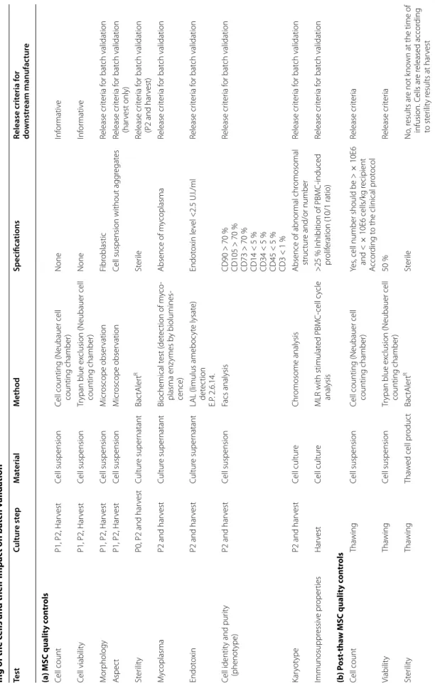
Table 1 Q ualit y c on tr ols p er formed a
t (a) the diff
er en t st eps of the cultur e and their impac t on ba tch v alida tion and qualit y c on tr ols p er formed a t (b ) the tha w -ing of the c
ells and their impac
t on ba tch v alida tion Test Cultur e st ep M at erial M ethod Specifica tions Release crit eria f or do wnstr eam manufac tur e (a) MSC qualit y c on tr ols Cell count P1, P2, Har vest Cell suspension
Cell counting (Neubauer cell counting chamber)
None Inf or mativ e Cell viabilit y P1, P2, Har vest Cell suspension Tr ypan blue ex
clusion (Neubauer cell
counting chamber) None Inf or mativ e M or phology P1, P2, Har vest Cell suspension M icr oscope obser vation Fibr oblastic Release cr iter ia f or bat ch validation A spec t P1, P2, Har vest Cell suspension M icr oscope obser vation
Cell suspension without agg
regat es Release cr iter ia f or bat ch validation (har vest only) St er ilit y P0, P2 and har vest Cultur e super natant Bac tAler t R St er ile Release cr iter ia f or bat ch validation (P2 and har vest) M ycoplasma P2 and har vest Cultur e super natant Biochemical t est ( det ec tion of m yco -plasma enz ymes b y biolumines -cence) Absence of m ycoplasma Release cr iter ia f or bat ch validation Endot oxin P2 and har vest Cultur e super natant
LAL (limulus ameboc
yt e lysat e) det ec tion E.P . 2.6.14. Endot oxin le vel <2.5 U .I./ml Release cr iter ia f or bat ch validation Cell identit y and pur ity (phenot ype) P2 and har vest Cell suspension Facs analysis CD90 > 70 % CD105 > 70 % CD73 > 70 % CD14 < 5 % CD34 < 5 % CD45 < 5 % CD3 < 1 % Release cr iter ia f or bat ch validation Kar yot ype P2 and har vest Cell cultur e Chr omosome analysis Absence of abnor mal chr omosomal struc tur e and/or number Release cr iter ia f or bat ch validation Immunosuppr essiv e pr oper ties Har vest Cell cultur e MLR with stimulat ed PBMC-cell c ycle analysis >25 % I nhibition of PBMC-induced pr olif eration (10/1 ratio) Release cr iter ia f or bat ch validation (b ) P ost-tha w MSC qualit y c on tr ols Cell count Tha wing Cell suspension
Cell counting (Neubauer cell counting chamber)
Yes
, cell number should be
> × 10E6 and < × 10E6 cells/k g r ecipient A ccor ding t o the clinical pr ot ocol Release cr iter ia Viabilit y Tha wing Cell suspension Tr ypan blue ex
clusion (Neubauer cell
counting chamber) 50 % Release cr iter ia St er ilit y Tha wing Tha w ed cell pr oduc t Bac tAler t R St er ile No , r esults ar e not k no wn at the time of infusion. C ells ar e r eleased accor ding to st er ilit y r esults at har vest
5B1 clone), vioblue-conjugated anti-CD14 (IgG2a, TÜK4 clone), vioblue-conjugated anti-CD34 (IgG2a, AC136 clone), vioblue-conjugated anti-CD3 (IgG2a, BW264/56 clone). Cells incubated with their corresponding isotype control (IgG1 PE, FITC and APC: IS5-21F5 clone, IgG2a vioblue and PerCP: S43.10 clone Miltenyi Biotec) were also included. Data were acquired on a Macsquant Flow Cytom-eter (Miltenyi Biotec) by collecting a minimum of 10,000 events and analyzed with Macsquantify software.
Mesenchymal stromal cells differentiation assays
Fat, bone and cartilage differentiation assays were
car-ried out as described by Pittenger et al. [2] and revealed
by staining with oil red O, alizarin red and toluidine blue, respectively. Differentiation media were home-made. MSC immunosuppression assays
1 × 10⁴ MSC were plated in triplicates in round-bottom
96-well plates (Becton–Dickinson) in a total volume of 100 μl of RPMI 1640 medium supplemented with 10 % FBS, 100 U/ml penicillin, 100 mg/ml streptomycin, l-glutamine (2 mM) (all from Lonza), sodium pyruvate (100 mM), non-essential amino acid (NEAA) (100 mM)
and 5 × 10⁻⁵ M β-mercaptoethanol (β-ME) (all from
Gibco, Merelbeek, Belgium). After 4-hour incubation, MSC were irradiated at 25 Gy using a 137Cs source (GammaCell 40, Nordion, Ontario, Canada).
Allogeneic human peripheral mononuclear cells (PBMC) were isolated from a blood sample (healthy
vol-unteer donor) by Ficoll PaqueR Plus density gradient.
PBMC (5 × 10⁴ or 1 × 10⁵) were then added to wells
in a total volume of 200 μl containing or not irradiated MSC, in the presence of anti-αCD3/CD28 microbeads (Invitrogen, Dynal A/S, Oslo, Norway). Co-cultures with-out anti-αCD3/CD28 microbeads were used as controls. Cells were then incubated at 37 °C in 5 % humidified air for 4 days. Cell cycle analysis of PBMC stimulated or not with anti-αCD3/CD28 microbeads and cultivated dur-ing 4 days with or without MSC were performed usdur-ing
CycleTESTR Plus DNA Reagent Kit
(Becton–Dickin-son). The percentage of cells in the different phases of the cell cycle was determined with the Macsquant Software (Miltenyi) or the Modfit Software (Becton–Dickinson). The effect of MSC on PBMC stimulation responses was calculated as percentage suppression compared with the proliferative response in the positive control without MSC (+- standard deviation of the mean). The positive control was set to 0 % suppression.
Cytogenetics
Karyotyping was performed at the Genetics Department of the Hospital by the Q-banding technique and analyzed
with CytovisionR software.
Microbiology testing
MSC sterility was assessed by bacterial culture
(aero-bia, anaerobia and fungi with BactalertR; Microbiology
department of the Hospital), mycoplasma screening
(luminometry, MycoalertR, Lonza; Microbiology
depart-ment of the Hospital) and endotoxin detection (limulus test, European pharmacopeia 2.6.14, Pharmacy depart-ment of the Hospital).
MSC release and thawing (Table 1b)
MSC are only prescribed and infused in the context of a clinical trial. Thus, patient eligibility/inclusion criteria, clinical evaluation and laboratory testing are protocol-specific and listed in each of the respective clinical trial descriptions. If the patient is eligible according to all pro-tocol criteria, he must sign the study informed consent form to allow release of the cells (a frozen MSC product is released for clinical use specifically for a patient according to the prescribed MSC dose and the patient weight).
If MSC are delivered to a patient inside the hospi-tal, they are thawed at the LTCG. Briefly, the MSC bag is protected in a sterile plastic bag and thawed in a 37 °C water bath for a few minutes. If the MSC bag is in a dual packaging (bags cryopreserved after April 2008), the bag is just immersed in the water bath a few seconds to take off easily the second packaging. Then the bag in its first packaging is quickly thawed in the water bath in a ster-ile protective bag. The bag is taken out of the water when the access sites are just thawed; there must still be small ice clots in the bag. Then, the bag is quickly transferred under the laminar flow and immediately perforated with a sampling-site coupler and diluted with a PBS buffer [1:0.75 (MSC solution:PBS)] (Clinical Grade, Miltenyi Biotec, Utrecht, The Netherlands) to avoid DMSO toxic-ity. Quality controls are performed after thawing of the
cells (Table 1b). Numeration and viability are assessed by
trypan blue coloration and cell count on a Neubauer cell counting chamber. We thus chose this method because it was easy and rapid (MSC must be infused as quickly as possible after thawing) and we did not have a flow cytom-eter available in the clean room to rapidly use other cell counting methods (7-AAD, PI). Besides, we do not rou-tinely re-assess phenotype, mycoplasma and endotoxins after thawing. Indeed freezing/thawing steps have been validated with all critical QC parameters meeting
eligibil-ity criteria (Table 2c). In addition, as thawed cells must be
infused as soon as possible, it is not possible to proceed with these QC before infusion. Moreover, the only new reagent introduced during the thawing step is PBS which is devoid of mycoplasma and endotoxins.
The cell product is then transferred in an appropri-ately labeled sterile transfer bag and transported to the hematology department of the hospital for infusion to
the patient. The released MSC product can also be trans-ferred frozen in a dry-shipper at −160 °C to another center for infusion after inclusion of a patient in a mul-ticenter clinical protocol. Upon preparation before ship-ment, the bag canister, dry shipper and protective box are labeled as described in the transportation SOP. The shipper is placed in a protective box and transported by a specialized transporter ordered by the destination hos-pital. In the recipient tissue bank, cells are then thawed according to the LTCG standard procedure.
Population doubling level (PDL) calculation
PDL was calculated according to the formula PDL = 3.322 (log Y−log I) where Y = number of cells harvested and I = number of cells inoculated at P1.
Doubling time (DT) estimation
Doubling time (DT) was calculated according to the for-mula DT = t × log (2)/log (number of cells harvested/ number of cells inoculated), where t is the time in hours between passage 1 and harvest of the cells.
Table 2 MSC culture process validation
Results of three large-scale MSC cultures for initial process validation: culture rates, phenotypes at final harvest and post-thaw parameters are reported
(a) Cultures rates
1st Exp. 2nd Exp. 3rd Exp.
BM sample (mL) 28 52 55
MNC on day 0 180 × 106 195 × 106 260 × 106
MSC on day 28 225 × 106 165 × 106 356 × 106
(b) Phenotypes
Marker Eligibility criteria (%) 1st Exp. (%) 2nd Exp. (%) 3rd Exp. (%) Conformity
CD73 >70 96.9 99.9 99.4 Ok CD105 >70 85.6 90.3 77.8 Ok CD90 >70 100 98.2 100 Ok CD34 <10 1.8 8.8 0.92 Ok CD45 <10 1.1 1.10 0.55 Ok HLA-DR <10 0.84 0.63 0.52 Ok CD80 <10 1.0 0.7 0.45 Ok CD31 <10 1.0 0.9 0.62 Ok (c) Post-thaw
Test Eligibility criteria 1st Exp. 2ndExp. 3rd Exp. Conformity
Initial cell number Na 100 × 106 70 × 106 55 × 106 Na
Initial viability Na 95.8 % 82.5 % 86.0 % Na
Post-thaw cell count Na 100 × 106 56 × 106 43 × 106 Na
Post-thaw cell viability >50 % 89.5 % 78.0 % 78.0 % Ok
Post-thaw cell culture expansion Na Ok Ok Ok Na
Sterility Sterile Ok Ok Ok Ok
Mycoplasma Absence of mycoplasma Ok Ok Ok Na
Endotoxin Endotoxin level <2.5 U.I./ml Ok Ok Ok Na
Phenotype CD73 >70 % 99.5 % 96.1 % 87.9 % Ok CD105 >70 % 93.6 % 70.3 % 70.0 % Ok CD90 >70 % 100 % 99.6 % 99.8 % Ok CD34 <10 % 3.1 % 2.7 % 2.6 % Ok CD45 <10 % 1.0 % 1.9 % 1.0 % Ok HLA-DR <10 % 1.0 % 1.2 % 1.0 % Ok CD80 <10 % 0.4 % 0.8 % 0.4 % Ok CD31 <10 % 0.6 % 1.2 % 0.6 % Ok
Statistics
Results are reported as mean ± standard error of the mean (SEM). Comparisons between conditions were made using Student unpaired t tests with GraphPad Prism 5.00 software (GraphPad Software, La Jolla, CA, USA).
Results
Large‑scale cultures—process validation
After a few small-scale MSC expansions to set up the process, three large-scale clinical MSC cultures were ini-tiated for validation with three different bone marrow (BM) samples obtained from healthy volunteer donors. All quality controls fulfilled pre-defined qualification
cri-teria (Table 2).
Freezing/thawing steps were also validated. For each previous culture, frozen MSC were thawed and criti-cal parameters were evaluated. Again, all quality
con-trols met eligibility criteria (Table 2c). Thawed cells were
also devoid of bacterial, mycoplasma and endotoxin contamination.
Shelf life determination of the product after thawing was also assessed. Cells from four different bags were left (or not = control) in the post-thaw buffer at room tem-perature for different times (1, 2, 4H). At each time point, cell count and viability were assessed. Viability and cell count seemed stable for 2 h but drop significantly when kept for longer periods. Cells were also seeded in culture flasks to test their proliferation potential. When replated, the cell proliferative potential was affected as early as after 1 h in the post-thaw buffer.
The effects of long-term cryopreservation were not sys-tematically addressed. However, three MSC bags were thawed after 7–8 years of storage and showed excellent post-thaw viability (80, 77 and 83 %, respectively) and recovery (83, 71 and 94 %, respectively). This indicates a good stability of the cell product during long-term stor-age in liquid nitrogen. Systematic evaluation of long-term MSC stability is scheduled for future batches produced under GMP recommendations.
MSC banking
After appropriate validation, we started in November 2006 a clinical-grade third party MSC bank following the EBMT manufacturing process.
During the past 7.5 years, 61 donors were screened. One donor was discarded for multiple allergies and col-lection was technically impossible for another one. From the 59 validated donors (36 females and 23 males), 70 large-scale MSC expansions were initiated and completed
(Table 3). Donors were between 18 and 52 years of age
(median 26.7 years). There is no standard for MSC donor age. Ten percent (7/68) of our donors were older than
40 years and 90 % were between 18 and 40, with a mean of 29 years. The older donors were collected at the begin-ning of our banking activity, but now we select donors younger than 40 years of age. Volumes of collected bone marrow ranged from 25 to 70 mL (median 50 mL) and total number of mononucleated cells obtained after ficoll
ranged from 124 to 956 × 106 (median 280 × 106).
All the initiated cultures except two gave rise to colo-nies and completed the MSC culture process resulting in 1 to 45 MSC unit bags per culture. A total of 464 MSC bags have been produced and stored from the 68
com-pleted cultures (Table 3; Fig. 2).
Table 3 MSC production between 2007 and 2015
Criteria Min. Max. Median
MSC culture parameters
Donor age (years) 18 52 26.7
BM sample volume (mL) 25 70 50 Mononuclear cells post-Ficoll (×106) 124 956 280
CFU-F (number per 2 × 10⁶ cells) 2 96 25
Final MSC yields (×106) 19 5431 546
PDL (P1-harvest) 2.38 7.18 4.69
DT in hours (P1-harvest) 46.8 141 71.7
Viability (%) 72 100 85
Number of aliquots (=bags)/culture 1 45 4 Number of cells/aliquot (×106) 19 189 110
Fig. 2 MSC banking: production and release activity. a Production:
From 59 validated donors, 68 cultures were completed allowing freezing of 464 MSC bags. From these, 430 are already validated, 18 non-compliant and 16 are still awaiting validation. b Release: Since 2007, 314 bags have been released. From those, 290 were MSC and 24 placebo; 187 were released on site and 103 were sent to other Belgian centers
Final yields ranged from 19 × 106 to 5431 × 106 cells
with a median of 546 × 106 cells per culture.
Popula-tion doubling levels (PDL) between passage 1 and pas-sage 3 (harvest) ranged from 2.38 to 7.18 with a median of 4.69. Doubling times (DT) ranged from 46.8 to 141 h with a median of 71.7 h. We didn’t observe any correla-tion between age and Colony-Forming Units or between CFU-F and yield of the culture/mL BM processed (data not shown). However, although sex didn’t influence the yield of CFU-F (P = 0.55), male BM samples gave rise to significantly higher total cell yields compared to cultures obtained from female BM samples (P = 0.0011).
Viability at harvesting was generally excellent with a mean of 88.9 ± 5.5 %. All the harvested MSC were fro-zen in adequate aliquots at −160 °C in liquid nitrogen. Aliquots contained various numbers of cells in order to cover a wide range of patient weights, considering that
the ideal MSC dose is ranging between 1–4 × 10⁶/kg
recipients. MSC were stored in bags containing between
19 × 106 and 189 × 106 cells with a mean of 132.8 × 10⁶
cells per bag. We did not freeze standardized quantities because the required cell count for infusion is defined by the protocol in which the patient is included as a weight-based dose. The acceptable dose is always within a speci-fied range, for example 1–2 or 3–4 × 10E6 cells/kg. The minimal cell count is the minimal acceptable dose of viable cells multiplied by the patient’s weight. So when a patient is included in a clinical trial, we choose an ali-quot containing the optimal amount of cells according to the dose prescribed in the study and the patient weight. In this context, cell doses as small as 20 × 10E6 cells can be stored and released for low-weight patients (pediatric cases).
All cultures were initiated with fresh cells but due to
technical limitations, a maximum of 300 T175 cm2 were
replated at passage 2. If cell number exceeded this capac-ity, the cells were frozen at P2 and thawed later to allow further expansion and final harvest. Each harvest gave rise to one MSC batch so that one culture can generate one or more batches.
The large range in the final yields is due to variability in the intrinsic quality of the collected BM and also to variability in the efficiency of the different FBS batches (always pre-validated but some were better than others). As explained in the text, two cultures did not expand and were discarded. No T flask was discarded. Noteworthy, the extreme expansion rates (low and high) were rare,
with most cultures (>80 %) yielding >200 × 106 cells.
Quality controls and compliance with release criteria Each MSC batch underwent extensive testing to verify its conformity with release criteria and was individually validated. EBMT release criteria were applied to release
batches but retrospective analysis revealed that cells were also compliant with the stricter ISCT phenotype criteria (>95 % expression CD90, CD105, CD73; <2 % for CD14, CD45, CD34 and <1 % for CD3). HLA-DR expression was also evaluated for all produced batches and was always below 2 % at harvest, but this was not a release criterion.
All but 3 batches satisfied to release criteria. Indeed, the batches from two cultures had to be discarded for non-conformity due to a donor constitutional anomaly detected by karyotypic analysis and further confirmed by analysis of donor lymphocytes. One batch was discarded due to a positive bacteriology result. MSC were also evaluated in a stimulation assay by incubation with
anti-CD3/CD28. As shown in Fig. 3, proliferation of
stimu-lated PBMC was significantly reduced by the addition of MSC [mean inhibition (10/1 ratio): 41 ± 9 %, range 25–57 %]. Thawed cells were cultured and also demon-strated good inhibitory properties (>25 % inhibition in a 10/1 ratio; data not shown). In summary, 464 MSC bags have been produced and frozen in our facility during
the past 7 years (Fig. 2). Among them, 430 (93 %) have
already been validated according to EBMT consortium release criteria and 16 are still awaiting validation.
Eight-een bags had to be discarded (3.9 %) (Fig. 2).
All placebo bags were sterile and fulfilled release criteria.
MSC release and thawing
All MSC products validated for clinical use were listed according to their cellular content, which enabled us to choose the adapted MSC product for each patient according to his/her weight and the dose specifications of the clinical trial in which the patient was included.
When a patient is included in a clinical trial, the selected MSC bag(s) is (are) released and thawed for infu-sion. Thawed MSC are quickly diluted with buffer and an aliquot is collected to assess MSC numeration and viabil-ity. If cell recovery is adequate for dose specifications and viability above 50 %, MSC infusion is allowed. Thawed cells must be infused as soon as possible and in all cases within 1 h after thawing. In order to respect the set time limit, before each scheduled infusion, we verify that the patient is in good condition and ready to receive the MSC before thawing the bag.
As of June 1 2015, 314 bags have been released (24 pla-cebo and 290 MSC bags) and infused to patients. Among these, 103 were transported frozen to other Belgian cent-ers before being thawed on site and 187 were prepared in
the LTCG (Fig. 2b).
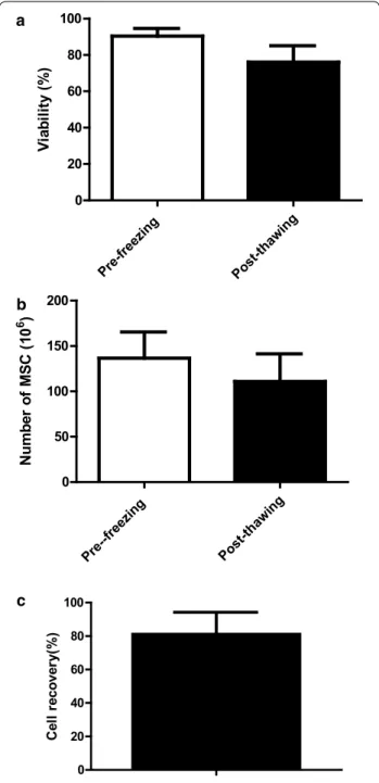
Mean viability at freezing was 90 ± 4 % (Fig. 4a),
rang-ing from 80 to 100 %. Viability dropped to 76 ± 9 % after thawing (range 50 % to 96 %). The mean decrease of via-bility during thawing was 14 % (P < 0.0001). However, all
thawed MSC units satisfied to release criteria as none showed viability below 50 %. To our knowledge, there is no standard in Europe for viability of such products and this 50 % viability cutoff was accepted by our regulatory authorities. However, the mean viability of our MSC
after thawing was 76 ± 9 % (Fig. 4a) which is quite higher
indeed. Noteworthy, very few bags showed viabilities between 50 and 60 % (3/167 = 1.8 %).
The mean cell content of the released bags was
136.6 ± 28.8 × 10⁶ cells (range 65 to 189 × 106) before
freezing (Fig. 4b) and 111 ± 30.5 × 10⁶ after
thaw-ing (range 45.5 to 189 × 106), with a mean recovery of
81 ± 13 % (range 50 to 115 %) (Fig. 4c).
All thawed bags (MSC and Placebo) were subjected to bacteriological analysis and results were all negative.
Post‑thaw MSC potency
In order to evaluate post thaw MSC immunosuppressive properties, five MSC bags were thawed in separate exper-iments. Cells were washed in tubes and resuspended
in complete media before being plated in T175 cm2
flasks. At different time points (day 1, 2, 3 and 4), cells were detached and evaluated for different parameters: recovery (compared to the number of seeded cells after
Fig. 3 MSC immunosuppressive properties. Inhibition of PBMC
prolif-eration by third party MSC: PBMC (100,000 or 50,000) were stimulated (S-PBMC) with anti-αCD3/CD28 microbeads during 4 days with or without irradiated (25 Gy) MSC (10/1 or 5/1 PBMC/MSC ratios) added at the beginning of the culture. Proliferation was assessed by analysis of the cell cycle by flow cytometry (N = 28). Result are expressed as the percentage of cells present in S + G2 M phases (a) and as the percentage of inhibition compared to the stimulated PBMC condition alone (b) for the 10/1 PBMC/MSC ratio
Pre-f reezin g Po st-thawi ng 0 20 40 60 80 100 Vi ab ilit y (% ) Pr e--freez ing Po st-thawi ng 0 50 100 150 200 Numb er of MS C (1 0 6 ) 0 20 40 60 80 100 Ce ll re co ve ry (% ) a b c
Fig. 4 MSC post-thaw viability and cell recovery. Pre- and post-thaw
MSC viability (a), MSC number (b), as well as recovery of cell number (c) (N = 170)
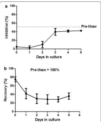
washing), viability, phenotype, differentiation and immu-nosuppressive properties. Thawed MSC demonstrated very poor PBMC inhibition capacities in MLR assays (day
1: 3 ± 4 %; day 2: 10 ± 7 %; (Table 4). Indeed, 3 days of
culture were necessary to restore a full and stable PBMC inhibition ability (day 3: 40 ± 9 %; day 4: 42 ± 2 %; day 5:
43 ± 1 %) (Table 4; Fig. 5).
Recoveries were very low until day 4 (Fig. 5) before a
slight increase thereafter indicating that cells had begun to re-proliferate. Thus, restoration of MSC immunosup-pressive properties correlated with a stop in MSC loss. Nevertheless, cells did not require re-culture to maintain their differentiation capacities towards adipocytic, osteo-blastic and chondroosteo-blastic lineages.
Discussion
The purpose of this paper was to demonstrate the feasibil-ity and describe the difficulties of setting up a large MSC bank from allogeneic donors for use in academic clinical trials. The constitution of a human third-party MSC bank from screened healthy volunteer donors and the follow-up of different aspects of this activity since 2007 are devel-oped. Results from 68 large scale clinical expansions are described and discussed. Methods of generating clinical-grade MSCs have already been described by other groups but, to our knowledge, this is the first paper describing
such a large and detailed banking experience [14, 15].
The absence of co-stimulatory molecules and human leucocyte antigen (HLA) class II molecules, as well as low HLA class I expression on MSCs, make them ideal for allogeneic use. MSC produced and used in our clini-cal trials are of allogeneic origin but do not seem to elicit immune responses. Indeed, HLA-mismatched MSC manufactured by the same process were shown to be weakly immunogenic after infusion into HSCT
recipi-ents [16]. One concern when expanding large number
of MSC is the number of passages required to meet dose requirements. Some studies indicate that MSC proper-ties change with passages and that immunosuppressive capacities become less potent when multiplying passages
[17, 18]. Von Bar et al. published recently clinical results
that suggest that acute GVHD patients treated with early passage MSC had a better survival than those treated
with late passage cells [19]. In our manufacturing
pro-cess, MSC are only cultured for three passages (14 days of primary culture and 2 passages of 7 days each). This is quite low and comparable to a MSC culture process yield-ing comparable cell numbers in four shorter passages
as reported by Sabatino et al. [14]. However, it is now
generally accepted that MSC will begin to senesce after a certain number of cell divisions and that this is best Table 4 MSC thawing: pre and post-thaw MSC
immuno-suppressive properties
Results are shown for MSC recovery, viability and immunosuppressive properties (%) at different times after thawing and replating in T-flasks with complete media
(%) Min. Max. Mean ± SD
Day 0 Recovery 61 84 72.8 ± 10.4 Viability 61 71 65.1 ± 4.4 Immunosuppression 5 5 5.0 ± 0.0 Day 1 Recovery 21 54 41.8 ± 12.9 Viability 86 91 88.8 ± 2.1 Immunosuppression 0 10 2.9 ± 4.1 Day 2 Recovery 19 40 30.3 ± 11.3 Viability 78 87 83.9 ± 4.0 Immunosuppression 4 19 10.3 ± 7.4 Day 3 Recovery 21 36 26.8 ± 10.8 Viability 74 97 85.5 ± 16.3 Immunosuppression 34 47 40.5 ± 8.6 Day 4 Recovery 21 34 28.3 ± 6.5 Viability 89 93 90.3 ± 2.3 Immunosuppression 40 43 41.8 ± 2.2 Day 5 Recovery 31 42 36.3 ± 7.5 Viability 85 91 88.0 ± 4.2 Immunosuppression 42 44 42.6 ± 1.3
Fig. 5 MSC post-thaw immunosuppressive properties. MSC bags
were thawed (N = 5), washed and replated in flasks in complete media. Immunosuppressive properties (a) and cell recoveries (b) were evaluated at different time points after thawing
evaluated by their population doubling level (PDL) rather than by the number of passages or the duration of cul-ture. As it is very difficult to evaluate the starting number of MSC in the initial culture (mixture of mononuclear cells), most labs start counting MSC cumulative popu-lation doubling at the end of the primary culture (first passage). The calculated PDL of our MSC culture ranged from 2.38 to 7.18 with a median of 4.69. This is consist-ent with standard MSC protocols showing 2.5–3 popu-lation doublings per passage. Moreover, the observed PDL are still below the PDL correlating with occurrence of MSC senescence (PDL from 10 to 40). However, the PDL of these cells in vivo before collection of the BM, which also depends on donor age, are not known. Dur-ing our large culture experience, we were able to produce 1 to 45 doses of MSCs from each BM sample in only 3 passages demonstrating the efficacy of the process. We consider that it is very important to produce MSC from multiple donors for two main reasons: First, to limit the number of passages and second to have a large variabil-ity of donors. Indeed, it has been suggested that if one donor is used to produce a multiplicity of MSC doses to treat a lot of patients, a potency bias may be observed
[20]. Indeed, MSC from different donors may have
dif-ferent interferon-gamma responsiveness (low or high IDO inducers) and thus different potency. The outcome of a patient receiving MSC from a low or high responder may be different. On the other hand, cells that are subject to a high proliferative pressure and late passage random donor MSC could be less effective than early passage
MSC [19]. These factors may explain the below
expecta-tion results of the phase III trial of random donor MSC in
steroid-resistant GVHD with the ProchymalR industrial
MSC product [20, 21]. Indeed, despite promising phase II
trials, this phase III clinical trial did not meet its primary
end point [22]. However, the product has been approved
in two countries based on statistically significant benefits observed at certain disease sites.
Despite low passage cultures, we have been able to produce 464 MSC doses in 68 cultures that allowed us to create an allogeneic “off-the-shelf” MSC bank with a large number of frozen aliquots.
Up to June 2015, 290 MSC bags have been released and safely infused to patients included in our 6 current clinical protocols. We have evaluated cell viability and recovery from these thawed MSC products. Viability of fresh cells (90.3 ± 4.3 %) dropped to 76 ± 9.1 % after thawing (mean loss 14 %), but remained sufficient for meeting release criteria. These results compare favorably with those of Polchow et al. who describe a 20 % loss of viability (from 84.4 ± 9.4 to 67.4 ± 7.6 %) with thawing after short-time cryopreservation of human umbilical
cord artery-derived cells (HUCAC) [23]. However, this
difference can be explained by the fact that these cells were of different origin and cryopreserved in vials and not in bags. This was also the case for other studies that are difficult to compare because of dysparities in origin or culture stage of cells, cryopreservation and thawing
methods [24–26].
Beside viability and recovery, freezing/thawing steps may also affect MSC potency. Indeed Galipeau et al. reported recently that post-thaw cells have impaired
immunosuppressive properties [27]. They demonstrate
that post-thawed MSC up-regulate heat-shock proteins, are refractory to interferon–γ-induced up-regulation of indoleamine 2,3-dioxygenase (IDO) and are compro-mised in suppressing CD3/CD28/-driven T cell prolifera-tion. These properties were fully restored following 24 h of post-thaw MSC culture. In order to assess whether potency of our MSC presented similar kinetics, we thawed MSC bags and seeded cells back in culture for different periods of time before evaluating their viability, proliferation and potency (immunosuppressive prop-erties and differentiation abilities). In accordance with Galipeau et al., we observed that after thawing, MSC are compromised in suppressing CD3/CD28/-driven T-cell proliferation. In our hands, 3 days of culture were nec-essary to fully restore stable immunosuppressive proper-ties. This correlated with a recovery of their proliferative potential, indicating that MSC probably need a 3-day recovery period after thawing. However, differentiation potential was maintained even when evaluated immedi-ately after thawing. Nevertheless, the relevance of these in vitro findings for the immunosuppressive efficacy of MSC is unknown. In vivo recovery of various cell prop-erties may be quite different in the recipient body envi-ronment. Indeed, Von Bahr et al. have demonstrated that in vitro testing of MSC inhibitory capacity in MLC or mitogenic response to PHA did not correspond with
in vivo responses [19]. MSC potency defined by MLR
assay may not be a good predictor of their in vivo effects. Krampera et al. recently published a working proposal of the ISCT for immunological characterization of MSC
[28]. It mentioned the urgent need as part of cell
man-ufacturing to develop and implement robust potency assays that may be associated with clinical effects and suggest different proposals including the use of IFN-γ and/or TNF-α in vitro primed MSC; investigation of cell-surface markers expression by flow cytometry for char-acterizing MSC immunological properties; the use of purified immune effector cells subsets in MLR instead of unselected peripheral blood mononuclear cells (PBMC), the of study some suppressor pathways induced by MSC such as IDO activation and the use of animal models to
evaluate MSC immunosuppressive properties The aim of standardization is to obtain reproducible and consist-ent in vitro data that reflect immunological properties of MSCs infused to patients.
In this paper, we report optimization of clinical-grade large-scale MSC expansion with the set-up of a MSC bank and summarize 8 years of experience. The technical process in place leads to per donor cell yields that are sufficient for therapeutic purposes (median of 546 × 10E6 MSC/cul-ture = 4 doses of 2 × 10E6 cells/kg for an average 75-kg patient). However, the manipulation of large quantities of T-flasks is a hurdle (time consuming, space consuming and open system) and, to avoid these disadvantages, we have
validated the fully automated closed Quantum®
bioreac-tor for our process and published our observations [29].
The major advantages of the bioreactor were that (1) cells
grow better in the Quantum® than in flasks, (2) working
time is shorter especially at the final harvest step, and (3) all the feeding tasks are done automatically. This system thus allows production of large quantities of MSC. However, the cost of the device counterbalances its advantages, so that we did not implement it in our manufacturing strategy.
In the meantime, MSC have become considered as advanced therapy medicinal products (ATMP) by the European Medicines Agency (EMA), are under Euro-pean Regulation N° 1394/2007 and must be produced in compliance with Good Manufacturing Practices (GMP). GMP requires thorough analysis of critical aspects such as donor screening, production processes and quality controls of expanded MSC (safety, identity and efficacy)
[30]. Our MSC are produced under strict and defined
parameters from donor screening to culture reagents and processes, using screened and irradiated FBS batches and subjected to extensive quality controls for safety (bacte-riological analysis, endotoxin and mycoplasma detec-tion), identity (FACS analysis according to ISCT criteria) and potency (immunosuppressive properties). Due to extensive washing and dilution of the cells before cryo-preservation, residual reagents (trypsin–EDTA, FBS, and penicillin/streptomycin) are present at extremely low lev-els (0.00138, 0.00024 and 0.000000286 %, respectively) after thawing. In addition, karyotype analysis performed on each batch has consistently shown genetic stability of the cells. We also showed that despite cell trapping in the lungs after intravenous infusion, this did not result in
deleterious effects on lung function ([31]. In an attempt
to improve aseptic conditions, GMP recommends the development of closed processes. For that purpose, we have successfully implemented in 2012 a new method for selection of mononuclear cells from BM samples
with the fully closed SepaxR device [32]. Similarly, as
explained earlier, we have validated the QuantumR
bio-reactor but didn’t implement it in our manufacturing
process because of cost concerns. However, closed sys-tems are not an absolute requirement of GMP and sev-eral groups have described GMP-compliant culture
processes in Cellstacks [33] or T-flasks [15]. We are now
in the last phase of GMP implementation in our process of MSC banking. Briefly, major changes applied to the process concern the environment (extensive monitor-ing, class B room with adapted controls of work environ-ment and equipenviron-ment, clothes, hygienic and safety rules, sanitization of the clean room and maintenance/checking of equipment), technical process (replacement of some reagents, use of cellstacks instead of T-Flasks, separa-tion of people involved in producsepara-tion and in quality con-trol), quality control (formal validation of all internal QC methods and use of GMP-certified laboratories for sub-contracted analyses, quarantine status before release of batches by a qualified person), reagents (quarantine sta-tus until release by the qualified person according to their specifications) and a considerable extension of the docu-mentation system and traceability of all processes. Conclusion
In conclusion, we report here an efficient clinical-grade MSC banking activity in place for more than 7 years. This activity now has to be performed in accordance with GMP requirements. A significant challenge remains the development and implementation of standardized potency assays and the demonstration of a correlation with relevant endpoints in clinical trials.
Abbreviations
MSC: mesenchymal stem/stromal cells; HLA: human leukocyte antigen; GVHD: graft-versus-host disease; CD: Crohn’s disease; RA: rheumatoid arthritis; ISCT: International Society of Cellular Therapy; LTCG: Laboratory of Cell and Gene Therapy; BM: bone marrow; GVHD: graft versus host disease; NMAHCT: non myeloablative hematopoietic cell transplantation; GVT: graft versus tumor; EBMT: European Group for Blood and Marrow Transplantation; HCT: hematopoietic cell transplantation; HSC: hematopoietic stem cells; CBT: cord blood transplantation; GMP: good manufacturing practise; UMN: urgent medical need; SOP: standard operating procedures; MNC: mononuclear cells; PBS: phosphate-buffer saline; DMEM: Dulbecco’s modified eagles medium; EDTA: ethylène diamine tetra acétate; FBS: foetal bovine serum; P/S: penicil-line/streptomycine; HAS: human serum albumin; DMSO: diméthylsulfoxyde; CFU-F: colony forming unit fibroblasts; MLR: mixed lymphocyte reaction; R&D: research and development; PBMC: peripheral blood mononuclear cells; HSCT: human stem cell transplantation; IDO: indoléamine oxidase; HUCAC: human umbilical cord artery derived cells; CPA: cryoprotectant; EMA: European medi-cines agency; ATMP: advanced therapeutic medicinal product.
Authors’ contributions
CL performed upscaling and validation of MSC clinical process, supervision and organization of the clinical-grade cultures until end 2012 and quality con-trols during all the study. AB assumed MSC production from 2012 until now. OG is the responsible of the quality aspect of MSC banking. OD is the Qualified Person of the lab. EB is the director of the lab and performed all the Bone Mar-row collections. YB is the head of the department and reviewed extensively the manuscript. All authors read and approved the final manuscript. Author details
1 Department of Hematology, Laboratory of Cell and Gene Therapy, CHU of Liège and University of Liège, CHU Sart-Tilman, 4000 Liège, Belgium.
2 Department of Medicine, Division of Hematology, CHU and University of Liège, Liège, Belgium.
Acknowledgements
We are very grateful to Delphine Bourgeois, Sylviane Simar, Tiffany Crepin, Amélie Halleux, Pascale Dufour and Amelie Lubcke for their technical assis-tance. We also thank Mauricette Jamar and Christian Herens for karyotype analysis. The authors are also grateful to Sandra Ormenese and Raafat Stephan from the Imaging and Flow Cytometry Platform of the GIGA for help with cell cycle analyses.
Availability of data and materials section
The dataset(s) supporting the conclusions of this article is (are) included within the article.
Competing interests
The authors declare that they have no competing interests. Ethics approval and consent to participate
The study was approved by the human and animal Ethics Committees of the University of Liege. Written informed consent was obtained from all bone marrow donors in accordance with the Declaration of Helsinki.
Funding
This work has been supported by Grants from the Belgian Federal Cancer Plan, the National Fund for Scientific Research (FNRS) and the Belgian Foundation against Cancer.
Received: 9 March 2016 Accepted: 3 May 2016
References
1. Friedenstein AJ, Petrakova KV, Kurolesova AI, Frolova GP. Heterotopic of bone marrow. Analysis of precursor cells for osteogenic and hematopoi-etic tissues. Transplantation. 1968;6:230–47.
2. Pittenger MF, Mackay AM, Beck SC, Jaiswal RK, Douglas R, Mosca JD, et al. Multilineage potential of adult human mesenchymal stem cells. Science. 1999;284:143–7.
3. Le BK, Ringden O. Mesenchymal stem cells: properties and role in clinical bone marrow transplantation. Curr Opin Immunol. 2006;18:586–91. 4. Le BK, Tammik L, Sundberg B, Haynesworth SE, Ringden O. Mesenchymal
stem cells inhibit and stimulate mixed lymphocyte cultures and mito-genic responses independently of the major histocompatibility complex. Scand J Immunol. 2003;57:11–20.
5. English K, French A, Wood KJ. Mesenchymal stromal cells: facilitators of successful transplantation? Cell Stem Cell. 2010;7:431–42.
6. Uccelli A, Moretta L, Pistoia V. Mesenchymal stem cells in health and disease. Nat Rev Immunol. 2008;8:726–36.
7. Di NM, Carlo-Stella C, Magni M, Milanesi M, Longoni PD, Matteucci P, et al. Human bone marrow stromal cells suppress T-lymphocyte proliferation induced by cellular or nonspecific mitogenic stimuli. Blood. 2002;99:3838–43.
8. Le BK, Tammik C, Rosendahl K, Zetterberg E, Ringden O. HLA expres-sion and immunologic properties of differentiated and undifferentiated mesenchymal stem cells. Exp Hematol. 2003;31:890–6.
9. Yi T, Song SU. Immunomodulatory properties of mesenchymal stem cells and their therapeutic applications. Arch Pharm Res. 2012;35:213–21. 10. Horwitz EM, Le BK, Dominici M, Mueller I, Slaper-Cortenbach I, Marini FC,
et al. Clarification of the nomenclature for MSC: The International Society for Cellular Therapy position statement. Cytotherapy. 2005;7:393–5. 11. Dominici M, Le BK, Mueller I, Slaper-Cortenbach I, Marini F, Krause D, et al.
Minimal criteria for defining multipotent mesenchymal stromal cells. The International Society for Cellular Therapy position statement. Cytother-apy. 2006;8:315–7.
12. Le Blanc K, Frassoni F, Ball L, Locatelli F, Roelofs H, Lewis I, et al. Mesenchy-mal stem cells for treatment of steroid-resistant, severe, acute graft-versus-host disease: a phase II study. Lancet. 2008;371:1579–86.
13. Baron F, Lechanteur C, Willems E, Bruck F, Baudoux E, Seidel L, et al. Cotransplantation of mesenchymal stem cells might prevent death from versus-host disease (GVHD) without abrogating graft-versus-tumor effects after HLA-mismatched allogeneic transplantation following nonmyeloablative conditioning. Biol Blood Marrow Transplant. 2010;16:838–47.
14. Sabatino M, Ren J, David-Ocampo V, England L, McGann M, Tran M, et al. The establishment of a bank of stored clinical bone marrow stromal cell products. J Transl Med. 2012;10:23.
15. Hanley PJ, Mei Z, da Graca Cabreira-Hansen M, Klis M, Li W, Zhao Y, et al. Manufacturing mesenchymal stromal cells for phase I clinical trials. Cyto-therapy. 2013;15:416–22.
16. Sundin M, Barrett AJ, Ringden O, Uzunel M, Lonnies H, Dackland AL, et al. HSCT recipients have specific tolerance to MSC but not to the MSC donor. J Immunother. 2009;32:755–64.
17. Banfi A, Muraglia A, Dozin B, Mastrogiacomo M, Cancedda R, Quarto R. Proliferation kinetics and differentiation potential of ex vivo expanded human bone marrow stromal cells: implications for their use in cell therapy. Exp Hematol. 2000;28:707–15.
18. Tanabe S, Sato Y, Suzuki T, Suzuki K, Nagao T, Yamaguchi T. Gene expression profiling of human mesenchymal stem cells for identifica-tion of novel markers in early- and late-stage cell culture. J Biochem. 2008;144:399–408.
19. von Bahr L, Sundberg B, Lonnies L, Sander B, Karbach H, Hagglund H, et al. Long-term complications, immunologic effects, and role of passage for outcome in mesenchymal stromal cell therapy. Biol Blood Marrow Transplant. 2012;18:557–64.
20. Galipeau J. The mesenchymal stromal cells dilemma—does a negative phase III trial of random donor mesenchymal stromal cells in steroid-resistant graft-versus-host disease represent a death knell or a bump in the road? Cytotherapy. 2013;15:2–8.
21. Martin PJ, Uberti JP, Soiffer RJ, Klingemann H, Waller EK, Daly AS, Her-rmann RP, Kebriaei P. Prochymal improves response rates in patients with steroid-refractory acute graft versus host disease (SR-GVHD) involving the liver and gut: results of a randomized, placebo-controlled, multicenter phase III trial in GVHD. Biol Blood Marrow Transplant. 2010;16(2):S169–70. 22. Chen GL, Paplham P, McCarthy PL. Remestemcel-L for acute
graft-versus-host disease therapy. Expert Opin Biol Ther. 2014;14:261–9.
23. Polchow B, Kebbel K, Schmiedeknecht G, Reichardt A, Henrich W, Hetzer R, et al. Cryopreservation of human vascular umbilical cord cells under good manufacturing practice conditions for future cell banks. J Transl Med. 2012;10:98.
24. Ginis I, Grinblat B, Shirvan MH. Evaluation of bone marrow-derived mes-enchymal stem cells after cryopreservation and hypothermic storage in clinically safe medium. Tissue Eng Part C Methods. 2012;18:453–63. 25. Chatzistamatiou TK, Papassavas AC, Michalopoulos E, Gamaloutsos C, Mallis P, Gontika I, et al. Optimizing isolation culture and freezing methods to preserve Wharton’s jelly’s mesenchymal stem cell (MSC) properties: an MSC banking protocol validation for the Hellenic Cord Blood Bank. Transfusion. 2014;54:3108–20.
26. Pravdyuk AI, Petrenko YA, Fuller BJ, Petrenko AY. Cryopreservation of alginate encapsulated mesenchymal stromal cells. Cryobiology. 2013;66:215–22.
27. Francois M, Copland IB, Yuan S, Romieu-Mourez R, Waller EK, Galipeau J. Cryopreserved mesenchymal stromal cells display impaired immuno-suppressive properties as a result of heat-shock response and impaired interferon-gamma licensing. Cytotherapy. 2012;14:147–52.
28. Krampera M, Galipeau J, Shi Y, Tarte K, Sensebe L. Immunological char-acterization of multipotent mesenchymal stromal cells—The Interna-tional Society for Cellular Therapy (ISCT) working proposal. Cytotherapy. 2013;15:1054–61.
29. Lechanteur C, Baila S, Janssens ME, Giet O, Briquet A, Baudoux E, et al. Large-scale clinical expansion of mesenchymal stem cells in the GMP-compliant, closed automated quantumR cell expansion system: compari-son with expansion in traditional T-flasks. J. Stem Cell Res Ther. 2014;4:222. 30. Sensebe L, Gadelorge M, Fleury-Cappellesso S. Production of
mesenchy-mal stromesenchy-mal/stem cells according to good manufacturing practices: a review. Stem Cell Res Ther. 2013;4:66.
31. Moermans C, Lechanteur C, Baudoux E, Giet O, Henket M, Seidel L, et al. Impact of cotransplantation of mesenchymal stem cells on lung function after unrelated allogeneic hematopoietic stem cell
• We accept pre-submission inquiries
• Our selector tool helps you to find the most relevant journal • We provide round the clock customer support
• Convenient online submission • Thorough peer review
• Inclusion in PubMed and all major indexing services • Maximum visibility for your research
Submit your manuscript at www.biomedcentral.com/submit
Submit your next manuscript to BioMed Central
and we will help you at every step:
transplantation following non-myeloablative conditioning. Transplanta-tion. 2014;98:348–53.
32. Guven S, Karagianni M, Schwalbe M, Schreiner S, Farhadi J, Bula S, et al. Validation of an automated procedure to isolate human adipose tissue-derived cells by using the Sepax(R) technology. Tissue Eng Part C Methods. 2012;18:575–82.
33. Fekete N, Rojewski MT, Furst D, Kreja L, Ignatius A, Dausend J, et al. GMP-compliant isolation and large-scale expansion of bone marrow-derived MSC. PLoS One. 2012;7:e43255.