Topically Applied Vitamin C Enhances the mRNA Level of Collagens I and
III, Their Processing Enzymes and Tissue Inhibitor of M
1atrix
Metalloproteinase 1 in the Human Dermis
1Betty V. Nusgens, Philippe Humbert,* André Rougier,† Alain C. Colige, Marek Haftek,‡ Charles A. Lambert, Alain Richard, †Pierre Creidi,* and Charles M. Lapière
Laboratory of Connective Tissues Biology, Tour de Pathologie, University of Liège, Sart Tilman, Belgium; *Department of Dermatology, Hôpital Saint Jacques, University of Franche-Comté, Besançon, France; †La Roche-Posay, Laboratoire Pharmaceutique, Asnières, France; ‡INSERM U346/CNRS, Hôpital E. Herriot, Lyon, France
Abstract
Ascorbic acid (vitamin C) is a cofactor required for the function of several hydroxylases and mono-oxygenases. It is not synthesized in humans and some other animal species and has to be provided by diet or pharmacologic means. Its absence is responsible for scurvy, a condition related in its initial phases to a defective synthesis of collagen by the reduced function of prolylhydroxylase and production of collagen polypeptides lacking hydroxyproline, therefore, they are unable to assemble into stable triple-helical collagen molecules. In fibroblast cultures, vitamin C also stimulates collagen production by increasing the steady-state level of mRNA of collagen types I and III through enhanced transcription and prolonged half-life of the transcripts. The aim of the experimental work has been to evaluate the effect on dermal cells of a preparation of vitamin C topically applied on one side vs placebo on the other side of the dorsal face of the upper forearm of postmenopausal women. Biopsies were collected on both sides and the level of mRNA measured by non competitive reverse transcription—polymerase chain reaction made quantitative by the simultaneous transcription and amplification of synthetic RNA used as internal standards. The mRNA of collagen type I and type III were increased to a similar extent by vitamin C and that of three post-translational enzymes, the carboxy- and amino-procollagen proteinases and lysyloxidase similarly increased. The mRNA of decorin was also stimulated, but elastin, and fibrillin 1 and 2 were not modified by the vitamin. The expression of matrix metalloproteinases 1, 2, and 9 was not significantly changed, but an increased level of tissue inhibitor of matrix metalloproteinase 1 mRNA was observed without modification of tissue inhibitor of matrix metalloproteinase 2 mRNA. The stimulating activity of topical vitamin C was most conspicuous in the women with the lowest dietary intake of the vitamin and unrelated to the level of actinic damage. The results indicate that the functional activity of the dermal cells is not maximal in postmenopausal women and can be increased.
Keywords: ADAMTS2 ; 'BMP'1; decorin ; elastin ; matrix metalloproteinases. Abbreviations:
28S rRNA, ribosomal RNA; sRNA, synthetic RNA; a1I, collagen I a1 chain; a1 III, collagen III a1 chain;
N-PCP, aminoprocollagen proteinase (ADAMTS2); C-PCP, carboxy-procollagen proteinase (BMP1); LO, lysyloxidase;
MMP-1, matrix metalloproteinase 1; MMP-2, matrix metalloproteinase 2; MMP-9, matrix metalloproteinase 9;
TIMP, tissue inhibitors of matrix metalloproteinases.
L-ascorbic acid (vitamin C) is an essential nutrient for some animal species and humans that lack the last enzyme in the pathway for its synthesis from glucose (Banhegyi et al, 1997). Vitamin C is required for the optimal functioning of several hydroxylases and mono-oxygenases (Padh, 1990). Its absence is responsible for scurvy, a disease characterized by altered functions of the connective tissues, including perifollicular hemorrhages and
defective healing. Scurvy "was a most usual cause of morbidity and mortality in sailors during the fifteenth century and up to the end of the eighteenth century in the British Navy (Thomas, 1997). It "was largely prevented by the introduction of lemon juice in the diet. At the present time, scurvy still exists in the Western "world (Hirschmann and Raugi, 1999), mainly in institutionalized patients, drug addicts, and alcoholics that consume food deprived of vitamin C. The minimal dietary intake of vitamin C to prevent scurvy is 6.5 mg per day (Bartley et al, 1953) and is barely large enough to prevent a drop of the body pool of the vitamin below 50%. The recommended daily intake to keep a saturated pool of the vitamin is much larger and about 80 mg (Levine et al, 1999) contained in five servings of vegetables (fresh or barely cooked) or fruits per day. In the absence of chemical supplementation saturation is rarely achieved.
It is presently known that vitamin C is required as a cofactor for the correct hydroxylation of prolyl and lysyl residues of the procollagen polypeptides (Kivirikko and Prockop, 1967) allowing their triple helical conformation in the cells (Rosenbloom et al, 1973) and the secretion, processing, and polymerization of these precursors to form the fibers ultimately conferring resistance to the tissues (Levene, 1975). In fibroblast cultures, vitamin C stimulates collagen biosynthesis (Peterkofsky, 1972) not only by promoting the activity of the hydroxylases, but also by increasing the steady-state level of the procollagen mRNA (Lyons and Schwarz, 1984; Schwarz, 1985; Pinnell et al, 1987). This depends on both an increased transcription of the genes and a stabilization of the transcripts to a similar extent for the mRNA of the main types of collagen (I and III) present in skin (Geesin et al, 1988).
The effect of vitamin C on the level of elastin mRNA in cultured fibroblasts is the reverse. It reduces transcription and decreases the stability of the transcripts (Davidson et al, 1997).
Besides its cofactor activity, vitamin C is also a free radical scavenger by its antioxidant properties (for a review see Sauberlich, 1994). It protects keratinocytes from the damage produced by ultraviolet A (Tebbe et al, 1997). Its beneficial activity as a photoprotectant (Darr et al, 1996) and anticancer agent (Pauling, 1991) has been demonstrated by dietary supplementation in humans and in animal species even in those that can synthesize the vitamin. A photoprotective effect has also been demonstrated after topical application (Darr et al, 1992; Colven and Pinnell, 1996). Vitamin C is also required to form competent barrier lipids in the epidermis (Ponec et al, 1997) by stimulating the synthesis of ceramides. It has also been shown to stimulate the barrier function of the endothelial cells (Utoguchi et al, 1995).
The aim of this study was to evaluate the effect on the dermal cells of vitamin C administrated by topical application on the skin of normal human volunteers by measuring: the steady-state level of the mRNA of procollagen I and III, their post-translational processing enzymes, carboxy-procollagen proteinase (Li et al, 1996), amino-procollagen proteinase (Colige et al, 1997) and lysyloxidase (LO) (Hamalainen et al, 1991), decorin, a collagen fiber-associated proteoglycan (Danielson et al, 1997), the main components of the elastic fibers, elastin and fibrillins 1 and 2 (Ramirez and Pereira, 1999) as well as the metalloproteinases and their physiologic inhibitors involved in the degradation of these matrix components (Nagase and Woessner, 1999). Such investigations performed on small biopsies were made possible by the use of quantitative reverse transcription-polymerase chain reaction (RT- PCR) controlled by original newly created internal standards of synthetic RNA.
MATERIALS AND METHODS
Selection and treatment of the volunteers
Ten human volunteers "were selected from a cohort of postmenopausal "women 50-60 y old (mean 55.3 y).
Most testers included in the study received a substitutive hormonal therapy, except tester no. 10 "who received only progesterone. No vitamin supplementation "was allowed during the test. The study "was conducted between December and June and the testers "were instructed to avoid sun exposure. No other topically applied preparation "was permitted. The treatment consisted of applying, at night, on the dorsal side of the upper forearm preparation A (active) containing 5% vitamin C on one side and preparation P (placebo) on the other side. The distribution of the preparations, active and placebo, "was randomly allocated and not known by the testers or by the investigators. The once-daily application "was repeated for 6 mo, the tester being examined after 3 and 6 mo for clinical observation and detection of potential side-effects. The tolerance "was perfect. At the termination of treatment, two 5 mm punch biopsies up to the hypodermis "were collected under local anesthesia at the site of the topical application. One biopsy "was used for measurement of mRNA and collagen extractability. The second biopsy "was used for classical morphology and electron microscopy to evaluate sun damage. The study "was conducted in agreement "with the Declaration of Helsinki and approved by the ethical committee of the
University Hospital Saint-Jacques in Besançon, France.
Preparation of the stabilized w/o emulsion containing 5% vitaminC
The active preparation (A) "was a solution of 5% L-ascorbic acid (vitamin C) in glycerol at pH 6.0 emulsified in
a silicone base and prepared under an atmosphere of nitrogen, commercially available (Active C, La Roche-Posay, France). The placebo (P) "was the same preparation devoid of vitamin C. The preparations "were kept in similar aluminum tubes preventing contact "with air. The stability of the preparations "was longer than 3 y at room temperature.
Extraction and purification of the RNA
Immediately after sampling, one biopsy from each side "was freed of adhering hypodermis, "wrapped in
aluminum foils, placed in a small vials dropped and kept in liquid nitrogen until use. Half of the biopsy "was ground in liquid nitrogen in a Microdismembrator S (Braun Biotech International, Melsungen, Germany) and the resulting powder collected in 2 ml of the lysis solution (5 M guanidium isothiocyanate, 0.1 M β-mercaptoethanol, 17 mM Na laurylsarcosyl, 25 mM Na citrate pH 7.0). After 15 min of agitation at room temperature the lysate "was overlaid on a cushion of 1.4 ml 5.7 M cesium chloride, 0.1 M ethylenediamine tetraacetic acid pH 7.0 and ultracentrifuged (110,000 x g, 18 h at 20°C) in a SW 60 rotor (Beckman L70M, Palo-Alto, CA). The pellet of RNA was rinsed in 70% ethanol, centrifuged at 6800 x g (4°C, 10 min), and dissolved in 500 µ1 RNAse-free distilled water. The concentration of RNA and its purity "were estimated by optical density at 260 nm and 260/280 nm ratio. The stock solutions of RNA "were diluted to a concentration of 4 ng per µ1, aliquoted, and stored at -80°C.
Reverse transcription-PCR assay
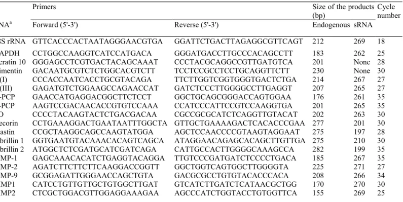
The specific oligonucleotide primers of the mRNA of interest "were around 24 bases long "with an A-T proportion close to 50%. The sequences shown in Table I "were chosen on different exons to allow discrimination of products amplified from potential contaminating DNA. The mRNA of interest and the 28S rRNA "were quantified in triplicate by reverse transcription—PCR using 10 ng of total RNA. The RNA "were reverse transcribed at 70°C for 15 min and amplified (94°C for 15 s, 66°C for 20 s, 78°C for 10 s) using the Gene Amp rTth Kit (Perkin Elmer, Branchburg, NJ), except matrix metalloproteinase (MMP) -2 that "was reverse-transcribed at 55°C for 30 min and amplified (94°C for 18 s, 62°C for 20 s, 68°C for 15 s) using the kit Titan (Boehringer, Mannheim, Germany). The number of amplification cycles used in this study is indicated in Table I. The efficiency of the reverse transcription and the amplification reactions "was monitored by adding in each tube an appropriate number of copies of a synthetic RNA (sRNA) that can be reverse-transcribed and amplified "with the primers pair used for the amplification of the mRNA of interest, but giving rise to a product slightly larger or smaller than the product amplified from the mRNA (Table I), allowing its discrimination after migration in a 10% polyacrylamide gel. The different sRNA "were generated from linearized template plasmid containing appropriate DNA inserts by the use of SP6 RNA polymerase (SP6/T7 transcription kit; Boehringer Mannheim), purified (High Pure RNA Isolation kit; Boehringer Mannheim) and quantitated2(manuscript in
preparation). The gels were stained with SyberGreen (Biorad, Hercule, CA) or Gelstar (FMC Bioproducts, Rockland, ME) and the intensity of the fluorescent signals measured in a Fluor-S-Multilmager (Biorad).
For each of the mRNA, the optimal conditions of reaction (temperature, number of cycles and choice of the reverse transcription-PCR kit) and the amount of internal standard "were determined taking into account the level of expression of the mRNA in human skin to obtain values "within the linear range of measurement as recommended by Freeman et al (1999) and illustrated in Fig 1. Each measurement "was normalized to the cotranscribed and coamplified internal standard. The normalized values "were expressed in arbitrary units (AU) per unit of 28S rRNA measured in the same dilution of RNA to correct for RNA input of each sample.
Measurement of collagen extractability
The remaining half of the biopsy "was used to measure collagen after sequential fractionation. The fragments of skin "were lyophilized, "weighed, and extracted at 4°C in 0.5 M acetic acid-HCl (HAc) at pH 2.0 for 24 h followed by extraction after pepsin digestion (200 µg per ml) in 0.5 M acetic acid-HCl pH 2.0 for a further 24 h.
2Lambert ChA, Colige AC, Maniglia S, Heyeres A, Munaut C, Lapière ChM. and Nusgens BV: Measurement of matrix metalloproteinases
by quantitative RT-PCR assay: Application to the study of gene regulation in fibroblasts by stress relaxation. J Invest Dermatol 110:612, 1998 (abstr.)
Aliquots of the extracts and the residual material "were hydrolyzed in 6 M HC1 and the concentration of hydroxyproline was determined by a colorimetric assay (Bergman and Loxley, 1963).
Morphologic analysis
The second biopsy from the placebo-treated side "was separated in two fragments perpendicularly to the stratum
corneum. One fragment "was fixed in Baker's solution for 3 h and embedded in paraffin. The other fragment "was further sectioned in smaller samples and fixed in 2% glutaraldehyde in cacodylate buffer for 6 h, postfixed in 1% osmium tetroxide, dehydrated in ethanol, and embedded in Epon. Ultrathin sections were counterstained with lead citrate and uranyl acetate. Five micrometer paraffin sections were stained with hematoxylin-eosin-saphran, orcein, or immunolabeled using monoclonal antibodies to vimentin (VIM 3B4, Progen, Heidelberg, Germany) and collagen IV (clone CIV22, Dako, Glostrup, Denmark), both revealed with biotin-streptavidin-alkaline phosphatase (LSAB kit, Dako). Light and electron microscopy were used to evaluate sun damage by comparison with nonexposed buttock skin of five age-matched individuals. Three parameters were analyzed: irregularity of the epidermal pigmentation and disorganization of the dendritic cells network, flattening of the epidermal-dermal junction and subepidermal hyaline bodies, and disorganization of the elastic fiber network. Each parameter was scored semiquantitatively (0, no alteration; 1, very light; 2, moderate; and 3, severe) and summed to determine the extent of solar alteration.
Statistical analysis Results were expressed as the ratio between the values measured in the skin sample treated
with the preparation containing vitamin C (A) and that treated with the placebo (P). The mean ratio was tested for statistical difference from 1 by using the onesided Student's t test for paired data.
Table I. Nucleotide sequence of the primers, size of the generated reverse transcription-PCR products and
aThe identity of the investigated RNA is described in the text.
Primers Size of the products
(bp)
RNAa Forward (5'-3') Reverse (5'-3') Endogenous sRNA
Cycle number
28S rRNA GTTCACCCACTAATAGGGAACGTGA GGATTCTGACTTAGAGGCGTTCAGT 212 269 18
GAPDH CCTGGCCAAGGTCATCCATGACA GGGATGACCTTGCCCACAGCCTT 183 262 25
Keratin 10 GGGAGCCTCGTGACTACAGCAAAT CCCTACGCAGGCCGTTGATGTCA 201 None 28
Vimentin GACAATGCGTCTCTGGCACGTCTT TCCTCCGCCTCCTGCAGGTTCTT 230 None 30
αl(I) CCCACCAATCACCTGCGTACAGA TTCTTGGTCGGTGGGTGACTCTGA 214 267 27
αl(III) GAGATGTCTGGAAGCCAGAACCAT GATCTCCCTTGGGGCCTTGAGGT 207 265 27
N-PCP GAACCATGAGGACGGCTTCTCCT GGCTGCAGCGGGACCAGTGGAA 176 261 35
C-PCP AAGTCCGACAACACCGTGTCCAAA CCATCCCATTCCGTCCAAGGTGA 201 265 35
LO CCCCTACAAGTACTCTGACGACAA CGCCGCGCATCTCAGGTTGTACAT 202 263 30
Decorin CCTGAAAGGACTGAATAATTTGGCTA GTTGCTGAAAAGACTCACACCCGAA 277 201 30
Elastin CCGCTAAGGCAGCCAAGTATGGA AGCTCCAACCCCGTAAGTAGGAAT 275 197 28
Fibrillin 1 GGTGAATGTACAAACACAGTCAGCA ATAGGAACAGAGCACAGCTTGTTGA 275 210 30
Fibrillin 2 ATGGCTCTCGATGCATCGATCAGA CATTGCCACTTGGGGCAAAGCCA 282 199 35
MMP-1 GAGCAAACACATCTGAGGTACAGGA TTGTCCCGATGATCTCCCCTGACA 185 267 35
MMP-2 AGATCTTCTTCTTCAAGGACCGGTT GGCTGGTCAGTGGCTTGGGGTA 225 271 27
MMP-9 GCGGAGATTGGGAACCAGCTGTA GACGCGCCTGTGTACACCCACA 208 266 34
TIMP1 CATCCTGTTGTTGCTGTGGCTTGAT GTCATCTTGATCTCATAACGCTGG 170 270 30
RESULTS
The biopsies contain similar proportions of dermal and epidermal RNA
The contribution of the dermis in the collected RNA was estimated by measuring the steady-state level of
vimentin (VIM) mRNA, the protein of the intermediate filaments of the mesenchymal cells whereas that of the epidermis was obtained by measuring the mRNA of keratin 10 (K10) (Table II). The mean ratio VIM/K10 was similar on both sides, that treated with vitamin C (A) and the controlateral side receiving the placebo (P), indicating a similar contribution of epidermal and dermal RNA in the samples; however, the values of VIM and K10 per unit of 28S rRNA were both increased in the vitamin C-treated side, which resulted in an A/P ratio significantly higher than 1.0. The mean value for the mRNA level of the housekeeping gene GAPDH per unit of 28S rRNA was very similar on both sides resulting in an A/P ration close to 1.0 (Table II).
The studied population is heterogeneous in terms of level of expression of all the investigated genes. This is clearly illustrated in Fig 2 for the procollagen processing enzyme, the C-PCP ranged in the studied population from 65 to 370 AU per unit of 28S rRNA. The individual variations were, however, similar on both arms resulting in a mean value almost identical for the group of samples collected on the right (R) and the left (L) arm and an R/L ratio close to 1.0. To account for that heterogeneity the results in the tables were mostly expressed by the ratio of the values observed in the vitamin C-treated side (active: A) to that of the placebo (P). In the example illustrated in Fig 2, the A/P ratios significantly higher than 1.0 indicate an increased steady-state level of C-PCP mRNA upon vitamin C treatment.
The mRNA levels of collagen type I and type III and that of three extracellular procollagen processing enzymes are coordinately increased by vitamin C
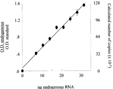
The steady-state level of the mRNA of the two main types of collagen in skin, types I and III, was measured by quantitative reverse transcription-PCR using specific sRNA as internal standards for the α1 chains of collagen I and III. The example of titration of the collagen polypeptides α1 (I) mRNA (Fig 1) illustrates the linearity, reproducibility, and sensitivity of the procedure.
The variability in the level of the two collagen mRNA was lower than that observed for C-PCP and similar on both sides (not illustrated). Six of the 10 testers displayed increased values for both types of collagen on the side treated with the active preparation and the mean A/P ratio of the whole group for both collagen type I and type III supported a stimulating effect of the vitamin (Table III).
This occurred to the same extent with the two types of collagen. The ratio between collagen I and collagen III was very similar on both sides in spite of the individual variability in the level of these mRNA within the group of testers.
The interindividual variability of LO was higher than that of C-PCP and N-PCP. The C-PCP mRNA enhancing activity of vitamin C is illustrated in Fig 2 by a statistically significant increase of the mean A/P ratio. The mean values of the A/P ratios were also significantly increased for the two other enzymes (N-PCP and LO) involved in the maturation of the (pro) collagen molecules (Table IV).
The concentration of collagen and its extractability are not significantly modified in the vitamin C treated skin.
The fraction of acetic acid-extracted collagen was very small in agreement with previously published data (Legrand et al, 1969). The proportion of extracted collagen and that remaining in the insoluble residue was similar in the two groups of samples and the mean A/P ratio did not significantly differ from 1 (Table V).
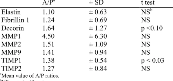
The mRNA of decorin and TIMP1 are increased by vitamin C but not that of elastin, fibrillins, MMP-1, MMP-2, MMP-9, and tissue inhibitor of matrix metalloproteinase (TIMP) 2 The mean value of the mRNA for elastin and fibrillin 1 collected from the vitamin C-treated "was not significantly increased as compared "with placebo (Table VI). The niRNA of decorin "was increased by the topical application of vitamin C. The difference "was, however, at the limit of significance. The transcript of fibrillin 2 "was not reproducibly detected.
Figure 1. Titration of the mRNA of collagen α1 (I). The ratio of the signals obtained for the amplification
products of increasing amounts of endogenous RNA with the addition of a constant amount (85,000 copies) of standard synthetic RNA increases linearly (r = 0.998) with the amount of endogenous RNA. Each value is the average of triplicate measurements ± SD.
Figure 2. Heterogeneity of the tested population. The mRNA level (in arbitrary units per unit of 28S sRNA) for
C-PCP varies among the testers but similarly on the right (R) and the left (L) sides resulting in a mean R/L ratio close to 1.0 (left part). The stimulating effect of topical vitamin C (in bold figures) is expressed by a mean ratio active/placebo (A/P) significantly higher than 1.0 (right part).
Table II. The RNA collected from the biopsies arises in a constant proportion from the epidermis (K10) and the dermis (VIMV)a VIM/K10 Tester A P K10 A/P VIM A/P GAPDH A/P 1 7.22 6.05 1.58 1.88 1.76 2 6.88 [14.10] b [2.64]b 1.29 0.69 3 5.27 5.63 0.93 0.87 0.82 4 4.53 2.68 0.90 1.53 1.16 5 4.82 4.18 1.17 1.35 1.51 6 2.65 4.31 1.68 1.03 0.87 7 3.59 6.34 1.88 1.06 1.35 8 3.77 5.36 1.17 0.83 0.62 9 3.21 4.48 1.23 0.88 0.64 10 4.19 4.93 1.46 1.24 1.14 Mean 4.61 4.88 1.33 1.20 1.06 ± SD ± 1.50 ± 1.12 ± 0.34 ± 0.33 ± 0.39 NSC NS p <0.01 p <0.05 NS
aK10 and VIM mRNA levels are significantly increased in vitamin C treated sides (A) as compared with placebo (P), whereas GAPDH
mRNA level is not modified by the vitamin.
bEctopic values excluded from mean and SD. cNS, not significant.
Table III. The topical application of vitamin C (A) increases the steady-state level of the mRNA of collagen I (α1
I) and collagen III (α1 III) in the same six of the 10 testers without disturbing the ratio between the two types of collagen (I/III) I/III Tester α1 I A/P α1 III A/P A P 1 1.60 1.34 2.91 2.45 2 1.38 1.64 2.58 3.07 3 0.92 0.91 2.84 2.75 4 1.55 1.56 3.01 3.02 5 1.74 1.59 2.72 2.49 6 0.89 0.96 2.59 2.80 7 1.24 1.24 2.55 2.56 8 0.71 0.62 3.02 2.64 9 0.83 0.78 2.76 2.57 10 1.62 1.45 3.04 2.72 Mean 1.25 1.21 2.80 2.71 ± SD ± 0.38 ± 0.37 ± 0.19 ± 0.21 p <0.04 p <0.06 NSa NS aNS, not significant.
Table IV. The mRNA steady-state level of the processing (N-PCP and C-PCP) and cross-linking (LO) enzymes is
increased on the side treated by vitamin C (A) by comparison with the side treated with placebo (P)
Tester N-PCP
A/P C-PCP A/P LO A/P
1 1.33 1.66 5.67 2 1.10 1.22 2.53 3 0.87 1.02 0.57 4 1.96 1.46 1.18 5 1.58 1.30 2.20 6 1.19 1.14 1.41 7 1.00 1.16 2.55 8 0.86 1.17 0.24 9 0.79 1.11 1.47 10 1.41 1.33 1.45 Mean 1.21 1.26 1.93 ± SD ± 0.37 ± 0.19 ± 1.52 p <0.06 p <0.01 p <0.05
Table V. Extractability of collagen in vitamin C-treated (A) and placebo-treated (P) skin
A (n = 10) P (n = 10) A/P (n = 10) t test 0.5 M HAc 8.3 ± 3.3a 8.7 ± 3.7 1.30 ± 1.20 NS Pepsin 55 ± 16 58 ± 16 0.98 ± 0.27 NS Insoluble 373 ± 103 349 ± 60 1.07 ± 0.23 NS Total 436 ± 110 416 ± 64 1.05 ± 0.21 NS
aIn µg of collagen per mg of tissue, dry weight.
Table VI. Expression of a series of other connective tissue related genes upon topical application of vitamin C
A/Pa ± SD t test Elastin 1.10 ± 0.63 NSb Fibrillin 1 1.24 ± 0.69 NS Decorin 1.64 ± 1.27 p <0.10 MMP1 4.50 ± 6.30 NS MMP2 1.51 ± 1.09 NS MMP9 1.41 ± 0.94 NS TIMP1 1.38 ± 0.54 p < 0.03 TIMP2 1.27 ± 0.84 NS
aMean value of A/P ratios. bNS, not significant.
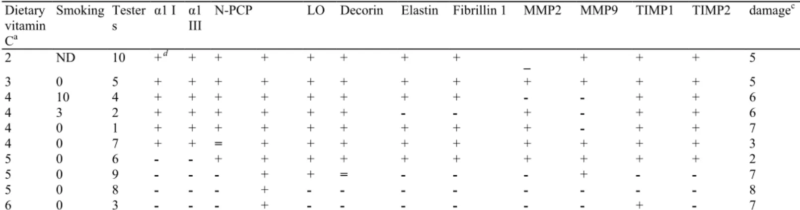
The expression of MMP-1 "was very small, barely detectable in most instances, "whereas a few samples displayed higher values. There "was no statistically valid difference between the two sides. The interindividual expression of MMP-2 "was also variable and no significant difference "was observed between the side treated "with the vitamin and the placebo. The same conclusions apply to MMP-9 (Table VI); however, the niRNA level of TIMP1 was increased on the side treated "with the active preparation (Table VI), but not that of TIMP2. Relationship between the metabolic effect of topical vitamin C, the dietary intake of vitamin C and the extent of actinic damage The dietary intake of vitamin C has been estimated after termination of the study by a questionnaire collecting semiquantitative information to estimate the consumption of fruits and vegetables. It is expressed as the sum of servings per day. Smoking "was also recorded as it increases the need for a higher intake of vitamin C to contribute to the pool. This information has been gathered in Table VII and correlated "with the potential of topical vitamin C to modulate the biosynthetic activity of the cells. It is "worth noting that the testers
that have the lowest score of dietary vitamin C intake are those in "which topically applied vitamin C displayed the most constant stimulation of the steady-state level of niRNA for collagens and their processing enzymes. All 10 subjects presented histologic and ultrastructural signs of actinic damage (Table VII). There "was, however, no correlation between its extent (from 2, very mild, to 8, extensive) and the responsiveness to the topically applied vitamin C.
Table VII. The testers with the lowest intake of vitamin C are most responsive to topically applied ascorbic acid
Dietary vitamin Ca Smoking Tester s α1 I α1 III
N-PCP LO Decorin Elastin Fibrillin 1 MMP2 MMP9 TIMP1 TIMP2 damagec
2 ND 10 +d + + + + + + + _ + + + 5 3 0 5 + + + + + + + + + + + + 5 4 10 4 + + + + + + + + - - + + 6 4 3 2 + + + + + + - - + - + + 6 4 0 1 + + + + + + + + + - + + 7 4 0 7 + + = + + + + + + + + + 3 5 0 6 - - + + + + + + + + + + 2 5 0 9 - - - + + = - - - + - - 7 5 0 8 - - - + - - - - - - - - 8 6 0 3 - - - + - - - - - - + - 7
aSemiquantitative estimation of the dietary intake of vitamin C expressed as the sum of servings of fruits and vegetables per day. Cigarettes
per day.
cSemiquantitative estimation (see Materiab and Methods). d+, A/P ratio > 1; -, A/P ratio < 1; =, A/P ratio = 1; ND, not determined.
DISCUSSION
The presented data demonstrate that daily topical application of a preparation containing 5% L-ascorbic acid enhances the steady-state level of procollagens I and III niRNA and that of their post-translational maturation enzymes. The niRNA level of the MMP responsible for the degradation of the extracellular matrix is not statistically modified, "whereas the niRNA of TIMP 1, a physiologic inhibitor of MMP, is increased. These modifications reflect the expression of an anabolic phenotype. This pattern of enhanced anabolic activity has been obtained by performing the niRNA measurements in a small cohort of only 10 female volunteers at the site of application of the active preparation compared "with the excipient alone on the opposite parallel site. This strategy allowed us to establish a ratio between paired values in the same individual, therefore, minimizing the bearing of a marked interindividual heterogeneity in the expression of most niRNA on the statistical significance of the results.
The reverse transcription—PCR procedure used for measuring the biosynthetic activity of the cells in small samples of tissues has been carefully controlled to generate comparative data "with a high degree of confidence. Our quantification procedure of ribosomal (28S rRNA) and specific niRNA by reverse transcription—PCR under noncompetitive conditions used an internal standard of sRNA and is most adequate to demonstrate small differences (Freeman et al, 1999). We have verified that in each biopsy the RNA arises in a similar proportion from the epidermis by measuring the niRNA of K10 and from the dermis by measuring the niRNA of VIM. This condition has to be fulfilled to permit a reliable estimation of the niRNA of interest on the basis of a unit amount of 28S rRNA. The validity of the procedure is supported by the constant ratio of collagen type I and type III mRNA level in the biopsies, similar to the ratio of these collagens in adult human skin (Epstein, 1974) and that produced by human fibroblasts in culture (Phillips et al, 1992).
In fibroblast cultures, the niRNA of the two main types of collagens are known to be coordinately upregulated by vitamin C (Geesin et al, 1988). This also seems to be the case in our in vivo study. Furthermore, the niRNA level of the three enzymes involved in the post-translational processing and cross-linking of the collagen molecules "was also stimulated by the vitamin C treatment suggesting the existence of a mechanism of regulation parallel to that operating for the collagen polypeptides. An increased activity of the N-PCP is known to exist in pathologic conditions "where the biosynthetic activity of the connective tissues is enhanced (Lapière and Piérard, 1974). The repercussion at the tissue level of the increased expression of procollagens niRNA in vitamin C
treated skin "was tentatively evaluated by measuring the amount of extractable collagen at acid pH (Legrand et al, 1969) and after pepsin digestion as an indirect indicator of collagen turnover rate, the newly synthetized collagen being less cross-linked and more extractable (Gross, 1959). In the adult human, the half-life of dermal collagen might be as long as 10 y by extrapolation of animal data (Nimni and Bavetta, 1964; Nimni M., personal communication) and the amount of replaced collagen about 10 µg per mg dry skin during the period of treatment by the vitamin. The 30% increase induced by vitamin C (3 µg in mass of "which only a small fraction can be extracted) is too small to be detected. The parameters estimating the level of cell activity are obviously more sensitive.
The daily requirement of dietary vitamin C to prevent scurvy, i.e., to permit at least the vital function of the collagen hydroxylases and other monooxygenases, has been estimated at less than 10 mg per day (i.e., 15 ml of lemon juice). Such a low intake is compensated by a sparing mechanism of cellular recycling of the oxidized vitamin by two enzyme systems (Banhegyi et al, 1997). A daily intake of 60—100 mg vitamin C is required to raise the serum level in humans to that of animals that synthesize L-ascorbate. This amount needs to be increased to 140 mg per day for smokers that seem to consume vitamin C to inactivate oxidants from tobacco smoke (Schectman, 1993). The concentration of ascorbate is greater in the skin than that in plasma, probably by a mechanism of active transport (Welch et al, 1995). Nevertheless, our data demonstrate that in postmenopausal "women the topical application of vitamin C is able to produce a coordinated increase in the steady-state level of the niRNA for collagen I and III as observed in six testers, and of their post-translational extracellular enzymes, as observed in at least, seven of the 10 testers. We speculated that the nonresponders might already have a concentration of the vitamin in skin great enough for maximal expression of the ascorbate-responsive niRNA. This hypothesis is supported by the ranking of the testers (Table VII) showing an inverse relationship between the responsiveness to vitamin C and the overall dietary intake of the vitamin. An indirect effect of vitamin C mediated by the epidermis, however, cannot be ruled out as the epithelial cells "were also stimulated by the topical application of the vitamin. Keratinocytes are known to produce in vitro several cytokines that are modulated by vitamin C (Tebbe et al, 1997).
The dermal signs of aging, photoinduced and chronologic, are to some extent comparable "with scurvy, i.e., atrophy, fragility, easy bruising, and palor. In aging, the density of the dermal collagen network diminishes by reduced metabolic activity (Legrand et al, 1969), and enhanced degradation by increased production of MMP (Millis et al, 1992), perhaps related to an oxidative stress (Sohal and Weindruch, 1996) similar to that induced by ultraviolet in fibroblasts (Wlaschek et al, 1995). The level of three MMPs involved in acute (Fischer et al, 1999) or chronic (Seite et al, 2000) skin alterations induced by ultraviolet irradiation "was not significantly modified by the topical application of the vitamin. The activity of these proteases is known to be reduced in vivo by vitamin A (Varani et al, 2000) through a mechanism of action probably different from that operating for the vitamin C. The expression of TIMP1, but not TIMP2, however, "was increased suggesting that the MMP activity in the vitamin C-treated side may be balanced by their physiologic inhibitor. Chronic photoaging also features an increased deposition of abnormal elastic fibers. The genes of elastin and fibrillin are known to be upregulated in chronically photodamaged skin (Bernstein et al, 1994). In vitro, vitamin C downregulates the biosynthesis of elastin by fibroblasts (Davidson et al, 1997) at the concentration that increases the level of niRNA for collagen in the same cells. In this study, the niRNA for elastin and fibrillins "was not modified by vitamin C. Glycosaminoglycans and proteoglycans also accumulate in photoaged skin (Bernstein et al, 1996). Decorin is a small proteoglycan closely associated "with the collagen fibrils. The increased level of its niRNA in the vitamin C-treated skin is at the limit of significance. The level of accumulated solar damage estimated in the placebo-treated side of the testers does not correlate "with the extent of responsiveness to the topically applied vitamin. This suggests that a reduced biosynthetic activity related to chronologic aging and/or a low tissue concentration of the vitamin are the likely targets of the topical treatment.
Acknowledgement
The excellent technical assistance of M.J. Nix and T. Heyeres in the preparation of RNA samples and reverse transcription-PCR assays is gratefully acknowledged. The help of A. Albert for the statistical analysis of the data has been greatly appreciated. We thank C. Clabeck for the preparation of the manuscript. This work was supported in part by grants of the Belgian FRSM and the Faculty of Medicine of the University of Liège.
References
Banhegyi G, Braun L, Csala M, Puskas F, Mandl J: Ascorbate metabolism and its regulation in animals. Free Rad Biol Med 23:793-803., 1997
Bartley W, Krebs HA, O'Brien JRP: Vitamin C requirement of human adults. Med Res Counc Spec Rep Ser 280:1-179, 1953
Bergman I, Loxley R: Two improved and simplified methods for the spectrophotometric determination of hydroxyproline. Anal Chem 35:1961-1965, 1963
Bernstein EF, Chen YQ, Tamai K, et al: Enhanced elastin and fibrillin gene expression in chronically photodamaged skin. J Invest Dermatol 103:182-186, 1994
Bernstein EF, Underhill CB, Hahn PJ, Brown DB, Uitto J: Chronic sun exposure alters both the content and distribution of dermal glycosaminoglycans. Br J Dermatol 135:255-262, 1996
Colige A, Li S-W, Sieron AL, Nusgens BV, Prockop DJ, Lapière CM: cDNA cloning and expression of bovine procollagen I N-proteinase: a new member of the superfamily of zinc-metalloproteinases with binding sites for cells and other matrix components. Proc Natl Acad Sci USA 94:2374-2379, 1997
Colven RM, Pinnell SR: Topical vitamin C in aging. Clin Dermatol 14:227-234, 1996
Danielson KG, Baribault H, Holmes DF, Graham H, Kadler KE, Iozzo RV: Targeted disruption of decorin leads to abnormal collagen fibril morphology and skin fragility, J Cell Biol 136:729-743, 1997
Darr D, Combs S, Dunston S, Manning T, Pinnell S: Topical Vitamin-C protects porcine skin from ultraviolet radiation-induced damage. Br J Dermatol 127:247-253, 1992
Darr D, Dunston S, Faust H, Pinnell S: Effectiveness of antioxidants (Vitamin C and E) with and without sunscreens as topical photoprotectants. Acta Derm Venereol (Stockh) 76:264-268, 1996
Davidson JM, LuValle PA, Zoia O, Quaglino D, Giro MG: Ascorbate differentially regulates elastin and collagen biosynthesis in vascular smooth muscle cells and skin fibroblasts by pretranslational mechanisms. J Biol Chem 272:345-352, 1997
Epstein EH: [alphal (III) ]3 Human Skin Collagen. Release by pepsin digestion and preponderance in fetal life. J Biol Chem 249:3225-3231,
1974
Fisher GJ, Talwar HS, Lin JY, Voorhees JJ: Molecular mechanisms of photoaging in human skin in vivo and their prevention by all-trans retinoic acid. Photochem Photobiol 69:154-187, 1999
Freeman WM, Walker SJ, Vrana KE: Quantitative RT-PCR: Pitfalls and potential. Biotechniques 26:112-125, 1999
Geesin JC, Darr D, Kaufman R, Murad S, Pinnell SR: Ascorbic acid specifically increases type I and type III procollagen messenger RNA levels in human skin fibroblast, J Invest Dermatol 90:420-424, 1988
Gross J: On the significance of the soluble collagens. In: Page IH (éd.). ConnectiveTissue, Thrombosis and Atherosclerosis. New York: Academic Press, 1959, pp 77-95
Hamalainen ER, Jones TA, Sheer D, Taskinen K, Pihlajaniemi T, Kivirikko KI:Molecular cloning of human lysyl oxidase and assignment of the gene to chromosome 5q23.3-31.2. Genomics 11:508-516, 1991
Hirschmann JV, Raugi GJ: Adult scurvy. J Am Acad Dermatol 41:895-906, 1999 Kivirikko KI, Prockop DJ: Enzymatic hydroxylation of proline and lysine in protocollagen. Proc Nat Acad Sci USA 57:782-789, 1967
Lapière ChM, Piérard GE: Skin procollagen peptidase in normal and pathologic conditions. J Invest Dermatol 62:582-586, 1974
Legrand Y, Lapière ChM, Pignaud G, Caen J: Microméthode d'extraction du collagène à partir de biopsie de peau humaine. Pathol Biol 17:991-993, 1969
Levene CI: Ascorbic acid and collagen synthesis in cultured fibroblasts. Ann N Y Acad Sci 258:288-306, 1975 Levine M, Rumsey SC, Daruwala Pv, Park JB, Wang Y: Criteria and recommendations for vitamin C intake. JAMA 281:1415-1423, 1999 Li SW, Sieron AL, Fertala A, Hojima Y, Arnold WV, Prockop DJ: The C-proteinase that processes procollagens to fibrillar collagens is identical to the protein previously identified as bone morphogenic protein-1. Proc Natl Acad Sci USA 93:5127-5130, 1996
Lyons BL, Schwarz RJ: Ascorbate stimulation of PAT cells causes an increase in transcription rates and a decrease in degradation rates of procollagen mRNA. Nucleic Acids Res 12:2569-2579, 1984
Millis AJ, Hoyle TM, McCue HM, Martini H: Differential expression of metalloproteinase and tissue inhibitor of metalloproteinase genes in aged human fibroblasts. Exp Cell Res 201:373-379, 1992
Nagase H, Woessner JF: Matrix metalloproteinases. J Biol Chem 274:21491-21494, 1999
Nimni ME, Bavetta LA: Collagen synthesis and turnover in the growing rat under the influence of methylprednisolone. Proc Soc Exp Biol Med 117:618-624, 1964
Padh H: Cellular functions of ascorbic acid. Biochem Cell Biol 68:1166-1173, 1990
Pauling L: Effect of ascorbic acid on incidence of spontaneous mammary tumors and UV-light-induced skin tumors in mice. Am J Clin Nutr 54:1252S-1255S, 1991
Peterkofsky B: the effect of ascorbic acid on collagen polypeptide synthesis and proline hydroxylation during the growth of cultured fibroblasts. Arch Biochem Biophγs 152:318-328, 1972
Phillips CL, Tajima S, Pinnell SPv: Ascorbic acid and transforming growth factor-beta1 increase collagen biosynthesis via different mechanisms—coordinate regulation of proalphal (I) and proalphal(III) collagens. Arch Biochem Biophγs 295:397-403, 1992
Pinnell SPv, Murad S, Darr D: Induction of collagen synthesis by ascorbic acid. Arch Dermatol 123:1684-1686, 1987
Ponec M, Weerheim A, Kempenaar J, Mulder AA, Gooris GS, Bouwstra J, Mommaas AM: The ormation of competent barrier lipids in reconstructed human epidermis requires the presence of vitamin C J Invest Dermatol 109:348-355, 1997
Ramirez F, Pereira L: The fibrillins. Int J Biochem Cell Biol 31:255-259, 1999
Rosenbloom J, Harsch M, Jimenez S: Hydroxyproline content determines the denaturation temperature of chick tendon collagen. Arch Biochem Biophγs 158:478-484, 1973
Sauberlich HE: Pharmacology of vitamin C. Annu Rev Nutr 14:371-391, 1994
Schectman G: Estimating ascorbic acid requirement for cigarette smokers. Ann N Y Acad Sci 686:335-345, 1993
Schwarz RJ: Procollagen secretion meets the minimum requirements for the rate-controlling step in the ascorbate induction of procollagen synthesis. J Biol Chem 260:3045-3049, 1985
Seite S, Colige A, Piquemal-Vivenot P, et al: A full-UV spectrum absorbing daily use cream protects human skin against biological changes occurring in photoaging. Photodermatol Photoimmunol Photomed 16:147-155, 2000
Sohal PvS, Weindruch R: Oxidative stress, caloric restriction and aging. Science 273:59-63, 1996
Tebbe B, Wu SL, Geilen CC, Eberle J, Kodelja V, Orfanos CE: L-ascorbic acid inhibits UVA-induced lipid peroxidation and secretion of IL-1 alpha and IL-6 in cultured human keratinocytes in vitro. J Invest Dermatol IL-108:302-306, IL-1997
Thomas DP: Sailors scurvy and science. J R Soc Med 90:50-54, 1997
Utoguchi N, Ikeda K, Saeki K, et al: Ascorbic acid stimulates barrier function of cultured endothelial cell monolayer. J Cell Phγsiol 163:393-399, 1995
Varani J, Warner RX, Gharaee-Kermani M, et al: Vitamin A: antagonizes decreased cell growth and elevated collagen-degrading matrix metalloproteinases and stimulates collagen accumulation in naturally aged human skin. J Invest Dermatol 114:480-486, 2000
Welch RV, Wang Y, Crossman AJr, Park JB, Kirk KL, Levine M: Accumulation of vitamin C (ascorbate) and its oxidized metabolite de hydro ascorbic acid occurs y separate mechanisms. J Biol Chem 270:12584-12592, 1995
Wlaschek M, Briviba K, Stricklin GP, Sies H, Scharffetter-Kochanek K: Singlet oxygen may mediate the ultraviolet A-induced synthesis of interstitial collagenase. J Invest Dermatol 104:194-198, 1995
![Table II. The RNA collected from the biopsies arises in a constant proportion from the epidermis (K10) and the dermis (VIMV) a VIM/K10 Tester A P K10 A/P VIM A/P GAPDH A/P 1 7.22 6.05 1.58 1.88 1.76 2 6.88 [14.10] b [2.64] b 1.29 0.69 3](https://thumb-eu.123doks.com/thumbv2/123doknet/5872812.143100/7.892.107.593.162.437/table-collected-biopsies-constant-proportion-epidermis-tester-gapdh.webp)