Université de Montréal
14-3-3zeta is required for PKA-dependent lipolysis in
mature adipocytes
Par
Abel Oppong
Faculté de médecine
Mémoire présenté à la Faculté des études supérieures
en vue de l’obtention du grade de Maître es science (M.sc)
en Sciences Biomédicales
option médecine expérimentale
Août, 2019
Université de Montréal
Faculté des études supérieures
Ce mémoire intitulé :
14-3-3zeta is required for PKA-dependent lipolysis in
mature adipocytes
Présenté par :
Abel Oppong
A été évalué par un jury composé des personnes suivantes
Dr Mathieu Ferron
Président-rapporteur
Dr Gareth Lim
Directeur de recherche
Dre Julie Lavoie
Résumé
Une augmentation de l’hypertrophie et l'hyperplasie des adipocytes est au cœur du développement de l'obésité. Nous avons déjà constaté que 14-3-3zeta (14-3-3ζ), une protéine d’échafaudage moléculaire, a plusieurs rôles essentiels dans l'adipogenèse. Cependant, les contributions de 14-3-3ζ dans la fonction des adipocytes matures ne sont pas connues. Les cellules 3T3-L1 et souris dépourvues de 14-3-3ζ dans les adipocytes (adi14-3-3ζKO) ont été utilisés pour examiner le rôle de 14-3-3ζ dans la lipolyse.
L’élimination de 14-3-3ζ dans les cellules 3T3-L1 par l’ARNi a réduit significativement la lipolyse stimulée par l'isoprotérénol (un agoniste bêta adrénergique), la forskoline (un activateur de l’adénylate cyclase) et le dibutyryl AMPc (dbcAMP). Les analyses par qPCR ont démontré des réductions significatives
d’adipose triglyceride lipase (Atgl) et lipase hormonsensible (Hsl) au niveau transcriptionnel. De plus, une réduction au niveau des substrats de la PKA phosphorylés et totaux tels que HSL et CREB, a été détectée par Western Blot dans les 3T3-L1 appauvris en 14-3-3ζ. Ces résultats in vitro ont été récapitulés in vivo, car des diminutions des taux phosphorylés et totaux de HSL ont été observés dans le tissu adipeux gonadique des souris adi14-3-3ζKO. Les souris adi14-3-3ζKO et les explants gonadiques ont également montré une lipolyse affaiblie après des injections i.p de l’agoniste bêta 3-adrénergique CL-316,243 et un traitement de l’isoprotérénol respectivement. De manière intéressante, une diminution de l’expression de 14-3-3ζ dans les cellules 3T3-L1 et les souris adi14-3-3ζKO a mené à une diminution des
caractéristiques des adipocytes matures telles que les niveaux d’ARNm de Pparg, Lpl et Fabp4, les niveaux de PPARγ, le contenu en triglycérides et l'incorporation de Oil Red-O. Collectivement, ces résultats démontrent que 14-3-3ζ joue un rôle essentiel en facilitant la lipolyse et en déterminant la maturité des adipocytes.
Abstract
Altered hypertrophy and hyperplasia of adipocytes lie at the core of the development of obesity. We previously demonstrated that the molecular scaffold 14-3-3zeta (14-3-3ζ) had essential roles in adipogenesis. However, the contributions of 14-3-3ζ to mature adipocyte function are not known. 3T3-L1 cells and tamoxifen-inducible adipocyte-specific 14-3-3ζ knockout mice (adi14-3-3ζKO) models were used to examine the roles of 14-3-3ζ in lipolysis. siRNA-mediated knockdown of 14-3-3ζ impaired lipolysis in 3T3-L1 cells stimulated by the beta-adrenergic agonist isoproterenol (Iso), forskolin (an activator of adenylyl cyclase) and dibutyryl cAMP (dbcAMP). qPCR analyses revealed significant
reductions in lipase transcript levels (Atgl and Hsl). Furthermore, reductions in the phosphorylated and total levels of PKA substrates such as HSL and CREB were detected in 14-3-3ζ-depleted 3T3-L1 lysates by immunoblotting. These findings were recapitulated in vivo, as reductions in phosphorylated and total HSL levels were detected in the gonadal adipose tissue of adi14-3-3ζKO mice. adi14-3-3ζKO mice and gonadal explants also displayed impaired lipolysis following i.p CL-316,243 (a beta-3 adrenergic agonist) injections and Iso treatment respectively. Interestingly, decreased 14-3-3ζ expression in 3T3-L1 cells and mice revealed reductions in characteristics of a mature adipocyte, such as Pparg, Lpl, and Fabp4
transcript levels, PPARγ levels, triglyceride content, and Oil Red O (ORO) incorporation. Collectively, these results demonstrate that 14-3-3ζ has essential roles in facilitating lipolysis and determining adipocyte maturity.
Table of Contents List of tables ... 7 List of figures ... 8 List of abbreviations ... 9 Acknowledgements... 11 1.0 Introduction ... 12
1.1 Overview of adipocyte development ... 13
1.1.1 Adipogenesis ... 13
1.1.2 General functions of adipocytes ... 19
1.1.3 Interactions between adipocytes and other cell-types ... 21
1.1.4 Functional differences between different adipose depots ... 24
1.1.5 Heterogeneity of adipose tissue ... 24
1.2 Lipid Homeostasis ... 27
1.2.1 Lipid droplets ... 27
1.2.2 Lipolysis ... 28
1.2.2.1 Key players in the lipolytic pathway ... 28
1.2.2.2 HSL ... 29 1.2.2.3 ATGL ... 30 1.2.2.4 MAGL ... 32 1.2.3 Lipogenesis ... 33 1.2.3.1 Overview of lipogenesis ... 33 1.2.3.2 SREBP-1c ... 35 1.2.3.3 ChREBP ... 36 1.2.3.4 ACC1 ... 37 1.2.3.5 FASN ... 37 1.3 14-3-3 proteins... 38 1.3.1 Structure ... 38 1.3.2 Function ... 39
1.3.3 Implication of 14-3-3 proteins on adipocyte development ... 40
6
2.0 Research Article... 42
2.1 Abstract ... 44
2.2 Introduction ... 45
2.3 Materials and methods ... 47
2.4 Results ... 51
2.5 Discussion ... 55
2.6 Figures and Tables ... 58
3.0 Discussion ... 73
4.0 Conclusion ... 79
List of Tables 1.0 Introduction Table 1 ... 21 2.0 Research Article Supplementary Table 1 ... 62 Supplementary Table 2 ... 63
List of Figures 1.0 Introduction Figure 1 ... 14 Figure 2. ... ……… 15 Figure 3 ... 29 Figure 4 ... 34 2.0 Research article Figure 1 ... 64 Figure 2 ... 65 Figure 3 ... 66 Figure 4 ... 67 Figure 5 ... 68 Figure 6 ... 69 Supplementary Figure 1 ... 70 Supplementary Figure 2 ... 71 Supplementary Figure 3 ... 72
List of abbreviations
ACC Acetyl-CoA carboxylase
ACLY ATP-citrate lyase
adi14-3-3ζKO Adipocyte-specific 14-3-3ζ knockout mice
Adrb Adrenergic β-receptor
APC Adipocyte precursor cell
ATGL Adipose triglyceride lipase
ATP Adenosine triphosphate
BAD Bcl-2-associated death promoter
BAT Brown adipose tissue
BMP Bone morphogenic protein
CDC25B Cell division cycle 25B
C/EBP CCAAT/enhancer-binding protein
cAMP Cyclic adenosine monophsophate
ChREBP Carbohydrate-response element binding protein
CREB cAMP-responsive element binding protein
CTBP C-terminal binding proteins 1 and 2 dbCAMP Dibutyryl cyclic adenosine monophosphate
DLK Delta-like-1
DPP4 Dipeptidyl peptidase 4
FACS Fluorescence activated cell sorting
FAP Fibro-adipogenci precursor
FASN Fatty acid synthase
FFA Free fatty acids
GAPDH Glyceraldehyde-3-phosphate dehydrogenase
GLUT4 Glucose transporter
GPCR G-protein coupled β-adrenergic receptor
gWAT Gonadal white adipose tissue
HFD High-fat diet
HSL Hormone sensitive lipase
IBMX Isobutylmethylxanthine
10
IPC Interstitial progenitor cell
IPGTT Intraperitoneal glucose tolerance test ITT Intraperitoneal insulin tolerance test
JNK JUN N-terminal kinase
KLF Kruppel-like factor
MAGL Monoacylglycerol lipase
MAPK Mitogen-activated protein kinase
MSC Mesenchymal stem cell
ORO Oil Red-O
PDGF Platelet-deteived growth factor
PFK-2 Phosphofructo-2-kinase/fructose-2,6-biphosphatase
PGC-1 Peroxisome proliferator-activated receptor gamma co-activator
PKA Protein kinase A
PKB Protein kinase B
PKC Protein kinase C
PLIN1 Peripilin 1
PPAR Peroxisome proliferator-activated receptor PRDM16 PRD1-BF1-RIZ1 homologous domain-containing 16
PREF Pre-adipocyte factor
qPCR quantitative polymerase chain reaction
ROCK Rho-associated kinase
SCD-1 Stearoyl-CoA desaturase
scRNAseq single cell RNA sequencing
SREBP Sterol response element binding protein
SVF Stromal vascular fraction
TCA Tricarboxylic acid (TCA)
TGF-β Transforming growth factor-beta
UCP1 Uncoupling protein 1
WAT White adipose tissue
WNT Wingless-type MMTV integration site signaling YWHA Tyrosine- and tryptophan-hyroxylase activator
Acknowledgements
I would first like to thank my research supervisor Dr. Gareth Lim for giving me the opportunity to do my master’s project in his laboratory. I thank him for his extensive support as a mentor who devoted countless hours to ensure that I could succeed in realizing this research project. I truly appreciate his mentoring approach over the last two years which included personally walking me through some experiments and readily making himself available for discussions about the current literature and my progress.
I also would like to thank Dr. Mathieu Ferron and Dr. Julie Lavoie for agreeing to evaluate my dissertation. I also want to thank them for the critiques and suggestions they gave while also being members of my Advisory Committee earlier during Graduate studies.
I would also like to thank my fellow lab members Dr. Yves Mugabo and Kadidia Diallo for their assistance and suggestions for improving and troubleshooting experiments. I could not ask for a better group of lab members to work with. I particularly want to thank Dr. Mugabo for being an exceptionally patient mentor who assisted and trained me with handling mice despite me having no prior experience with working with animals.
Finally, I would like to thank my parents and my sister Isabelle for their love and constant support throughout this journey.
13
1.1 Overview of adipocyte development and function 1.1.1 Adipogenesis
Adipogenesis is the process whereby pluripotent mesenchymal stem cells (MSCs) develop into mature adipocytes and consists of two phases: commitment to a cell lineage (or determination), and terminal differentiation. During adipogenesis, mesenchymal stem cells that undergo determination are committed to becoming adipocytes and lose the ability differentiate into other cell types. The pre-adipocytes then differentiate into mature pre-adipocytes and gain features necessary for lipid synthesis, hydrolysis and transport, glucose uptake, insulin sensitivity and adipokine secretion (Figure 1) [1].
14
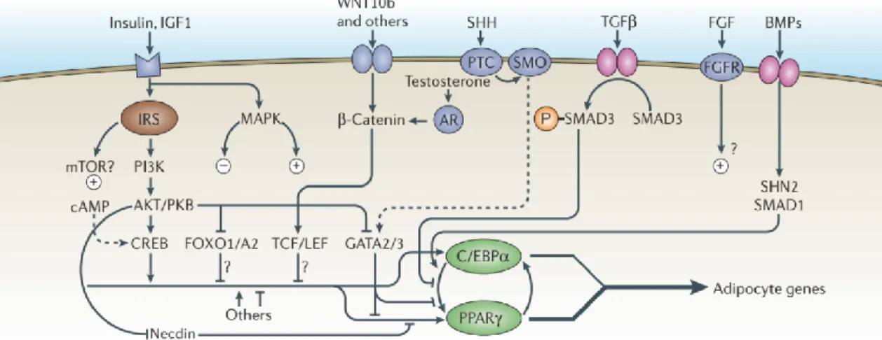
Figure 1: Overview of adipogenesis. Commitment of mesenchymal stem cells (MSCs) to the adipocyte lineage is mediated by factors such as bone morphogenetic protein 4 (BMP4). In contrast, β-catenin-dependent Wingless-type MMTV integration site (WNT) signaling inhibits the conversion of MSCs into pre-adipocytes by inhibiting the expression of the master transcription factors Peroxisome proliferator-activated receptor gamma (PPARγ) and CCAAT/enhancer-binding protein alpha (C/EBP-α). These transcription factors are induced by C/EBP-β and C/EBP-δ which are expressed during early adipogenesis. Terminal differentiation is driven by PPARγ and C/EBP-α promoting each other’s expression in a positive feedback loop and results in the induction of mature adipocyte genes such as
FABP4, adiponectin and GLUT4. The transcription factor PRD1-BF1-RIZ1 homologous domain-containing
16 (PRDM16) associates with PPARγ coactivator 1 alpha (PGC1α) to promote the expression of
thermogenic genes such as UCP1 and mediates the browning of white adipocytes that is induced by β3 agonists and cold exposure. The roles of the abovementioned factors and others in the regulation of adipogenesis are further discussed in the text below. Figure taken and modified from [2].
While MSCs mainly originate from bone marrow, they are also found in other organs, such as fat, lung, and skin [3]. MSCs are multipotent in that they can develop into precursors of multiple specialized cells, including adipocytes, myocytes, chondrocytes, and osteoblasts [4]. Specialized cells can be
categorized by the presence or absence of proteins in precursor cells that they originate from. For instance, brown adipocytes and myocytes derive from Myf5+ precursor cells whereas white and beige adipocytes and osteocytes derive from Myf5- precursor cells [5]. Lineage commitment of MSCs is also
15
regulated by several signaling pathways including Wingless-type MMTV integration site (WNT), transforming growth factor-beta/bone morphogenic protein (TGF-β/BMP) signaling, Notch, and Hedgehog. (Figure 2)
Figure 2: Signaling pathways involved in the regulation of adipogenesis. Signaling pathways affect adipogenesis by activating downstream transcription factors that promote or inhibit the expression of Peroxisome Proliferator-Activated Receptor gamma (PPARγ) and CCAAT/Enhancer-Binding Protein alpha (C/EBP-α). Signaling pathways that negatively regulate adipogenesis include Wingless-type MMTV integration site (WNT), transforming growth factor-beta (TGF-β), and sonic hedgehog (SHH). In the canonical WNT pathway, β-catenin recruits the transcription factor T cell factor (TCF) to activate WNT targets that inhibit adipogenesis. The transcription factors GATA2/3 and SMAD3, which are downstream of the SHH and TGF-β pathways respectively, inhibit adipogenesis by hindering C/EBP-α from binding to the Pparg promoter. Conversely, bone morphogenetic protein (BMP)2/4 promote adipogenesis through the activation of the transcription factor SMAD1. The transcription factor cyclic AMP (cAMP) response element-binding protein (CREB) promotes the transcription of C/EBP-β which in turn induces PPARγ expression during early adipogenesis. Image taken from [1].
WNT belongs to a family of glycoproteins that regulates MSC fate to either adipocyte or
osteoblast lineages. In the case of canonical (β-catenin-dependent) signaling, WNT ligands like WNT10β promote the nuclear translocation of β-catenin, resulting in the activation of WNT target genes that promote osteogenic differentiation and inhibits adipogenic differentiation via the suppression of the adipogenic master regulators, PPARγ and C/EBP-α [3] (Figure 2). In contrast, WNT-5β, a non-canonical WNT ligand promotes adipogenesis by inhibiting WNT10β-mediated signaling possibly through ROR receptors [3, 6]. The TGF-β family of secreted ligands have regulatory roles in cell proliferation and cell
16
differentiation [3]. This family is divided into 3 sub-types: TGFβ1-3, with BMPs belonging in the TGFβ1 sub-family. TGFβ/BMPs regulate adipogenic and osteogenic MSC differentiation through both the activation of SMAD transcription factors and SMAD-independent pathways such as the p38 mitogen-activated protein kinase (MAPK) pathways. Several BMPs including BMP2 and BMP4 are associated with promoting adipogenic differentiation of MSCs (Figure 2) [7, 8].
The Notch family of transmembrane proteins has also been implicated in regulating adipogenic differentiation of MSCs. Ross et al. showed that the Notch ligand Jagged1 blocks PPARγ and C/EBP-α expression in 3T3-L1 cells, a fibroblast cell line derived from Swiss 3T3 mouse embryos that can differentiate into adipocytes [9]. Another study demonstrated that inhibition of Notch via the
PI3K/AKT/mTOR pathway promotes autophagy-mediated adipogenic differentiation of MSCs [10]. Lastly, hedgehog secretory proteins have also been reported to induce osteogenic differentiation and inhibit adipogenesis in MSCs by decreasing PPARG and C/EBPA expression through GLI transcription factors [11].
Terminal differentiation is controlled by several transcription factors that temporally regulate a series of metabolic and adipokine gene-expression events. Induction and maintenance of this
transcriptional cascade is primarily driven by the expression and interactions of two crucial transcriptional regulators: Peroxisome Proliferator-activated Receptor gamma (PPARγ) and
CCAAT/Enhancer-Binding Protein (C/EBP). PPARγ is a ligand-activated transcription factor that belongs to the superfamily of hormone nuclear receptors. PPARγ exists as two isoforms in mice, PPARγ1 and PPARγ2. These isoforms differ in that the N-terminal region of PPARγ2 contains an additional 30 amino acids compared to PPARγ1. In contrast to PPARγ1 which is expressed in multiple tissues, PPARγ2
expression is adipocyte-specific. Upon activation by endogenous ligands (fatty acids such as nitrolinoleic acid), PPARγ forms a heterodimer with retinoid X receptor and binds to specific PPAR response element
17
regions of target genes involved in lipogenesis, lipolysis, and adipogenesis to increase the expression of their corresponding proteins, including FABP4, LPL, perilipin, and glycerol kinase [12].
Several in vivo models have demonstrated the role of PPARγ as a master regulator of adipogenesis. One of the earliest reports came from Barak et al. who generated chimeric Pparg
knockout mice by supplementing knockout embryos with wild-type tetraploid cells to rescue the Pparg knockout mice from an embryonic lethal phenotype [13]. The absence of BAT and WAT in the chimeric mutants suggested that PPARγ was required for adipogenesis. In another study, systemic deletion of PPARγ2 yielded severely lipodystrophic mice with virtually no WAT and smaller BAT. Growth retardation accounted for nearly half of these mice dying before reaching adulthood. Furthermore, compensation by muscle to consume excess lipids limited the extent of metabolic dysfunction in these mice as they were mildly glucose intolerant and did not develop fatty livers [14]. In a recent study, Wang and
colleagues observed that adipocyte-specific PPARγ knockout mice had almost no visible WAT or BAT at 3 months of age. These lipodystrophic mice also displayed severe metabolic phenotypes including insulin resistance, fatty liver and abnormalities in tissues that normally contain WAT such as bone and
mammary glands [15].
CCAAT/enhancer-binding proteins (C/EBPs) are a class of transcription factors that contain a basic leucine zipper domain. Among the 6 isoforms (α, β, δ, γ, ε, and ζ) comprising this family, α, C/EBP-β, and C/EBP-δ play key roles in stimulating adipogenesis. C/EBP-β and C/EBP-δ are maximally expressed during early adipogenesis and bind to the Pparg promoter to induce PPARγ expression [6].
Transcriptional regulation of C/EBP-β is mediated by factors including cAMP-responsive element binding protein (CREB) and STAT3, whereas MAPK and GSK3β regulate C/EBP-β phosphorylation which is
required for C/EBP-β binding to DNA [16]. While CEBP-δ expression was reported to be induced by glucocorticoids [17], little is known about C/EBP-δ regulation. C/EBP-α expression is later induced by PPARγ and participates in a positive-feedback loop with PPARγ by reciprocally binding to the PPARγ
18
promoter to reinforce PPARγ expression and drive adipogenesis (Figure 2) [18, 19]. In vivo models have also demonstrated the contributions of C/EBPs to adipogenesis. For instance, transgenic mice lacking C/EBP-α in all tissues except the liver had almost no subcutaneous, perirenal and epididymal WAT although fat was present in mammary glands. These mice also had elevated lipid levels in the serum [20]. In another study, global, double knockout mice that lacked C/EBP-β and CEBP-δ displayed reduced lipid accumulation in BAT and significantly reduced epididymal fat depots despite expressing normal levels of PPARγ and C/EBP-α [21]. This demonstrates that the induction of PPARγ and C/EBP-α alone is not enough for complete adipocyte differentiation to occur.
Other transcription factors contribute to the regulation of adipogenesis by promoting or inhibiting the expression and activity of PPARγ and C/EBP-α proteins. Kruppel-like factors (KLFs) are zinc-finger proteins that have established roles in regulating differentiation, proliferation and apoptosis [6]. The KLF family consists of both pro-adipogenic and anti-adipogenic members. KLFs that promote differentiation include KLF5, KLF6, and KLF15, whereas KLF2 and KLF7 inhibit adipogenesis. Upon induction by C/EBP-β and C/EBP-δ, KLF5 binds and activates the Pparg promoter to maintain the differentiated state [22]. The role of KLF5 in adipogenesis was recapitulated in vivo by Oishi and colleagues who showed that
heterozygous KLF5 knockout mice displayed impaired WAT development within the first few days after birth [22]. Heterozygous mice also weighed less than control littermates. In addition, WAT depots from the back and neck were significantly smaller and adipocytes contained either smaller or no lipid droplets compared to wild-type adipocytes.
KLF6 promotes adipogenesis in 3T3-L1 cells by inhibiting the expression of delta-like-1/pre-adipocyte factor-1 (DLK1/PREF-1), a transmembrane protein that inhibits delta-like-1/pre-adipocyte differentiation by activating the ERK/MAPK pathway to stimulate SOX9 expression [23]. In addition to promoting
adipocyte differentiation, KLF15 also induces the expression of glucose transporter 4 which is necessary for insulin-mediated glucose uptake. In contrast, KLF2 inhibits adipogenesis by repressing the Pparg
19
promoter [24]. Similarly, KLF7 decreases PPARγ and C/EBP-α expression [25]. GATA transcription factors also contain zinc finger domains and regulate cellular processes, such as proliferation and
differentiation, by binding to specific DNA sequences. GATA proteins such as GATA2 and GATA3 inhibit adipogenesis by directly binding to the Pparg promoter or by binding to C/EBP-α to decrease its transcriptional activity on the Pparg promoter [26].
In contrast to white adipogenesis, PPARγ and C/EBPs have less prominent roles in the differentiation of brown adipocytes when compared to the transcription factor PRD1-BF1-RIZ1
homologous domain-containing 16 (PRDM16), which was shown to be crucial to brown adipogenesis. In fact, PRDM16 was reported to act as a molecular switch controlling the fate of Myf5+ precursor cells between brown adipocyte and myocyte lineages [27]. While PRDM16 participates in the induction of related genes such as Pparg and Fabp4, it also induces the expression of brown adipocyte-specific and thermogenic genes, such as Ucp1 upon forming a transcriptional complex with C/EBP-β and PPARγ [6]. PRDM16 also associates with PPARγ co-activator 1α (PGC-1α) to induce the expression of genes related to mitochondrial biogenesis and adaptive thermogenesis in brown adipocytes [4]. In addition, PRDM16 inhibits the expression of white adipocyte genes, such as resistin and
angiotensinogen, by forming a transcriptional repression complex with C-terminal binding proteins 1 and 2 (CTBP1 and CTBP2) [28]. PRDM16 is also expressed in white adipocytes and plays a role in mediating the browning or “beiging” of white adipocytes stimulated by cues such as β3-adrenergic stimulation and cold exposure [29].
1.1.2 General functions of adipocytes
The regulation of energy homeostasis is principally mediated by three types of adipocytes: white, brown and beige. These adipocytes differ in several aspects, including morphology, function, and gene expression. White adipocytes are the principal component of white adipose tissue (WAT) which
20
specializes in the storage of lipids. White adipocytes have unilocular lipid droplets and few
mitochondria. In contrast, brown adipocytes specialize in thermogenesis and contain multilocular lipid droplets and a high density of mitochondria [30]. Brown adipocytes dissipate energy in the form of heat by the action of uncoupling protein 1 (UCP-1), which uncouples the electrochemical proton gradient from ATP production. Beige adipocytes are interspersed within WAT and acquire brown adipocyte-like features under conditions, such as cold stimulation and β3 adrenergic receptor stimulation [31]. In the basal state, beige adipocytes resemble white adipocytes; however, upon stimulation, beige adipocytes acquire the brown adipocyte phenotype, as both mitochondrial density and UCP-1 expression increase [32]. Removal of stimuli reverses the browning process thereby returning beige adipocytes back to the basal state [33].
The functions of adipose tissue extend beyond regulating lipid storage and energy dissipation as they also include protecting delicate organs such as the eye, protecting body parts from mechanical stress, moving skeletal components within joints, and sculpting facial features [5, 34]. Since the discovery of leptin, adipose tissue has been recognized as a major endocrine organ involved the regulation of whole-body glucose metabolism, systemic insulin sensitivity, and inflammatory responses [35]. White adipocytes also secrete a wide range of cytokines (also called adipokines) that affect whole-body metabolism and regulate inflammation. Leptin, which is perhaps the most renowned adipokine, is secreted in proportion to adipose tissue mass and plays an important role in regulating food intake. One of the well-known functions of leptin includes its binding to leptin receptors in the hypothalamus to reduce appetite and food intake [36]. Upon binding to its cognate receptors, ADIPOR1 and ADIPOR2, adiponectin promotes insulin sensitivity in tissues, such as the liver and skeletal muscle, through crosstalk between the adiponectin and insulin signaling pathways. More specifically, an adaptor protein (APPL1) that is initially bound to ADIPOR1/2 mediates the action of adiponectin by binding to IRS1/2, which promotes IRS1/2 binding to the insulin receptor to ultimately enhance insulin signaling [37]. In
21
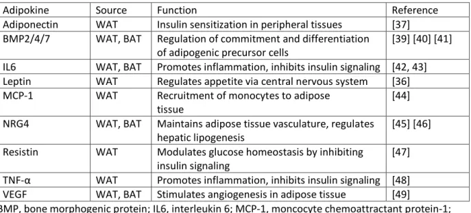
contrast, proinflammatory cytokines like resistin, tumor necrosis factor-α (TNF-α), and IL-6 contribute to insulin resistance by activating inflammation pathways involving c-Jun N-terminal kinase (JNK) and inhibitor of κβ (IKK) that result in increased serine phosphorylation of IRS molecules [38]. Other adipokines and their reported functions are listed in Table 1.
Table 1: Examples of adipokines secreted by WAT and BAT and their functions
Adipokine Source Function Reference
Adiponectin WAT Insulin sensitization in peripheral tissues [37] BMP2/4/7 WAT, BAT Regulation of commitment and differentiation
of adipogenic precursor cells
[39] [40] [41] IL6 WAT, BAT Promotes inflammation, inhibits insulin signaling [42, 43] Leptin WAT Regulates appetite via central nervous system [36] MCP-1 WAT Recruitment of monocytes to adipose
tissue
[44] NRG4 WAT, BAT Maintains adipose tissue vasculature, regulates
hepatic lipogenesis
[45] [46] Resistin WAT Modulates glucose homeostasis by inhibiting
insulin signaling
[47] TNF-α WAT Promotes inflammation, inhibits insulin signaling [48] VEGF WAT, BAT Stimulates angiogenesis in adipose tissue [49]
BMP, bone morphogenic protein; IL6, interleukin 6; MCP-1, moncocyte chemoattractant protein-1; NRG4, neuregulin 4; TNF-α, tumor necrosis factor alpha; VEGF, vascular endothelial growth factor
1.1.3 Interactions between adipocytes and other cell-types
Adipocytes have been shown to play additional roles in various biological processes through crosstalk with other cell types. In the context of obesity, elevated levels of proinflammatory adipokines promote the recruitment of monocytes to adipose depots to increase the inflammatory response and clear infections [50]. Macrophages are also recruited to adipose depots by free fatty acids (FFAs) during fasting and weight loss periods and take up nearby lipids to maintain circulatory free fatty acids levels [51]. In addition, M2 macrophages activated by cold exposure in an IL-4-dependent manner were shown to secrete catecholamines that promote the browning of white adipose tissue, confirming reciprocal crosstalk between macrophages and adipocytes [52]. Adipocyte progenitor cells were shown to promote
22
hair growth in mice by stimulating the expression of platelet-derived growth factor α (PDGF-α), which induces stem cell activation in hair follicles [5]. Adipocytes found in the epicardium are associated with increased atherosclerosis and blood pressure [53]. Furthermore, adipocytes regulate insulin sensitivity in skeletal muscle [54]. Adipocyte-myocyte interactions also involve fibro-adipogenic precursor (FAP) cells which exist in skeletal muscle and can differentiate into white adipocytes under conditions of metabolic dysfunction and muscular dystrophy. Undifferentiated FAPs were previously shown to be involved in muscle repair following muscle damage, whereby they proliferate, clear necrotic cells, and promote myogenesis in response to cytokine production at the site of injury [5].
Adipocytes in the bone marrow arise from multipotent precursor cells that commit to either the osteoblast or adipocyte lineages. This cell fate decision is thought to explain an inverse association between marrow fat and bone strength and density [55]. In addition, several studies have demonstrated reciprocal regulation between adipocytes and osteoblasts. For instance, Hamrick and colleagues
reported that leptin-deficient ob/ob mice treated with leptin displayed a loss in the size and number of bone marrow adipocytes as well as increased bone formation [56]. In contrast, Luo et al. showed that adiponectin negatively regulates bone formation by inhibiting the production of the osteoclastogenesis inhibitor, osteoprotegerin, in osteoblasts [57]. Conversely, osteocalcin which is secreted by osteoblasts, was reported by Ferron and colleagues to affect fat mass and improve insulin sensitivity in wild-type mice by stimulating the release of adiponectin in white adipose tissue [58]. Adipocytes have also been implicated in tumor development, as one in vitro study demonstrated that leptin promotes tumor proliferation by activating the ERK1/2 and JNK pathways [59]. Secretion of cytokines such as IL-6 and MCP-1 promote macrophage recruitment to proinflammatory environments that are optimal for tumor proliferation [60]. Cytokines also mediate tumor migration and homing to adipose depots (e.g. omental depot) that provide cancer cells with fatty acids to serve as fuel for rapid cell division [59]. Additionally, adipocytes secrete ECM components such as collagen VI and matrix metalloproteinase 11 that promote
23
ECM remodelling [59]. Adipocytes also secrete VEGF-A which stimulates angiogenesis to accommodate the increased demand for nutrients and O2 to be delivered at tumor sites [49].
The functional diversity of adipocytes is also reflected by the identification of adipocytes that are distinct from their traditional white, brown and beige counterparts. These include pink and yellow adipocytes that develop in mammary glands and in the bone marrow respectively. Mammary adipocytes are involved in the regulation of epithelial growth and epithelium function, and communicate with other cell types in mammary glands [61-63]. Lineage tracing studies by Cinti and colleagues led to the
observation that subcutaneous adipocytes in mammary glands convert into milk-secreting epithelial cells [64]. In this regard, mammary adipocytes were reported to dedifferentiate into fibroblast-like preadipocytes during lactation to allow for mammary alveolar structures to expand and develop milk-secreting properties in the ductal epithelium of the mammary gland [65, 66]. However, these findings were recently challenged in a report by Wang et al. who used a doxycycline-inducible adipocyte-specific tracking model to demonstrate that mammary adipocytes do not trans-differentiate into milk-secreting alveolar cells [66]. During the involution period, these preadipocytes re-differentiate into mature mammary adipocytes as mammary alveolar structures undergo apoptosis [66-68].
Bone marrow adipocytes have unique features compared to adipocytes found in other depots. For instance, marrow adipocytes are smaller and differ in fatty acid composition compared to other adipocytes [69]. Lipid mobilization doesn’t occur in marrow adipocytes during caloric restriction despite marrow adipocytes being able to hydrolyze lipids [70, 71]. In addition, progenitor cells of marrow adipocytes lack cell surface markers such as CD24 that are seen in progenitor cells for other adipose depots [55, 72]. Thus, the influence that adipocytes have on other cell types and tissues demonstrates that the function of adipose tissue is more complex than just regulating energy homeostasis.
24
1.1.4 Functional differences between different adipose depots
Adipose tissue can be further classified into depots according to their location. In the case of WAT, most depots are generally categorized as being visceral (vWAT) or subcutaneous (scWAT) tissue. In humans, vWAT includes the mesenteric, omental, retroperitoneal and epicardial depots. scWAT
encompasses depots found in the abdominal, gluteal, and femoral regions. All vWAT depots listed above are also found in mice except for epicardial depots [73]. Conversely, epididymal fat depots that are present in mice are not found in humans [5]. vWAT and scWAT also differ in physiological and metabolic function. For instance, vWAT adipocytes are more metabolically active and insulin-resistant than scWAT [74, 75]. Visceral adipocytes also display greater lipolytic activity, as they are more sensitive to
catecholamine-stimulated lipolysis, which is the hydrolysis of triglycerides into FFA and acylglycerols [31, 76]. Furthermore, inflammatory cells are more prevalent in vWAT compared to scWAT depots [77, 78]. scWAT depots are also more efficient in taking up FFA and triglycerides from circulation during the postprandial period [76]. These differences explain in part why vWAT is closely associated with insulin resistance, hyperglycemia, dyslipidemia and mortality in comparison to scWAT which is even associated with protection against cardiometabolic disease in obesity [31, 79]. In humans, BAT is distributed around the paravertebral, supraclavicular, and suprarenal regions in humans whereas in mice, BAT is divided into the interscapular and perirenal depots [73].
1.1.5 Heterogeneity of adipose tissue
Functional differences between adipose depots may result from heterogeneity among adipocyte precursor cells (APCs) that develop into different adipocyte populations. Methods such as lineage tracing, fluorescence activated cell sorting (FACS), and single-cell RNA sequence (scRNAseq) analysis have led to the identification of distinct APC populations and have advanced our understanding of the developmental origins of different adipose depots. One example is the case of Seale and colleagues who
25
used the Cre–LoxP system to investigate whether white and brown adipocytes shared a common precursor expressing Myf5, which was initially thought of being expressed solely in myocytes. Lineage tracing by Cre–LoxP is a method that uses reporter and Cre recombinase models to evaluate whether progeny cells express genes that are also expressed by precursor cells belonging to a specific lineage. White and brown adipocytes were determined to be derived from separate APCs that were Pax7-; Myf5 -and Pax7+; Myf5+ respectively [27, 80]. Furthermore, brown adipocytes and skeletal muscle were derived from a common precursor cell whose fate into either lineage was controlled by PRDM16 [27]. Lineage tracing also indicated that beige adipocytes that develop in WAT following stimulation with an β3-adrenergic agonist were not derived from Myf5+ precursors of intrascapular BAT but did resemble
Myf5- white adipocyte precursors [27].
In another study, Rodeheffer and colleagues used FACS, a technique that separates cells based on the absence or presence of cell-surface proteins, to characterize the preadipocyte lineage using APCs collected from the stromal vascular fraction (SVF) of murine adipose tissue. Rodeheffer et al. defined this population as being negative for CD31, CD45, and Ter119 which are respectively expressed in committed lineages for endothelial cells, macrophages and erythrocytes [30]. This population was positive for CD29, CD34, Sca1 and CD24 stem cell markers [81]. To confirm that adipocytes were derived from this population of cells, these precursor cells were transplanted into depots of lipodystrophic mice, and the successful reconstitution of WAT in these mice validated the identification of these precursor cells belonging to the adipocyte lineage [81]. In a later study, lineage tracing by the Cre–LoxP system was utilized to determine whether white adipocytes in mice could be derived from hematopoietic and endothelial lineages [82]. Results from that study indicated that WAT was not derived from either lineage, which was consistent with the earlier identification of an APC population by FACS that was devoid of endothelial and erythrocyte cell surface markers. Additional lineage tracing studies using
26
for the cell surface marker CD24. The expression of adipocyte-associated genes like Pparg and C/ebpa in CD24- APCs but not in CD24+ APCs led Rodeheffer and colleagues to the conclusion that CD24+ precursors
develop into CD24- precursors that are more committed to the preadipocyte lineage [82]. Additional
studies into the heterogeneous origins of WAT depots that utilized the lineage tracing by Cre–LoxP method demonstrated that APCs from respective depots originate from distinct areas of the mesoderm [30, 83, 84].
In a recent study conducted by Merrick et al., separation of fat cells with FACS followed by further grouping of cells by single-cell RNA sequencing (scRNAseq, a method that groups cells according to gene expression) led to the identification of three novel APC populations in mice and humans: interstitial progenitor cells (IPCs) expressing Dipeptidyl Peptidase-4 (DPP4), preadipocytes expressing intercellular adhesion molecule-1 (ICAM1), and group 3 cells expressing CD142 [85]. Based on in vivo cell transplantation studies, DPP4-expressing IPCs are progenitor cells for committed ICAM1- and CD142-expressing preadipocytes. Merrick et al. noted that group 3 APCs are present in the subcutaneous fat of mice but not humans. The idea of APC heterogeneity contributing to differences between adipose depots was supported by the finding that fewer IPCs were present in visceral depots than in
subcutaneous depots [85]. Adipocyte heterogeneity has not only been seen between different depots, but also among adipocytes within a single depot. Reports of mature white adipocytes isolated from a single depot displaying variable lipogenesis, insulin sensitivity and fatty acid uptake suggest that a single adipose depot may be comprised of several distinct adipocyte subtypes [86, 87]. Furthermore, Lee et al. utilized in vitro clonal cell analysis to identify three white adipocyte populations characterized by unique expression profiles of three gene markers: Wilms tumor 1 (Wt1, type 1), transgelin (Tagln, type 2) and Myxovirus 1 (Mx1, type 3) [88]. In addition, clonal cell analysis and lineage tracing models revealed differences in metabolism and gene expression across these three populations, and that WAT depots are comprised of these 3 distinct adipocyte populations that differ in abundance [88].
27
1.2 Lipid Homeostasis 1.2.1 Lipid droplets
Lipid droplets consist of a core composed of triacylglycerols (TAGs) and cholesterol esters enveloped by a monophospholipid layer and are coated with various proteins including ATGL (adipose triglyceride lipase) [89]. Lipid droplets are mainly found in adipocytes, hepatocytes, mammary epithelial cells and steroidogenic cells [89]. Furthermore, the composition of the lipid core varies between cell types. Lipid droplets are not found only in mammals, but also in plants, bacteria and insects [90, 91]. Lipid droplets were initially thought of only having roles in lipid storage and preventing lipotoxicity; however, recent reports of the presence of proteins such as Rab GTPases on the lipid droplet surface suggest that they have additional roles in lipid trafficking and lipid metabolism [92]. Proteins found on the surface of lipid droplets include perilipin A (PLIN1), adipocyte differentiation-related protein (ADRP or PLIN2), and tail-interacting protein of 47 kDa (TIP47 or PLIN3), all of which contain a conserved sequence region called the PAT domain [93].
Whereas PLIN3 is highly expressed in all tissues, PLIN1 and PLIN2 are mostly expressed in WAT and liver respectively [94]. In adipocytes, perilipin A regulates lipid metabolism by controlling the access of lipases to the lipid droplets. Upon phosphorylation by protein kinase A, perilipin no longer blocks lipases found on the surface of lipid droplets from hydrolyzing TAGs stored in the lipid core. ADRP, which was named based on its early induction during adipocyte differentiation, has roles in the formation and stabilization of small lipid droplets [94] and is replaced with PLIN1 as preadipocytes mature. [95, 96]. Despite initially being implicated with intracellular trafficking of lysozymes, PLIN3 was reported to contribute towards lipid droplet stabilization [97].
28
1.2.2 Lipolysis
1.2.2.1 Key players in the lipolytic pathway
While white adipose tissues serve as sites for the storage of excess energy in the form of triglycerides, they also regulate the release and mobilization of stored triglycerides to other tissues that require energy in a process called lipolysis [98]. Lipolysis is a chemical pathway whereby triglycerides are sequentially hydrolyzed into FFAs and acylglycerols. Lipolysis is modulated by catecholamines, which bind either to β-adrenergic receptors to stimulate lipolysis or α2-adrenergic receptors to inhibit lipolysis [99]. In the case of lipolysis, binding of endogenous catecholamines such as norepinephrine to G-protein coupled β-adrenergic receptors (GPCRs) causes the α-subunit of heterotrimeric G-proteins to dissociate from the β and γ subunits. As a result, the Gα subunit stimulates the activity of adenylyl cyclase,
increases production of the second messenger cAMP, activation of PKA, and the subsequent phosphorylation of HSL and transcription factors (Figure 3) [98]. Three enzymes are involved in the sequential hydrolysis of triglycerides to yield glycerol and FFA: adipose triglyceride lipase (ATGL), hormone sensitive lipase (HSL), and monoacylglycerol lipase (MAGL). ATGL catalyzes the hydrolysis of triacylglycerols (TAGs) into diacylglycerols (DAGs), which are subsequently hydrolyzed into
monoacylglycerols (MAGs) by HSL. Lastly, MAGL hydrolyzes MAGs into free fatty acids and glycerol. A study that used ATGL-deficient mice and small-molecule inhibitors for HSL suggests that ATGL and HSL account for at least 90% of lipid hydrolysis in murine WAT [100].
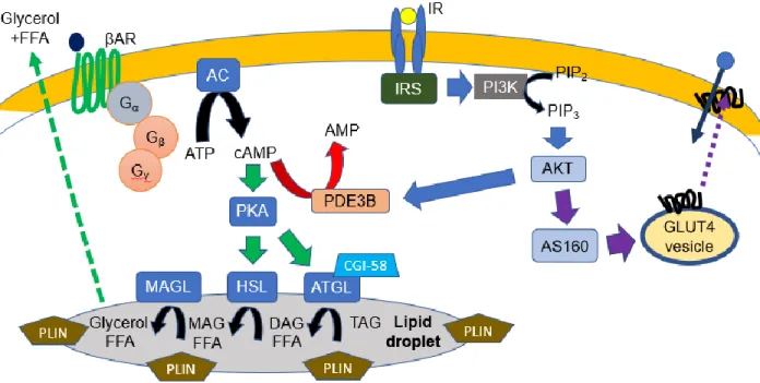
29
Figure 3: Lipolysis pathway in adipocytes. Stimulation of lipolysis involves catecholamines binding to β-adrenergic receptors and generation of the second messenger cAMP. Insulin signaling inhibits lipolysis through PI3K/AKT-mediated activation of PDE3B, which converts cAMP to AMP. AC, adenylyl cyclase; βAR, β-adrenergic receptor; CGI-58, comparative gene identification-58; HSL, hormone sensitive lipase; IR, insulin receptor; IRS, insulin receptor substrate; PDE3B, phosphodiesterase 3B; PIP2,
phosphatidylinositol 4,5-bisphosphate; PIP3, phosphatidylinositol (3,4,5)-trisphosphate; PLIN, perilipin
1.2.2.2 HSL
Although HSL has been shown to exhibit broad specificity in hydrolyzing TAGs, DAGs, and MAGs, it was reported to have greater specificity for DAGs which are hydrolyzed approximately 10 times faster than TAGs [101, 102]. Several findings challenged the initial belief that HSL was the sole rate-limiting enzyme involved in the hydrolysis of TAGs. One example was the difference in the relative fold increase in β-adrenergic stimulated lipolysis from basal conditions (50-fold) compared to the fold increase in the specific activity of HSL (less than 2-fold) upon stimulation [102]. Furthermore, Haemmerle and
colleagues observed that HSL knockout (HSL KO) mice only displayed a 30-40% reduction in FFA release compared to wild-type littermates. While these mice also displayed an accumulation of DAGs in adipose tissue, muscle, and testis, no TAG accumulation was observed in adipose or non-adipose tissue [103].
30
Taken together, these observations suggested that other lipases were involved in lipolysis (see sections 1.2.2.3 and 1.2.2.4).
Regulation of HSL activity is controlled by several factors. For instance, PKA has been shown to regulate HSL via phosphorylation at multiple sites including Ser650 in humans and Ser563, Ser659, and Ser660 in rat [104, 105]. Furthermore, β-adrenergic stimulation and insulin have opposing roles in modulating HSL activity, as the former condition strongly induces HSL activity and the latter inhibits its activity. Insulin inhibits lipolysis by activating AKT (also called PKB) and activates phosphodiesterase 3B which converts cAMP to 5’AMP (Figure 3) [106]. Under basal conditions, HSL resides in the cytoplasm and PLIN1 prevents HSL from accessing the lipid droplet. Upon stimulation of lipolysis, PKA
phosphorylates multiple PLIN1 residues, including Ser81, Ser222, Ser272, Ser433, Ser492 and Ser517. As a result, PLIN1 interacts with and activates phosphorylated HSL which is found at the lipid droplet and is no longer restricted from hydrolyzing lipid droplets [107, 108]. Although PLIN1 was initially believed to be essential for HSL translocation, Miyoshi and colleagues demonstrated by cell fractionation and confocal microscopy that translocation of HSL to the lipid droplet surface was still possible in the presence of PLIN1 devoid of its six PKA-binding sites. This suggests that PLIN1 and PKA are not the sole proteins that mediate the activation of HSL.
1.2.2.3 ATGL
ATGL is also known as desnutrin and patatin-like phospholipase domain containing 2 (PNPLA2) and is highly selective for TAGs as it hydrolyzes them 10 times faster than DAGs [109]. Reports of significant reductions in TAG hydrolase activity following ATGL immunoprecipitation from human and mouse adipose tissue lysates suggested that ATGL may be responsible for hydrolyzing most TAGs [109, 110]. The contributions of ATGL in lipolysis and its role as the principal enzyme implicated in the
31
hydrolysis of TAGs were further substantiated by studies involving ATGL knockout (ATGL KO) mice. Haemmerle and colleagues observed that ATGL KO mice displayed significant reductions in TAG hydrolase activity, enlarged adipose tissues, and accumulated lipids in the heart causing cardiac dysfunction [111]. ATGL KO mice were also cold-sensitive as they reduced their body temperature and oxygen consumption when fasted. This was explained by the failure of WAT and BAT to mobilize adequate amounts of FFA which was evident from the nearly 70% decrease in isoproterenol-stimulated FFA release and significantly reduced plasma FFA levels in ATGL KO mice [111].
At least two serine residues have been identified as phosphorylation sites in murine ATGL: Ser406 and Ser430 [89]. Similarly, Bartz et al. identified Ser404 and Ser428 as phosphorylation sites in human ATGL [89]. Despite the identification of these phosphorylation sites, PKA was shown not to play a role in ATGL phosphorylation [109]. Ahmadian et al. demonstrated that ATGL phosphorylation was mediated by AMP-activated kinase (AMPK) at Ser406 and resulted in an increase in lipase activity [112]. However, a recent study from Pagnon et al. called into question whether phosphorylation of murine ATGL was strictly PKA-independent. Pagnon and colleagues showed that whereas both PKA and AMPK were able to phosphorylate Ser406 in vitro, only PKA could phosphorylate Ser406 in vivo. This was based on observations of pharmacological PKA inhibitors blocking the increase in β-adrenergic-stimulated phosphorylation at Ser406. Furthermore, no increases in AMPK activity in adipose tissue of fasted mice was observed, despite increases in phosphorylation of Ser406 [113]. Thus, this study called into question the previously established roles (or lack thereof) of AMPK and PKA in regulating lipolysis by
phosphorylating mouse ATGL to modulate its activity.
Unlike HSL, ATGL requires a coactivator protein called comparative gene identification-58 (CGI-58) or α/β hydroxylase domain containing protein 5 (ABDH5) to achieve full TAG hydrolase activity. This was demonstrated in mouse models that reported up to a 20-fold increase in ATGL hydrolase activity in the presence of CGI-58 [114]. ATGL, which is found both in the cytoplasm and on the lipid droplet
32
surface, has also been reported to be indirectly regulated by perilipin. Studies point to perilipin regulating ATGL activity by controlling the accessibility of ATGL to its coactivator CGI-58 [115, 116]. In the basal state, perilipin binds to CGI-58 and prevents ATGL activation. In the stimulated state, PKA phosphorylates perilipin resulting in the dissociation of the perilipin – CGI58 complex. Consequently, CGI-58 is free to bind to and activate ATGL. Miyoshi and colleagues showed that PKA-mediated phosphorylation of perilipin at Ser517 was critical for ATGL activation as mutation of this site reduced forskolin-stimulated glycerol and FFA release by 95% in adipocytes derived from mouse embryonic fibroblasts [116].
1.2.2.4 MAGL
MAGL catalyzes the conversion of monoglycerides to glycerol and FFA. MAGL was previously reported to exhibit high specificity for MAGs [117]. Although MAGL is abundantly expressed in several tissues, the highest expression is seen in WAT. MAGL is found in the cytoplasm, plasma membrane and on the lipid droplet surface [118, 119]. In comparison to HSL and ATGL, very little is known about the regulation of MAGL [119]. In one study involving MAGL knockout mice, Taschler et al. reported a 35% reduction in stimulated lipolysis and MAG hydrolase activity [120]. This suggests that other recently discovered monoacylglycerol lipases including α/β hydrolase domain-containing protein 6 (ABHD6), ABDH12, and fatty acid amide hydrolase (FAAH) likely contribute to MAG hydrolysis. While the above-mentioned lipases were determined to display MAG hydrolase activity in vitro, the in vivo contributions and implications of these lipases to MAG hydrolysis remain to be elucidated. Lastly, interest in the contributions of MAGL outside of lipolysis stem from its reported role in degrading 2-arachidonylglycerol (2-AG), a signaling molecule belonging to the family of endogenous lipids known as endocannabinoids [121]. Endocannabinoids bind to G-protein coupled cannabinoid receptors to regulate energy
33
1.2.3 Lipogenesis
1.2.3.1 Overview of lipogenesis
De novo lipogenesis is a pathway whereby excess carbohydrates are used to synthesize free
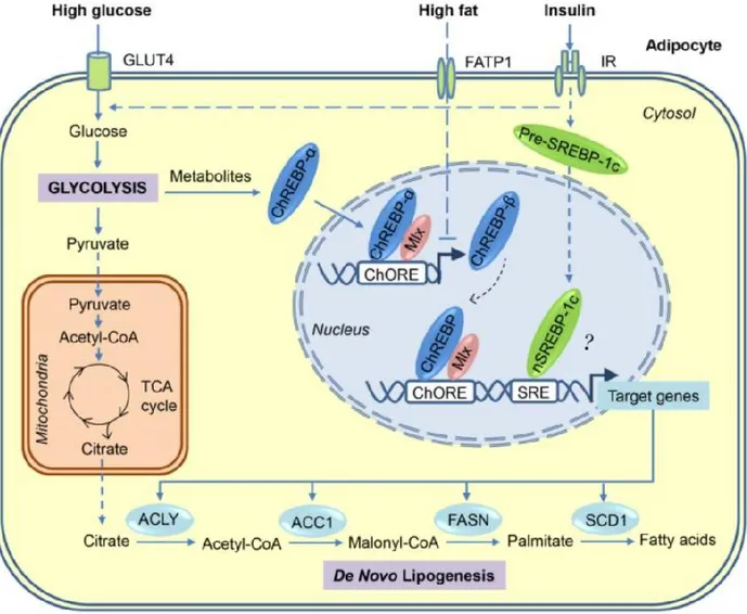
fatty acids, which are later converted into triglycerides for storage, particularly in white adipose tissue and the liver (Figure 4). Citrate produced from glucose undergoing glycolysis, pyruvate processing, and the tricarboxylic acid (TCA) cycle is shuttled from the mitochondria to the cytosol to generate acetyl-CoA by ATP-citrate lyase (ACLY). Fatty acids are then obtained from acetyl-CoA through a series of reactions catalyzed by the enzymes acetyl-CoA carboxylase (ACC1), fatty acid synthase (FASN), and stearoyl-CoA desaturase-1 (SCD-1). ACC1 and FASN are the two principle enzymes involved in the de novo lipogenesis pathway, as the former enzyme catalyzes the conversion of acetyl-CoA to malonyl-CoA and the latter enzyme converts malonyl-CoA into palmitate. The generation of palmitate, the first fatty acid product, is the rate-limiting step of the de novo lipogenesis pathway. Palmitate is elongated and saturated by enzymes including SCD-1 to generate various complex fatty acids, such as palmitoleic acid, steric acid, and oleic acid [123]. Regulation of these two principle enzymes are controlled by sterol-response-element-binding protein 1c (SREBP1c) and carbohydrate-response-sterol-response-element-binding protein (ChREBP). The roles of these transcription factors as the dominant regulators of de novo lipogenesis varies across different cell types, as SREBP1c is the main regulator in the liver and ChREBP is the main regulator in WAT [5].
34
Figure 4: The de novo lipogenesis pathway in adipocytes. Glucose is metabolized into pyruvate through glycolysis and is later processed into acteyl-coA which enters the TCA cycle to generate citrate. The enzymes ACLY, ACC1, FASN, and SCD-1 catalyze a series of reactions to convert citrate into palmitate which undergoes additional processing to synthesize various fatty acids. The expression of these enzymes is regulated by the transcription factors ChREBP and SREBP-1c, which in turn are respectively stimulated by high glucose and insulin. ACLY, ATP citrate lyase; ACC1, Acetyl-CoA carboxylase 1; ChREBP, carbohydrate-response element binding protein; ChORE, carbohydrate response element; FASN, fatty acid synthase; SREBP-1c, Sterol-responsive element binding protein; SRE, sterol response element; SCD-1, stearoyl-CoA desaturase. Image taken from [123].
35
1.2.3.2 SREBP-1c
Sterol-responsive element binding proteins (SREBPs) are a class of transcription factors that possess the basic helix-loop-helix-leucine zipper (bHLH-Zip) domain for binding to DNA. The SREBP family is comprised of three isoforms: SREBP-1a, SREBP-1c, and SREBP-2. SREBP-1a activates genes involved in cholesterol, fatty acid, and triglyceride synthesis. SREBP-1c and SREBP-2 have more restrictive roles, as they respectively promote expression of genes related to fatty acid synthesis and cholesterol synthesis [124]. SREBPs are initially synthesized in their inactive form consisting of an amino-terminal domain containing the bHLH-Zip region, a hydrophobic transmembrane region, and a carboxy-terminal regulatory domain. When intracellular cholesterol levels are low, the escort protein and sterol sensor SREBP cleavage-activating protein (SCAP) binds and directs SREBP to the Golgi apparatus where it is proteolytically cleaved by two proteases, Site-1 and Site-2 protease (S1P and S2P). The activated amino-terminal domain translocates to the nucleus and binds to specific sterol response elements (SRE) in promoter regions of genes associated with fatty acid and cholesterol synthesis to drive their
expression. Some target genes of SREBP-1c include ACLY, ACC1, FASN, and SCD-1 [124]. This process is also promoted by insulin. In hepatocytes, insulin also promotes transcription of SREBP-1c through the PI3K/AKT pathway that activates the downstream target mammalian target of rapamycin complex 1 (mTORC1) [125-127].
Loss of function and overexpression models for SREBP-1 have been utilized to examine its contributions to lipogenesis. Shimano and colleagues report that systemic deletion of SREBP-1
decreased hepatic lipogenesis but increased hepatic SREBP-2 levels and SREBP-2-mediated cholesterol synthesis. Interestingly, adiposity and lipogenic gene expression in WAT were not affected [128]. While adipocyte-specific SREBP knockout models have yet to be reported, adipocyte-specific SREBP
overexpression studies have been conducted for the SREBP-1a and SREBP-1c isoforms. In a study by Horton et al., increased lipogenesis, hypertrophy, and fatty acid release in adipose tissue were observed
36
[129]. In the case of SREBP-1c, Shimomura et al. observed impaired adipocyte differentiation, reduced adiposity, glucose intolerance and impaired insulin sensitivity [130]. When taken together, the loss of function and gain of function models for SREBP-1 suggest that SREBP-1 plays a minor role in adipocyte
de novo lipogenesis.
1.2.3.3 ChREBP
Carbohydrate-response element binding proteins (ChREBPs) are also transcription factors that have a bHLH/Zip motif. The N-terminus domain contains regions regulating its nuclear localization, whereas the C-terminus domain contains the bHLH/Zip motif required for DNA-binding [131, 132]. To date, two ChREBPs isoforms have been identified: ChREBP-α and ChREBP-β. ChREBP activity is
dependent on glucose levels. During fasting periods when glucose levels are low, elevations in plasma glucagon and epinephrine stimulate the activation of PKA, which along with AMPK, phosphorylates ChREBP-α. Consequently, the transcriptional activity of ChREBP-α is inhibited, and it is retained in the cytosol. During feeding and in high glucose conditions, intermediates of glucose metabolism, such as xylulose-5-phosphate (Xu-5-P) and glucose-6-phosphate (G6P), accumulate and activate protein phosphatase 2A, which dephosphorylates ChREBP-α. As a result, ChREBP-α translocates to the nucleus and binds to carbohydrate response elements in the promoter regions of ChREBP-α target genes
involved in glycolysis and fatty acid synthesis. These genes are the same previously mentioned targets of SREBP-1c. ChREBP-α also induces the transcription of ChREBP-β, which upon feed-forward stimulation of its own expression activates its respective set of gene targets (Figure 4) [133-135].
Global knockout of ChREBP in mice led to decreases in adiposity, increased glucose tolerance, and reduced insulin sensitivity [136]. However, adipocyte-specific deletion of ChREBP was reported to decrease sucrose-induced lipogenesis only in liver while also impairing glucose tolerance and insulin sensitivity [137]. Conversely, Nuotio-Antar et al. reported that mice overexpressing ChREBP in adipose
37
tissue displayed a lean phenotype while lipogenesis in adipose tissue increased. Other metabolic phenotypes included improved glucose tolerance and insulin sensitivity [138]. The reports of increased adipocyte de novo lipogenesis in the adipocyte-specific overexpression model for ChREBP but not SREBP-1c suggests that between the two transcription factors, ChREBP is the principal activator of de
novo lipogenesis in adipocytes.
1.2.3.4 ACC1
ACC1 catalyzes the carboxylation of acetyl CoA to produce malonyl CoA. In eukaryotes, ACC1 exists as a single multifunctional polypeptide that containing domains of a biotin carboxyl carrier protein, a biotin carboxylase, and a transcarboxylase. Each monomer is approximately 265 kDa. In contrast, these three domains correspond to three separate proteins in prokaryotes [139]. ACC1 expression is transcriptionally regulated by SREBP-1c and ChREBP as previously described (Figure 4). In addition, ACC1 activity is modulated post-translationally by AMPK-mediated phosphorylation. Activation of AMPK by glucagon and epinephrine results in the phosphorylation and inactivation of ACC1, whereas insulin-mediated activation of protein phosphatase 2A reverses this and activates ACC1. In addition, citrate, an upstream precursor in the de novo lipogenesis pathway, allosterically binds and partially activates ACC1 [123]. The important role of ACC1 in de novo lipogenesis was demonstrated by Mao et al. who reported that liver-specific ACC1 mice displayed reduced hepatic lipid content compared to control mice placed on a fat-free diet [140]. However, this protective reduction in hepatic lipid content was lost when the liver-specific knockout mice were put on high-fat diet.
1.2.3.5 FASN
FASN catalyzes the synthesis of palmitate from malonyl CoA and exists in two forms: FASN I (type 1) and FASN II (type 2). FASN I is found in animals and fungi and exists as a multifunctional homodimeric polypeptide [141, 142]. In plants and bacteria, FASN II exists as a series of individual
38
monofunctional enzymes whose domains are homologous to FASN I orthologs [143]. FASN is transcriptionally regulated by the previously mentioned transcription factors SREBP-1c and ChREBP (Figure 4). Mouse models have identified key roles of FASN in lipid synthesis and cellular signaling. In one study, liver-specific FASN knockout mice that were fed a zero-fat diet displayed exacerbated hepatic steatosis [144]. This suggests that the protection against steatosis observed in ACC1 knockout mice fed a fat-free diet is likely attributed to the reduction in malonyl-CoA production [145]. In another report by Lodhi et al, adipocyte-specific FASN knockout mice had a lean phenotype resulting from impaired PPARγ activation, and displayed increased energy expenditure and beiging of subcutaneous adipose tissue. activation. These mice were also protected from diet-induced obesity. In addition, decreased
transcriptional PPARγ activity and adipogenesis was shown to result from a reduction in the synthesis of PPARγ agonists such as specific alkyl ether lipids [146].
1.3 14-3-3 proteins 1.3.1 Structure
Since their original discovery in bovine brain homogenates over 50 years ago, a renewed interest has been taken in understanding the roles of molecular scaffolds belonging to the 14-3-3 protein family. The name 14-3-3 is derived from their discovery in the 14th fraction of DEAE-cellulose chromatography
and migration position 3.3 following gel electrophoresis [147]. 14-3-3 proteins are 28-33 kDa acidic proteins. In mammals, seven isoforms comprise the 14-3-3 protein family: β, γ, ε, η, σ, θ and ζ. Although these isoforms are highly conserved, their individual expression profiles vary between tissues and cell types [148]. 14-3-3 proteins often dimerize to form either homodimers or heterodimers in order to interact with client proteins [149, 150]. Each monomer consists of nine α-helices that are arranged in an antiparallel manner. Furthermore, a monomer consists of the N-terminus, a conserved core region, and the C-terminus. Although both the N-terminus and C-terminus display variability across the isoforms, only the N-terminus contains the residues required for dimerization [151, 152]. In the core region, four
39
α-helices, helices 3, 5, 7, and 9 arrange themselves in such a way that an amphipathic groove is formed, whereby one side has a cluster of polar residues and the other side has a cluster of non-polar residues [151]. This amphipathic groove comprises the ligand binding domain that allows the 14-3-3 proteins to interact with their target proteins. Two classical types of phosphorylation-dependent and high-affinity 14-3-3 binding motifs were identified: RSX pS/T XP (mode 1) and RXXX pS/T XP (mode 2) where pS/T and X represent phosphorylated serine or threonine and any amino acid respectively [153]. 14-3-3 proteins are also capable of binding to non-phosphorylated client proteins. Dimerization also allows 14-3-3 proteins to simultaneously bind to two targets [154]. These phospho-motifs are generated by kinases, such as PKA, AKT and PKC [147, 153]. Upon binding, 14-3-3 proteins can spatially and temporally control the localization of their binding partners, regulate the activity of their targets, and mediate protein interactions that involve the client protein [151, 152, 155].
1.3.2 Function
14-3-3 proteins are also called tyrosine and tryptophan hydroxylase activators (YWHAs). This alternative name comes from the discovery of their first reported function, which was the regulation of tyrosine and tryptophan hydroxylases [156]. Tyrosine and tryptophan hydroxylases catalyze the rate-limiting steps of catecholamine and serotonin synthesis, respectively. 14-3-3 proteins are often referred as molecular scaffolds due to their ability to interact with one or several target proteins implicated in a signaling pathway. For example, 14-3-3β and 14-3-3θ were reported to facilitate interactions between PKC and RAF-1 in the RAF/MEK/ERK cascade that regulates cell fate [157]. Various roles have been reported for 14-3-3 proteins with respect to cellular and organismal metabolism and include the
regulation of the cardiac isoform phosphofructo-2-kinase/fructose-2,6-biphosphatase (PFK-2) [158, 159] and glyceraldehyde-3-phosphate dehydrogenase (GAPDH) involved in gluconeogenesis and glycolysis [160-162]. Other 14-3-3 targets that have been cited include cell division cycle 25B (CDC25B) involved in progression of the cell cycle [163-165] and Bcl-2-associated death promoter (BAD) involved in apoptosis
40
and insulin secretion by pancreatic β-cells [166, 167]. Lim et al. previously reported on the metabolic contributions of the zeta isoform with respect to glucose homeostasis using a systemic knockout mouse model. They observed that 14-3-3ζ knockout mice had improved oral glucose tolerance despite
displaying insulin resistance. This was attributed to elevated levels of GLP-1 [168].
1.3.3 Implication of 14-3-3 proteins on adipocyte development
We previously demonstrated that 14-3-3ζ has essential roles in adipogenesis using a combination of
in vitro and in vivo models. We reported that only 14-3-3ζ was required for the differentiation of 3T3-L1
pre-adipocytes [169], and that 14-3-3ζ affects alternative splicing of Pparg mRNA during adipocyte differentiation [170]. We also reported that systemic 14-3-3ζ knockout (14-3-3ζKO) mice displayed significant reductions of fat specifically in visceral depots and had smaller gonadal adipocytes compared to wild-type littermates. [169]. Furthermore, transgenic mice overexpressing 14-3-3ζ gained more weight and fat mass compared to wild-type mice fed a high-fat diet (HFD). Metabolic phenotype studies revealed that 14-3-3ζ overexpression did not impair glucose tolerance or insulin sensitivity [169]. Regarding the insulin-mediated glucose uptake pathway that is dependent on GLUT4 in adipocytes, 14-3-3ζ was shown to interact with several targets including IRS1, IRS2, and AS160 [171, 172].
1.4 Hypothesis and objectives
We demonstrated in a previous report that 14-3-3ζ has essential roles in the development of adipocytes and adipose tissue [169, 170]. However, the contributions of 14-3-3ζ to the function of mature adipocytes are still not known. Furthermore, conclusions regarding the contributions of 14-3-3ζ to adipogenesis were based in part on mice models where 14-3-3ζ was either systemically deleted or systemically overexpressed. A limitation in using these models is that they do not allow for contributions of 14-3-3ζ specifically in adipocytes to be evaluated. This master’s project was pursued to address these outstanding issues.
41
Our hypothesis was that decreasing 14-3-3ζ expression in adipocytes will impair the function of mature adipocytes. In order to test this hypothesis, the first objective of this project is to obtain a clearer understanding of how 14-3-3ζ contributes to adipocyte-specific processes. We begin this endeavor by determining if 14-3-3ζ plays a role in lipolysis, as 14-3-3 protein binding motifs have been identified in ATGL and HSL [173]. This suggests that 14-3-3ζ may have regulatory roles in lipolysis. The second objective of this project is to assess whether the deletion of 14-3-3ζ specifically in adipocytes affects whole-body metabolism. This determination requires the metabolic characterization of adipocyte-specific 14-3-3ζ knockout mice (adi14-3-3ζKO) that will be used in this project.
43
14-3-3zeta is required for PKA-dependent lipolysis in mature adipocytes
Abel Oppong1, Kadidia Diallo1, Yves Mugabo1, Gareth Lim1
1 Department of Medicine, Université de Montréal and Centre de recherche du Centre hospitalier de
l’Université de Montréal (CRCHUM), 900 Saint-Denis St., Montreal, QC, Canada H2X 0A9
Contributions of:
AO: Performed experiments, analyzed data, and wrote the manuscript KD: Performed adipocyte morphological measurements (not included in memoire) YM: Assisted with in vivo experiments and technical assistance with TAG measurements GEL: Designed the study, analyzed data, and edited the manuscript. GEL is the guarantor of this work.
Corresponding author: Gareth Lim, Ph.D.
Centre de recherche du Centre hospitalier de l’Université de Montréal (CRCHUM) 900 Saint-Denis St., Montreal, QC, Canada H2X 0A9
Telephone: (514) 890-8000 Extension 12927 E-mail: [email protected]
Disclosure Statement: The authors have nothing to disclose
44
2.1 Abstract
The molecular scaffold, 14-3-3ζ, was previously reported to have roles in the development of
adipocytes. However, the contributions of 14-3-3ζ to processes such as lipolysis in mature adipocytes have yet to be elucidated. Herein, we demonstrate that 14-3-3ζ is necessary for lipolysis, as adipocyte-specific 14-3-3ζ knockout (adi14-3-3ζKO) mice and adipose tissue lacking 14-3-3ζ display impaired glycerol and FFA release following activation of the β3-adrenergic signaling pathway. Furthermore, HSL activation in gonadal adipose depots was decreased in adi14-3-3ζKO mice. These findings were
recapitulated in siRNA-transfected 3T3-L1 adipocytes that exhibited reductions in phosphorylated and total forms of PKA substrates including HSL and CREB. 3T3-L1 adipocytes depleted of 14-3-3ζ also displayed reductions in lipase mRNA and impaired lipolysis in response to multiple agonists including isoproterenol, forskolin and dbcAMP. In vitro mechanistic studies point to 14-3-3ζ regulating lipolysis in a PKA-dependent manner. In addition, 14-3-3ζ appears to have roles in determining adipocyte maturity as mature adipocyte features including Pparg mRNA and PPARγ expression and triacylglycerol content are reduced in adi14-3-3ζKO adipose depots and 14-3-3ζ-depleted 3T3-L1 adipocytes. Collectively, these findings reveal a novel role for 14-3-3ζ in regulating PKA-dependent lipolysis.
45
2.2 Introduction
The primary function of white adipose tissue is the regulation of energy homeostasis. White adipocytes specialize in both the storage of triacylglycerols (TAGs) and the mobilization of free fatty acids (FFAs) to peripheral tissues to accommodate increases in metabolic demand that occur during exercise and fasting [98]. In WAT, FFAs are generated by the hydrolysis of TAGs in a process known as lipolysis. Stimulation of the lipolytic pathway involves the binding of catecholamines to β-adrenergic receptors and the subsequent generation of the second messenger cyclic adenosine monophosphate (cAMP), which activates protein kinase A (PKA) [98]. TAG hydrolysis is mediated by three lipases: adipose triacylglycerol lipase (ATGL), hormone sensitive lipase (HSL), and monoacylglycerol lipase (MAGL). These lipases, respectively, catalyze the sequential conversion of TAGs into diacylglycerols (DAGs), monoacylglycerols (MAGs), and finally FFAs and glycerol. One of these lipases, HSL, has been shown to be directly phosphorylated by PKA [104, 105].
The 14-3-3 family of molecular scaffolds are implicated in several cellular processes, such as proliferation, apoptosis and metabolism [169, 174, 175]. This stems from their ability to interact with target proteins via phosphorylated serine and threonine motifs to regulate their localization, activity and interactions with other proteins [153, 154]. We have previously reported that only one of these seven mammalian isoforms, 14-3-3ζ, was required for adipogenesis [169]. However, the role of 14-3-3ζ in mature adipocyte function has not been examined. To that end, our focus turned to understanding the contributions of 3ζ to adipocyte-specific processes, such as lipolysis. The identification of the 14-3-3ζ binding motifs on ATGL and HSL suggests that 14-3-14-3-3ζ may have a regulatory role in lipolysis [173].