HAL Id: tel-02130620
https://tel.archives-ouvertes.fr/tel-02130620
Submitted on 16 May 2019HAL is a multi-disciplinary open access archive for the deposit and dissemination of sci-entific research documents, whether they are pub-lished or not. The documents may come from teaching and research institutions in France or abroad, or from public or private research centers.
L’archive ouverte pluridisciplinaire HAL, est destinée au dépôt et à la diffusion de documents scientifiques de niveau recherche, publiés ou non, émanant des établissements d’enseignement et de recherche français ou étrangers, des laboratoires publics ou privés.
Axonal homeostasis of VGLUT1 synaptic vesicles in
mice
Xiaomin Zhang
To cite this version:
Xiaomin Zhang. Axonal homeostasis of VGLUT1 synaptic vesicles in mice. Neurons and Cognition [q-bio.NC]. Université de Bordeaux, 2016. English. �NNT : 2016BORD0413�. �tel-02130620�
THÈSE PRÉSENTÉE POUR OBTENIR LE GRADE DE
DOCTEUR
DE
L’UNIVERSITÉ DE BORDEAUX
ÉCOLE DOCTORALE
SCIENCES DE LA VIE ET DE LA SANTE Neurosciences
Par ZHANG Xiaomin
Axonal homeostasis of VGLUT1 synaptic vesicles in mice
Sous la direction de : Dr. HERZOG Etienne Co-directeur : Prof. Dr. BROSE Nils
Soutenue le Mardi 14 Décembre, 2016 Membres du jury:
Mme. SANS Nathalie DR-INSERM Bordeaux Président
M. OHEIM Martin DR-CNRS Paris Rapporteur
M. MARTY Serge CR1-CNRS Paris Rapporteur
Mme. DANGLOT Lydia CR1-INSERM Paris Examinateur
M. PERRAIS David DR-CNRS Bordeaux Examinateur
M. HERZOG Etienne CR1-CNRS Bordeaux Directeur de thèse
Titre : Homéostasie axonale des vésicules synaptiques des
neurones excitateurs VGLUT1 chez la souris
Résumé :
Les vésicules synaptiques (VS), compartiment de stockage du neurotransmetteur, sont essentielles pour la transmission synaptique chimique entre les neurones. Lorsque le potentiel d'action traverse une terminaison pré-‐synaptique, l'afflux de Ca2+ peut
déclencher l’exocytose de VS qui sont ancrées et éventuellement amorcées à la zones active (ZA), et libérer leur contenu en neurotransmetteurs dans la fente synaptique. Ensuite, les molécules neurotransmetteurs se lient aux récepteurs situés dans la membrane post-‐synaptique et induisent des changements de conformation permettant la transduction du signal (échanges ioniques, voies de signalisation). Il existe de nombreuses études sur les mécanismes moléculaires en jeux lors de la neurotransmission. Concernant la partie pré-‐synaptique de ce processus, des études supplémentaires sont nécessaires pour mieux comprendre la dynamique de remplissage des VS en neurotransmetteurs, la distribution et la mobilité des VS dans l’axone.
Les neurotransmetteurs classiques sont concentrés dans les VS par l’action de transporteurs vésiculaires de neurotransmetteurs (VNT) contre un gradient électrochimique de protons généré par l’ATPase vacuolaire (v-‐ATPase). Le glutamate, neurotransmetteur excitateur principal, est transporté dans la VS par un transporteur vésiculaire du glutamate (VGLUT). Il existe 3 isoformes de VGLUT qui définissent 3 systèmes glutamatergiques anatomiques distincts dans le cerveau. Les neurones corticaux expriment fortement VGLUT1 qui représente ainsi le meilleur marqueur pour les VS dans ces neurones. Notre laboratoire a généré une souris gain de fonction VGLUT1venus qui permet de suivre VGLUT1 et les VS le portant par épi-‐fluorescence.
Dans un modèle ancien, les VS ont été considérées comme assignées à une seule terminaison et dans ce modèle chaque terminaison fonctionnait comme une unité autonome en relation directe et exclusive avec le corps cellulaire pour la synthèse et la dégradation de ses composants. Les VS accumulées dans le bouton synaptique étaient reparties selon trois contingents morphologiquement centrifuges à la ZA : le contingent immédiatement libérable, le contingent recyclant et le contingent de réserve. Mais les données obtenues récemment, nous indiquent que les VS ne sont pas organisées de façon laminaire dans le bouton et sont dynamiquement échangées entre plusieurs
boutons pré-‐synaptiques formés en passant le long des axones. Ce phénomène a conduit à la dénomination d’un contingent additionnel nommé le super contingent de VS («super-‐pool»). Notre laboratoire a participé à la caractérisation du super contingent vésiculaire in vitro et in vivo grace à l’utilisation des souris VGLUT1venus.
Finalement, la caractérisation des souris perte de fonction pour VGLUT1 (vglut1-‐/-‐) nous
a indiqué que la taille du super pool vésiculaire est augmentée alors que le nombre de vésicule à la synapse est diminué. Cela suggère que VGLUT1 joue un rôle dans la régulation de la taille du super contingent vésiculaire en plus de son rôle de transporteur vésiculaire du glutamate.
Mon travail de thèse s’est focalisé sur la mobilité des VS dans les axons, la caractérisation des mécanismes de régulation du super contingent impliquant VGLUT1 et l’élaboration de nouveaux outils pour caractériser le super contingent de VS.
Tout d’abord, j’ai engagé une étude des relations entre la structure de VGLUT1 et ses fonctions. J’ai mesuré l’échange vésiculaire entre le bouton pré-‐synaptique et le super contingent en utilisant la technique de FRAP pour récupération de fluorescence après photo-‐blanchiment (Fluorescence Recovery After Photobleaching). J’ai également analysé le mouvement des vésicules synaptiques du super contingent le long des axons par imagerie en temps réel. Les résultats montrent que les taux d’échanges vésiculaires sont dépendants du niveau d’expression de VGLUT1. Dans les cultures cellulaires VGLUT1-‐/-‐ le taux d’échange des VS est plus important que dans les cultures cellulaires
VGLUT1+/+. Il y a moins d’échange vésiculaire quand VGLUT1 est surexprimé. Par
conséquent, VGLUT1 semble participer à la régulation du stock de VS dans le bouton pré-‐synaptique.
Pour identifier les structures de VGLUT1 impliquées dans la régulation de la mobilité vésiculaire, nous avons généré un mutant dit “silencieux” de VGLUT1 (sVGLUT1). Ce mutant ne charge plus les VS en glutamate mais est exprimé normalement sur les VS. sVGLUT1 m’a permis d’identifier que la structure de VGLUT1 et non l’état de chargement du glutamate, est essentielle pour la régulation de la mobilité vésiculaire. Les 3 transporteurs VGLUTs présentent un cœur constitué des domaines transmembranaires très fortement identiques, mais ils sont très divergeant dans leurs parties N et C-‐terminal. Le motif SYGAT en position 540 s’avère être extrêmement conservé dans la partie C-‐terminal de toutes les isoformes de VGLUT. S540 est un site potentiel de phosphorylation. VGLUT1, à la différence des deux autres formes, semble
être spécialisé chez les mammifères par le gain de 2 motifs poly-‐proline spécifiques (PP1, PP2). Les motifs poly-‐proline contiennent une séquence consensus de liaison des protéines SH3. Des études antérieures ont montré que VGLUT1 peut interagir avec le domaine SH3 de l’endophilin A1. Cette interaction serait impliquée dans le processus de recyclage des vésicules synaptiques ou dans la réduction de la probabilité de libération des vésiculaires synaptiques.
Les cultures neuronales VGLUT1-‐/-‐ ont été transduites avec des mutants VGLUT1 et le
niveau d’expression de chaque mutant est contrôlé par l’expression de VGLUT1 endogène. De façon surprenante, VGLUT1 est une protéine phosphorylée mais S540 n’est pas un site phosphorylé. L’absence de PP2 augmente l’échange des VS avec le super contingent axonal.
Dans un second temps, j’ai généré une lignée de souris gain de fonction VGLUT1mEos2 afin
d’augmenter notre capacité à étudier le trafic vésiculaire et de caractériser le super contingent de VS. mEos2 est une protéine fluorescente verte photo-‐convertible en rouge par une illumination aux UV. Grâce à cette lignée, nous pouvons suivre des vésicules synaptiques individuellement avec des techniques de microscopie à super-‐résolution et faire face aux limites rencontrées lors du traçage des vésicules synaptiques VGLUT1venus.
Chez les animaux KI VGLUT1 mEos2, la protéine VGLUT1mEos2 remplace de façon précise VGLUT1. Mais un effet knock down a été retrouvé dans cette lignée. Comparé aux animaux possédant les allèles VGLUT1+/+, l’expression de VGLUT1 n’est que de 20-‐
30% chez les animaux VGLUT1Eo/Eo. Cela indique une faible efficacité de la synthèse
protéique ou une dégradation de la protéine altérée. Cependant VGLUT1mEos2 est
fonctionnel pour la neurotransmission. Contrairement au souriceaux VGLUT1-‐/-‐ qui
meurt 3 semaines après leur naissance, les souris VGLUT1Eo/Eo ne montrent aucun
déficit majeur comparés aux autres souris VGLUT1Eo/+ et VGLUT1+/+. Cela indique que le
la perte d’expression sévère de VGLUT1 n’a pas un impact majeur et permet de maintenir une activité glutamatergique de base chez la souris.
Avec VGLUT1mEos2, nous pouvons à la fois étendre la caractérisation du super-‐pool par le
traçage des VS sur de longues périodes au niveau d’un seul axone, mais aussi suivre des milliers de molécule de VGLUT1 individuellement par sptPALM (photoactivation localization microscopy). De plus, l’expression réduite de VGLUT1 dans cette lignée de souris peut être aussi un avantage pour étudier la relation entre l’expression de VGLUT1, la mobilité vésiculaire, et la neurotransmission.
En général, mon travail de thèse contribue à la connaissance de VGLUT1 dans la régulation de la mobilité des vésicules synaptiques et fournit de nouveaux outils pour l’investigation de la physiologie du super contingent vésiculaire. Cependant, des questions annexes restent à étudier. Il semble important d’arriver à comprendre le mécanisme d’action par lequel la partie c-‐terminale de VGLUT1 régule la taille du super contingent vésiculaire. Par ailleurs, le rôle du super contingent de VS sur l’activité neuronale et à fortiori sur le comportement reste inconnu. En utilisant ces nouveaux outils générés pendant ma thèse, il sera possible d’étudier la contribution du super-‐pool dans l’activité neuronale d’un comportement donné par le biais d’approche d’imagerie in vivo.
Mots clés :
mobilité des vésicules synaptiques, VGLUT1, super-pool, imagerie en temps réel, analyse structure-fonction.Title : Axonal homeostasis of VGLUT1 synaptic vesicles in
mice
Abstract :
Synaptic vesicles (SVs) are essential for neurotransmission, and more efforts are needed for better understanding their neurotransmitter content, release kinetics, distribution and mobility. SVs are not only clustered in presynaptic boutons, but also dynamically shared among multiple en passant presynaptic boutons, a phenomenon named SV super-‐ pool. Previous work from our laboratory suggested that the Vesicular GLUtamate Transporter 1 (VGLUT1) may play a role in regulating SV super-‐pool size beyond loading glutamate into SV.
My Ph.D project is focused on SVs mobility in axons. Firstly, I generated a VGLUT1mEos2
knock-‐in (KI) mouse line, which provides extended possibilities to study the SV trafficking and characterize SV super-‐pool. Secondly, I engaged in a thorough VGLUT1 structure-‐function analysis. I identified that VGLUT1 tends to cluster SVs in the presynaptic boutons and reduce SVs exchange with the super-‐pool via the second poly-‐ proline motif of its C-‐terminus.
Overall, my Ph.D work contributes to the knowledge of the role of VGLUT1 in regulating SVs mobility and provides new tools for the further investigations on SV super-‐pool physiology.
Keywords :
synaptic vesicle mobility, VGLUT1, super-pool, live imaging, structure-function analyses.Team Synapse in Cognition
Interdisciplinary Institute for NeuroScience
Université de Bordeaux
146 rue Léo – Saignat – 33077 Bordeaux Cedex
Acknowledgment
First and foremost, I would like to give my sincere thanks to Etienne Herzog for his great supervision and guidance throughout the past four years. With all the time and energy he invested on teaching, I benefited greatly both on scientific research and self-‐ confidence. I also would like to thank Nils Brose for his support and guidance, especially during my stay in Max Planck Institute of Experimental Medicine, Göttingen. He tried to solve all the problems and made my project smoothly ongoing.
I am very grateful to Martin Oheim, Serge Marty, Lydia Danglot, Nathalie Sans and David Perrais for accepting to be part of my Ph.D thesis jury and taking the time to evaluate and discuss this work.
Moreover, I would like to thank Dr. Yann Humeau for his support and useful suggestions to my project. In addition, he spent a lot of time to teach me the electrophysiological knowledge and techniques. As a great team leader, he organizes a relaxed lab atmosphere that I enjoyed the time that I worked in the lab.
I would like to thank all the members who are in or used to be in the team “Synapse in Cognition”. All of them helped and supported my work in the past 4 years. Maria Florencia Angelo, Christelle Martin, Marilyn Lepleux, Elisa Luquet, Chun-‐Lei Zhang, Hajer EI Oussini, Mattia Amie and Marianne Aincy, thank you all for the help on my work and the administrative issues. I really like the lovely moments we spent together with them in the past four years.
I would like to thank all the team members in Department of Molecular Neurobiology, MPI-‐EM, Göttingen. Especially, Noa Lipstein, thanks for her great teaching and help on generating the new mouse lines. Birgit Glaeser, Fritz Benseler, Sally Wenger, Dilja Krueger, Benjamin Cooper, Cordelia Imig, they helped me on the administrative issues and experimental techniques. And all the other team members in the department, I have a very nice memory of Göttingen with them.
Special thanks to Clémence Peyrot and Maria Victoria Fernandez Busch, for their 6 months internships and their great contributions on this project.
Many thanks to the PIV members, especially Elisabeth Normand and Méllissa Deshors, for taking care of the mouse lines, and Pierre Costet for the breeding of VGLUT1mEos2
mouse line. In addition, I would like to thank the technicians in the animal facility in Göttingen for their excellent work. Without them, there won’t be VGLUT1mEos2 knock-‐in
mouse line.
Thanks to all the members in Bordeaux Imaging Center. Christel Poujol, Fabrice Cordelières, Sébastien Marais and Patrice Mascalchi, for their help and technical assistance during my imaging experiments.
I would like to thank Rémi Galland, Corey Butler, Jean-‐Baptiste Sibarita, Pierre Bon and Baudouin Denis de Senneville for the pleasant collaboration with the ongoing work.
Delphine Bouchet, Béatrice Tessier and Angibaud Julie and many other colleagues in the institute, thank you all for your help on my experiments or the technical issues.
And all the friends in Bordeaux and Göttingen, thank you all for the encouragement and comfort when I was depressed or stressed. Especailly to Blanka Kellermayer, Azra Elia Zamri, Jennifer Day, Hung-‐en Hsia and Trayana Stankova for sharing the complains, dinners, trips and board games.
Last but not least, I would like to thank my parents for their understanding and support of my work. And many thanks to my best friend Dan Li, she is always available whenever I need her. And also thanks to my husband, for his excellent company.
Table of contents
Introduction ... 10
Neurotransmission in CNS ... 10
Cell organization in the central nervous system ... 10
Synapse and neurotransmission ... 10
Morphology ... 10
Molecular organization ... 12
Vesicular GLUtamate Transporter (VGLUT) ... 13
Vesicular neurotransmitter transporters ... 13
The lingering history of VGLUT cloning and characterization ... 13
How do VGLUTs uptake glutamate? ... 14
Complementary distribution patterns of VGLUTs ... 17
VGLUTs expression in development ... 18
Molecular structure of VGLUTs ... 19
VGLUT genetically modified models ... 20
VGLUT1 ... 20
VGLUT2 ... 21
VGLUT3 ... 22
Synaptic vesicles cycle and trafficking ... 23
The synaptic vesicle cycle ... 23
Exocytosis ... 23
Endocytosis ... 25
Synaptic vesicle pools ... 28
Axonal trafficking ... 29
Cytoskeleton of axon ... 29
Mechanism and Regulation of axonal transport ... 31
Energy supply of axonal transport ... 32
Long distance transport ... 32
Short distance transport ... 34
Regulation of vesicles trafficking ... 34
Scope of the thesis ... 35
Methodologies used for SV tracing ... 37
Molecular replacement by viral vectors transduction ... 37
Transfection ... 38
Viral transduction ... 38
Quantitative control of protein expression ... 38
Live imaging with spinning disk confocal microscope ... 39
Spinning disk confocal microscope ... 39
Live imaging to detect synaptic vesicles mobility ... 39
Generating VGLUT1mEos2 knock-‐in mouse line ... 40
Gene targeting by homologous recombination ... 40
Generation of knock in models with CRISPR/Cas9 systems ... 41
VGLUT1mEos2 KI mouse line………..41
Results ... 43
Article 1: VGLUT1 poly-‐proline domain controls synaptic vesicle super-‐pool size
in mammals ... 44
Article 2: Characterization of a VGLUT1mEOS2 knock in mouse line for the study synaptic vesicle mobility in excitatory neurons ... 70
General discussion and perspectives ... 91
VGLUT1mEos2 knock-‐in mouse ... 91
The complex photophysics of mEos2 ... 91
mEos2 induces the knock down of VGLUT1 ... 93
Impact of VGLUT1 expression level on neurotransmission ... 94
VGLUT1 and the synaptic vesicle super-‐pool ... 95
VGLUT1 and synaptic vesicles mobility ... 96
VGLUT1 and SVs cycle ... 97
VGLUT1 and phosphorylation ... 100
The exploration of super-‐pool ... 100
Reference………..102
Introduction
Neurotransmission in CNS
Cell organization in the central nervous system
In vertebrates and some invertebrates, brain is the main organ of the central nervous system (CNS). An adult human brain contains around 93 billion neurons and 112 billion glial cells (Herculano-‐Houzel, 2009). Neurons are highly polarized cells that mainly transmit information at axon varicosities/terminals and receive signals at dendrites; whereas, glial cells play important roles mainly in metabolic support, insulation, and guidance of development but may also impact the neurotransmission more directly (Zahs, 1998; Hatten, 1999; Auld and Robitaille, 2003). These cells form an extremely sophisticated, yet accurate and efficient network, which can produce a variety of complex responses to stimuli.
Neurons are polarized cells with a basal compartment consisting of the soma and dendrites, and an apical axonal compartment (Dotti and Banker, 1987). Dendrites usually extend for hundreds of micrometers and branch multiple times, whereas the axonal projection may travels for centimeters to meters for neurotransmission. Neurons are excitable cells that connect to each other to form networks and transmit electrical or chemical signals to other neurons at synapses.
Synapse and neurotransmission
Morphology
There are two types of synapses in the CNS: electrical synapses and chemical synapses. A minority of electrical synapses are composed of specialized cell-‐to-‐cell contacts known as nexuses or gap junctions which mediate the direct transfer of ions and small molecules between adjacent cytosols (Margiotta and Walcott, 1983). Voltage changes in the presynaptic cell can induce the same voltage changes in the postsynaptic cell. Unlike electrical synapse, chemical synapses leave a small gap between the presynaptic and postsynaptic compartments (Fig. 1). This gap, named “synaptic cleft”, is approximately 20 nm wide (Palay, 1956; Gray, 1959; Zuber et al., 2005). Chemical synapses convert the action potentials into chemical signals by releasing neurotransmitter into the
presynaptic cleft at the level of presynaptic boutons. Neurotransmitter molecules bind to receptors located in the plasma membrane of the postsynaptic dendrites (but also on other cells; Fig. 1) to induce an electrical response or achieve a secondary messenger pathway in the postsynaptic neuron (López-‐Muñoz and Alamo, 2009).
Fig 1. Chemical synapses. (A) Neurons contact target neurons at synapses for signal transmission. Chemical synapses (here the example of an excitatory synapse) comprise a presynaptic compartment with synaptic vesicles and mitochondria, and a postsynaptic element with receptors at the surface that are regulated by a wealth of scaffolding molecules assembled at the post-‐synaptic density. SVs fuse at active zone and release neurotransmitter in the synaptic cleft. Neurotransmitters bind with the receptors and induce and electrical response. (B) Ultrastructure of synapses in cryo-‐fixed neuropil (Korogod et al., 2015).
Chemical synapses consist of a presynaptic component that is formed by axonal varicosities/terminals, and a postsynaptic part, which mainly locates at dendritic shafts and spines (Fig 1). Using electron microscopy (EM), Gray identified two main categories of synapses: type I or asymmetric, which were later shown as glutamatergic and excitatory; and type II or symmetric, later shown to be GABAergic and inhibitory (Gray, 1959; Uchizono, 1965). The excitatory synapses are the majority and mainly contact spines on dendrites. They have a prominent postsynaptic density as seen by EM, in
contrast with inhibitory synapses, which have less dense postsynaptic specialization and are usually found on the cell body or proximal dendritic shaft (Colonnier, 1968). Through chemical fixation, synaptic vesicles (SVs) in the excitatory synapse appear in round shape and with a clear center under EM, whereas they are flattened and pleomorphic at inhibitory synapse (Uchizono, 1965; Colonnier, 1968; Korogod et al., 2015).
Molecular organization
At the molecular level, both sides of the synapse are highly specialized through a genetic program expressed by either cell and adjusted through trans-‐synaptic signaling (Brose, 2009). The presynaptic element is packed with macromolecular scaffolds, SVs and mitochondria (Palay, 1956; Gray, 1959; Perkins et al., 2015). SVs dock and fuse at the active zone (AZ) to release neurotransmitters following stimulations. The AZ is a strip of electron-‐dense material at the presynaptic plasma membrane (Fig. 1B), which provides both a structural and molecular organization for synaptic activity (Heuser et al., 1974; Südhof, 2012). The AZ is formed by a compact protein cytomatrix, which includes Piccolo/Aczonin, Bassoon, Rab3 interacting molecules (RIMs), Unc-‐13, RIM binding protein (RBP), Liprin-‐α, and CAST/ELKS/ERC among others (Südhof, 2012).
Dendritic spines are actin-‐enriched protrusions and are normally the postsynaptic targets of axon terminals releasing glutamate (glutamatergic). In the spine head (Fig. 1), a postsynaptic density (PSD), which comprises neurotransmitter receptors, signaling proteins and cytoskeletal proteins, is precisely apposed to the presynaptic AZ. The plasma membrane of PSD are enriched with 2 types of ionotropic glutamate receptors (AMPA: α-‐amino-‐3-‐hydroxy-‐5-‐methyl-‐4-‐isoxazolepropionic acid; NMDA: N-‐methyl-‐D-‐ aspartic acid and kainic acid) (Sheng and Hoogenraad, 2007; Harris and Weinberg, 2012).
In addition to the pre and postsynaptic compartments, in the cleft, there is a packed electron-‐dense material (Fig. 1B) that may participate to the maintenance of synapses such as cell adhesion molecules. These cell adhesion molecules include the neuroligins, SynCAMs/Necls, EphBs/ephrinBs, LRRTMs and netrin G ligands (NGLs/LRRC4s) (Sheng and Hoogenraad, 2007; Brose, 2009; Harris and Weinberg, 2012).
Vesicular GLUtamate Transporter (VGLUT)
Vesicular neurotransmitter transporters
Neurotransmitter molecules are packed into synaptic vesicles by the vesicular neurotransmitter transporter (VNT) with the driving force supplied by a vacuolar-‐type H+-‐ATPase (V-‐ATPase). V-‐ATPases are located at SVs membrane and generate an
electrochemical proton gradient (∆µH+) across the membrane (fig. 2A). Thus the vesicle
lumen is acidified to a pH value of ~5.5 (Moriyama et al., 1992; Edwards, 2007). ∆µH+
consists of two components, the membrane potential (∆Ψ) and the pH gradient (∆pH). According to the human genome nomenclature, vesicular transporters belong to three solute carrier (SLC) families. Vesicular monoamine transporters (VMAT) and vesicular acetylcholine transporter (VAChT) are SLC18 transporters. The vesicular monoamine and Ach transport depends more on ∆pH than ∆Ψ (EIDEN et al., 2004). Glutamate and the putative ATP transporter belong to SCL17 family and rely more on ∆Ψ than ∆pH (Reimer and Edwards, 2004). The SLC32 family comprises the vesicular inhibitory amino acid transporter (VIAAT, or VGAT for vesicular GABA transporter) (Gasnier, 2004). GABA transport with no net charge involves the movement of equal number of H+
and charge, making VGAT equally dependent on both ∆pH and ∆Ψ (Edwards, 2007; Anne and Gasnier, 2014).
The lingering history of VGLUT cloning and characterization
Glutamate is transported into SVs by vesicular glutamate transporters (Fig. 2A). Molecularly, VGLUT1 (Fig. 2B) was first cloned by similarity to the kidney Na+
dependent phosphate co-‐transporter. Expression of VGLUT1 mRNA in Xenopus oocytes resulted in a Na+ dependent transport of inorganic phosphate (PI). Then, VGLUT1 was
formed to be highly expressed in brain and thereby confoundedly named BNPI for Brain specific Na+/PI cotransporter (Ni et al., 1994). Some years later, the group of Robert Edwards could show that BNPI mostly locates at SVs of excitatory synapses instead of plasma membrane where Na+ dependent transporters are normally found (Bellocchio et
al., 1998). Later on, BNPI was shown to uptake glutamate in SVs and renamed as VGLUT1 (Bellocchio et al., 2000; Takamori et al., 2000). Soon after, a highly homologous protein named differentiation-‐associated Na+/PI cotransporter (DNPI) was discovered and identified as VGLUT2 (Fremeau et al., 2001; Herzog et al., 2001; Takamori et al., 2001). In 2002, the third isoform was identified by homology cloning in a rat
hippocampal cDNA library. Upon vesicular glutamate uptake confirmation, it was named VGLUT3 (Gras 2002; Takamori 2002). VGLUT3 differs from the other two isoforms, as it acts proeminently at neurons previously known to release another neurotransmitter (GABA, acetycholine, serotonin) (Gras et al., 2002; Herzog et al., 2004a).
To date, the Pi transport function of VGLUTs remains as an open question. Though, it was shown that Pi transport by VGLUT2 is resistant to inhibitors and mutations that could alter glutamate uptake (Juge et al., 2006), no other work identified this transport activity and could assign a physiological function to it.
How do VGLUTs uptake glutamate?
Glutamate is the main excitatory neurotransmitter in CNS. Together with other amino-‐ acid neurotransmitters, glutamate was originally considered to be released directly from the cytoplasm of nerve terminals (De Belleroche and Bradford, 1977). But later on, with highly purified SVs, it was demonstrated that glutamate is uploaded into SVs and released through exocytosis (Naito and Ueda, 1983). In addition, SV glutamate transporter does not recognize aspartate or glutamine, and has a marked preference for L-‐glutamate over D-‐glutamate (Naito and Ueda, 1983; Maycox et al., 1988).
As described before, V-‐ATPase builds up a proton electrochemical gradient (∆µH+)
across the vesicle membrane for transporting neurotransmitters (Moriyama et al., 1992; Forgac, 1999). Unlike other vesicular neurotransmitter transport, glutamate uptake depends predominantly on ∆Ψ rather than ∆pH (Maycox et al., 1988; Edwards, 2007). Wether VGLUTs use intraluminal protons in an antiport mechanism to load glutamate against its concentration gradient inside the SV lumen remains an open question as well as the stoechiometry of glutamate transport (Fig 2A). Chloride (Cl-‐) provides charge
compensation for the electrogenic H+-‐ATPase, enabling the acidification of synaptic
vesicles (Maycox et al., 1988).
VGLUT activity has a biphasic dependence on extra vesicular Cl-‐ (Naito and Ueda, 1985).
It reaches maximal transport activity when Cl-‐ is 2-‐4 mM, but declines with rising Cl-‐
concentration from 10-‐100 mM and very low in the absence of external Cl-‐ (Naito and
Ueda, 1983; Bellocchio et al., 2000). At low concentrations, Cl-‐ appears to act
allosterically (Hartinger and Jahn, 1993). Whereas, at high concentrations, it inhibits glutamate uptake, presumably by dissipating the driving force ∆Ψ. The intracellular Cl-‐
Clcn3-‐/-‐ mice, display an reduced SV acidification in the presence of ATP (Stobrawa et al.,
2001). But later on, a study showed that VGLUT1 knock out abolishes nearly completely the Cl-‐ dependent acidification of SVs (Schenck et al., 2009). These authors also
demonstrated that Cl-‐ transport was conducted by VGLUT itself and high luminal Cl-‐
concentration activates the uptake of glutamate (Schenck et al., 2009). The result, is consistant with other studies that support the presence of an allosteric binding site of Cl-‐
on VGLUTs (Hartinger and Jahn, 1993; Preobraschenski et al., 2014; Eriksen et al., 2016; Farsi et al., 2016)
V-‐ATPases activity builds up ∆pH, which in turn limits H+ pump activity and glutamate
uptake. The cation/H+ exchange could dissipate ∆pH while maintaining ∆Ψ, and thereby
facilitate glutamate accumulation in vesicles. Indeed, manipulating presynaptic K+ at the
calyx of Held influenced vesicle quantal size, demonstrating that, K+/H+ exchange
regulates glutamate release(Goh et al., 2011). Later on, it was identified that K+/H+
exchange may be carried by VGLUTs (Preobraschenski et al., 2014). One hypothesis is that, there are three binding sites in VGLUTs: one cationic and two anionic sites (Preobraschenski et al., 2014). But the exact locations of these binding sites are still unknown. Recently, Eriksen and colleagues generated plasma membrane targeted VGLUT1, -‐2 and -‐3 expressed in Xenopus Oocytes. The resulting “inside-‐out” vesicles configuration with VGLUT luminal side facing the extracellular space, enabled electrophysiological recording of VGLUTs associated currents. They show that VGLUTs exhibit a Cl-‐ conductance allosterically activated by luminal H+ as well as Cl-‐. In addition,
luminal H+ and Cl-‐ activate glutamate transport (Eriksen et al., 2016).
The multiple transport modes of VGLUTs ensure that the transporters have high degree of flexibility to adapt with the changing ionic environment during SV cycle and glutamate loading states (Preobraschenski et al., 2014). Based on the investigations up to now, the Cl-‐ conductance by VGLUTs is readily accepted. More investigations need to
focus on confirming whether VGLUTs indeed mediate multiple transport activities with distinct driving forces and exploring the exact mechanisms (Takamori, 2016).
Fig. 2 Vesicular glutamate transport. (A) VGLUT locates at synaptic vesicle and mediates glutamate loading into vesicles. A V-‐ATPase generates an electrochemical gradient by transporting protons into the vesicle in an ATP dependent fashion. Glutamate transport mainly depends on ∆Ψ rather than ∆pH. Cl-‐ and K+ also seem to contributes to glutamate loading but
this is still debated. (B) Schematic transmembrane profile of VGLUT1. VGLUT1 contains 12 transmembrane domains. N-‐ and C-‐ termini are exposed to the cytoplasmic side. (C) Alignment of VGLUT C-‐terminal sequences in various species. A highly conserved SYGAT motif (double arrow) exists in all VGLUT homologues and two poly proline domains (asterisks) are specific to mammalian VGLUT1 proteins (Vinatier et al., 2006).
Complementary distribution patterns of VGLUTs
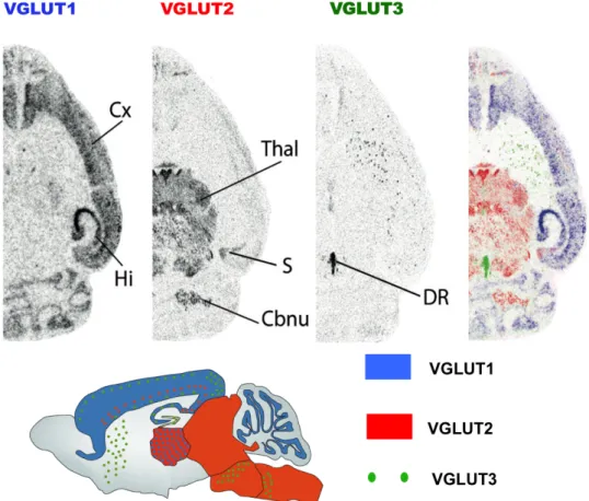
In mammals, there are three isoforms of VGLUT: VGLUT1 (SLC17A7), VGLUT2 (SLC17A6) and VGLUT3 (SLC17A8); these VGLUTs share the same glutamate transport mechanisms, but distinct regional distributions (Fig. 3).
VGLUT1 and VGLUT2 are largely expressed to distinct populations of excitatory neurons in a complementary pattern (Herzog et al., 2001; Kaneko and Fujiyama, 2002; Takamori, 2006). In situ hybridization shows that VGLUT1 mRNA is mainly located in olfactory bulb, neo-‐cortex, cerebellar cortex, medial habenula, pontine nucleus and hippocampus (including subiculum); and lower expression in lateral septum and thalamic nuclei (Fremeau et al., 2001; Herzog et al., 2001; Varoqui et al., 2002; Boulland et al., 2004). Whereas, VGLUT2 mRNA is detected in all thalamic nuclei (except reticular nucleus typically GABAergic), hypothalamus, inferior and superior colliculi, deep cerebellar nuclei and many neurons from the brainstem. Besides, low levels of VGLUT2 were also detected in hippocampus, some cortical areas and amygdala, (Fremeau et al., 2001; Herzog et al., 2001; Boulland et al., 2004). It was soon observed that VGLUT1 is present at synapse known to exhibit low release probability, whereas VGLUT2 is present at those that are known to exhibit high release probability (Fremeau et al., 2001; Varoqui et al., 2002). Immunofluorescence studies suggested a seggregation of synaptic proteins selectively at VGLUT1-‐positive synapse and not at VGLUT2-‐positive contacts (Shank PSD scaffold proteins, (Heise et al., 2016); Endophilin1A (Vinatier et al., 2006)). This further suggests that the molecular organization of VGLUT1 and VGLUT2 synapses may differ and support different modalities of glutamatergic transmission.
Unlike VGLUT1 and VGLUT2, which are mainly expressed in asymmetric synapses, VGLUT3 is expressed at symmetric synapse. VGLUT3 mRNA is found in sparse populations of neurons at regions or in patterns not typically associated to glutamatergic transmission (Gras et al., 2002; Takamori, 2006). VGLUT3 was shown at serotoninergic neurons of raphe nuclei, Cholinergic neurons of the striatum, and subclasses of GABA interneurons (cerebral cortex and hippocampus) (Gras et al., 2002; Herzog et al., 2004a; Gras et al., 2005; Mestikawy et al., 2011). Moreover, VGLUT3 is found in subgroups of primarily glutamatergic neurons in the raphe, habenula, hypothalamus, olfactory tubercles and inner hair cells of the cochlea (Gras et al., 2002; Herzog et al., 2004a; Ruel et al., 2008; Seal et al., 2008). More recently, VGLUT1 and VGLUT2 were also described in some Ach and GABA neurons in CNS (Herzog et al.,
2004b; Zander et al., 2010; Fattorini et al., 2015; Granger et al., 2016). VGLUT2 is even suspected to be expressed in some noradrenaline, adrenaline or dopamine neurons (Stornetta et al., 2002; Kawano et al., 2006; Dal Bo et al., 2008; Mestikawy et al., 2011). The role of VGLUT co-‐localization with other classical VNT in the same synapse is under debate. It may serve as an enhancer to uptake and release of both neurotransmitters at synapses (Gras et al., 2008), and/or provide glutamate as a co-‐transmitter with distinct target or function (Mestikawy et al., 2011).
Fig. 3 Distribution patterns of the Three VGLUTs. In situ hybridization shows mRNA distribution of VGLUT1, VGLUT2 and VGLUT3 (image from Salah EI Mestikawy). VGLUT1 mainly labels cerebral and cerebellar cortex, while VGLUT2 mainly labels subcortical regions. VGLUT3 is depicting sparse populations of noncanonical glutamatergic neurons.
VGLUTs expression in development
The functional difference between VGLUTs is further enforced by the fact that the three transporters have different regulations during development.
VGLUT2 is expressed at high levels from early prenatal developmental stages and increases slightly to reach a plateau at P21 (Gras et al., 2005). VGLUT2 transcripts are transiently observed in the future VGLUT1 territory before fading away between P7 and P21 (Miyazaki et al., 2003; Fremeau et al., 2004; Gras et al., 2005). Very low levels of
VGLUT1 are detected from E15 to E18 in rat brain. After birth, it massively increases its expression level and replaces most VGLUT2 expression in cortex and hippocampus (Boulland et al., 2004; Miyawaki, 2005). While, VGLUT3 expression at birth is around 60% of adult level, it peaks transiently between P5 and P10 due to transient expression at Purkinjee cells of the cerebellum, and raises again steadily from P15 to adulthood (Boulland et al., 2004; Gras et al., 2005). The transient expression demonstrates that the transmitters release is changed during the development, and the glutamate release plays a role in the refinement of synaptic connection (Gillespie et al., 2005; Gras et al., 2005).
Molecular structure of VGLUTs
Each isoform of the VGLUT polypeptides comprises of 12 transmembrane domains (TMDs, Fig. 2B) with cytosolic N and C termini. The core segment of the transporters share a high degree of amino-‐acid identity. Correspondingly, they share nearly identical glutamate transport kinetics as discussed here above. The 3D structure of VGLUTs remains unknown. However, VGLUT1 and VGLUT2 are already constructed by homology modeling based on the cytosol-‐open crystallographic structure of E. Coli glycerol-‐3-‐ phosphate transporter (GlpT) (Juge et al., 2006; Almqvist et al., 2007). These studies show that the residues H128 in TMD2, R184 and E191 in TMD4 of VGLUT2(Juge et al., 2006), and equivalent residence of H120, R176 and E183 in VGLUT1(Almqvist et al., 2007) are essential for L-‐glutamate transport. Moreover, there are two docking substrates in VGLUT1 structure: R80 and R314 as the central binding site, and R176 and H120 as the upper binding site. These two docking substrates need similar energy for L-‐ glutamate binding. But when Pi is docked, all docked molecules are completely clustered around R314 (Almqvist et al., 2007). Herman et. al mutated VGLUT2 successively R88A, R184A, and R322A, only the triple mutant failed to rescue the glutamate transport and release (Herman et al., 2014).
The N-‐terminal and C-‐terminal sequences of VGLUTs are highly divergent (Fig. 2C), which may contribute to functional difference by interacting with different proteins of the cytosolic compartment (Takamori, 2006). There is a highly conserved motif 540-‐ SYGAT (Fig. 2C) in the C-‐terminal end of all VGLUT isoforms and it is shared by most species (Vinatier et al., 2006). The S540 is a predicted GSK-‐3 substrate, fitting the consensus sequence S/T-‐X-‐X-‐X-‐S/T(P); it may participate in proteins interaction or SVs trafficking (Santos et al., 2014). Several dileucine-‐like motifs exist only in VGLUT1,
which are involved in VGLUT1 endocytosis at SVs. Foss et. al showed that the 504-‐ SEEKCGFV motif at C-‐terminal acts largely via assembly protein complex 2 (AP-‐2) and N-‐terminal motif use AP-‐1 in trafficking regulation, but VGLUT2 mostly depends on the C-‐terminal motif (Foss et al., 2013). VGLUT1 C-‐terminal also contains two poly-‐proline motifs (PP1, PP2), which are not present in the other isoforms. PP1 and PP2 contain the consensus site for SH3 protein interaction domains (PXXP). Several studies showed VGLUT1 could interact with endophilin A1 SH3 domain at PP2/P554 (De Gois et al., 2006; Vinatier et al., 2006; Voglmaier et al., 2006). VGLUT1 might recruit endophilin, which is an essential component of clathrin-‐mediated endocytosis, to speed up the endocytosis of SVs through AP2 pathway (Voglmaier et al., 2006). Later on, Weston and colleagues reported that VGLUT1/endophilin A1 interaction reduces SVs release probability, yet the detailed mechanism for this phenomenon remains unknown (Weston et al., 2011).
VGLUT genetically modified models
To better understand VGLUTs function and properties, various genetically modified models were generated. We will now discuss the achievements obtained from these models on VGLUTs function at multiscale levels.
VGLUT1
In 2004, Fremeau et al. and Wojcik et al. first revealed the VGLUT1 contributions to CNS with VGLUT1 knockout (KO) mice (Fremeau et al., 2004; Wojcik et al., 2004). Due to the VGLUT2 dominant expression during post-‐natal development, VGLUT1 KO pups don’t show a significant deficit before the end of the second week of life, but start to have a poor food intake afterwards and eventually die at around three weeks after birth. With special food intake care, VGLUT1 KO pups survive for a long time (Fremeau et al., 2004; Wojcik et al., 2004). The synapse number and global morphology in VGLUT1 KO mice and culture is similar to that of WT by ultrastructural analysis, but SVs number in the presynaptic terminal and SVs tonicity are significantly reduced (Fremeau et al., 2004; Siksou et al., 2013). As expected, neurotransmission is impaired when VGLUT1 is lacking: the frequency and amplitude of miniature excitatory synaptic current (mEPSC) are reduced in VGLUT1 KO neuronal culture. The remaining signal may be attributed to VGLUT2 expression (Wojcik et al., 2004). In contrast, Drosophila DVGLUT knock-‐down mutants one unit of VGLUT seemed already enough to fill completely SVs (Daniels et al.,
2006). Meanwhile, overexpression of VGLUT1/DVGLUT resulted in increased quantal content of glutamate (Daniels et al., 2004), but normal mEPSC frequency. In addition, the absence of VGLUT1 doesn’t alter the expression of the other two isoforms of VGLUT or most other synaptic proteins (Wojcik et al., 2004; Siksou et al., 2013).
Half expression of VGLUT1 in mice (vglut1+/-) exhibits no apparent phenotypic
abnormalities during development and adulthood (Tordera et al., 2007). Yet, detailed analysis of reduced VGLUT1 expression was shown to cause increased anxiety, impair reversal learning and working memory (Tordera et al., 2007; Inta et al., 2012; Granseth et al., 2015). The LTP induction is significantly decreased in the CA1 region of vglut1+/-‐
slices, this is accompanied by a deficit in spatial reversal learning (Balschun et al., 2010). After exposure to chronic mild stress, vglut1+/-‐ mice surprisingly show an up regulation
of VGLUT2 and an hyperlocomotion (Garcia-‐Garcia et al., 2009).
VGLUT1 is a specific maker for glutamatergic SVs in CNS. Our laboratory generated a VGLUT1Venus knock-‐in (KI) mouse line. With this KI mouse line, our laboratory
contributed to the characterization of SV sharing among presynaptic boutons along the same axon both in vitro and in vivo (Herzog et al., 2011). This new pool of SVs transiting in the axons was named “super-‐pool” (Staras et al., 2010). In line with this work, vglut1-/-
phenotypes were revisited to unravel that SV super pool size is increased while SV cluster is reduced (Siksou et al., 2013). Recently, with VGLUT1Venus mice, a study showed
that SVs in mossy fiber terminals have a high mobility and the diffusion limits vesicle reloading during sustained high frequency stimulations (Rothman et al., 2016).
VGLUT2
VGLUT2 deletion in mice results in perinatal lethality, which is due to the respiratory failure (Moechars et al., 2006; Wallén-‐Mackenzie et al., 2006). Further investigations show that, the neural circuits in the location of the pre-‐Bötzinger (PBC) inspiratory rhythm generator fail to become active (Wallén-‐Mackenzie et al., 2006). Similar to VGLUT1 KO condition, the mEPSC amplitude and frequency in vglut2-‐/-‐ are reduced
significantly (Moechars et al., 2006; Wallén-‐Mackenzie et al., 2006). In addition, VGLUT2 absence also seem to cause reduced SVs number and abnormal elongated shape in the terminal (Wallén-‐Mackenzie et al., 2006).
Behavioral analysis of vglut2+/-‐ mice showed unchanged motor function, learning and
memory and inflammatory pain, but impairements in the development and maintenance of neuropathic pain (Moechars et al., 2006). VGLUT2 conditional KO mice provides