HAL Id: tel-01599241
https://tel.archives-ouvertes.fr/tel-01599241
Submitted on 2 Oct 2017HAL is a multi-disciplinary open access archive for the deposit and dissemination of sci-entific research documents, whether they are pub-lished or not. The documents may come from teaching and research institutions in France or abroad, or from public or private research centers.
L’archive ouverte pluridisciplinaire HAL, est destinée au dépôt et à la diffusion de documents scientifiques de niveau recherche, publiés ou non, émanant des établissements d’enseignement et de recherche français ou étrangers, des laboratoires publics ou privés.
Multiscale organization of the human cortex : from
anatomo-functional cognitive networks to gene
expression
Claudia Cioli
To cite this version:
Claudia Cioli. Multiscale organization of the human cortex : from anatomo-functional cognitive net-works to gene expression. Imaging. Université Pierre et Marie Curie - Paris VI, 2015. English. �NNT : 2015PA066412�. �tel-01599241�
Université Pierre et Marie Curie
École doctorale informatique, télécommunications et électronique Laboratoire d’Imagerie Biomédicale (LIB)
Équipe Systèmes dynamiques anatomo-fonctionnels chez l’homme, altération et récupération fonctionnelle (ADSH)
Organisation multi-échelle du cortex humain: des
réseaux anatomo-fonctioneles à l’expression des gènes
Par Claudia CIOLI
Thèse de doctorat de Sciences et technologies de l’information et de la communication
Dirigée par Yves Burnod et Habib Benali Présentée et soutenue publiquement le Septembre 30, 2015
Devant le jury composé de:
POTIER, Marie-Claude, DR CNRS Rapporteur
TORO, Roberto, Prof. Universidad de Valparaíso, Chili Rapporteur
COLLIOT, Olivier, DR CNRS Examinateur
FROUIN, Vincent, Chercheur-Ingénieur CEA Examinateur
ABDI, Hervé, Prof. University of Texas, USA Examinateur
BOURGINE, Paul, Prof. Open University, UK Examinateur
BENALI, Habib, DR INSERM Directeur de Thèse
Université Pierre et Marie Curie
École doctorale informatique, télécommunications et électronique Laboratoire d’Imagerie Biomédicale (LIB)
Équipe Systèmes dynamiques anatomo-fonctionnels chez l’homme, altération et récupération fonctionnelle (ADSH)
Multiscale organization of the human cortex: from
anatomo-functional cognitive networks to gene
expression
Par Claudia CIOLI
Thèse de doctorat de Sciences et technologies de l’information et de la communication
Dirigée par Yves Burnod et Habib Benali Présentée et soutenue publiquement le Septembre 30, 2015
Devant le jury composé de:
POTIER, Marie-Claude, DR CNRS Rapporteur
TORO, Roberto, Prof. Universidad de Valparaíso, Chili Rapporteur
COLLIOT, Olivier, DR CNRS Examinateur
FROUIN, Vincent, Chercheur-Ingénieur CEA Examinateur
ABDI, Hervé, Prof. University of Texas, USA Examinateur
BOURGINE, Paul, Prof. Open University, UK Examinateur
BENALI, Habib, DR INSERM Directeur de Thèse
Abstract
This work is conceived in the present panorama of fast development of large databases gathering experimental results about the organization of the human brain at different scales. This abundance of information calls for an intra and inter-disciplinary effort aimed to syn-thesize this information in a coherent way.
The aim of this thesis was to contribute to this effort for knowledge synthesis to better un-derstand the multiscale organization of the cerebral cortex. The work followed two paths: an intra-disciplinary effort to bring together results produced by the brain imaging com-munity with particular focus on Resting State and Task Based MRI experiments; an inter-disciplinary attempt to draw a link between the anatomo-functional organization of the cortex as emerging from brain imaging studies and the cortical patterns of gene expression as revealed by recently published atlases of the adult human brain transcriptome.
The thesis is organized into three parts: Part I is devoted to the study of the anatomo-functional organization of the human cortex starting from brain imaging studies.
In the first chapter of Part I we showed using multiscale clustering, that anatomo-functional networks (including Resting State and Task Based networks (referred to also as cognitive networks) as obtained in brain imaging experiments) are topologically organized across the cortex as two rings and that these two rings correspond to two different ways of informa-tion processing. A first ring called VSA–Visual Somatosensory Auditory– carries out real time, high fidelity processing of sensory-motor and multimodal information. A second ring called PTF–Parieto, Temporo, Frontal– and while it encompasses the cognitive networks implementing language, memory and emotions, it is responsible for multi-temporal infor-mation processing and characterized by a stronger autonomous component.
In the second chapter of Part I we deepened the analysis of the organization of these net-works at a finer level. To do so we built a graph of topographical similitude among several hundreds of cognitive networks covering all the domains, sensory-motor, language, mem-ory, emotional and vital needs. This analysis allowed not only to find again the ring organi-zation but also to establish relations and continuity across different cognitive domains. We finally propose a global scheme to describe the organization of cognitive networks across the anatomo-functional regions of the cortex. This scheme is determined by: 1) poles of
connections of cerebral areas with the rest of nervous structures; 2) functional gradients connecting these poles within parietal, frontal and temporal regions.
In Part II we studied the link between cortical gene expression and the anatomo-functional organization of the cortex both in term of their topography and in term of their function, focusing in particular on information processing and memory formation.
In the first chapter of Part II we showed using multidimensional statistical techniques –such as CA and DiCA – that the ensemble of genes the most differentially expressed across the cortex are organized according to the ring architecture. The advantage of this method is to reveal the members of the gene families the most spatially differentiated. We focused on family of genes with specific synaptic properties in neuronal information processing namely ionic channels and proteins implied in neurotransmitter release. We showed that the expression of different protein isoforms is coherent with the information processing performed by each of the two rings. We found proteins supporting precise and high fidelity information processing (such as ionic channel SCN1A, KCNA1 and release proteins SYT2 and CPLX1), more expressed across VSA networks, while proteins favoring spontaneous activity production (such as ionic channel SCN3A, KCNG1 and release proteins SYT5 and CPLX3), more expressed in the PTF ensemble.
In the second chapter of Part II we studied on a finer scale this dual organization analyzing the topographical relation between specialized networks–visual, motor, language, memory etc.–and the proteins controlling information processing (ionic channels and neurotrans-mitter release), short term memory (calcium-dependent kinases), long term memory (actin, tubulin) and development (guidance and growth factors). Short-term and long term memory involves a cascade of transformation: glutamate NMDA receptor activation signaling co-occurrence of inputs, calcium inflow prolonged by CaMK auto-phosphorylation producing a short term memory, and actin polymerization increasing the spine surface for long-term memory encoding. The expression of genes coding for these 3 families of proteins is high-est in the cerebral cortex, with a differential distribution of subunits in the cortical regions. The glutamate receptor NR2B responsible of sustained activities of working memory and strong plasticity is higher in cognitive regions PTF; NR2A which produces shorter activa-tion and limits plasticity is higher in sensorymotor VSA regions; GRIN3A, which delay maturation is more expressed in PTF. The CaMK isoforms CaMK2D higher in cognitive regions PTF and CaMK2G higher in sensorymotor regions VSA produce two modes of actin polymerization, with CaMK2D favoring strong consolidation. There is also a con-trast between temporal and frontal regions. CaMK1G in frontal regions produces longer duration of short term memory than CaMK4 in temporal regions. Genes coding for pro-teins regulating the actin network, are either more expressed in the frontal regions (ACTN2 gene coding for alpha-actinin2), or in the temporal regions (actin-related protein ACTR3) suggesting different mode of long-term memory encoding. Furthermore, genes coding for alpha and beta tubulin, which regulate the microtubule dynamics, and thus axon and
den-drites growth, are also more expressed in the frontal regions where very slow maturation allows for learning-dependent network remodeling.
In Part III we present the platform that we developed to favor knowledge integration be-tween databases of cognitive networks and databases of gene expression and to foster re-search. The platform is based in part on the studies presented in Part I and Part II.
In perspective we show how the type of analyses we perform could help to better under-stand, in Alzheimer disease, the relation between the gradient associated to gene expression and the pattern of propagation of the disease across cortical regions. Similarly the prefer-ential expression of genes implicated in ASD could help to better understand the cognitive networks and cognitive functions which are impaired. Finally a more precise knowledge of the genes the most expressed in different cortical areas can help adapting the parameters necessary to model the dynamic processes (activation, plasticity) in the different areas of the cortex.
Résumé
Ce travail est conçu dans le panorama actuel de développement rapide de grandes bases de données qui rassemblent des ensembles de résultats expérimentaux sur l’organisation anatomo-fonctionnelle du cerveau humain à différentes échelles; l’abondance d’informations demande un effort intra et interdisciplinaire pour les synthétiser de façon cohérente. Le but de cette thèse est de contribuer à cet effort de synthèse. Le travail suit deux chemins: l’effort intra)disciplinaire pour relier et synthétiser les résultats produits par la commu-nauté de l’imagerie cérébrale, avec une focalisation particulière sur les Réseaux de Re-pos et les Réseaux Cognitifs; l’effort inter-disciplinaire pour relier l’organisation anatomo-fonctionnelle du cortex cérébral telle qu’elle émerge des ensembles de résultats en im-agerie cérébrale, et les expressions des gènes révélées par les bases de données publiées très récemment sur le transcriptome humain.
Cette thèse est organisée en trois parties: la Partie I est consacrée à l’étude de l’organisation anatomo-fonctionnelle du cortex à partir des études d’imagerie cérébrale.
Dans le premier chapitre de la partie I, nous montrons en utilisant des méthodes de cluster-ing multi-échelle que les réseaux anatomo-fonctionnels (incluant les réseaux de repos et les réseaux activés par des tâches cognitives) sont topologiquement organisés sur la surface cor-ticale comme deux anneaux entrelacés, et que ces deux anneaux implémentent deux modes de traitement de l’information. Le premier anneau appelé VSA (Visuel-Somatomoteur-Auditif) effectue des traitements sensorimoteurs (et multimodaux) en temps réel, avec une haute fidélité par rapport aux entrées et sorties reliées au monde extérieur. Un second anneau, appelé PTF (Parieto-Temporo-Frontal) qui implémente le langage, la mémoire sé-mantique et épisodique, les émotions, les fonctions et rythmes vitaux) effectue des traite-ments multi-temporels, plus autonomes par rapport aux sources extérieures.
Dans le second chapitre de la Partie I, nous approfondissons l’analyse de ces réseaux à un niveau de résolution plus fin. Pour cela, nous construisons un graphe des similitudes topographiques entre des centaines de réseaux cognitifs dans tous les domaines, sensori-moteurs, pour le langage, la mémoire, les émotions, les besoins vitaux, etc. Cette analyse nous permet de retrouver l’organisation d’ensemble en deux grands anneaux, mais aussi d’établir des relations de continuité ’ corticotopique’ entre les différents domaines cog-nitifs. Nous décrivons finalement un schéma d’ensemble pour décrire l’organisation des
réseaux cognitifs à partir de régions anatomo-fonctionnelles de référence. Ce schéma est déterminé par: 1) des pôles de connection des aires cérébrales avec les autres structures nerveuses; (2) des gradients fonctionnels reliant ces pôles par les réseaux entre régions pariétales, frontales, et temporales.
Dans la Partie II, nous étudions les liens entre l’expression corticale des gènes et l’organisation anatomo-fonctionnelle du cortex, à la fois en terme de similitude topographique et de con-gruence de fonction, en se focalisant en particulier sur le traitement de l’information et la mémorisation.
Dans le premier chapitre de la Partie II nous montrons, en utilisant des techniques statis-tiques multidimensionnelles - tels que l’Analyse des Correspondances, et l’Analyse Dis-criminante des Correspondances – que l’ensemble des gènes les plus différentiellement ex-primés sur le cortex sont organisés suivant l’architecture en deux anneaux. L’avantage de cette méthode statistique est de révéler les gènes les plus différenciés dans chaque famille de gènes. Nous nous focalisons sur les familles de gènes spécialisés dans le traitement de l’information, comme les canaux membranaires et les protéines contrôlant la libération des vésicules de neurotransmetteurs. Nous montrons que l’expression des différents isoformes, compte tenu de leurs propriétés fonctionnelles, est cohérent avec les modes spécifiques de traitement de l’information dans chacun des deux anneaux. Nous trouvons des protéines plus spécialisées pour le traitement de l’information précis dans le temps et fidèle par rap-port aux entrées (canaux SCN1A, KCNA1, contrôlant la libération SYT2, CPLX1), plutôt dans l’anneau VSA qui effectue les traitements en temps réel avec le monde extérieur, et des protéines plus spécialisées dans l’activité plus soutenue et plus spontanée (canaux SCN3A, contrôlant la libération CPLX3) dans l’anneau PTF qui effectue les traitements multi-temporels, plus autonomes par rapport au monde extérieur.
Dans le second chapitre de la Partie II, nous étudions à une échelle spatiale plus fine cette organisation en deux anneaux, en différenciant les régions anatomo-fonctionnelles spécial-isées (pour le langage, la mémoire, les émotions, les besoins vitaux, etc. ). En plus des qui contrôlent le traitement de l’information (canaux ioniques et libération du neurotrans-metteur), nous nous focalisons aussi sur celles qui contrôlent la mémoire à court terme (ré-cepteur glutamate NMDA et CaMK calcium-dependent kinases), la mémoire à long terme (cytosquelette actine et tubuline), et le développement (facteurs de croissance et facteurs de guidage des axones). La mémorisation à court et long terme se fait par une cascade de trans-formations: le récepteur NMDA au glutamate signale la co-occurrence des entrées et génère un flux entrant de calcium, qui produit l’autophosphorylation des CaMK (mémoire à court terme) et la polymérisation de l’actine qui augmente la surface des épines dendritiques et donc le poids synaptique (support de la mémoire à long terme). L’expression des gènes qui codent ces trois familles de protéines est plus fort dans le cortex cérébral, avec une distri-bution différentielle des sous-unités et des isoformes dans les régions corticales. Ainsi le récepteur au glutamate NR2B, responsable des activités soutenues de la mémoire de tra-vail, et d’une plasticité très forte, s’exprime plus dans PTF, alors que le récepteur NR2B,
qui produit des activations plus courtes et moins de plasticité, s’exprime plus dans les ré-gions VSA. Le gène GRIN3A, qui retarde la maturation, s’exprime plus dans PTF. Pour les isoformes de la CaMK, CaMK2D s’exprime plus dans les régions cognitives PTF, et CaMK2G s’exprime plus dans les régions sensori-motrices VSA. Ces deux isoformes de la CaMK produisent deux formes différentes de polymérisation de l’actine, avec la CaMK2D qui favorise des consolidations fortes, avec des mémoires persistantes. Il existe aussi un contraste entre les régions pariétales et frontales. La CaMK1G dans les régions frontales produit des mémorisations à court terme plus longues, de l’ordre de l’heure, alors que la CaMK4, dans les régions temporales, produit des mémorisations à court terme plutôt de l’ordre de la minute. Les gènes qui codent les protéines qui régulent les réseaux d’actine, sont soit plus exprimées dans les régions frontales (ACTN2 codant l’alpha-actinine 2), soit dans les régions temporales (actin-related protein ACTR3), suggérant des modes différents de mémorisation de l’information. De plus, les gènes qui codent pour les différents iso-formes des tubulines alpha et beta, qui régulent finement la dynamique des microtubules, et donc la croissance des axones et des dendrites, sont aussi plus exprimés dans les régions frontales où la maturation lente des neurones pendant deux décennies permet de modeler fortement les réseaux en fonction de l’apprentissage.
Dans la Partie III, nous présentons la plate-forme que nous avons développée, pour intégrer dans une même représentation les donnée`s d’imagerie cérébrale et les données d’expression génétique, et pour faciliter la recherche présentée dans les Parties I et II.
En perspective, nous montrons comment les analyses que nous faisons peuvent aider à mieux comprendre la relation, dans la maladie d’Alzheimer, entre les gradients d’expression des gènes et la propagation de la pathologie dans les réseaux corticaux. De même dans le spectre autistique, l’expression preferentielle des gènes à risque peut aider à mieux com-prendre les reseaux cognitifs et les fonctions cognitifs qui sont déficitiares. Finalement une meilleure connaissance des gènes les plus exprimés dans les différentes régions corti-cales peut aider à adapter les paramètres des modèles des processus dynamique (activation, plasticité) aux caractéristiques spécifiques des réseaux corticaux.
Contents
1 Introduction 1
1.1 Motivation . . . 1
1.2 Context . . . 5
1.2.1 Brain imaging databases . . . 5
1.2.2 Gene expression: the Allen Institue’s human brain transcriptome database . . . 7
1.3 This Dissertation: contents . . . 8
1.3.1 Anatomo-functional organization of the cortex: the dual intertwined rings architecture . . . 9
1.3.2 Anatomo-functional organization of cortical gene expression . . . . 9
1.3.3 Heuristic: focusing on expression of families of genes known to be involved in information processing and memory formation . . . 10
1.4 This Dissertation: plan . . . 12
1.5 This Dissertation: personal contributions . . . 17
1.6 Publications from this Dissertation . . . 18
1.6.1 Journal articles . . . 18
1.6.2 Peer-Reviewed Abstracts . . . 18
1.6.3 Others . . . 19
I Cortical organization of anatomo-functional networks: rings and gradients 21 2 The dual intertwined rings architecture 23 2.1 Graphical abstract . . . 23
2.2 Abstract . . . 23
2.3 Introduction . . . 24
2.4 Results. . . 27
2.4.1 Extraction of the most representative RSNs in the group: robustness 27 2.4.2 RSNs Specialization: overlap between RSNs and TBNs . . . 28
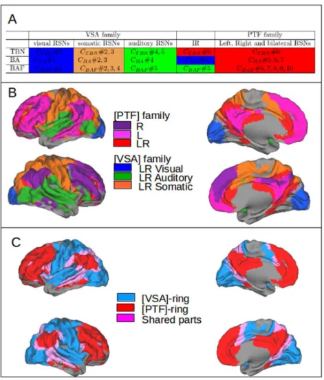
2.4.3 RSNs topography: overlap between RSNs and the Brodmann Areas. 30 2.4.4 Corticotopy of the RSNs clusters: the dual intertwined rings archi-tecture. . . 31
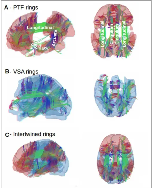
2.4.5 Anatomical connections within and between the dual rings.. . . 35
2.5 Discussion. . . 36
2.5.1 Method of RSNs extraction and RSNs coverage . . . 37
2.5.2 Comparison of the dual intertwined architecture with other RSNs results . . . 38
2.5.3 Real-time integration in the VSA ring and multi-temporal integra-tion in the PTF ring . . . 40
2.5.4 Topological advantages of the intertwining . . . 42
2.5.5 Development of the dual intertwined rings architecture . . . 42
2.5.6 Evolution of the dual intertwined ring architecture . . . 43
2.5.7 Conclusion . . . 43
2.6 Materials and Methods . . . 43
2.6.1 Resting-state database . . . 43
2.6.2 Pre-processing . . . 44
2.6.3 Functional network identification by spatial Independent Compo-nent Analysis (NEDICA) . . . 44
2.6.4 Clustering methods . . . 44
2.6.5 RSNs specialization: overlap with TBNs . . . 45
2.6.6 RSNs distribution: overlap with BAs regions . . . 45
2.6.7 Anatomical Fiber tracting . . . 46
2.6.8 Representation of the results . . . 47
3 Gradients of cognitive networks 51 3.1 Graphical abstract . . . 52
3.2 Abstract . . . 53
3.3 Introduction . . . 54
3.3.1 fMRI: single study vs meta-analysis . . . 55
3.3.2 Data-driven meta-analysis: the Neurosynth framework . . . 55
3.4 Results. . . 57
3.4.1 Lexical Map: 1000 cognitive terms . . . 57
3.4.2 Topographic Map: 1000 cognitive categories . . . 60
3.4.3 Anatomo-functional gradients of cognitive tasks across the cortex . 62 3.5 Methods . . . 73
3.5.1 Neurosynth: inverse inference maps . . . 74
3.5.2 CorText: lexical extraction . . . 75
3.5.3 Cognitive terms: lexical and topographical similarity . . . 76
II Cortical organization of gene expression in relation with
anatomo-functional networks 79
4 Cortical gene expression match the temporal properties of
large-scale functional networks 81
4.1 Graphical abstract . . . 82
4.2 Abstract . . . 83
4.3 Introduction . . . 83
4.4 Materials and Methods . . . 87
4.4.1 Data Base . . . 87
4.4.2 Labeling of cortical regions by VSA or PTF rings . . . 87
4.4.3 Statistical Analysis I: Correspondence Analysis (CA) . . . 87
4.4.4 Statistical Analysis II: Discriminant Correspondence Analysis (DiCA) 88 4.5 Results. . . 89
4.5.1 Gene expression spontaneously separates the rings . . . 89
4.5.2 Cortical gene expression predicts the assignment of cortical regions to the rings . . . 90
4.5.3 Correlation between gene expressions . . . 92
4.5.4 The proteins coded by genes most differentiated between cortical regions belong to families of proteins involved in neuronal informa-tion processing such as ionic channels and neurotransmitter release 92 4.5.5 Determination of genes the most expressed in VSA and PTF for the ionic channels and transmitter release functional classes . . . . 94
4.6 Discussion . . . 98
4.6.1 Sodium and potassium channels . . . 100
4.6.2 Calcium channels . . . 101
4.6.3 Synaptotagmins . . . 101
4.6.4 Complexins . . . 102
4.6.5 Synaptobrevins . . . 102
4.7 Conclusions . . . 103
4.8 Remark: Regions are organized according to gradients based on their gene expression profile . . . 103
5 Gradients of synaptic genes properties across cognitive networks 107 5.1 Graphical abstract . . . 108
5.2 Abstract . . . 109
5.3 Introduction . . . 110
5.4 Materials and Methods . . . 112
5.4.1 Data Base . . . 112
5.4.2 Anatomo-functional regions . . . 112
5.4.3 Statistical Analysis: Discriminant Correspondence Analysis (DiCA)113 5.5 Results. . . 113
5.5.1 Single gene expression profiles. . . 115
5.5.2 Organization of brain regions by gene expression . . . 116
5.5.3 Organization of cortical regions by gene expression . . . 118
5.5.4 Differential expression of genes in families involved in information processing and memory formation . . . 120
5.5.5 Gradients of gene expression in the cerebral cortex . . . 137
5.6 Discussion. . . 139
5.6.1 Main axes of gene expression in cerebral regions and cortical regions139 5.6.2 Neoteny of language regions . . . 141
5.6.3 Gradients of gene expression . . . 142
5.6.4 Multiscale anatomo-functional congruence . . . 143
5.6.5 Transcription factor FOXP2 and growth factor MET: language and autism . . . 156
5.7 Conclusions . . . 156
5.8 Remark I: The anatomo-functional synapse: multiscale scenarios in differ-ent cortical regions . . . 157
5.8.1 Perform a visuomotor task: cortical motor, visual and parietal vi-suomotor regions . . . 158
5.8.2 Memorize-recognize faces and events: temporal fusiform and parahip-pocampal regions . . . 159
5.8.3 Produce goal-directed sequences of actions: superior frontal regions 160 5.8.4 Produce sentences: network relating audition-phonation, temporal and frontal regions . . . 163
5.9 Remark II: Synaptic proteins evolution and the tethering hypothesis . . . . 165
III Tools development 169 6 LinkRbrain: a web-based platform to analyze spatial correlation of gene expression and cognitive networks 171 6.1 Graphical abstract . . . 171 6.2 Abstract . . . 171 6.3 Introduction . . . 172 6.4 Data sources . . . 174 6.5 Methods . . . 174 6.5.1 Text mining . . . 175
6.5.2 From probes to expression of genes . . . 175
6.5.3 Topographical distances . . . 175
6.5.4 Visualization . . . 176
6.6 LinkRbrain as a multi-scale, integrative explorer . . . 178
6.6.1 From cognitive functions to functional-anatomical architecture . . . 179
6.7 LinkRbrain as comparator of brain networks of cognitive functions . . . 182
6.8 LinkRbrain as a comparator of user results with the literature . . . 182
6.9 Conclusion . . . 183
7 Conclusions and future work 187 7.1 Conclusions . . . 187
7.2 Future work . . . 192
7.2.1 Alzheimer disease . . . 192
7.2.2 Autism Spectrum Disorder . . . 194
7.2.3 Models . . . 196
Bibliography 201
A Supplementary Material 227
1
Introduction
This work is conceived in the present panorama of fast development of large databases gathering experimental results about the organization of the human brain at different scales. This abundance of information calls for an intra and inter-disciplinary effort aimed to syn-thesize this information in a coherent way.
In this dissertation we explore the relations between brain imaging and transcriptomics databases, to better understand the multiscale organization of the cerebral cortex. We study the relation between gene expression and cognitive networks, both in term of their cortical topography and in term of their function, focusing in particular on information processing and memory formation.
1.1
Motivation
Fig. 1.1.:Spatiotemporal multiscale organization of the cortex
Figure 1.1 summarizes the multiscale functional architecture of the human brain, start-ing from the behavioral and cognitive level, and zoomstart-ing toward the cerebral and cortical networks, and then toward the local networks of cortical columns, neurons, synapses,
Fig. 1.2.: The spatiotemporal domain of neuroscience and the evolution of the main tech-niques of investigation. Source: [236]
til molecules within synapses. All these levels have been studied in term of information processing and memory formation. For example, at the behavioral and cognitive levels, psychology and artificial intelligence describe interaction between visual and motor infor-mation to manipulate objects, between phonation and audition to communicate, and at the same time, information storage to memorize new words and rules through verbal commu-nication.
At the brain level, brain imaging techniques, for example fMRI techniques, have revealed a very large set of sensorymotor and cognitive networks specialized for information pro-cessing and storage, as for example visuomotor propro-cessing in parietal areas, speech and sentence processing in dorsal temporal areas and ventral frontal regions, episodic memory in ventral temporal regions, working memory and rules processing in dorsal frontal areas, etc.
At the finer level of local neuronal networks, neurophysiology have shown that all forms of processing and memory are based on prototypical networks of neurons, the cortical columns. Cortical columns are processing units which combine locally different types of information (sensory, motor or cognitive) provided by different layers of the column, and store the congruences among these different types of information, as for example the con-gruency between visual, proprioceptive, tactile and motor information.
At the level of a single neuron, electrophysiological techniques have revealed how infor-mation is processed by the axodendritic system thanks to a set of ionic channels, and how memory processes are determined by pre and post synaptic plasticity and remodeling as a consequence of the coactivation of several synapses belonging to the same neuron.
Molecular biology and genetics have deciphered the function of proteins involved in infor-mation processing and memory forinfor-mation at the synaptic level. Among these proteins are ionic channels, receptors, cytoskeleton proteins and transcription factors. These families of proteins are composed by several isoforms or subunits differentiated among them by the
fine tuning of their molecular structure. This variety of molecular structures confers to these families of proteins a large repertoire of temporal and dynamical properties. Figure1.1 il-lustrates two important questions: 1) what is the spatial relation between gene expression and the anatomo-functional organization of the cerebral cortex; 2) what is the functional relation between the synaptic proteins coded by these genes and the specialized functions of cognitive networks which are spatially matched with the gene expression patterns. To understand how the cortex works and how it gives rise to the variety of human behaviors, it is necessary to understand how these different scales interact together. To this purpose, researchers have combined a variety of techniques at different scale in space and time (see for a set of techniques Figure1.2from [236]) to find out which are the relations between distribution and dynamics of proteins at the synaptic and neuronal level, how proteins in-teractions shape the way neurons process and store information, how different neurons and networks of neurons are reciprocally organized in maps and cognitive networks, which are the anatomical and genetic pathways leading to the genesis of neuronal and cognitive networks.
1.1.0.1 Multiscale research strategies
Since many diseases are due to molecular deficits and have effects at all levels, synaptic, neuronal, networks and cognitive, researchers have developed integrated mutiscale strate-gies to understand and cure these diseases.
For example, in Alzheimer disease, researchers have found specific molecules which are dysfunctional, like beta-amyloid peptide and tau proteins. They produce lesions at the neuronal level (reviewed in [73]): Tau accumulate in the cell body of the neuron as neu-rofibrillary tangle, in the dendrites as neuropil threads, and in the axons forming the senile plaque. At the neuronal circuit level, these lesions produce losses of synapses and neurons. At the level of cortical networks, there is a progression of the tau pathology which involves specific networks from the entorhinal cortex, through the hippocampus, to the isocortex. At the psychological and clinical level, there are progressive deficits starting from memory losses to language alterations and then to sensorymotor dysfunctions.
As another example, in Autism, researchers (see for example [262,221]) have found a vari-ety of genes which are dysfunctional, like cell-cell adhesion proteins, receptors of transcrip-tion factors, axon guidance proteins, etc. They have synthesized their combined synaptic and cellular effects by showing that the main category of genes associated with ASD is related to the development and function of neuronal circuits, and in synaptogenesis, such as, cell adhesion molecules which are major organizers of excitatory glutamatergic and inhibitory GABAergic synapses, and contribute to the activity-dependent formation of neu-ronal circuits. This set of genes is responsible after birth for synaptic homeostasis that allows neurons to maintain an optimal level of activity which plays an important role in the
activity-dependent refinement of brain connections during development and the first years of life. Furthermore, at the network level, they found that there is a dysmaturation of corti-cal thickness in the temporal lobes and within these the fusiform and middle temporal gyri. These brain regions are crucial to social cognition which is a specific cognitive deficits in ASD.
Beyond multiscale strategies, researchers have formed large consortia to share their data in genetics and brain imaging.
For example ADNI (Alzheimer Disease NeuroImaging) is a global research consortium to share genetic and brain imaging data on Alzheimer Disease. This multisite, longitudinal study assesses clinical, brain imaging, genetic and biospecimen biomarkers through the process of normal aging to early mild cognitive impairment (EMCI), to late mild cognitive impairment (LMCI), to dementia or AD. The IMAGEN consortium is a European research project investigating mental health and risk taking behaviour in teenagers. Research meth-ods include psychological measures (self-report questionnaires, behavioural assessment, interviews), neuroimaging of the brain, as well as blood sampling for genetic analyses. The ENIGMA Consortium [258] produce large-scale collaborative analyses of neuroimag-ing and genetic data to relate measures of brain volume, integrity, receptor distribution, or chemical composition, with functions of candidate genes—such as growth factors, tran-scription factors, guidance molecules, or neurotransmitters and their transporters. Many of these had already been implicated in the risk for psychiatric illness, and imaging offered the opportunity to study differences in brain connectivity or function, in carriers of genetic variants associated with disease risk. These consortia have shown a set of specific relation between genes and networks, including anatomical, functional, and cognitive networks. Concerning Anatomical networks, researchers working with ADNI data have shown that cortical thickness in different regions allows to automatically discriminate between patients with Alzheimer’s disease (AD) or mild cognitive impairment (MCI) and elderly controls (CN). By cumulating data from ADNI, IMAGEN, and 3 other consortia (30000 individu-als from 50 cohorts with MRI scans) researchers have conducted genome-wide association studies of the volumes of seven subcortical regions [120]. They found that genetic variants influencing the volumes of the putamen and caudate nucleus clustered near developmental genes that regulate apoptosis, axon guidance and vesicle transport. The ENIGMA con-sortium (ENIGMA) has performed a genome-wide association study GWAS identifying common variants in the genome associated with hippocampal volume, subcortical volumes and white matter microstructure to understand how schizophrenia, bipolar illness, major depression and attention deficit/hyperactivity disorder (ADHD) affect the brain.
Concerning Functional networks, researchers working on IMAGEN fMRI data and the Allen Gene expression atlas [222] have shown that functional Resting State Networks are correlated with a set of 136 genes significantly enriched for ion channels and axonal
con-nectivity. Polymorphisms in this set of genes significantly affect Resting State functional connectivity in a large sample of healthy adolescents, showing that RSN correlate with the orchestrated activity of dozens of genes linked to ion channel activity and synaptic function. Concerning cognitive networks, analysis of the IMAGEN data showed that a significant proportion of the brain response to facial expressions is predicted by common genetic variance (captured by ∼ 500,000 single nucleotide polymorphisms) in a subset of 25 regions constituting the face network [66].
The recent development of open databases about brain imaging and transcriptomics gives a unique opportunity to connect the anatomo-functional organization of the cortex with the topographic organization of gene expression. Previous studies showed that different cerebral structures (e.g., hippocampus, cerebellum) present characteristic gene expression, and that this is true, even if with weaker differences for distinct areas of the cerebral cortex [117, 101]. However what is poorly understood is the relation between the functional role of proteins and gene expression at the synaptic and cellular level and the functional specialization of different cognitive and sensorymotor cerebral networks.
The aim of this thesis was to contribute to this effort for knowledge synthesis following two paths: an intra-disciplinary effort to bring together results produced by the brain imaging community with particular focus on Resting State and Task Based MRI experiments; an inter-disciplinary attempt to draw a link between the anatomo-functional organization of the cortex as emerging from brain imaging studies and the cortical patterns of gene expres-sion as revealed by recently published atlases of the adult human brain transcriptome. This thesis is organized into three parts: Part I of the thesis will be devoted to the study of the anatomo-functional organization of the human cortex starting from brain imaging stud-ies; Part II will study the link between cortical gene expression and the anatomo-functional organization of the cortex; Part III will introduce a platform developed to favor knowledge integration and to foster research based in part on the studies presented in Part I and Part II. In the next section we will give some context for Part I and Part II.
1.2
Context
1.2.1
Brain imaging databases
1.2.1.1 Anatomical and Resting State networks (RSNs): human connectome
databases
There is a consensus on the anatomical organization of cortical areas. Historically, based on pure anatomical observations the human cerebral cortex have been divided in four main lateral cortical lobes: frontal lobe, parietal lobe, occipital lobe and temporal lobe; a mesial
lobe, the limbic lobe and an internal portion the insular cortex. This represent the first level of the topographic organization of the human cortex. At a smaller scale each lobe is char-acterized by crests (gyri) and valleys (sulci) result of the grey matter folding. The main gyri and sulci are stable across individuals. Gyri are not only different for their position across the different cortical lobes but also because they differ for their cytoarchitectural organization. Brodmann Areas (BAs) describe the heterogeneity of cortical areas cytoar-chitectonic and their boundaries follow mainly those of the main sulci. These areas are connected among them by local cortico-cortical connection and long range connections linking non contiguous regions. Diffusion Tensor Imaging (DTI) technique has brought a large number of results on the precise anatomy of these long-range connections, and these data are shared by the scientific community. Another method to analyze the functional relations between areas are the Resting State Networks (RSNs). Cerebral activity of par-ticipants is measured with an MRI scan while people are not performing any particular task. The Human Connectome project (HCP) has provided open databases of experimen-tal results on anatomical connections and Resting State Networks. Another fundamenexperimen-tal initiative in the field of data sharing with particular focus on Resting State Networks is the International Neuroimaging Data-sharing Initiative(INDI).
1.2.1.2 Task based cognitive networks (TBNs): the Neurosynth database
Functional Magnetic Resonance Imaging (fMRI) maps cognitive tasks (human behavior) to cortical anatomy. Participants to fMRI experiments are asked to perform task according to a predefined paradigm while scanning their brain activity. The result of the experiment consists in so called Task Based Networks (TBNs) relating cortical areas to the task per-formed. TBNs studies are important not only to get spatial information about cognitive networks organization but also to have some hints about the time-scale associated with different tasks and consequently with the cortical regions implementing them. Trying to solve the problem of poor statistical power of experiments–given the small cohorts of par-ticipants (N∼10)–the differences in experimental paradigms and MRI machine models it was started a movement of meta-analysis studies. Statistical methods were developed to exploit the peaks of activations extracted manually from a set of trustworthy studies about a task of interest to obtain more statistical sound results. The BrainMap project [157], was the first to try to scale up this process accumulating several hundreds of articles manually tagged according to top-down ontologies with the cognitive task performed in the experi-ment and the MNI/Talairach coordinates for the peaks of activations found as the result of the experiment. In 2011 Tal Yarkoni implemented an automated and open source frame-work to perform meta-analysis of large chunks of fMRI literature collecting a database of about 4000 articles in its first release (now around 10000): the Neurosynth platform. The framework include a set of scripts able to automatically extract peaks of activations from tables found in fMRI articles, do a basic ranking of terms used all along the text of the pa-per based on their frequency of occurrence and create a database relating each papa-per with
its doi (digital object identifier) to a set of coordinates and a set of terms. Together with his collaborator he also implemented a set of statistical scripts to analyze this database and return meta-analytic probability maps for each term, based on thousands of papers.
1.2.2
Gene expression: the Allen Institue’s human brain
transcriptome database
Neurons are very specialized cells and as such they undergo a massive process of RNA transcription. Since in general they do not duplicate their chromatin is constantly unfolded and ready for transcription operations [243]. This transcription activity is fundamental for the production of proteins regulating all neuronal processes from the more general as metabolism (90% of neuronal metabolism is devoted to maintain a functional resting state potential) to the more specific such as information processing and memory formation. It is well known in mice at least for genes implied in cortical morphogenesis that they are expressed according to gradients [219, 218]: it means that their expression is differential across the cortex with poles of maximal expression and dieing out moving away from these regions of preferential expression. Moreover there is evidence–from comparative studies of PSD composition in vertebrates–that synaptic composition in mammals (mouse) not only varies according to the neuronal phenotype like (for example if they belong to glutamatergic or GABAergic neurons [218]), but also in relation to their position across the cortical sheet [297].
In humans only recently for the strong postmortem constraint has been possible to study the whole genome transcriptome. In particular the Allen Institute was the first to produce and make available to the scientific community a detailed atlas of human cerebral transcrip-tome (ABA) in healthy adults (2 complete brains and 4 left halves). Previously only large anatomical areas had been analyzed as for example in [142, 50]. For each specimen the ABA contains measures conducted for about 60000 probes corresponding to about 21000 genes in at least 180 samples for cortical hemisphere. While these data represent a unique source to study gradient of gene expression across the cortex and it is not difficult to pro-duce systematic analyses for the entire bulk of data, a very challenging task is to make sense of these results. Three are the main difficulties underlying the interpretation path: the first is that a large proportion of genes is still part of the so called Ignorome [205] so that their function is currently unknown; the second is that the cellular function of genes is not homogeneously studied across different tissues and developmental stages; for exam-ple CAMKs proteins playing a central role in neurons for short memory formation had been studied till not long ago preeminently in cardiac tissues. Third databases such as GeneCardsor Gene Ontology, while they represent a precious source to have ideas about family of genes, general properties and related pathways, are still too weak in capturing and offering to the user precise and systematically exploitable information about differences in
functionality of genes according to differences in molecular structure, tissue of expression and developmental stage.
1.2.2.1 Neurosynth and the Allen Brain Institue database of gene expression
Recently Tal Yarkoni extended the Neurosynth platform to include transcriptomics data from the Allen Brain Institute. In [84] they quantify the spatial similarity between over 20,000 genes studied by the ABI and 48 psychological topics derived from lexical analysis of neuroimaging articles. The result is a comprehensive set of gene/cognition mappings called the Neurosynth-gene atlas. In [84] it is shown how the platform is able to indepen-dently replicate known gene/cognition associations (e.g., between dopamine and reward). Furthermore the framework allows to discover new associations between genes and cogni-tive networks representing thus a method to generate hypotheses about the genetic dimen-sion of functional networks.
1.3
This Dissertation: contents
This project started in 2012 one year after the publication of Neurosynth [295] and the year of the first paper by the Allen Institute [117]. This conjuncture was a unique opportunity to start studying in a systematic way the eventual relations between the organization of cognitive networks and the spatiotemporal properties of proteins.
The conjunction of these two projects brought us together data coming from two far away worlds –brain imaging and molecular biology– by providing us a common reference frame where to integrate data in a spatiotemporal fashion: the spatial part represented by the MNI coordinates system and the temporal part represented by the time constants proper to pro-teins and dynamics of protein networks. The data coming from the Allen Institute were indeed unique in their nature since it was the first study to provide the mRNA expression of the entire human genome on a comprehensive grid of samples across all cerebral structures. This type of information is complementary respect to one obtained to GWAS-like analysis. The two main questions we wanted to answer using these data were: which is the anatomo-functional organization of the human cortex and which is the relation between gene ex-pression and this anatomo-functional organization in term of processing and storage of information. In order to answer this question we adopted a strategy consisting in starting with the correlation of the databases at a large scale and successively zooming in towards finer level of spatial resolution.
1.3.1
Anatomo-functional organization of the cortex: the dual
intertwined rings architecture
We first studied the organization of cortical anatomo-functional networks using two sets of databases and statistical techniques. A first study aimed to interpret a group of reproducible Resting State Networks. The comparison of these networks with Task Based Networks (as from [157]) and fibers tracts allowed us to reveal two large ensemble of regions, called the two rings VSA and PTF, opposed by their topography and by the type of functions implemented: real-time, stimulus driven for VSA, multi-temporal, more spontaneous and internally driven for PTF. We called this anatomo-functional organization the two inter-twined rings architecture.
A second study parallel to the first one started from the database of Neurosynth and used text mining and meta-analytic methods allowed us to extract about 1000 cognitive net-works. The analysis of the cortical overlap of these networks performed using community detection algorithms revealed again a main superstructure made of the same two large clus-ters and at the same time revealed the finer structure of cognitive networks organized within each of the two large clusters.
Finally based on a spatial reference frame represented by a set of anatomo-functional re-gions we proposed a global scheme do describe the organization of cognitive networks across the cortex. This scheme is determined by: 1) poles of connections of cortical areas with the rest of the cerebral regions: the visual pole, the motor pole, the auditory pole, the pole of vital functions linked to the Hypothalamus, the pole of episodic memory linked to the Hippocampus, the pole of emotional recruitment linked to the Amygdala; 2) func-tional gradients connecting these poles in correspondence of parietal, frontal and temporal regions.
1.3.2
Anatomo-functional organization of cortical gene expression
When the first result of the Allen Institute on the systematic analysis of gene expression across the human cerebral cortex [117] was published, we were struck by the similitude be-tween the topography of gene expression (obtained for a set of about 1000 genes) and the anatomo-functional architecture forming two intertwined rings that we had just defined. We then studied the relation between the cortical gene expression patterns and the two intertwined rings architecture by means of multivariate statistical techniques such as Corre-spondence Analysis (CA) and Discriminant CorreCorre-spondence Analysis (DiCA). Results con-firmed that the main organization of gene expression is given by the dichotomy between the anotomo-functional regions belonging to the two rings. In particular DiCA analysis allowed in conjunction with the bootstrap resampling technique, to identify which genes were the most associated (preferentially expressed in) with one of the two rings.
We then investigated gene expression gradients at a finer spatial scale in relation with
nitive networks made of doublets or triplets of cortical regions forming a tessellation of the cortex. Results revealed four main gradients of gene expression: two gradients related to the two rings, a gradient maximal in VSA and a gradient maximal in PTF ring, and two gradients related to a ventro-dorsal organization of the cerebral cortex, a gradient maximal in ventral regions (occipito temporal) and a gradient maximal in dorsal regions (frontal, motor and parietal). These four groups identify four groups of genes. This analysis gave us a list of genes maximally expressed in each of the four poles.
We then wanted to understand the meaning for this particular configuration looking at the function of these genes: is there a functional correspondence between the role of a protein at the neuronal level and the cognitive functions a region is involved in? This relation if it exist was not trivial. Databases reporting proteins properties are usually not precise enough and they do not take into account how different proteins can be expressed in different tis-sues or at different developmental stages still playing a role.
1.3.3
Heuristic: focusing on expression of families of genes known
to be involved in information processing and memory
formation
Given the difficulties encountered in interpreting the results about genes’ distribution, we had to elaborate a more precise strategy. Since the main result was that genes are orga-nized in two ensembles of cortical regions related to two distinct way of processing and storing information (the two intertwined ring architecture), we should find gene families differentially expressed participating to synaptic dynamics for information processing and memory.
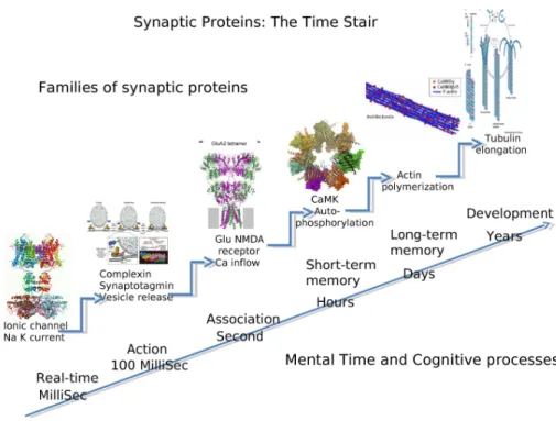
We thus focused on families of proteins (and the genes they are coded by) with known synaptic function in information processing and memory. Experimental results indeed show that memory and information processing at the synaptic and cellular level (e.g., LTP) can be seen as a cascade of molecular transformations with temporal effects lasting longer through the successive steps of the cascade. We exemplified this cascade by a stair in Figure1.3: at each step a new molecular process with a longer duration can be triggered by the previous one, from millisecond, the operation time of typical ionic channels, up to years in well consolidated memories stored in spines and dendritic morphologies. For each step of the stairs we identified protein families with a well established role in informa-tion processing and storage. This is clearly far from being exhaustive but it allowed us to verify if indeed there was a correlation between the function of key proteins for informa-tion processing and storage, and their poles of expression in correspondence to particular anatomo-functional regions. It is then possible to simply add new families to be more and more precise.
Starting from the first step of Figure 1.3we find the families of ionic channels subunits: SCNs (sodium channels), KCNs (potassium channels) and CACNs (calcium channels). These proteins are the basic bricks of neuronal information processing since controlling the propagation of action potentials in axons and the interactions of post-synaptic poten-tials in dendrites. The KCN family counts alone about 100 members with different tempo-ral characteristics important for the regulation and duration of stimuli effects. The precision reached by some family members can be of the order of milliseconds allowing ultra precise synchronization between different sensory-motor functions.
Another process important in shaping information processing is presynaptic neurotrans-mitter release. There are several families regulating the complex dynamic of this process and their role is to control on one hand the precision of release with respect to an affer-ent action potaffer-ential, on the other hand the degree of spontaneity that is the capacity of the synaptic button to release neurotransmitter independently from the arrival of an action potential. Among these families two are particularly interesting, synaptotagmins (SYTs) and complexins (CPLXs) which molecular and functional properties have been understood recently.
The third step relating information processing to memorization is controlled by NMDA glutamate receptor able to modify the influx of calcium when there is a simultaneous pres-ence of pre-synaptic activity (glutamate release) and post-synaptic (voltage depolarization). This hebbian association at the molecular level make of NMDA glutamate receptor the principal doorway toward memorization.
We studied the results for different families of glutamate and GABA receptors. NMDA receptors in the synapse are strongly influenced by the activation of receptors for neuro-modulators such as serotonin, dopamine, acetylcholine and norepinephrine. There exists a tight association at the molecular level between NMDA glutamate receptors and neuro-modulator receptors, with specific effect depending upon the different receptor isoforms. We analyzed the distribution of families of receptor for these four neuromodulators (not represented on the figure).
Short term memory is the capacity of a neuron to activate and stay active also when the stimulus is not there anymore. This capacity has its molecular support in the process of autophosphorylation of CaMKs proteins: thanks to the calcium influx produced by NMDA receptors, CaMKs modulate its phosphorylation in a prolonged way as a threshold function of calcium level. Here again, we analyzed the different CaMK isoforms which can support different forms of short term memory.
Long-term memory has been linked to the molecular process of actin polymerization which changes the shape of synaptic spines. This change in spine configuration leads to a modified synaptic weight and so to a different importance for that input to the neuron. Researchers
have found a strong link between different state of polymerization of actin in relation with different types of CaMKs and different types of actin networks ( like actinin ACTNs, actin-related proteins ARPs and ACTRs). Short term and long term memories in human interfere with the maturation of neuronal networks (growth of dendrites and axons), as in frontal cor-tex, lasting decades. A next step relating long term memory and network construction is controlled by microtubules networks . These microtubules have dynamical properties of elongation strongly dependent on their composition on tubulin subunits (TUBs.) They can play an important role in learning-dependent reorganization of the axodendritic neural network. Indeed, studies in adults (Allen, more studies) show that protein which play a role in the construction of neuronal networks during development keep playing a major role during adulthood in the homeostatic processes necessary to neuronal functioning and synaptic plasticity. We focused on protein families known to be particularly important for the development of the cortical neural network, with two families of growth factors, epi-dermal growth factors (EGFs) and fibroblast growth factors (FGFs), and three families of protein involved in cellular recognition and axon guidance: semaphorins (SEMAs), ephrins (EPHs) and C-lectins (CLECs). The choices do not pretend to be exhaustive but allow to evaluate the distribution of the protein isoforms of playing an important role in information processing and memory at different time scales.
With this scheme in mind, the essential problem to solve becomes the comprehension of the relation between the properties of genes differentially distributed in the cortex and the properties of corresponding cognitive networks, in term of memorization and information processing as exemplified in Figure 1.4. The statistical analyses reveal which members of the family are more expressed in a given anatomo-functional region of the cortex (map of the differential expression for isoforms/subunits of a family of genes). Comparison of functional properties of different isoforms differentially expressed (Pubmed) allowed to propose a multiscale scenario between the different protein cellular properties and different information and memory processing underlying cognitive functions.
1.4
This Dissertation: plan
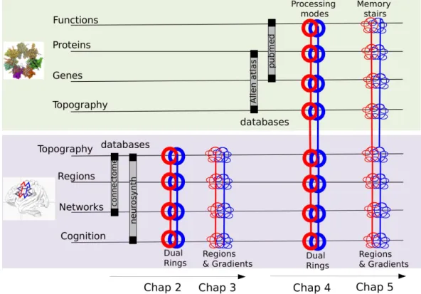
The presentation of the result is organized in direct relation with the scientific production developed in the course of my PhD; in particular each sets of results is presented in the form of an article. Each chapter is introduced by a brief introduction and a graphical abstract. We added at the end of the papers supplementary remarks that can be eventually skipped. Chapters are organized as in the following (see also Figure1.5).
Part I: Cortical organization of anatomo-functional networks: rings and gradients
Fig. 1.3.:Stairway to memory formation: from information processing to synaptic remodeling. Experimental results show that memory and information processing at the synaptic and cellular level (e.g., LTP) can be seen as a cascade of molecular transformations with temporal effects lasting longer through the successive steps of the cascade.
Fig. 1.4.:The central question of this dissertation: which is the relation between the cellular
prop-erties of genes differentially expressed across the cortex and the functional propprop-erties of cognitive networks, in term of memorization and information processing?
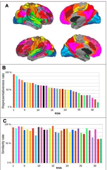
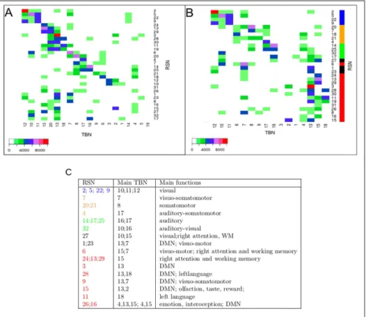
Chapter 2: The dual intertwined rings architecture of anatomo-functional and cog-nitive cortical networks. In this chapter we used a multi-scale clustering approach to interpret Resting State Networks (RSNs) from an anatomo-functional point of view and shed light on the question how does the brain integrate multiple sources of information
to support normal sensori-motor and cognitive functions? To do so we extracted using ICA (Independent Component Analysis) 32 highly intersubject reproducible RSNs–using independent databases provided by the 1000 Functional Connectomes Project–and we com-pared them with: (i) Brodmann Areas (BAs) in order to better capture their topographi-cal/cytoarchitectonic relation across the cortex, (ii) with fiber bundles of white matter (as from Human Connectome Project) to complete the anatomical view on a 3-dimensional base; (iii) with Task Based Networks (TBNs – as from BrainMap) to better understand their cognitive-neuronal function and have ideas on the characteristics time constants. The results tell us that anatomo-functional networks are topologically organized across the cor-tex as two rings (from comparison to BAs and fiber bundles) and that these two rings correspond to two different ways of information processing (from comparison with TBNs). A first ring called VSA–Visual Somatosensory Auditory– carries out real time, high fi-delity processing of sensory-motor and multimodal information. A second ring called PTF–Parieto, Temporo, Frontal–is responsible for multi-temporal information processing and while it encompasses the cognitive networks implementing language, memory and emotion it is characterized by a stronger autonomous component. The results of this work were published in Mesmoudi et al. PlosOne, 2013.
Chapter 3: Organization of the gradients of cognitive networks on the cortical surface. In this chapter we investigate how cognitive networks are organized across cortical regions. Using the Neurosynth database, we first built a lexical map (graph) of 1000 cognitive cat-egories (concerning sensory, motor, language, emotion, semantic and episodic memory, etc..), and build a topographical map (graph) of all the overlaps between the corresponding cognitive networks. At a global level we found that the main organization of these cognitive networks (in term of overlaps) reproduces the two intertwined rings architecture, obtained with a totally different method. Furthermore, this graph shows many overlaps between dif-ferent cognitive networks revealing the continuity of underlying neural processes. A a finer level to better understand the organization of cognitive networks across the cortical surface we chose a set of 21 anatomo-functional regions, (1) forming like BAs a tesselation of the cortex covering the entire cortical surface without overlaps, (2) organized as Brodmann areas along the main cortical gradients , (3) directly comparable with the tesselation and the sampling proposed in the Allen atlas of genetic expression, (4) forming a more precise description of the dual ring architecture. (5) differentiated by a global sensory-motor or cognitive specialization. Cognitive networks are organized on the gradients formed within and between the 21 anatomo-fonctional cortical regions. Each anatomo-functional region represents a pole for specific groups of cognitive tasks. We propose a scheme do describe the organization of cognitive networks across the cortex. This scheme is determined by 1) poles of connections of cerebral regions with the other brain regions: 2) the functional gra-dients connecting these poles along parietal, frontal and temporal gragra-dients. These results were partially published in Cioli et al. OHBM 2013 – Seattle - USA, 2013.
Part II: Cortical organization of gene expression in relation with anatomo-functional networks
Chapter 4: Cortical gene expression match the temporal properties of large-scale functional networks. In this chapter we studied the cortical organization of gene expres-sion for about 1000 genes found as the most differentially expressed by the Allen Institute. To do so we used mRNA postmortem measures of local expression as provided by the Allen Institute. Samples are reported across about 400 cortical regions of two complete human brains of healthy adult donors. Using multidimensional statistical techniques –such as CA and DiCA – that the 1000 genes the most differentially expressed across the cortex are or-ganized according to the two intertwined rings architecture. Among these 1000 genes we found genes playing fundamental roles in neuronal information processing (such as Ionic Channels) and neurotransmitter release (such as Synaptotagmins, Synaptobrevins and Com-plexins). We found that members of a same family of genes have their pole of expression in different rings. We adopted then a new heuristic consisting in analyzing entire families of genes with a known neuronal function to find eventual differences between the molecular properties of member preferentially expressed in different rings. In this chapter we ana-lyzed family of genes with specific synaptic properties of neuronal information processing namely ionic channels and proteins implied in neurotransmitter release. The advantage of this method is to reveal the members of the families the most spatially differentiated. We showed that the expression of different isoforms is coherent with the information process-ing performed by each of the two rprocess-ings. We found proteins supportprocess-ing precise and high fidelity information processing, more expressed across VSA networks while proteins fa-voring spontaneous activity production more expressed in the PTF ensemble. The results presented in this chapter were published in Cioli et al. PlosOne, 2014.
Chapter 5: Gradients of synaptic genes properties across cognitive networks. In this chapter we studied the relation between cortical gradients of gene expression and anatomo-functional cortical networks a finer scale. The interest was to go closer to the spatial scale of cognitive networks to study if it exists a relation between cognitive functions and groups of genes. Since cognitive functions often overlap among them we chose to define a set of disjoint anatomo-functional regions based on the Allen Institute anatomical labels. We used this time the entire set of genes measured by the Allen Institute. We studied these data in relation to the anatomo-functional cortical regions by means of multivariate statistical technique called Discriminant Correspondence Analysis (DiCA). For completeness to bet-ter understand the overall picture we also studied gene expression in relation to the entire set of cerebral structures. To draw a link between genes’ pole of expression and anatomo-functional regions this time at the anatomo-functional level we focused the analysis of the results on three class of genes: genes involved in information processing, genes participating to memory formation (short term and long term) and genes taking part to networks gene-sis. The results obtained confirm that: 1) the patterns of expression of the human genome
across cerebral areas are mainly dictated by anatomical and cytoarchitectural constraints; 2) the differential gene expression across the cerebral cortex also follows anatomy and cy-toarchitectonic more than functional specialization; 3) the cortical regions with the most distinctive patterns of gene expression are also the more phylogenetically ancient namely visual and motor areas on the VSA side and parahippocampal and temporal pole regions on the PTF side. Furthermore we found that regions implicated in the implementation of language result the most average regions in term of gene expression. Finally the systematic study of families of genes coding for proteins covering central roles in synaptic activity (in-formation processing, neurotransmission, short and long term memory, networks genesis and homeostasis) showed strong congruence between the preferential expression of genes, the cellular properties of the proteins they code, and the cognitive function implemented in anatomo-functional regions. These results point out to the existence of differential synapses whose composition in terms of proteins varies based on the anatomo-functional specializa-tion of the region they belong to. The results presented in this chapter are in course of submission (Cioli et al.).
Part III: Tools development
Chapter 6: LinkRbrain: web-based platform to analyze spatial correlation of gene ex-pression and cognitive networks.In this chapter we propose a web-based platform meant to integrate the scientific knowledge about brain organization called LinkRbrain. In partic-ular in this first version of the platform we gathered fMRI task-based results (as obtained in Chapter 2), standard anatomical labels (Talairach Atlas) and gene expression data (Allen Institute Transcriptome Atlas). These different sources of data are linked together thanks to a common spatial framework given by the MNI system of coordinates. The platform can provide information about similarities among groups of cognitive networks, groups of genes or a group of networks and a group of genes. This information is delivered to the user in four different ways: 3D and 2D visualization directly on the brain, as a graph or as a list stating distribution similarities. Finally the platform allows to compare new data provided by the user against the core data of the platform itself. The description of the platform has been published Mesmoudi et al. Journal of Neuroscience Methods, 2015.
Chapter 7: Conclusions and future work. In perspective we show how the type of analyses we perform could help to better understand, in Alzheimer disease, the relation between the gradient associated to gene expression and the pattern of propagation of the disease across cortical regions. Similarly the preferential expression of genes implicated in ASD could help to better understand the cognitive networks and cognitive functions which are impaired. Finally a more precise knowledge of the genes the most expressed in different cortical areas can help adapting the parameters necessary to model the dynamic processes (activation, plasticity) in the different areas of the cortex.
Fig. 1.5.:Plan of the dissertation: this scheme shows the progression of the work in the different
chapters. First we analyzed the brain imaging databases at two level of resolution (two large scale regions and then 21 anatomo-functional regions) and then we analyzed the correlation between gene expression and anatomo-functional regions at these two leveles of resolution.
1.5
This Dissertation: personal contributions
Chapter 2: My contribution was to systematically study the cortical organization of cogni-tive networks using the Neurosynth database as described in Chapter 3. This work allowed to compare cognitive networks obtained from two independent databases: the BrainMap database (obtained with top-down methods) and the Neurosynth database obtained in a bottom-up fashion.
Chapter 3: I was the principal contributor of the work presented in this chapter. The results presented were partially published in Cioli et al.,OHBM 2013 and they will be further structured for future publication.
Chapter 4: I was the main contributor of the work described in this chapter; the results are published in Cioli et al.,PlosOne 2014.
Chapter 5: I was the main contributor of the work described in this chapter; the results are in course of submission as Cioli et al..
Chapter 6: My contribution for this chapter is represented by: 1) the cognitive networks (as derived in Chapter3) used in the platform; 2) the discussion/proposition of methods to compare and display data.
1.6
Publications from this Dissertation
The results of this dissertation appeared in the following publications:
1.6.1
Journal articles
• Mesmoudi S., Rodic M., Cioli C., Cointet J. P., Yarkoni T., Burnod Y., LinkRbrain: Multi-scale data integrator of the brain, Journal of Neuroscience Methods, Volume 241, 15 February 2015, Pages 44-52.
DOI:http://dx.doi.org/10.1016/j.jneumeth.2014.12.008.
• Cioli C., Abdi H., Beaton D., Burnod Y., Mesmoudi S., Differences in Human Cor-tical Gene Expression Match the Temporal Properties of Large-Scale Functional Networks. PLoS ONE 9(12): e115913. 2014.)
DOI:http://dx.doi.org/10.1371/journal.pone.0115913.
• Mesmoudi S., Perlbarg V., Rudrauf D., Messe A., Pinsard B., Hasboun D., Cioli C. et al., Resting State Networks’ Corticotopy: The Dual Intertwined Rings Architecture. PLoS ONE 8(7): e67444. 2013.
DOI:http://dx.doi.org/10.1371/journal.pone.0067444
1.6.2
Peer-Reviewed Abstracts
• Cioli C., Abdi H., Beaton D., Burnod Y., Mesmoudi S., Integration of functional cerebral networks and genetic expression: the dual intertwined rings architecture of the cerebral cortex for real-time vs multi-temporal information processing, Pro-ceedings in the 21th Annual Meeting of the Organization for Human Brain Mapping, Honolulu, USA, 2015.
• Cioli C., Cointet J. P., Mogoutov A., Reuillon R., Messé A., Benali H., Rudrauf D., Yarkoni T., Burnod Y., Mesmoudi S., Systematic analysis of task-based fMRI database: comparison between lexical and topographic maps, Proceedings in the 19th Annual Meeting of the Organization for Human Brain Mapping, Seattle, USA, 2013.


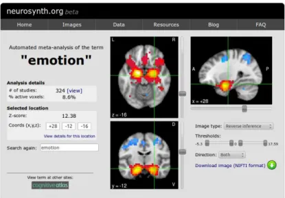
![Fig. 3.2.: (source: [295]) a) Neurosynth pipeline: once selected a term to meta-analyze, Neurosynth identi- identi-fies all the papers present in the database reporting the term chosen and the coordinates of peaks of activations present in those papers](https://thumb-eu.123doks.com/thumbv2/123doknet/14740892.755031/74.892.259.686.96.397/neurosynth-pipeline-selected-neurosynth-database-reporting-coordinates-activations.webp)
