HAL Id: hal-02102973
https://hal.archives-ouvertes.fr/hal-02102973
Submitted on 17 Apr 2019
HAL is a multi-disciplinary open access archive for the deposit and dissemination of sci-entific research documents, whether they are pub-lished or not. The documents may come from teaching and research institutions in France or abroad, or from public or private research centers.
L’archive ouverte pluridisciplinaire HAL, est destinée au dépôt et à la diffusion de documents scientifiques de niveau recherche, publiés ou non, émanant des établissements d’enseignement et de recherche français ou étrangers, des laboratoires publics ou privés.
visceral fat mass loss in obese Zucker rats without
modulating gut microbiota
Florie Maillard, Emilie Vazeille, Pierre Sauvanet, Pascal Sirvent, Lydie
Combaret, Antoine Soudrille, Vivien Chavanelle, Richard Bonnet, Yolanda
Otero, Geoffrey Delcros, et al.
To cite this version:
Florie Maillard, Emilie Vazeille, Pierre Sauvanet, Pascal Sirvent, Lydie Combaret, et al.. High intensity interval training promotes total and visceral fat mass loss in obese Zucker rats without modulating gut microbiota. PLoS ONE, Public Library of Science, 2019, 14 (4), �10.1371/journal.pone.0214660�. �hal-02102973�
High intensity interval training promotes total
and visceral fat mass loss in obese Zucker rats
without modulating gut microbiota
Florie MaillardID1,2*, Emilie Vazeille2,3, Pierre Sauvanet2,4, Pascal Sirvent1,
Lydie Combaret5, Antoine Sourdrille1, Vivien Chavanelle1, Richard Bonnet2,6, Yolanda Fernandez Otero1, Geoffrey Delcros1, Nicolas Barnich2☯, Nathalie Boisseau1☯
1 Universite´ Clermont Auvergne, Laboratoire des Adaptations Me´taboliquesàl’Exercice en conditions Physiologiques et Pathologiques (AME2P), Clermont-Ferrand, France, 2 Universite´ Clermont Auvergne/ Inserm U1071; USC-INRA 2018, Microbes, Intestin, Inflammation et Susceptibilite´ de l’Hoˆte (M2iSH), Clermont-Ferrand, France, 3 Universite´ Clermont Auvergne, Inserm, 3iHP, CHU Clermont-Ferrand, Service d’He´pato-Gastro Ente´rologie, Clermont-Ferrand, France, 4 Universite´ Clermont Auvergne, CHU Clermont-Ferrand, Service de chirurgie digestive, Clermont-Clermont-Ferrand, France, 5 Universite´ Clermont Auvergne, INRA, UNH, Unite´ de Nutrition Humaine, CRNH Auvergne, Clermont-Ferrand, France, 6 Department of
Bacteriology, CHU Clermont-Ferrand, Clermont-Ferrand, France ☯These authors contributed equally to this work.
*Florie.maillard@gmail.com
Abstract
Aims
Increased visceral adipose tissue and dysbiosis in the overweight and obese promote chronic inflammation. The aim of this study was to compare the effects of moderate-intensity continuous training (MICT) and high-intensity interval training (HIIT) on the gut-adipose tis-sue cross-talk in obese Zucker rats.
Methods
Obese male Zucker rats (n = 36) were divided in three groups: MICT (12m.min-1for 51min), HIIT (6 sets at 18 m.min-1for 4min followed by 3min at 10m.min-1) and controls (CONT; no exercise). The animals ran on a treadmill 5 days/week for 10 weeks. Body composition, gly-caemic control, lipid profile, inflammation, lipolysis signalling in subcutaneous and visceral adipose tissue, intestinal permeability (tight junctions and plasma lipopolysaccharide bind-ing protein; LBP), and gut microbiota composition were assessed in the three groups.
Results
After 10 weeks of exercise, total and epididymal fat mass decreased only in the HIIT group. Theα/βadrenergic receptor RNA ratio in subcutaneous adipose tissue increased only in the HIIT group. The expression level of phosphorylated hormone-sensitive lipase was not modi-fied by training. Both HIIT and MICT decreased inflammation (plasma myeloperoxidase and keratinocyte-derived chemokine secretion in adipose tissue) and improved glucose metabo-lism. Zonula occludens-1 and occludin were upregulated in the HIIT group. Plasma LBP
a1111111111 a1111111111 a1111111111 a1111111111 a1111111111 OPEN ACCESS
Citation: Maillard F, Vazeille E, Sauvanet P, Sirvent
P, Combaret L, Sourdrille A, et al. (2019) High intensity interval training promotes total and visceral fat mass loss in obese Zucker rats without modulating gut microbiota. PLoS ONE 14(4): e0214660.https://doi.org/10.1371/journal. pone.0214660
Editor: Marcia B. Aguila, Universidade do Estado
do Rio de Janeiro, BRAZIL
Received: December 1, 2018 Accepted: March 19, 2019 Published: April 9, 2019
Copyright:© 2019 Maillard et al. This is an open access article distributed under the terms of the
Creative Commons Attribution License, which permits unrestricted use, distribution, and reproduction in any medium, provided the original author and source are credited.
Data Availability Statement: All the data have
been added in an excel file,S1 Table. The data uploaded under the fileS1 Tableconstitutes our minimal data set. All relevant data are within the manuscript and supporting information files.
Funding: This study was supported by the “Region
Auvergne-Rhoˆne-Alpes” (PREVAMIC project) and I-SITE project (CAP 2025). The funders had no role in study design, data collection and analysis,
was similarly reduced in both training groups. HIIT and MICT did not affect gut microbiota composition.
Conclusion
In obese Zucker rats, HIIT and MICT improved inflammation and glucose metabolism. In contrast, only HIIT decreased total and visceral fat mass. These adaptations were not asso-ciated with modifications in gut microbiota composition.
Introduction
Obesity has dramatically increased worldwide in recent decades and is now a critical health problem. In 2014, 1.9 billion people were overweight and among them 600 million were obese in developed and developing countries [1]. Obesity is characterized by a low-grade inflamma-tion state [2,3]. Abdominal fat mass (FM), especially visceral FM, is associated with metabolic disorders and inflammatory state to a greater extent than total and subcutaneous FM [4]. Vis-ceral FM is characterized by a greater secretory activity (free fatty acids, TNF-α, IL-6, IL-8,
etc.) that promotes insulin resistance, chronic low-grade inflammation and risks of
cardiovas-cular disease [5,6].
More recently, gut microbiota has been recognized as a major actor in pathological condi-tions associated with obesity and its related complicacondi-tions [7,8]. This relationship was con-firmed by increased adiposity in germ-free mice after faecal microbiota transplant from obese mice (ob/ob) [9]. Moreover, a reduced microbial diversity and dysbiosis (i.e., a microbial
imbalance, characterized by an alteration in the Bacteroidetes/Firmicutes ratio) have been observed in overweight or obese individuals [10] and in animal models of obesity [11]. Gut microbiota is an exteriorized organ that can interact directly with other organs. Butyrate, pro-pionate and acetate, the main short-chain fatty acids (SCFAs) produced by microbial fermen-tation, favourably influence the host metabolism by producing anorexigenic hormones via colonic epithelial cells [12,13], increasing the energy expenditure [14] and reducing inflamma-tion [15]. High-fat diets induce dysregulation of intestinal permeability, promoting an increase in plasma lipopolysaccharide (LPS) (i.e., a constituent of Gram-negative bacteria), a condition
defined as metabolic endotoxemia [16], and triggering inflammation and insulin resistance [17].
Regular physical activity, alone or combined with energy restriction, is an effective way to prevent and/or reduce excess adiposity [18]. Traditionally, Moderate Intensity Continuous Training (MICT) is recommended for the overweight or obese. However, this exercise modal-ity has little effect on weight and FM loss [19,20]. In the last few years, High Intensity Interval Training (HIIT) has grown in popularity as a time-efficient and powerful strategy to reduce total and abdominal/visceral FM [21,22]. Adrenergic receptors (α/β AR ratio) are probably involved in such adaptations, but other, still unknown mechanisms could contribute to FM loss. In this context, modulation of gut microbiota composition by physical activity is an inter-esting hypothesis. Numerous studies have demonstrated that moderate-intensity training can favourably alter gut microbiota composition in humans [23] and in animal models [24–31]. Most studies with rodents used MICT protocols that involve treadmill running or spontaneous activities on a running wheel. Only two studies investigated the effects of HIIT on gut micro-biota composition. Batacan etal. found slight differences in gut microbiota phylotype in
Wis-tar rats after MICT and HIIT without any association with FM loss [32]. Similarly, despite decision to publish, or preparation of the
manuscript.
Competing interests: The authors have declared
changes in gut microbiota composition, Denou etal. detected no FM loss after HIIT in
C57BL/6 mice [33].
The aim of the present study therefore was to compare the effect of HIIT and MICT pro-grammes on total and visceral FM loss in Zucker rats, a genetic model of obesity. We hypothe-sized that HIIT is more effective than MICT in decreasing total and visceral FM. We also aimed to determine whether gut microbiota modulation is involved in the reduction of the amount of adipose tissue, and whether the metabolic and inflammatory profiles are improved more effectively by HIIT than by MICT.
Material and methods
Ethical approval
The experimental protocol was approved by the “Comite´ d’e´thique en expe´rimentation ani-male” of Auvergne (C2EA-02, approval number: 3075–2015120813375547) and was in accor-dance with the current legislation on animal experimentation (Guide for the care and use of laboratory animals, Eighth edition 2011). All efforts were made to protect animal welfare and to minimize suffering at each step of the protocol. The animals were sacrificed by cervical dis-location following isoflurane anaesthesia.
Animals
Seventy-five 8-week-old male Zucker rats from Charles River Laboratories were individually housed with a reversed light-dark cycle in a temperature-controlled room (21˚C). After 1 week of treadmill acclimatization, rats most proficient at running were selected and randomly assigned to one of the three groups: HIIT (n = 12), MICT (n = 12) or CONT (no exercise) (n = 12).
Exercise training
Exercise training was performed on a motorized treadmill at 0˚ inclination 5 days/week (Mon-day to Fri(Mon-day) for 10 weeks. Both groups started with a warm-up at 10 m.min-1for 5min. In the HIIT group, rats ran 6 sets of 4min at 18m.min-1followed by 3min at 10m.min-1. In the MICT group, animals ran at 12m.min-1for 51min. The protocols were originally designed to have the same total running distance for all groups, as proposed by Metz etal. 2005 [34], Kapravelou etal. 2015 [35] and Haram etal. 2009 [36]. CONT rats were placed in the training room during the sessions to expose them to the same environment and for the same time as the HIIT and MICT groups.
Food intake, weight and body composition
Food (3% fat, 16% protein, 60% carbohydrates, 5% minerals, and 4% fibres; SAFE A04, France) and water were providedad libitum. Food intake was recorded once a week (on Thursday).
Weight was recorded weekly during the 10 weeks of training (W0 to W10). At week 0, 2, 5, 8 and 10, body composition was measured by MRI (Echo Medical Systems, Houston, TX), and epididymal fat pads were weighedpost-mortem.
Indirect calorimetry
At week 10 (end of the training), the rats were placed in indirect calorimetric cages (TSE Sys-tems, Bad Homburg, Germany) for 48h (24h of familiarization and 24h of measurements) withad libitum access to food and water. Metabolic measurements (O2and CO2consumption,
was determined as the ratio of produced CO2(VCO2) over consumed O2(VO2). Data were
analysed over 24h, and in 12h-light and 12h-dark conditions.
Oral Glucose Tolerance test (OGTT)
OGTTs were performed at the beginning and at the end of the study. After 6h of fasting fol-lowed by oral gavage of glucose (4.0 g.kg lean mass-1), glycaemia was monitored with a gluc-ometer (Accu-chek Performa, Roche Diagnostics, Basel, Switzerland) and tail blood samples taken at 15, 30, 60, 90 and 120min post-gavage. The area under the curve for glucose (AUC) and thenetAUC (after subtraction of the baseline glucose concentration) were calculated. The
homeostatic model assessment for insulin resistance (HOMA-IR) index was used to assess IR as follows: fasting insulin (mU.L-1)× fasting glucose (mmol.L-1
)/ 22.5 [37]
Post-mortem blood samples and plasma measurements
At the end of the study, blood was collected and centrifuged at 2000g for 10min for plasma sep-aration. All samples were frozen at -80˚C until analysis. Plasma insulin was measured with the Ultrasensitive Insulin ELISA Kit (ALPCO, Salem, NH, USA). Lipid profile was determined by quantifying plasma lipoproteins and lipids with commercial kits following the manufacturers’ instructions: triglycerides (Max Discovery, Austin, USA), LDL (Crystal Chem, Downers Grove, USA), HDL (Crystal Chem, Downers Grove, USA) and total cholesterol (Max Discov-ery, Austin, USA). LBL plasma level was measured with the LBP ELISA Kit for various species (Hycult Biotech, Netherland) following the manufacturer’s instructions.
Plasma myeloperoxidase (MPO), and cytokine and free fatty acid (FFA)
release from adipocytes
Plasma MPO concentration was measured using a commercial ELISA Kit (R&D Systems). At the end of the study, adipose tissue was collected and a known amount of each fat pad (subcutaneous and visceral) was placed in Dulbecco’s Modified Eagle Medium (DMEM) with antibiotics (50mg.ml-1gentamicin) or in KBEBS-Ringer’s solution (pH = 7.4). After overnight incubation at 37˚C, 5% CO2,samples were centrifuged and supernatants frozen at -80˚C.
Cyto-kines (KC and IL-6) secreted by tissues were quantified in these supernatants diluted in reagent diluent (1% BSA) using an ELISA Kit (R&D systems). FFA were analysed with a com-mercial kit (Wako Chemicals, Richmond, USA), according to the manufacturer’s instructions.
Microbiota composition analysis
Rat colons were transferred in ZR BashingBead Lysis Tubes (0.1 & 0.5 mm, Zymo Research) with lysis buffer (Maxwell RSC Buffy Coat DNA) and homogenized using a Precellys homoge-niser (2X 15 seconds followed by 2min rest). Lysis tubes were centrifuged at 14000g at 4˚C for 3min, and supernatants were collected in new tubes and centrifuged again to ensure that all beads were removed. The supernatants were then placed in cartridges in Maxwell RSC Instru-ment (Promega) to extract DNA. DNA concentration was determined by Qubit Fluorometric Quantitation (Invitrogen) and DNA quality was assessed by spectrophotometry (260/280 and 260/230 ratios, Nanodrop). The variable regions V3-V4 of bacterial 16S rRNA genes were amplified from the purified DNA using the following primers: Forward CTTTCCCTACACG ACGCTCTTCCGATCTACGGRAGGCAGCAG, and Reverse GGAGTTCAGACGTGTGCTCT TCCGATCTTACCAGGGTATCTAATCCT. All PCR amplifications were performed with MTP Taq DNA Polymerase and 10X MTP Taq Buffer (Sigma, D7442-1500U) and the following cycling conditions: 94˚C for 1min, followed by 30 cycles of 94˚C for 1min, 65˚C for 1min, and
72˚C for 1min, and a final elongation step at 72˚C for 10min. Illumina sequencing was per-formed in collaboration with the GeT core facility (Toulouse). Paired-end read assembly, qual-ity and length filtering, OTU picking (97% sequence identqual-ity threshold) and chimera removal were performed with UPARSE [38]. OTUs with low counts (<0.1% of the total number of sequences per sample) were excluded. Sequences of samples with over 6000 reads were loaded into the QIIME 1.9.1 pipeline for diversity analysis [39]. Taxonomy assignment was performed with the SILVA database 132 (https://www.arb-silva.de/). Alpha diversity of bacterial commu-nities was assessed from four different indexes including richness and/or evenness (Chao1, Shannon, Simpson and evenness). The Kruskal–Wallis test was used to estimate alpha diversity differences between groups. Beta diversity was used to analyse the dissimilarity between the groups’ membership and structure. Accordingly, abundance-weighted and/or phylogenetic-weighted distance matrices were generated on the basis of Bray-Curtis and phylogenetic-weighted/ unweighted UniFrac distances and reported according to principal coordinate analysis (PCoA). Permutational analysis of variance (PERMANOVA with 999 permutations) was used to determine significant differences between groups. Significance testing for taxon abundance was performed with a Wilcoxon rank sum test and the Bonferroni procedure to correct p-val-ues. P-values �0.05 were considered significant.
Faecal short-chain fatty acid (SCFA) concentration
Weighted faecal samples were reconstituted in 200μl Milli-Q water, disrupted, incubated at 4˚C for 2h and centrifuged at 12000g at 4˚C for 15min. Supernatants were weighed and satu-rated phosphotungstic acid solution was added (1g for 100μL). After overnight incubation at 4˚C, samples were centrifuged again and SCFA concentrations were determined by gas chro-matography (Nelson 1020, Perkin-Elmer, St Quentin en Yvelines, France) as previously described [40].
Protein extraction and western blotting
Adipose tissue (subcutaneous and visceral) and colon samples were homogenized in 500μl of lysis buffer (Tris 25mM, EDTA 1mM, EGTA 5mM, MgCl2 0.1mM, Glycerol 10%, NaCl 150mM, Nonidet P-40 1%, SDS 1%) supplemented with freshly added protease inhibitor cock-tail (cOmplete, Mini, EDTA-free Protease Inhibitor Cockcock-tail, Roche), Sodium Orthovanadate (1mM), PMSF (1mM) and N-Ethylmaleimide (5mM). The homogenates were then centri-fuged at 10 000 rpm at 4˚C for 5min. A small aliquot (20μl) was used for protein concentration with the DC Protein Assay (Bio-Rad, USA). The rest was frozen at -80˚C until use.
Proteins were separated on 12% SDS-PAGE gels, transferred to nitrocellulose membranes, and blocked with 5% BSA in Tris buffered saline (pH 8) containing 0.05% Tween 20 (TBST) at room temperature under agitation for 1h. Membranes were then incubated with diluted pri-mary antibodies against phospho-HSL (Cell Signaling Technology), occludin (1:500 dilution; 33–1500; Invitrogen) or ZO-1 (1:500 dilution; 61–7300; Invitrogen) at 4˚C under agitation overnight. After three washes with TBST, membranes were incubated with secondary antibod-ies in TBST at room temperature under agitation for 1h. Antibody interactions were detected with the Enhanced Chemiluminescence Detection Kit (Amersham Biosciences, RPN2108) fol-lowed by the Bio-Rad ChemiDoc imaging system. Data for pHSP were normalised to total pro-tein loading using the Stain-Free Blot system (Bio-Rad, USA). ZO-1 and occludin propro-tein contents were normalised to GAPDH expression. Band densities were analysed with Image J software.
Quantitative Real-Time PCR
Total RNA was extracted from adipose tissues using TRIzol (Invitrogen, Life Technologies) and was reverse transcribed using the High Capacity cDNA Transcription Kit (Applied Biosystems, Life Technologies). Expression of the genes encodingα AR and β AR was analysed with the SYBR Green qPCR Master Mix (applied biosystems) and a CFX Bio-Rad system. The fold induc-tion was calculated using theCt method as follows: ΔΔCt = (Cttarget gene-Cthousekeeping gene)treatment
- (Cttarget gene-Cthousekeeping gene)nontreatment, and the final data were derived from 2−ΔΔCT.
Statistical analysis
All statistical analyses were performed with Statistica software (version 12). Data were pre-sented as the mean± standard deviation (SD). Normal data distribution was tested using the Kolmogorov–Smirnov test and the homogeneity of variance from the F-test. In the absence of normal distribution or variance homoscedasticity, the data were log-transformed before analy-sis. A one-way ANOVA (group effect) or ANOVA with repeated measures was used to deter-mine significances (time (T) and group (G) effects & G x T interactions), followed by a Newman–Keuls post-hoc test when a significant effect was found. To assess beta- diversity, distance matrices between samples were generated on the basis of Bray-Curtis and weighted/ unweighted UniFrac distances and reported according to principal coordinate analysis (PCoA). Analyses and graphical outputs were performed in R version 3.3.2. Differences with a p value �0.05 were considered statistically significant.
Results
HIIT is a time- efficient strategy to decrease total and epididymal fat mass
After 1 week of treadmill acclimatization, adult Zucker rats were randomly divided in three groups: HIIT (n = 12), MICT (n = 12) or CONT (no exercise) (n = 12). No significant differ-ence in food intake between groups was observed during the study period (Fig 1).
Despite our attempt to match running distances between groups, rats in the MICT group ran greater distances (23.3± 1.1 vs 21.4 ± 2.3 km) (p �0.05) and for a longer time (1941 ± 88 vs 1510± 114 min) (p �0.001) than rats in the HIIT group. After 10 weeks of training, body
Fig 1. Weekly food intake. Data are expressed as the mean± SD.
weight and total lean mass were comparable between groups (Fig 2A and 2B). However, total FM was lower in the HIIT than in the CONT group at week 5 (p �0.05) and week 8 (p �0.01), and in the HIIT group than in the MICT group at the end of the training protocol (p �0.05) (Fig 2C). At the end of the study, epididymal (visceral) adipose tissue was reduced only in the HIIT group (p �0.05) (Fig 2D).
Gut microbiota composition and faecal short chain fatty acid
concentration are not modified by exercise training
To investigate the effect of the two exercise modalities on gut microbiota composition, the eubacterial 16S rRNA genes present in colon tissue were sequenced. All alpha diversity indexes (only Chao1 index is shown inFig 3A) did not show any significant difference in species rich-ness between the three groups at the end of the training programme (week 10). Beta-diversity analysis based on the uniFrac distance coupled with principal coordinate analysis (PCoA) showed no clustering of samples (Fig 3B). Univariate analysis of the abundance of major tax-ons (relative abundance >1%) %) identified no significant differences between groups. Simi-larly, the faecal concentrations of the major SCFAs (butyrate, acetate and propionate) were not significantly different in the three groups (Fig 3C).
HIIT stimulates tight junction protein synthesis more efficiently than
MICT, but both training modalities decrease plasma lipopolysaccharide
binding protein (LBP)
At the end of the training programme, zonula occludens-1 (ZO-1) (MICT vs CONT; p �0.05 and HIIT vs CONT; p �0.005) and occludin (MICT vs CONT; p� 0.01 and HIIT vs CONT;
Fig 2. Effect of 10 weeks of exercise training on weight and body composition. Body weight (A), total lean mass (B), total fat mass (C) and epididymal fat
mass (D). Data are expressed as the mean± SD. § p �0.05 CONT vs. HIIT; § § p �0.01 CONT vs. HIIT £ p �0.05 HIIT vs. MICT and�p �0.05.
p �0.001) were upregulated in both training groups, particularly in the HIIT group (Fig 4A, 4B and 4C). In addition, occludin expression level was significantly correlated with epididymal adipose tissue depots (r = -0.5; p �0.05) and total FM change (FM at week 10—FM at baseline) (r = -0.4; p �0.05). Occludin and ZO-1 were also negatively related to IL-6 secretion in epidid-ymal adipose tissue (r = -0.4, p �0.05 for both). Finally, expression of LBP (a marker of obesity [41,42] and metabolic endotoxemia [43]) was reduced by about 50% in both HIIT and MICT groups (p� 0.01 vs CONT) (Fig 4D).
MICT and HIIT induce anti-inflammatory effects at the systemic and
adipose tissue levels
At the end of the training programme (week 10), secretion of free fatty acids (FFA) was reduced in subcutaneous adipocytes (p �0.05 HIIT and MICT vs CONT), but not in the epi-didymal tissue (Fig 5A and 5B). Conversely, keratinocyte-derived chemokine (KC) secretion was reduced in the epididymal adipose tissue (p �0.05 HIIT and MICT vs CONT), but not in subcutaneous adipocytes (Fig 5C and 5D). On the other hand, IL-6 secretion in the two types of adipose tissue was not significantly different from that in the CONT group (Fig 5E and 5F). At the systemic level, the plasma concentration of myeloperoxidase (MPO) decreased in both MICT and HIIT groups after 10 weeks of training (p �0.05 vs CONT) (Fig 5G). Plasma MPO levels were positively associated with plasma LBP levels (r = -0.5; p �0.001).
Fig 3. Effect of 10 weeks of exercise training on gut microbiota and faecal concentration of short-chain fatty acids (SCFAs). Gut microbiota composition:
Chao1 index (A); Principal Component Analysis (PCoA) of the Bray-Curtis distances (β-diversity) (B); SCFA concentration in faeces after 10 weeks of training (C). Data are expressed as the mean± SD.
Only HIIT increases the
α2 AR/β3 AR ratio in subcutaneous adipose tissue
At the end of the training programme, theα/β adrenergic receptor RNA ratio in subcutaneous adipose tissue was greater in the HIIT group than in the CONT (p �0.001) and MICT (p �0.01) groups, but not in epididymal adipose tissue (p �0.05) (Fig 6A and 6B). The level of phosphorylated hormone-sensitive lipase (phospho-HSL) in subcutaneous (Fig 6C) and epi-didymal adipose tissue (Fig 6D) was not modified by exercise training.
HIIT and MICT improve glucose metabolism, but do not modify the lipid
profile
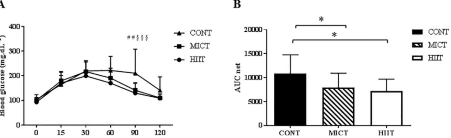
Fasting glycaemia did not differ between the three groups at the end of the study (week 10) (Table 1). Repeated ANOVA measures of the results of the oral glucose tolerance test (OGTT) performed at the end of the training programme showed a significant time x group interaction at 90min (p �0.01) with higher values in the CONT than in the MICT and HIIT groups (Fig
Fig 4. Effect of 10 weeks of training on intestinal permeability. Zonula occludens-1 (ZO-1) (A) and occludin (B) expression were assessed by western
blotting. Representative images of the western blot results (C). Plasma levels of lipopolysaccharide binding protein (LBP) determined by ELISA (D). Data are expressed as the mean± SD;�p �0.05,��p �0.01 and���p �0.001.
7A). The net area under the curve (netAUC) was lower in the MICT and HIIT groups than in
the CONT group (p �0.05) (Fig 7B). After 10 weeks, the plasma level of LDL cholesterol was higher in the MICT than in the CONT group (p �0.05). Conversely, plasma total cholesterol, HDL cholesterol, triglycerides and FFA were not modified by physical training (Table 1).
HIIT and MICT modify the resting respiratory exchange ratio, but not the
mean daily spontaneous physical activity and energy intake
After 10 weeks of training, the mean daily total energy expenditure and food intake (for the 12h-light period and during 24h) were comparable in the three groups (Table 2). Conversely, for the 12h-dark period, spontaneous physical activity was increased in the two training groups (p �0.05), and energy intake was higher in the MICT than in the CONT group (p �0.05).
At the end of the study, the respiratory exchange ratio (RER), evaluated by indirect calorim-etry in mice housed in calorimcalorim-etry cages for 24h, was significantly higher in the two training groups (0.97± 0.02 in the HIIT and 0.97 ± 0.02 in the MICT vs 0.93 ± 0.05 in the CONT group; p � 0.05) (Table 2).
Fig 5. Effect of 10 weeks of exercise training on adipose tissue and systemic inflammation. Free fatty acid (FFA) secretion by subcutaneous adipose tissue
(A) and visceral adipose tissue (B). Keratinocyte-derived chemokine (KC) secretion by subcutaneous adipose tissue (C) and visceral adipose tissue (D). IL-6 secretion by subcutaneous adipose tissue (E) and visceral adipose tissue (F). Plasma myeloperoxidase (MPO) concentration (G). Data are expressed as the mean± SD.�p �0.05 vs CONT.
Discussion
This study in Zucker rats, a genetic model of obesity, confirms that HIIT is more efficient than MICT in decreasing total FM, in particular visceral adipose tissue, which is responsible for numerous metabolic complications. In contrast, both exercise training modalities reduced sys-temic and adipocyte inflammation and improved glucose metabolism. Finally, endotoxemia
Fig 6. Effect of 10 weeks of exercise training on lipolysis factors. Theα-adrenergic receptor/β adrenergic receptor (α AR/β AR) ratio determined by q-PCR in subcutaneous adipose tissue (A) and epididymal adipose tissue (B). Level of phosphorylated hormone-sensitive lipase (phospho-HSL) in subcutaneous adipose tissue (C) and epididymal adipose tissue (D). Data are expressed as the mean± SD;��p �0.01 and���p �0.001.
https://doi.org/10.1371/journal.pone.0214660.g006
Table 1. Lipid and glycaemic profiles after 10 weeks of training (mean± SD). Total Chol (mg.dL-1) LDL (mg.dL-1) HDL (mg.dL-1) TG (mg.dL-1) FFA (mmol.L-1) Insulin (ng.mL-1)
Glucose (mmol.L-1) HOMA-IR
CONT 227.1± 49.9 260.2± 74.0 69.0± 36.0 603.0± 470.6 556.5± 442.4 2.5± 1.6 5.7± 1.1 17.4± 10
MICT 217.8± 33.4 314.5± 43.7#£ 80.2
± 19.3 426.5± 155.8 415± 141.7 3.4± 3.6 5.6± 0.6 25.0± 31.1
HIIT 209.2± 40.2 270.1± 52.5 74.9± 26.6 450.8± 94.9 493.9± 163.9 2.6± 3.7 5.3± 0.7 17.1± 23.1 Chol, cholesterol; LDL, low-density lipoproteins; HDL, high-density lipoproteins; TG, triglycerides; FFA, free fatty acids; HOMA-IR, homeostatic model assessment of insulin resistance.
# p �0.05 CONT vs MICT £ p �0.05 MICT vs HIIT
reduction was comparable in both training groups (LPB expression), although HIIT increased the synthesis of tight junction proteins to a greater extent than did MICT. These positive adap-tations observed in the HIIT group, in which rats ran shorter distances than those in the MICT group, demonstrate the time efficiency of this exercise modality.
MICT protocols are still traditionally recommended for sedentary overweight or obese individuals to reduce FM. However, a growing body of evidence shows that HIIT can be a more amusing and time-efficient exercise modality to lose total and visceral adipose tissue [21,22,44]. Our present results in obese Zucker rats confirm that HIIT (6 sets at 18 m.min-1for 4min followed by 3min at 10 m.min-1for 10 weeks) significantly reduces total and epididymal FM compared with MICT (12 m.min-1for 51min for 10 weeks). Other studies also demon-strated a greater effect of HIIT on total and abdominal FM loss than with MICT in different animal models of obesity. Wang etal. showed that in mice fed a high-fat diet, the adiposity
index (44% and 53%, respectively) was lower in the HIIT (10 x 4min [85–90% VO2max]/2min
active rest [5 m.min-1]) than in the MICT group (65–70% of VO2max [distance-matched
con-tinuous running]) [45]. Similarly, in male Sprague-Dawley rats fed a high-fat diet, HIIT (30sec [32 m.min-1]/10sec passive recovery, 5˚ slope) decreased epididymal FM, whereas MICT (16 m.min-1for 40min, 5˚ slope) had no effect [46]. In the sole study, to our knowledge, to assess the effects of an HIIT protocol (4min at 65–80% of VO2max/3min at 50–65% of VO2max) in
Zucker rats, the authors observed reduced FM and abdominal fat pad in the HIIT group (5.7 ±0.2 vs. 6.4±0.2g in the control group without exercise) [35].
Fig 7. Effect of 10 weeks of exercise training on glucose tolerance. Blood glucose monitoring during the oral glucose tolerance test (A) and net area under the
curve (netAUC) for glucose (B) at the end of the 10 weeks of exercise training in the three groups. Data are expressed as the mean± SD. ## p �0.01 CONT vs
MICT; § § § p �0.001 CONT vs HIIT and�p �0.05.
https://doi.org/10.1371/journal.pone.0214660.g007
Table 2. Indirect calorimetry results after 10 weeks of training (mean± SD).
24 hours 12h-light (08:05–20:00) 12h-dark (20:05–8:00)
EE/LM (kJ/ g) RER Spontaneous activity (m) Food intake (g) EE/LM (kJ/ g) RER Spontaneous activity (m) Food intake (g) EE/LM (kJ/ g) RER Spontaneous activity (m) Food intake (g) HIIT 0.93± 0.05 0.97± 0.02 211.1± 40.9 22.31± 3.3 0.50± 0.03 0.97± 0.03 131.1± 31.0 13.69± 2.2 0.42± 0.02 0.96± 0.03 73.9± 12.9 7.91± 1.8 MICT 0.93± 0.08 0.97± 0.02 184.9± 49.0 21.79± 2.7 0.50± 0.05 0.97± 0.03 112.4± 28.8 13.78± 2.7 0.43± 0.03 0.96± 0.02 67.6± 21.9 8.36± 1.41 CONT 0.95± 0.12 0.93± 0.05#$ 171.8 ± 46.5 19.05± 4.2 0.51± 0.07 0.94± 0.04 113.5± 37.9 12.57± 2.9 0.43± 0.06 0.91± 0.06#$ 50.5 ± 14.8#$ 6.36 ± 2.5# EE: energy expenditure, LM: Lean mass, RER: respiratory exchange ratio.
# p �0.05 CONTvs. MICT
$ p �0.05 CONTvs. MICT; p �0.05 MICT vs. HIIT
However, the mechanisms by which HIIT decreases abdominal and in particular visceral FM are still unknown. Here, we assessed the potential role of the gut microbiota in such adap-tations. In our study, exercise (MICT and HIIT) did not have any effect on gut microbiota composition (quantity and function). In addition, the main faecal SCFAs (acetate, propionate and butyrate) were not modified by the two exercise modalities. This result is surprising because previous studies have often reported that bacterial richness and function are improved by regular physical activity, and that some bacterial species related to the anti-inflammatory profile, health and leanness could also be increased by exercise [27,28,30,31,47–49]. To investi-gate the effect of exercise on gut microbiota, MICT training or spontaneous exercise (sponta-neous activity wheel) are generally used, and only two studies have tested the effect of a HIIT programme on gut microbiota modulation. The first one found an increase in the Bacteroi-detes/Firmicutes ratio and inLactobacillus, Bacteroidales and Dorea species in mice after HIIT
training, without any change in the epididymal FM, suggesting there is no correlation between these variables [33]. The second study reported slight differences in gut microbiota composi-tion in rats undergoing MICT and HIIT (increase inParasutterella excrementihominis and Lactobacillus johsonii in the MICT group, and in Clostridium saccharolyticum in the HIIT
group). Unfortunately, the authors did not measure total and visceral FM [32]. In Zucker rats, only one study has assessed modulation of gut microbiota after an MICT training programme (12.5 m.min−1for 30min, 5 days per week for 4 weeks). At the end of the training period, the authors found that the composition of the gut microbiota had changed [26]. However, this analysis concerned only faecal samples of three animals/group, whereas in our study, 16S sRNA gene expression in colon tissue was investigated, which is a more suitable assay [50,51].
In our study, the extreme amount of adipose tissue in Zucker rats could mask the changes in gut microbiota composition induced by regular exercise [23,52,53]. Nevertheless, Lamour-eux etal. observed minor effects of spontaneous exercise on gut microbiota composition also
in normal-weight C57BL/6 mice [24]. Hence, other factors than weight or total FM could explain the absence of modulation of gut microbiota after MICT or HIIT in our study. As most of the publications on exercise and gut microbiota are observational studies, the potential links between physical activity and gut microbiota modulation are still unknown and mecha-nistic investigations in animal models are needed. The lack of protocol standardization for gut microbiota analysis (DNA extraction, regions analysed, bioinformatics analysisetc.) could also
complicate interpretation of the results. In addition, following a perturbation, gut microbiota can return to its initial functional or taxonomical composition following a perturbation, which is defined as resilience. As we did not analyse gut microbiota during the training programme, we do not know whether gut microbiota is resilient or resistant to training interventions.
Intestinal permeability is regulated by the tight-junction proteins claudin, occludin, and ZO-1. In obesity, their expression, localization and distribution are altered [7], leading to an association between obesity (FM) and intestinal permeability [54]. In our study, the colon expression of occludin and ZO-1 was increased at the end of the HIIT and, to a lesser extent, the MICT programme. Moreover, the amount of epididymal adipose tissue was negatively associated with their expression level, reinforcing the beneficial effect of HIIT. Holland etal.
showed that 10 days of exercise in Sprague–Dawley rats (30m.min-1for 60min, 5 days/week) reduces 24h post-exercise intestinal inflammation and reinforces the intestinal barrier function [55]. In agreement, our study showed a negative correlation between the expression of occlu-din and ZO-1 and IL-6 secretion by the epididymal adipose tissue. Moreover, secretion of KC (the analogue of human IL-8) was decreased in the epididymal adipose tissue. In humans, plasma IL-8 levels are high in obese individuals and are associated with abdominal adiposity and insulin sensitivity [56]. Neels etal. showed in genetically and diet-induced obese mice an
after KC treatment, adipogenesis is not directly affected, but inflammatory factors (MCP-1, IL-6, TNF-α) are increased in adipose tissue [57], leading to low-grade systemic inflammation. Similarly, we observed that HIIT and MICT can decrease systemic inflammation, as indicated by the lower levels of MPO, a biomarker of inflammation and cardiovascular risks [58].
Intestinal permeability promotes metabolic endotoxemia, which is defined as an increase in LPS plasma levels [16,59]. When LPS is in the bloodstream, it is recognized by LBP and forms LBP-LPS complexes. LBP levels are correlated with abdominal FM[60], making of LBP a good obesity marker [41,42]. In our study, plasma LBP was similarly reduced by MICT and HIIT. This positive exercise effect is supported by the finding that plasma LPS is reduced in male Wistar rats after chronic (1h/day, 5 days/week, for 8 weeks) and acute swimming exercise (two 3h bouts, separated by a 45min rest period) [61].
A second hypothesis concerning the greater impact of HIIT on visceral fat mass loss could be related to a greater lipolytic activity. Adipocyte lipolysis is regulated by pro-lipolytic path-ways mediated by theβ AR and natriuretic peptide receptors and by anti-lipolytic pathways viaα AR and insulin through insulin receptor substrate-1 (IRS-1). As β AR expression is higher in visceral than in subcutaneous adipose tissue [62], the higher catecholamine produc-tion induced by HIIT [63] could explain the greater reliance on visceral FM during HIIT. In our study, HIIT (but not MICT) increased theα AR/β AR mRNA ratio in subcutaneous adi-pose tissue, suggesting a greater anti-lipolytic activity. Although we expected a greater lipolytic effect in the visceral than in subcutaneous adipose tissue, this result is interesting. Indeed, a greater ability to increase subcutaneous adipose tissue can prevent FM ectopic deposition and visceral adipose tissue development, as previously described. Moreover, although theα AR/β AR mRNA ratio in visceral adipose tissue was not changed by physical training, the receptor sensitivity could be modified. Exercise concomitantly reducesα AR sensitivity and increase β AR sensitivity, as already shown in the subcutaneous adipose tissue [64,65] of obese subjects. To evidence a possible increase in lipolytic activity in visceral adipose tissue, we measured the phosphorylation level of HSL. HSL and adipose triglyceride lipase (ATGL) are responsible for more than 90% of triglyceride hydrolysis [66], but only HSL induces PKA-dependent lipolysis [67]. However, the amount of phospho-HSL was not influenced by physical activity.
Both exercise modalities (MICT and HIIT) decreased the glucose AUCnetafter OGTT and
improved glucose utilization at rest (as shown by the respiratory exchange ratio at the end of the study). The improved glucose metabolism in our study can be partly explained by the reduction of KC secretion in the adipose tissue and by the decrease in LBP levels [57,61]. The positive association between LBP and the glucose AUCnetsupports this hypothesis (r = 0.4).
The present study has certain limitations that should be considered. First, MICT and HIIT protocols were not based on the animals’ VO2consumption and VCO2production. The
proto-cols were originally designed to have the same total running distance for all groups as imple-mented by Metz et al. in 2005 [34]. The intensity and the duration chosen for each modality had already been adopted in other studies [34–36]. Second, diet composition did not strictly comply with the feeding recommended by the American Institute of Nutrition (AIN-93M) [68]. The three groups nevertheless received the same diet preparation (SAFE 04), and the dif-ferences in body composition and metabolic profiles observed between groups were therefore induced by physical activity. A third possible limitation concerns the time spent (48h) in meta-bolic chambers to determine energy expenditure and substrate oxidation. Owing to the num-ber of animals, it was not feasible for us to keep the rats for a period of 72h as usually
recommended. However, we took care to start the measurements after 24h of acclimatization, and all the groups were compared in the same conditions. Fourth, considering the model of obesity used (Zucker rat), it would have been interesting to determine the plasma level of lep-tin. However, the total amount of blood recovered after sacrifice required us to make certain
choices. A lack of leptin response has been documented in obese Zucker rats [69], which carry a mutation (fa) in the leptin receptor gene [70]. Thus, even though leptin is produced, its effect is blunted by the mutation. The last limitation of our study concerns the statistical methods used with such a small sample of animals. In the absence of ‘normal distribution’ and ‘variance homoscedasticity’, we elected to transform the data with log-transformation and not by non-parametric tests since repeated values were compared. After transformation, the Newman-Keuls post hoc test was maintained for analysis.
In conclusion, in this genetic model of obesity, HIIT led to a reduction in total and visceral FM more efficiently than MICT. Contrary to our initial hypothesis, gut microbiota composi-tion is not involved in exercise-induced FM loss in obese Zucker rats. The decrease in FM could be explained by specific changes in the lipolysis pathway, including the sensitivity of adrenergic or insulin receptors, lipase activities or mitochondrial adaptations. On the other hand, the excess of adipose tissue in Zucker rats could limit gut microbiota modulation, sug-gesting that diet-induced obesity models would be more suitable to investigate the potential link between FM loss and gut microbiota. Finally, despite the lack of HIIT-induced changes in microbiota composition, we found that both training methods (MICT and HIIT) promoted total FM loss and decreased inflammation, improving intestinal permeability, decreasing LBP and enhancing glucose metabolism. As the HIIT programme was based on running a shorter distance for a shorter time, this modality is as a time-efficient strategy against obesity.
Supporting information
S1 Table. Data availability.(XLSX)
Author Contributions
Conceptualization: Richard Bonnet, Nicolas Barnich, Nathalie Boisseau. Formal analysis: Florie Maillard, Emilie Vazeille, Geoffrey Delcros.
Investigation: Florie Maillard, Emilie Vazeille, Pierre Sauvanet, Antoine Sourdrille, Vivien
Chavanelle.
Project administration: Emilie Vazeille, Pascal Sirvent, Lydie Combaret, Yolanda Fernandez
Otero, Nicolas Barnich, Nathalie Boisseau.
Writing – original draft: Florie Maillard, Nicolas Barnich, Nathalie Boisseau.
Writing – review & editing: Florie Maillard, Emilie Vazeille, Pascal Sirvent, Lydie Combaret,
Vivien Chavanelle, Richard Bonnet, Yolanda Fernandez Otero, Nicolas Barnich, Nathalie Boisseau.
References
1. Organization WH. Fact sheet: Obesity and Overweight 2016. Available: (Available from:)http://www. who.int/mediacentre/factsheets/fs311/en/. Accessed 23 Mar 2018.
2. Esser N, Legrand-Poels S, Piette J, Scheen AJ, Paquot N. Inflammation as a link between obesity, met-abolic syndrome and type 2 diabetes. Diabetes Res Clin Pract. 2014; 105: 141–150.https://doi.org/10. 1016/j.diabres.2014.04.006PMID:24798950
3. Pereira SS, Alvarez-Leite JI. Low-Grade Inflammation, Obesity, and Diabetes. Curr Obes Rep. 2014; 3: 422–431.https://doi.org/10.1007/s13679-014-0124-9PMID:26626919
4. Wajchenberg BL. Subcutaneous and visceral adipose tissue: their relation to the metabolic syndrome. Endocr Rev. 2000; 21: 697–738.https://doi.org/10.1210/edrv.21.6.0415PMID:11133069
5. Wronska A, Kmiec Z. Structural and biochemical characteristics of various white adipose tissue depots. Acta Physiol Oxf. 2012; 205: 194–208.https://doi.org/10.1111/j.1748-1716.2012.02409.xPMID: 22226221
6. Despre´s J-P, Lemieux I. Abdominal obesity and metabolic syndrome. Nature. 2006; 444: 881–887. https://doi.org/10.1038/nature05488PMID:17167477
7. Everard A, Cani PD. Diabetes, obesity and gut microbiota. Best Pract Res Clin Gastroenterol. 2013; 27: 73–83.https://doi.org/10.1016/j.bpg.2013.03.007PMID:23768554
8. Tang WHW, Kitai T, Hazen SL. Gut Microbiota in Cardiovascular Health and Disease. Circ Res. 2017; 120: 1183–1196.https://doi.org/10.1161/CIRCRESAHA.117.309715PMID:28360349
9. Turnbaugh PJ, Ley RE, Mahowald MA, Magrini V, Mardis ER, Gordon JI. An obesity-associated gut microbiome with increased capacity for energy harvest. Nature. 2006; 444: 1027–1031.https://doi.org/ 10.1038/nature05414PMID:17183312
10. Claesson MJ, Jeffery IB, Conde S, Power SE, O’Connor EM, Cusack S, et al. Gut microbiota composi-tion correlates with diet and health in the elderly. Nature. 2012; 488: 178–184.https://doi.org/10.1038/ nature11319PMID:22797518
11. Murphy EF, Cotter PD, Healy S, Marques TM, O’Sullivan O, Fouhy F, et al. Composition and energy harvesting capacity of the gut microbiota: relationship to diet, obesity and time in mouse models. Gut. 2010; 59: 1635–1642.https://doi.org/10.1136/gut.2010.215665PMID:20926643
12. Chambers ES, Morrison DJ, Frost G. Control of appetite and energy intake by SCFA: what are the potential underlying mechanisms? Proc Nutr Soc. 2015; 74: 328–336.https://doi.org/10.1017/ S0029665114001657PMID:25497601
13. Kaji I, Karaki S, Kuwahara A. Short-chain fatty acid receptor and its contribution to glucagon-like pep-tide-1 release. Digestion. 2014; 89: 31–36.https://doi.org/10.1159/000356211PMID:24458110
14. Gao Z, Yin J, Zhang J, Ward RE, Martin RJ, Lefevre M, et al. Butyrate Improves Insulin Sensitivity and Increases Energy Expenditure in Mice. Diabetes. 2009; 58: 1509–1517. https://doi.org/10.2337/db08-1637PMID:19366864
15. Nakajima A, Nakatani A, Hasegawa S, Irie J, Ozawa K, Tsujimoto G, et al. The short chain fatty acid receptor GPR43 regulates inflammatory signals in adipose tissue M2-type macrophages. PloS One. 2017; 12: e0179696.https://doi.org/10.1371/journal.pone.0179696PMID:28692672
16. Cani PD, Amar J, Iglesias MA, Poggi M, Knauf C, Bastelica D, et al. Metabolic endotoxemia initiates obesity and insulin resistance. Diabetes. 2007; 56: 1761–1772.https://doi.org/10.2337/db06-1491 PMID:17456850
17. Hersoug L-G, Møller P, Loft S. Gut microbiota-derived lipopolysaccharide uptake and trafficking to adi-pose tissue: implications for inflammation and obesity. Obes Rev Off J Int Assoc Study Obes. 2016; 17: 297–312.https://doi.org/10.1111/obr.12370PMID:26712364
18. Johns DJ, Hartmann-Boyce J, Jebb SA, Aveyard P, Behavioural Weight Management Review Group. Diet or exercise interventions vs combined behavioral weight management programs: a systematic review and meta-analysis of direct comparisons. J Acad Nutr Diet. 2014; 114: 1557–1568.https://doi. org/10.1016/j.jand.2014.07.005PMID:25257365
19. Shaw K, Gennat H, O’Rourke P, Del Mar C. Exercise for overweight or obesity. Cochrane Database Syst Rev. 2006; CD003817.https://doi.org/10.1002/14651858.CD003817.pub3PMID:17054187
20. Wu T, Gao X, Chen M, van Dam RM. Long-term effectiveness of diet-plus-exercise interventions vs. diet-only interventions for weight loss: a meta-analysis. Obes Rev. 2009; 10: 313–323.https://doi.org/ 10.1111/j.1467-789X.2008.00547.xPMID:19175510
21. Boutcher SH. High-intensity intermittent exercise and fat loss. J Obes. 2011; 2011: 868305.https://doi. org/10.1155/2011/868305PMID:21113312
22. Maillard F, Pereira B, Boisseau N. Effect of High-Intensity Interval Training on Total, Abdominal and Vis-ceral Fat Mass: A Meta-Analysis. Sports Med Auckl NZ. 2018; 48: 269–288.https://doi.org/10.1007/ s40279-017-0807-yPMID:29127602
23. Allen JM, Mailing LJ, Niemiro GM, Moore R, Cook MD, White BA, et al. Exercise Alters Gut Microbiota Composition and Function in Lean and Obese Humans. Med Sci Sports Exerc. 2017;https://doi.org/10. 1249/MSS.0000000000001495PMID:29166320
24. Lamoureux EV, Grandy SA, Langille MGI. Moderate Exercise Has Limited but Distinguishable Effects on the Mouse Microbiome. mSystems. 2017; 2.https://doi.org/10.1128/mSystems.00006-17PMID: 28845459
25. Lambert JE, Myslicki JP, Bomhof MR, Belke DD, Shearer J, Reimer RA. Exercise training modifies gut microbiota in normal and diabetic mice. Appl Physiol Nutr Metab Physiol Appl Nutr Metab. 2015; 40: 749–752.https://doi.org/10.1139/apnm-2014-0452PMID:25962839
26. Petriz BA, Castro AP, Almeida JA, Gomes CP, Fernandes GR, Kruger RH, et al. Exercise induction of gut microbiota modifications in obese, non-obese and hypertensive rats. BMC Genomics. 2014; 15: 511.https://doi.org/10.1186/1471-2164-15-511PMID:24952588
27. Kang SS, Jeraldo PR, Kurti A, Miller MEB, Cook MD, Whitlock K, et al. Diet and exercise orthogonally alter the gut microbiome and reveal independent associations with anxiety and cognition. Mol Neurode-gener. 2014; 9: 36.https://doi.org/10.1186/1750-1326-9-36PMID:25217888
28. Liu Z, Liu H-Y, Zhou H, Zhan Q, Lai W, Zeng Q, et al. Moderate-Intensity Exercise Affects Gut Micro-biome Composition and Influences Cardiac Function in Myocardial Infarction Mice. Front Microbiol. 2017; 8: 1687.https://doi.org/10.3389/fmicb.2017.01687PMID:28919891
29. Liu W-X, Wang T, Zhou F, Wang Y, Xing J-W, Zhang S, et al. Voluntary exercise prevents colonic inflammation in high-fat diet-induced obese mice by up-regulating PPAR-γactivity. Biochem Biophys Res Commun. 2015; 459: 475–480.https://doi.org/10.1016/j.bbrc.2015.02.047PMID:25701789
30. Evans CC, LePard KJ, Kwak JW, Stancukas MC, Laskowski S, Dougherty J, et al. Exercise prevents weight gain and alters the gut microbiota in a mouse model of high fat diet-induced obesity. PloS One. 2014; 9: e92193.https://doi.org/10.1371/journal.pone.0092193PMID:24670791
31. Mika A, Van Treuren W, Gonza´ lez A, Herrera JJ, Knight R, Fleshner M. Exercise is More Effective at Altering Gut Microbial Composition and Producing Stable Changes in Lean Mass in Juvenile versus Adult Male F344 Rats. PloS One. 2015; 10: e0125889.https://doi.org/10.1371/journal.pone.0125889 PMID:26016739
32. Batacan RB, Duncan MJ, Dalbo VJ, Tucker PS, Fenning AS. Effects of high-intensity interval training on cardiometabolic health: a systematic review and meta-analysis of intervention studies. Br J Sports Med. 2017; 51: 494–503.https://doi.org/10.1136/bjsports-2015-095841PMID:27797726
33. Denou E, Marcinko K, Surette MG, Steinberg GR, Schertzer JD. High-intensity exercise training increases the diversity and metabolic capacity of the mouse distal gut microbiota during diet-induced obesity. Am J Physiol Endocrinol Metab. 2016; 310: E982–993.https://doi.org/10.1152/ajpendo.00537. 2015PMID:27117007
34. Metz L, Vermaelen M, Lambert K, Broca C, Sirvent P, Raynaud E, et al. Endurance training increases lactate transport in male Zucker fa/fa rats. Biochem Biophys Res Commun. 2005; 331: 1338–1345. https://doi.org/10.1016/j.bbrc.2005.04.054PMID:15883022
35. Kapravelou G, Martı´nez R, Andrade AM, Nebot E, Camiletti-Moiro´n D, Aparicio VA, et al. Aerobic inter-val exercise improves parameters of nonalcoholic fatty liver disease (NAFLD) and other alterations of metabolic syndrome in obese Zucker rats. Appl Physiol Nutr Metab Physiol Appl Nutr Metab. 2015; 40: 1242–1252.https://doi.org/10.1139/apnm-2015-0141PMID:26509584
36. Haram PM, Kemi OJ, Lee SJ, Bendheim MØ, Al-Share QY, Waldum HL, et al. Aerobic interval training vs. continuous moderate exercise in the metabolic syndrome of rats artificially selected for low aerobic capacity. Cardiovasc Res. 2009; 81: 723–732.https://doi.org/10.1093/cvr/cvn332PMID:19047339
37. Matthews DR, Hosker JP, Rudenski AS, Naylor BA, Treacher DF, Turner RC. Homeostasis model assessment: insulin resistance and beta-cell function from fasting plasma glucose and insulin concen-trations in man. Diabetologia. 1985; 28: 412–419. PMID:3899825
38. Edgar RC. UPARSE: highly accurate OTU sequences from microbial amplicon reads. Nat Methods. 2013; 10: 996–998.https://doi.org/10.1038/nmeth.2604PMID:23955772
39. Caporaso JG, Kuczynski J, Stombaugh J, Bittinger K, Bushman FD, Costello EK, et al. QIIME allows analysis of high-throughput community sequencing data. Nat Methods. 2010; 7: 335–336.https://doi. org/10.1038/nmeth.f.303PMID:20383131
40. Lan A, Bruneau A, Bensaada M, Philippe C, Bellaud P, Rabot S, et al. Increased induction of apoptosis by Propionibacterium freudenreichii TL133 in colonic mucosal crypts of human microbiota-associated rats treated with 1,2-dimethylhydrazine. Br J Nutr. 2008; 100: 1251–1259.https://doi.org/10.1017/ S0007114508978284PMID:18466653
41. Kim KE, Cho YS, Baek KS, Li L, Baek K-H, Kim JH, et al. Lipopolysaccharide-binding protein plasma levels as a biomarker of obesity-related insulin resistance in adolescents. Korean J Pediatr. 2016; 59: 231–238.https://doi.org/10.3345/kjp.2016.59.5.231PMID:27279888
42. Moreno-Navarrete JM, Ortega F, Serino M, Luche E, Waget A, Pardo G, et al. Circulating lipopolysac-charide-binding protein (LBP) as a marker of obesity-related insulin resistance. Int J Obes 2005. 2012; 36: 1442–1449.https://doi.org/10.1038/ijo.2011.256PMID:22184060
43. Gonza´ lez-Sarrı´as A, Romo-Vaquero M, Garcı´a-Villalba R, Corte´s-Martı´n A, Selma MV, Espı´n JC. The Endotoxemia Marker Lipopolysaccharide-Binding Protein is Reduced in Overweight-Obese Subjects Consuming Pomegranate Extract by Modulating the Gut Microbiota: A Randomized Clinical Trial. Mol Nutr Food Res. 2018; e1800160.https://doi.org/10.1002/mnfr.201800160PMID:29665619
44. Maillard F, Rousset S, Pereira B, Traore A, de Pradel Del Amaze P, Boirie Y, et al. High-intensity inter-val training reduces abdominal fat mass in postmenopausal women with type 2 diabetes. Diabetes Metab. 2016; 42: 433–441.https://doi.org/10.1016/j.diabet.2016.07.031PMID:27567125
45. Wang N, Liu Y, Ma Y, Wen D. High-intensity interval versus moderate-intensity continuous training: Superior metabolic benefits in diet-induced obesity mice. Life Sci. 2017; 191: 122–131.https://doi.org/ 10.1016/j.lfs.2017.08.023PMID:28843495
46. Shen Y, Xu X, Yue K, Xu G. Effect of different exercise protocols on metabolic profiles and fatty acid metabolism in skeletal muscle in high-fat diet-fed rats. Obes Silver Spring Md. 2015; 23: 1000–1006. https://doi.org/10.1002/oby.21056PMID:25864958
47. Queipo-Ortuño MI, Seoane LM, Murri M, Pardo M, Gomez-Zumaquero JM, Cardona F, et al. Gut Micro-biota Composition in Male Rat Models under Different Nutritional Status and Physical Activity and Its Association with Serum Leptin and Ghrelin Levels. PLoS ONE. 2013; 8.https://doi.org/10.1371/journal. pone.0065465PMID:23724144
48. Allen JM, Mailing LJ, Cohrs J, Salmonson C, Fryer J, Nehra V, et al. Exercise training-induced modifica-tion of the gut microbiota persists after microbiota colonizamodifica-tion and attenuates the response to chemi-cally-induced colitis in gnotobiotic mice. Gut Microbes. 2017; 0.https://doi.org/10.1080/19490976.2017. 1372077PMID:28862530
49. Lee JR, Muckerman JE, Wright AM, Davis DJ, Childs TE, Gillespie CE, et al. Sex determines effect of physical activity on diet preference: Association of striatal opioids and gut microbiota composition. Behav Brain Res. 2017; 334: 16–25.https://doi.org/10.1016/j.bbr.2017.07.018PMID:28743600
50. Codling C, O’Mahony L, Shanahan F, Quigley EMM, Marchesi JR. A molecular analysis of fecal and mucosal bacterial communities in irritable bowel syndrome. Dig Dis Sci. 2010; 55: 392–397.https://doi. org/10.1007/s10620-009-0934-xPMID:19693670
51. Parthasarathy G, Chen J, Chen X, Chia N, O’Connor HM, Wolf PG, et al. Relationship Between Micro-biota of the Colonic Mucosa vs Feces and Symptoms, Colonic Transit, and Methane Production in Female Patients With Chronic Constipation. Gastroenterology. 2016; 150: 367–379.e1.https://doi.org/ 10.1053/j.gastro.2015.10.005PMID:26460205
52. Yang Y, Shi Y, Wiklund P, Tan X, Wu N, Zhang X, et al. The Association between Cardiorespiratory Fit-ness and Gut Microbiota Composition in Premenopausal Women. Nutrients. 2017; 9.https://doi.org/10. 3390/nu9080792PMID:28757576
53. Cronin O, O’Sullivan O, Barton W, Cotter PD, Molloy MG, Shanahan F. Gut microbiota: implications for sports and exercise medicine. Br J Sports Med. 2017; 51: 700–701. https://doi.org/10.1136/bjsports-2016-097225PMID:28077354
54. Ferraris RP, Vinnakota RR. Intestinal nutrient transport in genetically obese mice. Am J Clin Nutr. 1995; 62: 540–546.https://doi.org/10.1093/ajcn/62.3.540PMID:7661115
55. Holland AM, Hyatt HW, Smuder AJ, Sollanek KJ, Morton AB, Roberts MD, et al. Influence of endurance exercise training on antioxidant enzymes, tight junction proteins, and inflammatory markers in the rat ileum. BMC Res Notes. 2015; 8: 514.https://doi.org/10.1186/s13104-015-1500-6PMID:26423686
56. Bruun JM, Verdich C, Toubro S, Astrup A, Richelsen B. Association between measures of insulin sensi-tivity and circulating levels of interleukin-8, interleukin-6 and tumor necrosis factor-alpha. Effect of weight loss in obese men. Eur J Endocrinol. 2003; 148: 535–542. PMID:12720537
57. Neels JG, Badeanlou L, Hester KD, Samad F. Keratinocyte-derived Chemokine in Obesity. J Biol Chem. 2009; 284: 20692–20698.https://doi.org/10.1074/jbc.M109.018556PMID:19494115
58. Olza J, Aguilera CM, Gil-Campos M, Leis R, Bueno G, Martı´nez-Jime´ nez MD, et al. Myeloperoxidase is an early biomarker of inflammation and cardiovascular risk in prepubertal obese children. Diabetes Care. 2012; 35: 2373–2376.https://doi.org/10.2337/dc12-0614PMID:22912422
59. Cani PD, Bibiloni R, Knauf C, Waget A, Neyrinck AM, Delzenne NM, et al. Changes in gut microbiota control metabolic endotoxemia-induced inflammation in high-fat diet-induced obesity and diabetes in mice. Diabetes. 2008; 57: 1470–1481.https://doi.org/10.2337/db07-1403PMID:18305141
60. Saghafi-Asl M, Amiri P, Naghizadeh M, Ghavami SM, Karamzad N. Association of endotoxaemia with serum free fatty acids in metabolically healthy and unhealthy abdominally obese individuals: a case-control study in northwest of Iran. BMJ Open. 2017; 7: e015910. https://doi.org/10.1136/bmjopen-2017-015910PMID:28487462
61. Oliveira AG, Carvalho BM, Tobar N, Ropelle ER, Pauli JR, Bagarolli RA, et al. Physical Exercise Reduces Circulating Lipopolysaccharide and TLR4 Activation and Improves Insulin Signaling in Tissues of DIO Rats. Diabetes. 2011; 60: 784–796.https://doi.org/10.2337/db09-1907PMID:21282367
62. Rebuffe´ -Scrive M, Andersson B, Olbe L, Bjo¨rntorp P. Metabolism of adipose tissue in intraabdominal depots of nonobese men and women. Metabolism. 1989; 38: 453–458. PMID:2725284
63. Trapp EG, Chisholm DJ, Boutcher SH. Metabolic response of trained and untrained women during high-intensity intermittent cycle exercise. Am J Physiol Regul Integr Comp Physiol. 2007; 293: R2370– 2375.https://doi.org/10.1152/ajpregu.00780.2006PMID:17898114
64. Moro C, Pillard F, De Glisezinski I, Harant I, Rivière D, Stich V, et al. Training enhances ANP lipid-mobi-lizing action in adipose tissue of overweight men. Med Sci Sports Exerc. 2005; 37: 1126–1132. PMID: 16015128
65. Richterova B, Stich V, Moro C, Polak J, Klimcakova E, Majercik M, et al. Effect of endurance training on adrenergic control of lipolysis in adipose tissue of obese women. J Clin Endocrinol Metab. 2004; 89: 1325–1331.https://doi.org/10.1210/jc.2003-031001PMID:15001629
66. Schweiger M, Schreiber R, Haemmerle G, Lass A, Fledelius C, Jacobsen P, et al. Adipose triglyceride lipase and hormone-sensitive lipase are the major enzymes in adipose tissue triacylglycerol catabolism. J Biol Chem. 2006; 281: 40236–40241.https://doi.org/10.1074/jbc.M608048200PMID:17074755
67. Zimmermann R, Strauss JG, Haemmerle G, Schoiswohl G, Birner-Gruenberger R, Riederer M, et al. Fat mobilization in adipose tissue is promoted by adipose triglyceride lipase. Science. 2004; 306: 1383– 1386.https://doi.org/10.1126/science.1100747PMID:15550674
68. Reeves PG, Nielsen FH, Fahey GC. AIN-93 purified diets for laboratory rodents: final report of the American Institute of Nutrition ad hoc writing committee on the reformulation of the AIN-76A rodent diet. J Nutr. 1993; 123: 1939–1951.https://doi.org/10.1093/jn/123.11.1939PMID:8229312
69. Cusin I, Rohner-Jeanrenaud F, Stricker-Krongrad A, Jeanrenaud B. The weight-reducing effect of an intracerebroventricular bolus injection of leptin in genetically obese fa/fa rats. Reduced sensitivity com-pared with lean animals. Diabetes. 1996; 45: 1446–1450. PMID:8826985
70. Phillips MS, Liu Q, Hammond HA, Dugan V, Hey PJ, Caskey CJ, et al. Leptin receptor missense muta-tion in the fatty Zucker rat. Nat Genet. 1996; 13: 18–19.https://doi.org/10.1038/ng0596-18PMID: 8673096