HAL Id: hal-01329664
https://hal.sorbonne-universite.fr/hal-01329664
Submitted on 9 Jun 2016
HAL is a multi-disciplinary open access
archive for the deposit and dissemination of sci-entific research documents, whether they are pub-lished or not. The documents may come from teaching and research institutions in France or abroad, or from public or private research centers.
L’archive ouverte pluridisciplinaire HAL, est destinée au dépôt et à la diffusion de documents scientifiques de niveau recherche, publiés ou non, émanant des établissements d’enseignement et de recherche français ou étrangers, des laboratoires publics ou privés.
Mutation in lamin A/C sensitizes the myocardium to
exercise-induced mechanical stress but has no effect on
skeletal muscles in mouse
Marie-Elodie Cattin, Arnaud Ferry, Alban Vignaud, Nathalie Mougenot,
Adeline Jacquet, Karim Wahbi, Anne T. Bertrand, Gisèle Bonne
To cite this version:
Marie-Elodie Cattin, Arnaud Ferry, Alban Vignaud, Nathalie Mougenot, Adeline Jacquet, et al.. Mu-tation in lamin A/C sensitizes the myocardium to exercise-induced mechanical stress but has no effect on skeletal muscles in mouse. Neuromuscular Disorders, Elsevier, 2016, �10.1016/j.nmd.2016.05.010�. �hal-01329664�
Accepted Manuscript
Title: Mutation in lamin A/C sensitizes the myocardium to exercise-induced mechanical stress but has no effect on skeletal muscles in mouse
Author: Marie-Elodie Cattin, Arnaud Ferry, Alban Vignaud, Nathalie Mougenot, Adeline Jacquet, Karim Wahbi, Anne T. Bertrand, Gisèle Bonne
PII: S0960-8966(16)30186-9
DOI: http://dx.doi.org/doi: 10.1016/j.nmd.2016.05.010 Reference: NMD 3189
To appear in: Neuromuscular Disorders
Received date: 17-4-2016 Accepted date: 18-5-2016
Please cite this article as: Marie-Elodie Cattin, Arnaud Ferry, Alban Vignaud, Nathalie Mougenot, Adeline Jacquet, Karim Wahbi, Anne T. Bertrand, Gisèle Bonne, Mutation in lamin A/C sensitizes the myocardium to exercise-induced mechanical stress but has no effect on skeletal muscles in mouse, Neuromuscular Disorders (2016), http://dx.doi.org/doi: 10.1016/j.nmd.2016.05.010.
This is a PDF file of an unedited manuscript that has been accepted for publication. As a service to our customers we are providing this early version of the manuscript. The manuscript will undergo copyediting, typesetting, and review of the resulting proof before it is published in its final form. Please note that during the production process errors may be discovered which could affect the content, and all legal disclaimers that apply to the journal pertain.
Mutation in lamin A/C sensitizes the myocardium to exercise-induced mechanical stress but has no effect on skeletal muscles in mouse
Marie-Elodie Cattin1+*, Arnaud Ferry1,2, Alban Vignaud1,#, Nathalie Mougenot3, Adeline
Jacquet3, Karim Wahbi1,2,4, Anne T. Bertrand1, Gisèle Bonne1.
1
Sorbonne Universités, UPMC Univ Paris 06, INSERM UMRS_974, CNRS FRE 3617,
Center of Research in Myology, F-75013, Paris, France.
2 , Paris, F-75006 France
3
Sorbonne Universités, UPMC Univ Paris 06, INSERM UMS28 Phénotypage du petit animal,
Faculté de Médecine Pierre et Marie Curie, F-75013, Paris, France. 4
AP-HP Groupe Hospitalier Cochin-Broca-Hôtel Dieu, Service de Cardiologie, 75014 Paris,
France
+ Current address: University of Ottawa Heart Institute, Ottawa, Ontario, Canada
# Current address: Généthon, R&D department, Evry, F-91002, France *
To whom correspondence should be addressed: Marie-Elodie Cattin, University of Ottawa
Heart Institute, 40 Ruskin St, Ottawa, Ontario, K1Y 4W7, Canada. Tel.: +1-613-761-4274;
Highlights
The impact of exercise on heart and muscles of LmnadelK32/+
mice was investigated.
Strenuous chronic exercise led to earlier onset of the cardiomyopathy in LmnadelK32/+ mice.
Skeletal muscle structure and function was not impacted by exercise-induced stress.
Abstract
LMNA gene encodes lamin A/C, ubiquitous proteins of the nuclear envelope. They
play crucial role in maintaining nuclear shape and stiffness. When mutated, they essentially
lead to dilated cardiomyopathy with conduction defects, associated or not with muscular
diseases. Excessive mechanical stress sensitivity has been involved in the pathophysiology.
We have previously reported the phenotype of LmnadelK32 mice, reproducing a mutation found
in LMNA-related Congenital Muscular Dystrophy patients. Heterozygous LmnadelK32/+ (Het)
mice develop a progressive dilated cardiomyopathy leading to death between 35 and 70
weeks of age. To investigate the sensitivity of the skeletal muscles and myocardium to
chronic exercise-induced stress, Het and wild-type (Wt) mice were subjected to strenuous
running treadmill exercise for 5 weeks. Before exercise, the cardiac function of Het mice was
similar to Wt-littermates. After the exercise-period, Hetmice showed cardiac dysfunction and
dilation without visible changes in cardiac morphology, molecular remodelling or nuclear
structure compared to Wt exercised and Het sedentary mice. Contrary to myocardium,
skeletal muscle ex vivo contractile function remained unaffected in Het exercised mice. In
conclusion, the expression of the LmnadelK32 mutation increased the susceptibility of the
myocardium to cardiac stress and led to an earlier onset of the cardiac phenotype in Het mice.
Key words: A-type lamin, L-CMD, dilated cardiomyopathy, mechanical stress, chronic
1. Introduction
Lamin A and C are proteins of the type V intermediate filament family. They result
from alternative splicing of LMNA gene transcript [1]. They assemble in a highly organized
structure that forms a meshwork beneath the inner nuclear membrane: the nuclear lamina. The
physiological roles of lamin A/C are not yet all fully understood but major advances have
been made in the past decade, increasing the complexity of these proteins. Their filamentous
nature and the regular organization of the meshwork, together with their numerous
interactions with many proteins of the nuclear membrane (e.g; Emerin, SUN proteins, nuclear
pore complexes among many others) provide structure and stiffness to the nucleus. Therefore,
lamin A/C plays fundamental role in the nuclear and cellular mechanical resistance. They also
largely interact with proteins of the nucleoplasm and with chromatin, influencing all the
nuclear functions (DNA replication, RNA transcription, chromatin organization among
others), extending their properties to regulation of gene expression and transmission of
signaling cascades [2]. By providing physical coupling between the cytoplasm and the nuclear
interior, lamins have been shown to integrate external mechanical cues, allowing force
transmission from the outside of the cell to adapt gene expression to face changes in cellular
environment [3].
To date, more than 500 mutations in LMNA gene have been reported
(http://www.umd.be/LMNA/ and G Bonne, R Ben Yaou, personal communications) and lead
to a wide spectrum of diseases collectively referred to as laminopathies [4]. These pathologies
involve either specific tissues (striated muscles, adipose tissue, peripheral nerves) or several
systems (progeroid syndromes) with overlapping phenotypes. Among all LMNA mutations
identified in patients, more than 60% lead to striated muscle-specific diseases [5]. Four
striated muscle laminopathies have been described involving different muscles and of
dystrophy type 1B (LGMD1B) [7], LMNA-related congenital muscular dystrophy (L-CMD)
[8] and dilated cardiomyopathy with conduction disease (DCM-CD) [9]. This last entity can
be of diverse severity but is a common feature to all laminopathies affecting striated muscles
[10].
The mechanisms by which mutations in lamin A/C, proteins ubiquitously expressed in
differentiated cells, lead to tissue-specific diseases remain unclear. Based on their crucial
roles in mechanical adaptation and signaling transmission to gene expression, two major
non-exclusive hypotheses are now collectively accepted (reviewed in [5] and [11]): (i) the ‘ u u l hyp h ’ p p h f h m mp fu f h u l l m
would be to maintain the structural integrity of cells, particularly cells subjected to constant
mechanical stress (as striated muscles). Lamin dysfunction due to mutation would lead to
cellular weakness and/or lack of cellular abilities to properly adapt to the microenvironment,
ultimately promoting death and/or inadequate adaptation when cells are subjected to high
mechanical stress (reviewed in [3]) (ii) h ‘g gul hyp h ’ proposes that altered
patterns of gene expression, due to genome modifications and to improper
regulation/transmission of signaling pathways, induced by the expression of mutant lamin,
might be the underlying causes of the diseases [12].
We reported the LmnadelK32 mouse model (deleted for lysine in position 32 of lamin
A/C) [13, 14], mutation responsible for severe form of EDMD and L-CMD in patients [8,
15-18]. The homozygous LmnadelK32/delK32 mice die at 2 weeks of age due to severe global
maturation defects and metabolic disorders [13]. The heterozygous LmnadelK32/+ mice (referred
to as Het mice) were the first Lmna knock-in mice that present a cardiac phenotype at the
heterozygous state. They develop a progressive cardiac systolic dysfunction followed by
ventricle dilation (DCM) without rhythm or conduction defect, evolving to death between 35
As mentioned above, it has been shown that intact nuclear lamina network is crucial
for the maintenance of proper cellular architecture and mechanical stiffness notably in
muscular cells. Because the deletion of lysine 32 leads to an important reduction of the
protein stability and content in tissue [13, 14], we hypothesized that the expression of mutant
delK32-lamin A/C would increase the vulnerability of mechanically challenged tissues. In the
present study, we investigated the sensitivity of the skeletal muscles and myocardium of Het
mice to exercise-induced mechanical stress. To unravel the effect of myocardial stress on the
development of the dilated cardiomyopathy observed in Het mice, we studied 17 week-old
Het and wild-type (Wt) mice, before the onset of any cardiac dysfunction [14]. Hetand Wt
mice were subjected to strenuous treadmill protocol (5 weeks, 5 days/week, 45 min/day at
speed ranging from 16 to 21 m/min), cardiac and skeletal muscles function and structure were
2. Materials and Methods
2.1. Animals and treadmill protocol
LmnadelK32 knock-in mice in a C57Bl/6_129/SvJ genetic background were generated
and genotyped as previously described [5]. Mutant and WT male littermates were studied
according to protocols approved by the European legislation (L358-86/609/EEC). The
phenotype of Het mice has been reported previously [14]. Forced exercise was performed on
a rodent motorized treadmill (Bioseb LE8710M; 0% incline). Exercise performance of 17
week-old Het and Wt mice was assessed before the 5-week chronic exercise period by an
exercise tolerance test. This tolerance test consisted in graded increments of running speed
from 7 to 21 m/min in 15 minutes after which the speed was kept constant at 21 m/min and
time to exhaustion was recorded. Exhaustion occurred when mice were unable to maintain the
running speed despite electrical stimuli received from a grid at the base of each lane. Mice
were then randomly assigned into four groups: Wt sedentary S, n=4), Wt exercised
(Wt-E, n=5), Het sedentary (Het-S, n=9) and Het exercised (Het-(Wt-E, n=9). For strenuous chronic
exercise training, mice ran at speed ranging from 16 to 21 m/minat 0% incline for 45 min, 5
sessions/week for 5 weeks (Fig. 1A). The protocol included 3 days of acclimatization and 15
min of warm-up per session.
All the analyses (in vivo, ex vivo and in vitro) have been performed blindly.
2.2. Cardiac function measurement
Transthoracic echocardiography was performed at room temperature on sedated mice
(0.5% isofluorane) using an echocardiography-Doppler (General Electric Medical systems Co,
Vivid 7 Dimension/Vivid 7 PRO) with a probe emitting ultrasounds with 9-14 MHz
2.3. Muscle contractile properties
The isometric contractile properties of soleus muscles were studied in vitro as
previously described [19]. Briefly, Soleus muscles were soaked in an oxygenated Krebs
solution (95% O2 and 5% CO2) containing 58.5 mM NaCl, 24 mM NaHCO3, 5.4 mM KCl,
1.2 mM KH2PO4, 1.8 mM CaCl2, 1 mM MgSO4, and 10 mM glucose, pH 7.4, and
maintained at a temperature of 22°C. One of the muscle tendons was attached to a lever arm
of a servomotor system (300B Dual-Mode Lever; Aurora Scientific). After equilibration (30
min), electrical stimulation was delivered through electrodes running parallel to the muscle.
1-ms pulses were generated by a high power stimulator (701B; Aurora Scientific). The maximal
force (P0) was measured at L0 (optimal muscle length for maximal force production) during
isometric contractions in response to electrical stimulation (frequency of 50–125 Hz; train of
stimulation of 1500 ms). Fatigue resistance was determined after a 5-min rest period. Muscles
were stimulated at 75 Hz during 500 ms every 1.6 s for 3 min. The time for initial force to fall
by 20% was determined. Soleus muscles were weighted and sP0 (specific maximal force) was
calculated by dividing P0 by the estimated cross-section area (CSA) of the muscle. Muscle
cross-sectional area was calculated using the following equation: CSA = (muscle mass, in
gram)/[1.06 g/cm3 × (optimal fiber length, in cm)]. 1.06 g/cm3 is the muscle density. The
optimal fiber length is calculated as 0.70 × Lo. 0.70 represents the ratio of the optimal fiber
length to the Lo of the soleus muscle.
2.4. Histology and immunochemical analysis
Fresh heart and Plantaris muscle samples were snap frozen in liquid-nitrogen-cooled
isopentane (VWR International, Fontenay-sous-Bois, France), and stored at -80°C until
further processing. Three animals were analysed in each group. Frozen sections (8 µm) of
and Hematein/eosin, respectively. Sections were analysed by light microscopy. For
immunohistochemical analysis, tissue sections were fixed for 10 min in 100% acetone at
-20°C (sections were not fixed before vinculin staining) and incubated for 30 min with
blocking solution (5% bovine serum albumin IgG-free in PBS) at room temperature. Primary
mouse IgG1 anti-vinculin monoclonal Ab (1:250, Sigma Aldrich) and goat anti-lamin B1
polyclonal Ab (1:100, Santa Cruz) were diluted in blocking solution and sections were
incubated for 1h30 at room temperature. Sections were washed three times with PBS and
incubated with secondary Ab (1:500, Alexa fluor 568-conjugated donkey anti-goat IgG and
Alexa fluor 488-conjugated goat anti-mouse IgG1) for 30 min at room temperature. Cardiac
sections were mounted with mounting medium (Vectashield) with
40,6-diamidino-2-phenylindole dihydrochloride (DAPI) and images were acquired with a Carl Zeiss Axiophot1
fluorescence microscope. Collagen proportion, cardiomyocytes cross-section areas and nuclei
length were measured using NIS Software. Proportion of lengthened nuclei was evaluated by
counting all nuclei with length over 20 µm (95th percentile of Wt Sedentary mice nuclei
length distribution).
2.5. mRNA analysis
Heart were harvested and rapidly frozen in liquid-nitrogen. Total RNA was extracted
using RNeasy fibrous tissue mini kit (Qiagen) and qPCR was performed as described in [14].
The sequences of oligonucleotides used for qRT-PCR analysis are listed in the Supplemental
Table 1. Five animals in Wt-E, Het-E, Het-S groups and four animals in Wt-S group were
analysed.
2.6. Statistical analysis
Differences between groups were assessed with Sigmastat software using two-way
and two-way ANOVA on Ranks for the data that were not normally distributed. Probability
3. Results
3.1. Exercise ability
Prior to the 5-week forced-exercise period, the overall ability of Wt and Het mice to
exercise was assessed using an exercise tolerance test. Wt and Het mice were able to sustain
exercise for an average time of 55 and 53 min, respectively (Fig. 1B). This test indicated that
the running ability and endurance was similar in Het and Wt mice, confirming that Het mice
did not show major muscular, cardiac or metabolic defects at that age, as described previously
[14]. Mice were then randomly assigned into four groups: Wt sedentary (Wt-S, n=4), Wt
exercised (Wt-E, n=5), Het sedentary (Het-S, n=9), Het exercised (Het-E, n=9); Wt and Het
exercised mice were subjected to the treadmill running protocol. The average running time
per session was similar in Het-E and Wt-E throughout the 5-week period (Fig. 1C).
3.2. Survival during the 5 weeks study
Over the 5-week period of the study, two Het mice died: one sedentary (Het-S) and
one exercised (Het-E) Het mice. The Het-E mouse died at day 37, after the 5-week exercise
period was completed (Fig. 1A). Therefore, the exercise training did not increase the mortality
of Het mice in this study.
3.3. Muscular function
The Het mice do not show skeletal muscle dysfunction or histological abnormalities in
sedentary conditions [14]. After the 5-week exercise period, we assessed the ex vivo
contractile properties of the Soleus muscle (slow-twitch muscle) and the histological features
of the Plantaris muscle (fast-twitch muscle), two muscles recruited during exercise. Soleus
muscle did not show any contractile dysfunction or gross morphological abnormalities in
structure of the Plantaris muscle was preserved in Het-E mice compared to Wt and Het-S
(Fig. 2). These results indicated that exercise-induced stress did not affect skeletal muscle
structure and function in this study.
3.4. Cardiac function
All echocardiographic parameters were similar in Het and Wt mice before the 5-week
exercise period, as expected at this age (Table 2, [14]). All cardiac parameters of Wt mice
were identical at baseline and after the 5-week period of the study without any difference
between sedentary and exercised mice. After the 5-week exercise period (22 weeks of age),
the fractional shortening was decreased in Hetmice (exercised and sedentary) compared to Wt
mice (FS: 34±6.1% vs 40.6±4.7%, p=0.008), indicating a cardiac contractile dysfunction in
Het mice. This result was in accordance with the natural time course of the DCM progression
in Het mice [14]. The cardiac wall thickness in diastole (IVSd) was similar in all groups,
indicating no sign of cardiac hypertrophy in exercised mice.
The exercise-induced stress resulted in a trend toward decreased cardiac function in
Het-E mice compared to Het-S mice (FS: 31.7±6.21% vs 36.3±5.5%, respectively, p=0.091)
and significant increase in ventricular dilation in both diastole and systole in Het-E mice when
compared to Het-S mice (LVEDD: p<0.01 vs Het-S; LVESD: p<0.05 vs Het-S; Table 2 and
Figure 3A-B). The Het-E mice also demonstrated significant depression in cardiac function
when compared to Wt-E mice (lower fractional shortening and thinner interventricular septum
in systole (IVSs)), and ventricular dilation (p<0.01 and p<0.05, respectively; Table 2 and
Figure 3A-B).
In our first description of the Het mice [14], we reported that the ventricular dilation
was observed after 30 weeks of age in Het male mice and, importantly, the decrease in FS
always occurred before the ventricular dilation. In the present study, the results indicated that
and decreased FS), suggesting that the expression of the mutant delK32-lamin A/C weakened
the myocardium.
3.5. Cardiac morphology
Despite contractile dysfunction and relative dilation of the left ventricle, heart weight
to tibia length ratio was similar in Het-S when compared to the other groups (Fig. 3C),
indicating no cardiac hypertrophy. Myocardial structure was further examined by
immunohistology. The distribution of cardiomyocyte cross-section area (CSA) was similar in
all groups, despite a trend toward a higher proportion of smaller CSA in Het mice (Fig. 3D-E).
We investigated whether myocardial matrix remodeling could explain contractile dysfunction
observed in Het-S mice. Interstitial fibrosis was quantified using Sirius red staining of
collagen (Fig. 3F-G) d h xp f T f m g G w h F β g (Tgfb1 and
Tgfb2), increased during matrix remodeling, was measured by qRT-PCR (Fig. 3H-I). Both
parameters were not statistically different between groups.
3.6. Cardiomyonuclei morphology
Modifications of nuclear morphology are frequently associated with A-type lamins
deficiency. Particularly, fragile and elongated nuclei have been reported in different cell types
[13, 20-22]. We previously described such elongated nuclei in the myocardium of Het mice
[14]. We measured nuclei length in the heart of Het and Wt mice exercised or sedentary (Fig.
4). The results indicated that, despite a similar overall distribution of nuclear length in Het
and Wt mice (Fig. 4B), both Het-S and Het-E mice showed a higher proportion of elongated
nuclei (length >20 μm, Fig. 4A and C). However, exercise had no additional effect on this
parameter in Wt or in Het mice.
Expression levels of molecular markers specific of cardiac remodeling, i.e., Nppa,
Nppb, Myh6 and Myh7 were assessed. In addition, expression of mechanosensitive early
genes Egr1 and Iex1, previously used as markers for mechanical defective sensing or
resistance in Lamin mutant mice [23], was measured. Both Egr1 and Iex1 showed similar
expression levels in all groups (Fig. 5A-B), even if Egr1 expression tended to be higher in the
heart of Het-E mice. Ventricular expression of natriuretic peptides reflects ventricular stretch
and remodeling processes. They are expressed at low level in normal conditions but increase
under pathological stress conditions. Here, the expression of both Nppa and Nppb was not
statistically different between groups (Fig. 5C-D). Beta-myosin heavy chain (Myh7) was
highly increased in Het mice, without effect of exercise. Whereas, the alpha-myosin heavy
chain expression (Myh6) was similar in all groups (Fig. 5E-F). These results indicated that
contractile proteins (myosin heavy chains) expression was altered in Het mice, accounting for
the early stage of cardiac dysfunction. However, despite the myocardial sensitivity to
exercise-induced stress, there was no significant effect of exercise on cardiac remodeling
4. Discussion
The Het mice develop a progressive cardiac dysfunction followed by ventricle dilation
(DCM) without rhythm or conduction defect, evolving to death between 35 and 70 weeks of
age [14]. Here we investigated the impact of chronic exercise-induced mechanical stress on
cardiac and skeletal muscle function of Het mice. The major findings of this study are: (i)
strenuous exercise did not increase the mortality of Het mice but (ii) led to an anticipation of
the onset of the cardiac phenotype as shown by the earlier left ventricle dilation; (iii)
importantly, skeletal muscle structure and function remained unaltered under
exercise-induced stress.
A large variety of imposed exercise protocols can be found in the literature to
challenge cardiac and skeletal muscle function in mouse. Depending on the nature of the
exercise (forced exercise on treadmill, voluntary wheel or swimming), the intensity and the
frequency, different effects have been observed in terms of cardiac response to stress. Some
authors reported moderate cardiac hypertrophy; others showed that heart size and morphology
remained unaffected [24-27]. Because of the lack of consensus regarding strenuous exercise
protocols, we designed a protocol relatively similar to the one applied to heterozygous
Lmna8-11 mice [28]. These mice expressed a truncated form of lamin A/C at low level [29,
30]. They develop early cardiac rhythm and conduction abnormalities and a dilated
cardiomyopathy [28, 29] of milder severity compared with the DCM observed in Het mice
[14]. Chandar and colleagues showed previously that regular moderate exercise had beneficial
effect on the DCM in Lmna8-11 mice, slowing down the progression of the contractile
dysfunction and reducing the ventricle dilation [28]. They also evaluated the impact of
exercise intensity on those mice and showed that the benefit of exercise was lost under
strenuous exercise training. However, mechanical stress induced by this intense exercise did
had a significant impact on the cardiac phenotype. We showed that the expression of the
LmnadelK32 mutation increased the susceptibility of the myocardium to cardiac stress:
exercise-induced stress modified the time course of DCM (dilation occurring earlier in Het-E when
compared to Het-S mice) and led to a more severe cardiac phenotype. This result is in
accordance with our previous report showing that mutant LmnadelK32 is highly deleterious for
the heart [14]. Function and ventricle dimensions were altered before myocardial structural
abnormalities could be seen, suggesting fine intrinsic alterations under mechanical stress in
Het hearts. Further analyses are needed to delineate the mechanisms responsible for this
anticipation of the onset of DCM in Het mice.
Many studies have shown that lamin A/C expression level is cell-type dependent and
associates with stiffness of the nucleus and of the tissue [31]. Particularly, cells isolated from
Lmna8-11 mice have altered nuclear shape and chromatin organization, and show increased
deformability and reduced viability in response to biaxial strain in vitro [32]. In vivo, lamin
A/C deficiency leads to impaired response to pressure-overload induced mechanical stress,
linked to the inability of cardiomyocytes to properly activate mechanosensing pathways [33,
34]. We recently reported that human myoblasts from patients harbouring LMNAdelK32/+
mutation demonstrated an inability to properly sense and mediate mechanical cues coming
from their microenvironment [35]. This feature was associated with altered shape and
elongated nuclei, as observed in Het hearts. Therefore, cardiac dilation observed in Het-E
mice might result from inadequate sensing and/or remodelling to cope with increased
myocardial stress. We previously showed that delK32-lamin was unstable and subjected to
constant degradation in the cardiac and skeletal muscle tissue [13, 14], leading to a reduction
of 50% of lamin A/C protein level in Het mice. Level of lamin A protein correlates with
stiffness of the tissue, stiffer tissues which sustain high stress and mechanical tension
to specific tissue response to their mechanical environment and allows to maintain tissue
homeostasis. The decreased amount of lamin A/C, together with the negative impact of
mutant delK32 lamin in Het hearts [14], is therefore likely to affect myocardial resistance to
exercise-induced mechanical stress and could explain the earlier onset of the DCM observed
in this study.
Exercise training is approved as therapeutic intervention for stable NYHA class I-III
heart failure patients. It has been shown to reduce mortality and hospitalization in patients
suffering from mild to moderate heart failure [37]. The beneficial effects of exercise are
largely documented and include improvement of endothelial dysfunction, reduction of
neurohormonal hyperactivation, anti-inflammatory effects and improvement of general
hemodynamics parameters in patients with heart failure [38, 39]. However, intensive exercise
could also have a negative impact on the heart, especially in a cardiomyopathic context. For
instance, competitive sport activities must be discouraged for patients suffering from or
diagnosed with genetic conditions associated with hypertrophic cardiomyopathy or DCM, in
which intensive exercise increases the risk of sudden death due to ventricular arrhythmia. For
these patients only recreational and/or low-moderate dynamic sports are recommended [40,
41]. Contraindications have also been raised for patients with complex ventricular arrhythmia
at rest or appearing with exertion [38], conditions associated with LMNA-related DCM-CD
[10]. In laminopathies, the effect of exercise on cardiac function has not been studied
extensively. In a study evaluating the long-term outcome and risk stratification in patients
with cardiolaminopathies, Pasotti and colleagues identified an increased risk of major cardiac
events (e.g. sudden cardiac death) in LMNA-positive subjects [42]. They concluded that high
dynamic competitive sports should be discouraged for these patients. We previously reported
that the Het mice represent a valuable model for the study of Lmna-related DCM [14]. Here
phenotype, reinforcing the idea that LMNA-related DCM constitutes a non-common subset of
DCM and heart failure. Therefore the present study underlines that, even if considered
beneficial for cardiac management of heart failure, exercise might be recommended with
caution in LMNA-related DCM patients. In addition, the effect of stress upon myocardial
function appears to be mutation-dependent, as Lmna8-11/+ and LmnadelK32/+ mice were
differentially affected by exercise-induced stress.
Importantly, in contrast to myocardial susceptibility, exercise-induced mechanical
stress had no effect on skeletal muscle in Het mice in this study, confirming that delK32
Acknowledgements
We thank Maud Beuvin for technical assistance with the histology and Anaïs Lagrange for
her assistance in conducting the exercise protocol. This work was financially supported by the
Institut National de la Santé et de la Recherche Médicale; the Sorbonne
Universités-Université Pierre et Marie Curie Paris 06, the Centre National de la Recherche Scientifique
and Association Française contre les Myopathies (AFM).
Disclosures
None declared
References
[1] Lin F, Worman HJ. Structural organization of the human gene encoding nuclear lamin A and nuclear lamin C. J Biol Chem 1993;268:16321-6.
[2] Choi JC, Worman HJ. Nuclear envelope regulation of signaling cascades. Adv Exp Med Biol 2014;773:187-206.
[3] Fedorchak GR, Kaminski A, Lammerding J. Cellular mechanosensing: Getting to the nucleus of it all. Prog Biophys Mol Biol 2014;115:76-92.
[4] Worman HJ, Bonne G. "Laminopathies": a wide spectrum of human diseases. Exp Cell Res 2007;313:2121-33.
[5] Bertrand AT, Chikhaoui K, Ben Yaou RB, Bonne G. Clinical and genetic heterogeneity in laminopathies. Biochem Soc Trans 2011;39:1687-92.
[6] Bonne G, Di Barletta MR, Varnous S, et al. Mutations in the gene encoding lamin A/C cause autosomal dominant Emery-Dreifuss muscular dystrophy. Nat Genet 1999;21:285-8.
[7] Muchir A, Bonne G, van der Kooi AJ, et al. Identification of mutations in the gene encoding lamins A/C in autosomal dominant limb girdle muscular dystrophy with atrioventricular conduction disturbances (LGMD1B). Hum Mol Genet 2000;9:1453-9. [8] Quijano-Roy S, Mbieleu B, Bonnemann CG, et al. De novo LMNA mutations cause a
new form of congenital muscular dystrophy. Ann Neurol 2008;64:177-86.
[9] Fatkin D, MacRae C, Sasaki T, et al. Missense mutations in the rod domain of the lamin A/C gene as causes of dilated cardiomyopathy and conduction-system disease. N Engl J Med 1999;341:1715-24.
[10] Cattin ME, Muchir A, Bonne G. 'State-of-the-heart' of cardiac laminopathies. Curr Opin Cardiol 2013;28:297-304.
[11] Azibani F, Muchir A, Vignier N, Bonne G, Bertrand AT. Striated muscle laminopathies. Semin Cell Dev Biol 2014;29:107-15.
[12] Kind J, van Steensel B. Genome-nuclear lamina interactions and gene regulation. Curr Opin Cell Biol 2010;22:320-5.
[13] Bertrand AT, Renou L, Papadopoulos A, et al. DelK32-lamin A/C has abnormal location and induces incomplete tissue maturation and severe metabolic defects leading to premature death. Hum Mol Genet 2012;21:1037-48.
[14] Cattin ME, Bertrand AT, Schlossarek S, et al. Heterozygous LmnadelK32 mice develop dilated cardiomyopathy through a combined pathomechanism of haploinsufficiency and peptide toxicity. Hum Mol Genet 2013;22:3152-64.
[15] D'Amico A, Haliloglu G, Richard P, et al. Two patients with 'Dropped head syndrome' due to mutations in LMNA or SEPN1 genes. Neuromuscul Disord 2005;15:521-4. [16] Muchir A, Medioni J, Laluc M, et al. Nuclear envelope alterations in fibroblasts from
patients with muscular dystrophy, cardiomyopathy, and partial lipodystrophy carrying lamin A/C gene mutations. Muscle Nerve 2004;30:444-50.
[17] Tan D, Yang H, Yuan Y, et al. Phenotype-Genotype Analysis of Chinese Patients with Early-Onset LMNA-Related Muscular Dystrophy. PLoS One 2015;10:e0129699. [18] Vytopil M, Ricci E, Dello Russo A, et al. Frequent low penetrance mutations in the
Lamin A/C gene, causing Emery Dreifuss muscular dystrophy. Neuromuscul Disord 2002;12:958-63.
[19] Agbulut O, Vignaud A, Hourde C, et al. Slow myosin heavy chain expression in the absence of muscle activity. Am J Physiol Cell Physiol 2009;296:C205-14.
[20] Lammerding J, Schulze PC, Takahashi T, et al. Lamin A/C deficiency causes defective nuclear mechanics and mechanotransduction. J Clin Invest 2004;113:370-8. [21] Nikolova V, Leimena C, McMahon AC, et al. Defects in nuclear structure and
function promote dilated cardiomyopathy in lamin A/C-deficient mice. J Clin Invest 2004;113:357-69.
[22] Arimura T, Helbling-Leclerc A, Massart C, et al. Mouse model carrying H222P-Lmna mutation develops muscular dystrophy and dilated cardiomyopathy similar to human striated muscle laminopathies. Hum Mol Genet 2005;14:155-69.
[23] Muchir A, Pavlidis P, Decostre V, et al. Activation of MAPK pathways links LMNA mutations to cardiomyopathy in Emery-Dreifuss muscular dystrophy. J Clin Invest 2007;117:1282-93.
[24] Ericsson M, Andersson KB, Amundsen BH, et al. High-intensity exercise training in mice with cardiomyocyte-specific disruption of Serca2. J Appl Physiol 2010;108:1311-20.
[25] Kemi OJ, Ceci M, Wisloff U, et al. Activation or inactivation of cardiac Akt/mTOR signaling diverges physiological from pathological hypertrophy. J Cell Physiol 2008;214:316-21.
[26] Kemi OJ, Loennechen JP, Wisloff U, Ellingsen O. Intensity-controlled treadmill running in mice: cardiac and skeletal muscle hypertrophy. J Appl Physiol 2002;93:1301-9.
[27] Nakamura A, Yoshida K, Takeda S, Dohi N, Ikeda S. Progression of dystrophic features and activation of mitogen-activated protein kinases and calcineurin by physical exercise, in hearts of mdx mice. FEBS Lett 2002;520:18-24.
[28] Chandar S, Yeo LS, Leimena C, et al. Effects of mechanical stress and carvedilol in lamin A/C-deficient dilated cardiomyopathy. Circ Res 2010;106:573-82.
[29] Wolf CM, Wang L, Alcalai R, et al. Lamin A/C haploinsufficiency causes dilated cardiomyopathy and apoptosis-triggered cardiac conduction system disease. J Mol Cell Cardiol 2008;44:293-303.
[30] Jahn D, Schramm S, Schnolzer M, et al. A truncated lamin A in the Lmna -/- mouse line: implications for the understanding of laminopathies. Nucleus 2012;3:463-74. [31] Swift J, Discher DE. The nuclear lamina is mechano-responsive to ECM elasticity in
[32] Lammerding J, Hsiao J, Schulze PC, Kozlov S, Stewart CL, Lee RT. Abnormal nuclear shape and impaired mechanotransduction in emerin-deficient cells. J Cell Biol 2005;170:781-91.
[33] Cupesi M, Yoshioka J, Gannon J, Kudinova A, Stewart CL, Lammerding J. Attenuated hypertrophic response to pressure overload in a lamin A/C haploinsufficiency mouse. J Mol Cell Cardiol 2010;48:1290-7.
[34] Ho CY, Jaalouk DE, Vartiainen MK, Lammerding J. Lamin A/C and emerin regulate MKL1-SRF activity by modulating actin dynamics. Nature 2013;497:507-11.
[35] Bertrand AT, Ziaei S, Ehret C, et al. Cellular microenvironments reveal defective mechanosensing responses and elevated YAP signaling in LMNA-mutated muscle precursors. J Cell Sci 2014;127:2873-84.
[36] Swift J, Ivanovska IL, Buxboim A, et al. Nuclear lamin-A scales with tissue stiffness and enhances matrix-directed differentiation. Science 2013;341:1240104.
[37] O'Connor CM, Whellan DJ, Lee KL, et al. Efficacy and safety of exercise training in patients with chronic heart failure: HF-ACTION randomized controlled trial. Jama 2009;301:1439-50.
[38] Piepoli MF, Conraads V, Corra U, et al. Exercise training in heart failure: from theory to practice. A consensus document of the Heart Failure Association and the European Association for Cardiovascular Prevention and Rehabilitation. Eur J Heart Fail 2011;13:347-57.
[39] Piepoli MF. Exercise training in chronic heart failure: mechanisms and therapies. Neth Heart J 2013;21:85-90.
[40] Maron BJ, Chaitman BR, Ackerman MJ, et al. Recommendations for physical activity and recreational sports participation for young patients with genetic cardiovascular diseases. Circulation 2004;109:2807-16.
[41] Pelliccia A, Fagard R, Bjornstad HH, et al. Recommendations for competitive sports participation in athletes with cardiovascular disease: a consensus document from the Study Group of Sports Cardiology of the Working Group of Cardiac Rehabilitation and Exercise Physiology and the Working Group of Myocardial and Pericardial Diseases of the European Society of Cardiology. European heart journal 2005;26:1422-45.
[42] Pasotti M, Klersy C, Pilotto A, et al. Long-term outcome and risk stratification in dilated cardiolaminopathies. Journal of the American College of Cardiology 2008;52:1250-60.
Figures Legends
Figure 1. Schematic representation of the protocol and running ability of Wt and Het mice. (A) After 3 days acclimatization, cardiac function was measured using
echocardiography. Exercise tolerance test was performed and mice underwent the 5-week
exercise period. After 24h rest, the cardiac function was assessed using echocardiography.
After 48h recovery post-echocardiography, mice were euthanized; tissues collected and
skeletal muscle function (Soleus) was assessed ex vivo. (B) Exercise tolerance test performed
before 5-week exercise period (n=9 Wt and 18 Het). Mean SEM. (C) Running time at each session of exercise period (n=5 Wt and 9 Het). Mean SEM.
Figure 2. Skeletal muscle histology of Wt and Het sedentary and exercised mice.
Plantaris d w h H m / . l = 50μm.
Figure 3. Cardiac function and morphology of Wt and Het sedentary and exercised mice after the 5-week exercise period. (A) Fractional shortening (FS), **p<0.01 compared to Wt
exercised mice. Mean SEM. (B) Left ventricular diameter in diastole, ##p<0.01 compared to Het sedentary mice. Mean SEM. (C) Heart weight (HW) to tibia length (TL) ratio in sedentary (n=4 Wt and 8 Het) and exercised mice (n=5 Wt and 8 Het). Mean SEM. (D) Heart sections stained with anti-vinculin antibody, labelling cardiomyocytes membranes
(green). Sed = sedentary mice; Ex = Exercised mice. Nuclei are counterstained with DAPI ( lu ). l = 50μm. (E) Distribution of cardiomyocytes cross-section areas (n=1124 302 cardiomyocytes measured per animal). (F) Heart sections stained with Sirius Red showing
collagen ( d). l = 100μm. (G) Quantification of collagen staining normalized to total
area. Mean SEM. (H-I) mRNA level of Transforming Growth Factor beta 1 (H) and beta 2 (I) genes. Mean SEM.
Figure 4. Cardiac nuclei morphology of Wt and Het sedentary and exercised mice. (A)
Heart sections stained with anti-lamin B1 antibody, labelling nuclear membrane. Sed = d y m ; Ex = Ex d m . l = 50μm. (B) u f u l l g h ( =272
89 nuclei measured per animal). (C) Proportion of elonga d u l (l g h > 20μm). M
SEM.
Figure 5. Expression of cardiac remodeling markers. (A-B) mRNA level of “m h ” g . A E ly g w h p 1 (Eg 1); B Imm d ly g
response X-1 (Iex1). (C-D) mRNA level of natriuretic peptides genes. C, Atrial natriuretic
peptide precursor (Nppa); D, Brain natriuretic peptide precursor (Nppb). (E-F) mRNA level
of myosin heavy chain genes. E, alpha-isoform (Myh6); F, beta-isoform (Myh7). *p<0.05 vs
Table 1. Ex vivo Soleus muscle contractile properties of Wt and Het mice after the 5-week exercise protocol. (number of animals). L0: optimal muscle length; sP0: specific maximal force. Mean SEM. Two-way ANOVA, p>0.05 for all comparisons.
Sedentary Exercised Wt (4) Het (8) Wt (5) Het (8) Muscle weight (mg) 13.6 0.6 12.8 0.5 13.2 1.0 12.8 0.5 L0 (mm) 13.0 0.0 12.9 0.2 12.9 0.1 13.3 0.04 sP0 (mN/mm2) 176.6 7.1 167.1 10.4 148.4 16.6 184.2 7.5 Fatigue (sec) 35.9 1.9 32.8 1.6 33.2 3.3 31.8 2.2
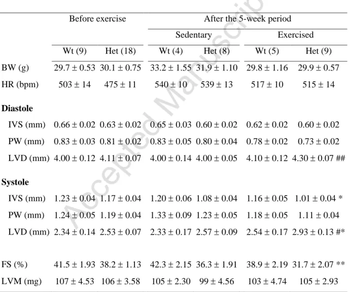
Table 2. Echocardiographic parameters of Wt and Het male mice before (17 weeks of age) and after 5 weeks of exercise protocol (22 weeks of age). (number of animals). BW:
body weight, IVSd: Interventricular septum thickness in diastole, PWd: Posterior wall thickness in diastole, LVEDD: Left ventricular end-diastolic diameter, IVSs: Interventricular septum thickness in systole, PWs: Posterior wall thickness in systole, LVESD: Left ventricular end-systolic diameter, LVM: Left ventricular mass, FS: Fractional shortening, HR: Heart rate. *p<0.05, **p<0.01 compared to Wt exercised mice, #p<0.05, ##p<0.01 compared to Het sedentary mice. ANOVA, Holm-Sidak test. Mean SEM.
Before exercise After the 5-week period Sedentary Exercised Wt (9) Het (18) Wt (4) Het (8) Wt (5) Het (9) BW (g) 29.7 0.53 30.1 0.75 33.2 1.55 31.9 1.10 29.8 1.16 29.9 0.57 HR (bpm) 503 14 475 11 540 10 539 13 517 10 515 14 Diastole IVS (mm) 0.66 0.02 0.63 0.02 0.65 0.03 0.60 0.02 0.62 0.02 0.60 0.02 PW (mm) 0.83 0.03 0.81 0.02 0.83 0.05 0.80 0.04 0.78 0.02 0.73 0.02 LVD (mm) 4.00 0.12 4.11 0.07 4.00 0.14 4.00 0.05 4.10 0.12 4.30 0.07 ## Systole IVS (mm) 1.23 0.04 1.17 0.04 1.20 0.06 1.08 0.04 1.16 0.05 1.01 0.04 * PW (mm) 1.24 0.05 1.19 0.04 1.33 0.09 1.23 0.05 1.18 0.05 1.11 0.04 LVD (mm) 2.34 0.14 2.53 0.07 2.33 0.17 2.57 0.09 2.54 0.17 2.93 0.13 #* FS (%) 41.5 1.93 38.2 1.13 42.3 2.15 36.3 1.91 38.9 2.19 31.7 2.07 ** LVM (mg) 107 4.53 106 3.58 105 2.30 99 4.56 103 4.74 105 2.93