HAL Id: inserm-00154872
https://www.hal.inserm.fr/inserm-00154872
Submitted on 15 Jun 2007
HAL is a multi-disciplinary open access
archive for the deposit and dissemination of
sci-entific research documents, whether they are
pub-lished or not. The documents may come from
teaching and research institutions in France or
abroad, or from public or private research centers.
L’archive ouverte pluridisciplinaire HAL, est
destinée au dépôt et à la diffusion de documents
scientifiques de niveau recherche, publiés ou non,
émanant des établissements d’enseignement et de
recherche français ou étrangers, des laboratoires
publics ou privés.
VGLUT1 and VGLUT2 in Parkinson disease.
Alireza Kashani, Catalina Betancur, Bruno Giros, Etienne Hirsch, Salah El
Mestikawy
To cite this version:
Alireza Kashani, Catalina Betancur, Bruno Giros, Etienne Hirsch, Salah El Mestikawy. Altered
ex-pression of vesicular glutamate transporters VGLUT1 and VGLUT2 in Parkinson disease.. Neurobiol
Aging, 2007, 28 (4), pp.568-78. �10.1016/j.neurobiolaging.2006.02.010�. �inserm-00154872�
Altered expression of vesicular glutamate transporters
VGLUT1 and VGLUT2 in Parkinson disease
Alireza Kashani
a, Catalina Betancur
a, Bruno Giros
a, Etienne Hirsch
b, Salah El Mestikawy
a,*
a INSERM U513, Faculté de Médecine, Université Paris XII, 8 rue du Général Sarrail, 94010 Créteil Cedex, France b INSERM U679, CHU Pitié-Salpêtrière, 47 boulevard de l’Hôpital, 75013 Paris, France
* Corresponding author. Tel: +33 1 49 81 36 06; fax: +33 1 49 81 36 85. E-mail address: salah.elmestikawy@im3.inserm.fr (S. El Mestikawy)
HAL author manuscript inserm-00154872, version 1
Abstract
Glutamatergic pathways play a key role in the functional organization of neuronal circuits involved in Parkinson disease (PD). Recently, three vesicular glutamate transporters (VGLUT1-3) were identified. VGLUT1 and VGLUT2 are responsible for the uploading of glutamate into synaptic vesicles and are the first specific markers of glutamatergic neurons available. Here, we analyzed the expression of VGLUT1 and VGLUT2 in autopsy tissues of PD patients and matched controls using Western blot and immunoautoradiography. VGLUT1 and VGLUT2 expression was increased in the parkinsonian putamen by 24% and 29%, respectively (p<0.01). In contrast, only VGLUT1 was dramatically decreased in the prefrontal and temporal cortex of PD patients (~50%, p<0.01 and p<0.001 respectively). These findings demonstrate the existence of profound alterations of glutamatergic transmission in PD, which are likely to contribute to the motor and cognitive impairments associated with the disease, and should thus be taken into account in the treatment of PD.
Keywords: Human, Parkinson disease; Vesicular glutamate transporter; VGLUT1; VGLUT2; Prefrontal cortex; Temporal cortex; Putamen; Western blot; Immunoautoradiography
1. Introduction
Glutamate is the primary excitatory neurotransmitter in the mammalian central nervous system (CNS). This amino acid, used by more than 40% of neurons, is involved in all the physiological functions of the brain. Research on glutamatergic neurons was hindered until recently by the lack of specific markers. Recently, three subtypes of vesicular glutamate transporters (VGLUT1-3) have been identified [21]. These proteins share similar structural and pharmacological properties and are responsible for the uploading of glutamate into pre-synaptic vesicles [5,23,29,52-54]. VGLUT1 and VGLUT2 are specific markers of canonical glutamatergic neurons [5,23,29,54] and show a distinct, complementary distribution in the CNS [23,29]. Neurons expressing VGLUT1 are found in the cerebral and cerebellar cortices as well as in the hippocampus and thalamus [6,29,43], while VGLUT2 is utilized by a majority of subcortical excitatory neurons [23,29,43,54]. Although VGLUT1 and VGLUT2 transcripts are not expressed by striatal neurons, the striatum receives two major, topographically organized, glutamatergic afferents: the cortico-striatal pathway, which is VGLUT1-positive and makes contact predominantly on the head of dendritic spines of GABAergic medium spiny neurons [29,33,34,50]; and the thalamo-striatal pathway, a VGLUT2-positive excitatory input that originates in the intralaminar and ventral motor nuclei and terminates on the dendritic shaft of medium-size projecting neurons [50,51].
Parkinson disease (PD) is classically characterized as a disorder resulting from the degeneration of dopaminergic neurons in the pars compacta of the substantia nigra. However, glutamatergic pathways play a leading role in the structural and functional organization of the cortico-baso-cortical loops involved in PD [3,30]. Dysregulations of glutamatergic systems have been suggested by studies of animal models of PD [9,16,36,37]. Furthermore, in patients with PD, histological studies have shown
that the total number of glutamatergic neurons is reduced by 50% in the intralaminar thalamic nuclei [26,27]. In addition, using morphological criteria, an 88% increase in glutamatergic perforated synapses was reported in the putamen of PD patients [4]. However, the impact of PD on glutamatergic pathways has never been monitored directly, due to the lack of specific markers of glutamatergic neurons. The recent identification of VGLUT1 and VGLUT2 allows for the first time the direct assessment of glutamatergic systems in PD. In this report, we studied the distribution of VGLUT1 and VGLUT2 in the basal ganglia, prefrontal and temporal cortices, and centromedian thalamic nucleus in post-mortem tissue of PD patients and control subjects.
2. Methods
2.1. Human brain samples
The brain samples used for this study belonged to 23 controls free of neurological disorders and 20 patients with confirmed clinical and post-mortem diagnosis of PD (Table 1). Brain aliquots and sections were obtained from the Brain Bank of INSERM U679 (Pitié-Salpêtrière Hospital, Paris, France). Four regions were analyzed using Western blot: dorsolateral prefrontal cortex (Brodmann area A9), temporal cortex (anterior part of the first temporal gyrus), putamen and centromedian nucleus of the thalamus. For each brain region studied, the entire region represented within a 1 cm-thick section was dissected at –20°C taking care not to include the white matter. The dissected material was crushed and homogenized to a powder using a frozen hammer and stored at –80°C until used. In addition, the expression of VGLUT1 and VGLUT2 was studied with immunoautoradiography on fresh frozen 10 µm sections (3 sections per subject) at the level of the putamen, caudate nucleus, and internal and external parts of the globus pallidus.
2.2. Antiserums
Selective anti-VGLUT1 and anti-VGLUT2 antiserums were obtained by immunization of rabbits with the corresponding peptides, as already described [29]. The anti-VGLUT1 antiserum was obtained by immunizing rabbits against the peptide CGLAPSYGATHSTVQPPR and the antiserum directed against VGLUT2 was obtained after immunization with the peptide HEDELDEETGDITQNYINY. These epitopes were selected from regions with minimal sequence similarity between VGLUT1 and VGLUT2. Accordingly, we have never observed cross-reactivity between the anti-VGLUT1 and VGLUT2 antiserums. Moreover, the subtype specificity of each antiserum has been assessed previously in BON cells stably expressing each subtype and in brain extracts by Western blotting [29].
The anti-VGLUT2 antiserum was affinity purified [25]. Other antiserums were from the best available commercial sources. For Western blotting, antiserums were used at the following concentrations: anti-VGLUT1 1:8000; anti-VGLUT2 1:500; anti-synaptophysin 1:50000 (mouse monoclonal; Chemicon, Temecula, CA); and anti-α-tubulin 1:20000 (mouse monoclonal; Sigma). For immunoautoradiography, both the anti-VGLUT1 and anti-VGLUT2 antiserums were used at 1:2000.
2.3. Western blots
Frozen powdered tissue homogenates (~20 mg) were homogenized by sonication in PBS containing protease inhibitors (Complete, Roche, France) and the total extract was used for Western blot experiments. Protein concentration was measured with the Bio-Rad Protein Assay Kit (Bio-Rad, France). The quantity of protein in post-mortem extracts from control and Parkinsonian tissues was not significantly different.
Equal concentrations of protein (5 µg per lane) were separated by SDS-PAGE (NuPage Bis-Tris 10%, InVitrogen
,
France) and transferred to a nitrocellulose membrane (0.4 µm pore size, InVitrogen). Protein loading was controlled by ponceau staining. Non-specific sites on nitrocellulose membranes were blocked for 1 h at room temperature with either: i) PBS containing Tween 20 (0.1%), and 5% nonfat dry milk for VGLUT1, synaptophysin and α-tubulin detection, or ii) PBS containing Tween 20 (0.5%), 5% nonfat dry milk and 5% bovine serum albumin for VGLUT2. The membranes were incubated overnight with primary antibodies at 4°C in PBS containing Tween 20 (0.1%), and 1% nonfat dry milk for the labeling of VGLUT1, synaptophysin and α-tubulin, or in PBS containing Tween 20 (0.5%), 1% nonfat dry milk and 1% bovine serum albumin for VGLUT2. Bound antibodies were detected with horseradish peroxidase-conjugated anti-rabbit or anti-mouse IgG antibodies (Sigma; 1:20000) and visualized by enhanced chemiluminescent detection (ECL plus Western Blotting detection system, Amersham Biosciences).2.4. Immunoautoradiography
Immunoautoradiographic labeling was performed as already described [28]. Briefly, on the day of the experiment sections were air dried and immersed in paraformaldehyde (4%) in PBS. Non-specific binding was saturated with PBS containing bovine serum albumin (3%), goat serum (1%) and NaI 1 mM (buffer A). Sections were incubated overnight at 4°C with buffer A supplemented with anti-VGLUT1 or –VGLUT2 serums and then with anti-rabbit [125I]IgG (0.25 µci/ml; Amersham). After rinsing and drying, sections were exposed to X-ray films (Biomax, Kodak) for 4-7 days.
2.5. Image analysis
Western blots and immunoautoradiograms were digitalized as 16-bit images (65,536 gray levels) using an Umax PowerLook 1100 scanner (Willich, Germany). The computerized images were transferred to the software MCID Image (Imaging Research, St. Catharines, ON, Canada) and optical density (O.D.) was measured. Film exposures were selected in order to maintain the grey levels of the tissue within the dynamic range of the film. In addition, MCID has a special function to verify if any part of the quantified region is saturated. Regions of interest were determined as delineated by the doted lines on Figure 1 and the density of the entire area was measured. The background density was determined at the level of the internal capsule for immunoautoradiograms and on the film for Western blots. This “zero” value was automatically subtracted from the selected area or band by MCID. In Western blot experiments, the density of various bands was normalized to that obtained for the α-tubulin band in the same sample. Exposure time of the film after the ECL reaction was selected in order to avoid saturation of the signal. Results are expressed as means of optical density (in arbitrary units) ± SEM.
2.6. Statistical analysis
Comparison between group means was performed with the Student’s t test. The Pearson’s coefficient was used for correlation analyses between protein levels and age, post-mortem delay, duration of treatment with L-Dopa and disease duration. Statistical analyses were performed with Statview 3.0 software (San Diego, CA).
3. Results
3.1. Characteristics of patients and brain samples
The demographic and clinical characteristics of patients and controls are shown in Table 1. The levels of VGLUT1 and VGLUT2 were compared by Western blot in postmortem samples of controls and PD subjects in frontal and temporal cortices, putamen and centromedian nucleus of thalamus. These tissues originated from 15 patients with PD (9 males, 6 females), aged 52 to 83 years old (mean age: 70.2 ± 2.4 years; mean disease duration: 11.1 ± 1.6 years and mean L-dopa treatment duration 6.9 ± 1.2 years) and 18 control subjects (10 males, 8 females), aged 45 to 91 years old (75.2 ± 2.7 years). The post-mortem delay ranged from 3 to 24 h (10.2 ± 2.0 h) in PD patients and from 2.2 to 25 h (10.2 ± 1.7 h) in controls.
The two glutamate vesicular transporters were also visualized on sections taken at the level of the dorsal putamen. These sections were obtained from the brains of five patients (4 males, 1 female) aged 66 to 78 years old (mean age: 71.0 ± 2.3 years) and five controls (2 males, 3 females) aged 57 to 94 years old (mean age: 76.6 ± 6.5 years).
The disease duration in the entire patient population ranged from 2 to 25 years. All patients except three were treated with L-dopa (mean treatment duration: 7.3 ± 1.1 years; Table 1). As shown in Table 2, L-dopa therapy duration, disease duration, age, gender and post-mortem delay did not appear to have a significant effect on the levels of VGLUT1, VGLUT2, synaptophysin and α−tubulin detected by Western blot in our population.
3.2. Specificity of the antiserums
As shown in Figure 1a, one major broad band of the expected molecular weight (50-60 kDa) and a smear are visible in Western blots of rat total brain extracts with the anti-VGLUT1 antiserum (lane 1). The major band corresponds to specific labeling, since the same 50-60 kDa protein is observed with a commercially available anti-VGLUT1 antiserum directed against a different epitope (Chemicon, Fig. 1a, lane 2). The equivalent protein is also visualized in an extract of control human putamen (Fig. 1a, lane 3), and is no longer detected when the antiserum has been exhausted with its corresponding antigen (Fig. 1a, lane 4). In total rat brain samples, our immunopurified anti-VGLUT2 antiserum recognizes two bands (Fig. 1a, lane 5): a large prominent band of 55-60 kDa and a narrower one at 62 kDa. Only the 55-60 kDa protein is present on Western blots probed with the Chemicon anti-VGLUT2 antiserum (Fig 1a, lane 6). This band is also detected with our immunopurified anti-VGLUT2 antiserum in human striatal extracts (Fig. 1a, lane 7). As expected for a specific labeling, this band disappears when the serum has been exhausted with its antigen (Fig. 1a, lane 8). Thus, the major bands
visualized in lanes 3 and 7 correspond to human VGLUT1 and VGLUT2. The same pattern is observed in all the regions studied (see also Fig. 2 and 3).
The regional distribution of VGLUT1 and VGLUT2 was visualized by immunoautoradiography on human brain sections (Fig. 1b and c). Both transporters are abundant in the cerebral cortex, caudate nucleus and putamen (Fig. 1b and c). Interestingly, only VGLUT2 is observed at a moderate level in the globus pallidus; the expression is higher in the internal than in the external segment (Fig. 1c). No labeling is observed in the white matter or when the antiserums have been pre-incubated with their cognate antigen (Fig. 1d and e). Taken together, these results indicate that there is no cross reactivity between VGLUT1 and VGLUT2 and that each labeling is specific.
3.3. Expression of VGLUT1 and VGLUT2 in the basal ganglia of PD patients
In the basal ganglia, VGLUT1 and VGLUT2 expression was analyzed by Western blot and immunoautoradiography (Fig. 2). Representative Western blots are shown in Figure 2a. In these samples, both VGLUT1 and VGLUT2 appear to be more abundant in the putamen of PD patients than in controls. In contrast, α-tubulin, an index of cellular integrity, was unchanged (Fig. 2a). Synaptophysin has often been used as an index of the integrity of synaptic terminals. As already reported by others [24,41], we observed no modification of synaptophysin labeling in the putamen of PD patients (Fig. 2a).
The increase in VGLUT1 and VGLUT2 expression was also visualized by immunoautoradiography (Fig. 2b). As can be seen in Figure 2b, VGLUT1 and VGLUT2 levels are increased in the caudate and the putamen of a patient with PD.
Quantification of the Western blot and immunoautoradiographic experiments is shown in Figure 2c. Statistical analyses of these data are summarized in Table 3. Neither synaptophysin nor α-tubulin were significantly altered (not shown). Similarly, the disease did not modify significantly VGLUT1 and VGLUT2 expression in the caudate nucleus, or VGLUT2 in the globus pallidus (internal and external segments, Table 3). However, in the putamen, VGLUT1 and VGLUT2 levels detected by Western blot were significantly increased by 24% and 29%, respectively (p<0.01 in both cases, Fig. 2c and Table 3). Despite the higher resolution of immunoautoradiography, the variability of the measurements is somewhat increased, as can be judged from the dispersion of the values and the increased size of the error bars in Figure 2c. Nonetheless, in all the regions examined, the changes observed by immunoautoradiography are compatible with those observed by Western blotting (Fig. 2c). In particular, VGLUT2 expression, measured by immunoautoradiography in the putamen, was increased by 21% in PD subjects (p<0.04). The level of VGLUT1 was also increased by 25%, but the difference did not reach statistical significance.
3.4. Expression of VGLUT1 and VGLUT2 in the cortex and thalamus of PD patients
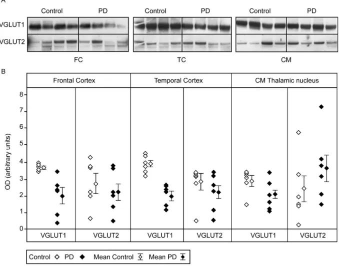
In the prefrontal and temporal cortex the levels of VGLUT1 and VGLUT2 were measured by Western blotting (Fig. 3a). Representative immunoblots are shown in Figure 3a. The blots from six controls and six patients were quantified and the data are summarized in Figure 3b and Table 3. In these areas, the levels of synaptophysin and α-tubulin were not significantly modified by the pathology (not shown). In contrast, a strong and significant reduction of VGLUT1 was observed in the prefrontal
cortex (-45%, p<0.01) and temporal cortex (-47%, p<0.001) of PD patients (Fig. 3b and Table 3). Interestingly, the dispersion of the values of VGLUT1 expression was markedly increased in the PD prefrontal cortex (Fig. 3b). One patient appears to express VGLUT1 levels similar to those observed in controls, whereas all the remaining samples are significantly lower than controls.
VGLUT2 expression was decreased by 20% and 22% in the prefrontal and temporal cortices of PD patients when compared to control subjects, but these differences were not statistically significant (Fig. 3b and Table 3). Finally, no significant changes were observed in VGLUT1, VGLUT2, synaptophysin or α-tubulin levels in the centro-median nucleus of the thalamus in PD (Fig.3b and Table 3).
4. Discussion
Using VGLUT1 and VGLUT2 as markers, we determined for the first time the distribution of glutamatergic terminals in the human caudate-putamen, globus pallidus, thalamus, prefrontal and temporal cortices. As observed in rodents, high levels of both transporters are found in the caudate-putamen, whereas only VGLUT2 is expressed in the globus pallidus complex [29,54]. Thus, selection pressure has conserved the anatomical distribution of both VGLUTs in humans and rodents. Using post-mortem tissues from control subjects and patients with PD, our results showed that PD induces striking quantitative changes in the expression of the two major subtypes of vesicular glutamate transporters. We observed a significant increase in both VGLUT1 and VGLUT2 in the putamen (+24% and +29%), whereas VGLUT1 was severely down-regulated in prefrontal and temporal cortical terminals (-45% and -47%). Since the patient and control populations were well matched for age and post-mortem delay, the differences between the two groups cannot be attributed to age or postmortem degradation. Notably, the changes in VGLUT1 and VGLUT2 expression were not correlated with disease duration or age, suggesting that they may already be present at early stages of the disease. However, due to the limited number of brain samples in each group, this interpretation will require confirmation in larger populations. Whether these changes in VGLUT1 and VGLUT2 expression are specific to PD is still an open question. To clarify this point, the status of VGLUT1 and VGLUT2 in other neurodegenerative diseases, such as progressive supranuclear palsy, cortical-basal ganglionic degeneration or Alzheimer disease, as well as in other areas of the parkinsonian brain, will have to be investigated.
4.1. PD increases the expression of VGLUT1 and VGLUT2 in the putamen but not in the globus pallidus
In the rat putamen, VGLUT2 is preferentially found within thalamo-striatal projections [33,34]. Our results show a 30% increase in VGLUT2 in the putamen of PD patients. This increase might appear paradoxical in light of previous studies showing a 40%-50% neuronal loss in the centromedian thalamic nucleus of PD subjects [26,27]. However, centromedian glutamatergic neurons projecting to the putamen are parvalbumin-negative and these neurons seem to be relatively spared by PD [48,49]. Moreover, the parafascicular/centromedian complex is hyperactive in 6-hydroxydopamine (6-OHDA) treated rats [45]. Together with our findings, these data suggest that VGLUT2 expression is increased in surviving centromedian thalamic glutamatergic neurons in PD.
Numerous studies in humans and animal models have reported that nigro-striatal dopaminergic depletion over-activates the cortico-striatal pathway [7,8]. In experimental models of PD, dopamine deficiency in the putamen induced by 6-OHDA lesion of the nigrostriatal tract, leads to increased release of glutamate in the striatum [39], increased spontaneous electrical activity of striatal neurons [7], and an increase in the number of asymmetrical synapses associated with a perforated postsynaptic density [42]. In individuals with PD, imaging studies have demonstrated a relative hypermetabolism in the putamen [18,32]. Furthermore, an hyperactivation of striatal glutamatergic terminals was also documented by morphological criteria and electron microscopy. Indeed, an 88% increase in the number of cortico-striatal perforated synapses was reported in the putamen of patients with PD [4]. Given that previous studies in rats have shown that the glutamatergic cortico-striatal pathway is VGLUT1-positive [29,33,34], these data are in agreement with our results showing increased VGLUT1 expression in the putamen of patients with PD. The activation of the cortico-striatal pathway can be the result of at least three mechanisms: i) an increased firing rate of neurons, ii) an increased number of glutamatergic terminals, and/or iii) an increase in the quantal size of glutamate release. The increased amplitude and frequency of the spontaneous glutamate-induced depolarization of medium spiny neurons in the dopamine-denervated rat striatum [7,10] are in favor of the first hypothesis. Moreover, we observed no change in synaptophysin labeling, as already reported in at least two other independent studies [24,41], suggesting that in the putamen, the total number of synapses is not altered by PD. Thus, the increase in VGLUT1 demonstrated herein could tentatively be related to an increased concentration of VGLUT1 per terminal. Further studies will be needed in order to assess the second and third hypothesis.
It is well established that PD results in an increased electrical activity of the subthalamic nucleus (STN) [30]. This STN stimulation leads in turn to the hyperactivation of its glutamatergic projection to the internal and external segments of the globus pallidus (GPi and GPe). Surprisingly, no significant changes of VGLUT2 expression were observed in the GPi and GPe in individuals with PD. Further investigations of the impact of PD on VGLUT2 expression in basal ganglia will be needed to clarify this issue.
VGLUT3, the third subtype of vesicular glutamate transporter, is expressed in cholinergic interneurons in the striatum [25]. In PD, the dopaminergic deafferentation of the caudate-putamen results in hypersensitivity of these cholinergic neurons, and anticholinergic agents have been successfully employed in the management of PD. Despite its relevance in PD, we have been unable to determine VGLUT3 levels in the human putamen and caudate nucleus because the various anti-VGLUT3 antiserums that were tested during the course of the present study were not able to recognize the human ortholog. The determination of VGLUT3 expression in the human caudate-putamen in PD is a key challenge for the near future.
4.2. PD decreases the expression of VGLUT1 in the prefrontal and temporal cortex
The major finding of the present study is the demonstration of a nearly 50% reduction in VGLUT1 in the prefrontal and temporal cortices of PD patients. Our results are indirectly supported by brain imaging studies showing decreased regional cerebral blood flow and metabolic rate in the prefrontal and temporal cortices of patients with PD [13,17,31]. This hypoactivity of subregions of the cerebral
cortex has been hypothesized to ultimately lead to rigidity and akinesia as well as subtle cognitive dysfunction and/or dementia in individuals with PD [3,14]. Although the expression of VGLUT2 in the cortex also appeared to be decreased (-20%), the effect was not statistically significant. These findings suggest a more severe alteration of VGLUT1-positive cortical interneurons compared with VGLUT2-positive thalamo-cortical neurons.
The mechanisms leading to a cortical decrease in VGLUT1 in PD could involve: i) loss of glutamatergic neurons, ii) loss of glutamatergic terminals, and iii) hypo-activity of glutamatergic neurons. Neuronal loss and the presence of Lewy bodies have been reported in the neocortex of PD patients at late phases of the disease [35]. In addition, an inhibition of the thalamo-cortical glutamatergic pathway has been proposed in PD [3,14], which could ultimately lead to the adaptive changes of VGLUT1 expression in cortical glutamatergic interneurons described here. We did not observe changes in synaptophysin levels in the prefrontal and temporal cortices, suggesting that there is no loss of synapses in these areas. Our results suggest that the dramatic cortical decrease in VGLUT1 could be related to reduced concentration of vesicular transporter per terminal rather than to a change in the absolute number of glutamatergic nerve endings. However, this hypothesis needs to be verified and extended by future immunohistochemistry and electron microscopy studies, with direct assessment of the number of synapses in PD patients.
Notably, our data indicate that cortical glutamatergic interneurons and efferent pyramidal cells from the cerebral cortex are differentially affected by PD. In the first case, VGLUT1 expression in terminals of cortico-cortical neurons is severely down-regulated. In contrast, in the cortico-striatal pathway, which seems to be hyperactive in PD, VGLUT1 expression in putamen nerve endings is enhanced. Thus, fine and apparently opposite regulations of discrete glutamatergic neuronal subpopulations take place in the cortex of patients with PD.
4.3. Clinical implications
What can be the consequences of altered VGLUT1 expression in PD? Several recent studies suggest that both the strength of glutamatergic neurotransmission and the quantal size are proportional to the expression level of VGLUT1 [12,55,56]. Thus, it can reasonably be surmised that the changes in the concentration of VGLUT1 in PD result in parallel changes in glutamatergic transmission. Specifically, the strength of glutamatergic transmission is likely to be reduced in the frontal and temporal cortex of patients with PD.
VGLUT1, VGLUT2 and VGLUT3 delineate three distinct glutamatergic systems in the CNS; VGLUT1 is the major excitatory system in the brain. Indeed, 80% of vesicular glutamate total uptake is mediated by VGLUT1 [22]. Thus, a 50% decrease in VGLUT1 in the prefrontal and temporal cortices of PD subjects must result in a major down-regulation of glutamatergic transmission in the fronto-temporal cortex. This decrease in VGLUT1 expression does not seem to be correlated with the age of the patient or the duration of the disease, suggesting that it could be already present during the early phases of the disease. PD patients have difficulties in the planning and anticipatory aspects of voluntary movement during the early stages of the disease. Moreover, severe deficits in visuospatial functions have been reported early in PD [19]. Patients also exhibit deficits in executive functions, and are particularly impaired in tasks such as the Wisconsin card sorting [38] and the Tower of London
[46]. Furthermore, dementia is common among aged patients with PD, with an average prevalence of 40% in cross-sectional studies [11] and a cumulative prevalence approaching 80% [1]. These cognitive deficits have been tentatively attributed to dopaminergic, noradrenergic, serotononinergic and cholinergic dysfunctions [44]. Based on our results, we suggest that the cognitive/motor deficits of PD patients could also be related to the massive glutamatergic deficit in the prefronto-temporal cortex. The clinical data of patients and controls included in this study were obtained retrospectively in order to warrant a good anatomo-clinical correlation, so no information was available concerning their level of cognitive impairment and further investigations will be necessary to confirm this hypothesis.
Current pharmacological treatments of PD focus on dopamine-replacement strategies. However, clinical responses to dopaminergic medications are incomplete and long-term therapy with L-dopa is associated with intractable adverse effects, such as extrapyramidal disorders [2]. It is thus necessary to explore new approaches for the treatment of this disease. Our study indicates that the glutamatergic systems could be prime pharmacological targets for the treatment of PD. Initial attempts, performed in animal models, have suggested that motor alterations in PD could benefit from treatment with glutamate antagonists [20,40,47]. Our results suggest that direct or indirect stimulation of glutamatergic transmission in the cerebral cortex could help to alleviate the cognitive and motor impairments observed in elderly patients with PD. For instance, high frequency electrical stimulation of the prefrontal cortex could be beneficial for the treatment of cognitive impairment in PD. This therapeutical strategy has been utilized recently to alleviate motor parkinsonian symptoms in a nonhuman primate model [15].
4.4. Conclusion
The studies reported here show specific regional effects on the expression of the two major vesicular glutamate transporters in PD, with a decrease in VGLUT1 in the prefrontal and temporal cortices and an increase in VGLUT1 and VGLUT2 in the putamen. We argue that these glutamatergic dysfunctions, and the decrease in glutamatergic transmission in the cerebral cortex in particular, should be taken into account in the treatment of the motor and cognitive deficits of PD patients. Our findings support a fundamental role for the glutamatergic alterations in PD symptoms and provide new insights into the molecular neuropathological changes that contribute to PD.
Acknowledgments
This work was supported by Institut National de la Santé et de la Recherche Médicale (INSERM) and by grants from Association France Parkinson and Fédération pour la Recherche sur le Cerveau. AK was supported by a fellowship from Association France Alzheimer.
References
[1] Aarsland D, Andersen K, Larsen JP, Lolk A, Kragh-Sorensen P. Prevalence and characteristics of dementia in Parkinson disease: an 8-year prospective study. Arch Neurol 2003;60:387-92.
[2] Ahlskog JE, Muenter MD. Frequency of levodopa-related dyskinesias and motor fluctuations as estimated from the cumulative literature. Mov Disord 2001;16:448-58.
[3] Albin RL, Young AB, Penney JB. The functional anatomy of basal ganglia disorders. Trends Neurosci 1989;12:366-75.
[4] Anglade P, Mouatt-Prigent A, Agid Y, Hirsch E. Synaptic plasticity in the caudate nucleus of patients with Parkinson's disease. Neurodegeneration 1996;5:121-8.
[5] Bellocchio EE, Reimer RJ, Fremeau RT Jr, Edwards RH. Uptake of glutamate into synaptic vesicles by an inorganic phosphate transporter. Science 2000;289:957-60.
[6] Bellocchio EE, Hu H, Pohorille A, Chan J, Pickel VM, Edwards RH. The localization of the brain-specific inorganic phosphate transporter suggests a specific presynaptic role in glutamatergic transmission. J Neurosci 1998;18:8648-59.
[7] Calabresi P, Mercuri NB, Sancesario G, Bernardi G. Electrophysiology of dopamine-denervated striatal neurons. Implications for Parkinson's disease. Brain 1993;116:433-52.
[8] Calabresi P, Pisani A, Mercuri NB, Bernardi G. The corticostriatal projection: from synaptic plasticity to dysfunctions of the basal ganglia. Trends Neurosci 1996;19:19-24.
[9] Carlsson M, Carlsson A. The NMDA antagonist MK-801 causes marked locomotor stimulation in monoamine-depleted mice. J Neural Transm 1989;75:221-6.
[10] Centonze D, Calabresi P, Giacomini P, Bernardi G. Neurophysiology of Parkinson's disease: from basic research to clinical correlates. Clin Neurophysiol 1999;110:2006-13.
[11] Cummings JL. Intellectual impairment in Parkinson's disease: clinical, pathologic, and biochemical correlates. J Geriatr Psychiatry Neurol 1988;1:24-36.
[12] Daniels RW, Collins CA, Gelfand MV, Dant J, Brooks ES, Krantz DE, DiAntonio A. Increased expression of the drosophila vesicular glutamate transporter leads to excess glutamate release and a compensatory decrease in quantal content. J Neurosci 2004;24:10466-74.
[13] Defebvre L, Lecouffe P, Destee A, Houdart P, Steinling M. Tomographic measurements of regional cerebral blood flow in progressive supranuclear palsy and Parkinson's disease. Acta Neurol Scand 1995;92:235-41. [14] DeLong MR. Primate models of movement disorders of basal ganglia origin. Trends Neurosci
1990;13:281-5.
[15] Drouot X, Oshino S, Jarraya B, Besret L, Kishima H, Remy P, Dauguet J, Lefaucheur JP, Dolle F, Conde F, Bottlaender M, Peschanski M, Keravel Y, Hantraye P, Palfi S. Functional Recovery in a Primate Model of Parkinson's Disease following Motor Cortex Stimulation. Neuron 2004;44:769-78.
[16] Dunah AW, Wang Y, Yasuda RP, Kameyama K, Huganir RL, Wolfe BB, Standaert DG. Alterations in subunit expression, composition, and phosphorylation of striatal N-methyl-D-aspartate glutamate receptors in a rat 6-hydroxydopamine model of Parkinson's disease. Mol Pharmacol 2000;57:342-52.
[17] Eberling JL, Richardson BC, Reed BR, Wolfe N, Jagust WJ. Cortical glucose metabolism in Parkinson's disease without dementia. Neurobiol Aging 1994;15:329-35.
[18] Eidelberg D, Moeller JR, Dhawan V, Spetsieris P, Takikawa S, Ishikawa T, Chaly T, Robeson W, Margouleff D, Przedborski S, Fahn S. The metabolic topography of parkinsonism. J Cereb Blood Flow Metab 1994;14:783-801.
[19] Emre M. What causes mental dysfunction in Parkinson's disease? Mov Disord 2003;18 Suppl 6:S63-71. [20] Engber TM, Papa SM, Boldry RC, Chase TN. NMDA receptor blockade reverses motor response alterations
induced by levodopa. Neuroreport 1994;5:2586-8.
[21] Fremeau RT Jr, Voglmaier S, Seal RP, Edwards RH. VGLUTs define subsets of excitatory neurons and suggest novel roles for glutamate. Trends Neurosci 2004;27:98-103.
[22] Fremeau RT Jr, Kam K, Qureshi T, Johnson J, Copenhagen DR, Storm-Mathisen J, Chaudhry FA, Nicoll RA, Edwards RH. Vesicular glutamate transporters 1 and 2 target to functionally distinct synaptic release sites. Science 2004;304:1815-9.
[23] Fremeau RT Jr, Troyer MD, Pahner I, Nygaard GO, Tran CH, Reimer RJ, Bellocchio EE, Fortin D, Storm-Mathisen J, Edwards RH. The expression of vesicular glutamate transporters defines two classes of excitatory synapse. Neuron 2001;31:247-60.
[24] Girault JA, Raisman-Vozari R, Agid Y, Greengard P. Striatal phosphoproteins in Parkinson disease and progressive supranuclear palsy. Proc Natl Acad Sci U S A 1989;86:2493-7.
[25] Gras C, Herzog E, Bellenchi GC, Bernard V, Ravassard P, Pohl M, Gasnier B, Giros B, El Mestikawy S. A third vesicular glutamate transporter expressed by cholinergic and serotoninergic neurons. J Neurosci 2002;22:5442-51.
[26] Henderson JM, Carpenter K, Cartwright H, Halliday GM. Loss of thalamic intralaminar nuclei in progressive supranuclear palsy and Parkinson's disease: clinical and therapeutic implications. Brain 2000;123:1410-21. [27] Henderson JM, Carpenter K, Cartwright H, Halliday GM. Degeneration of the centre median-parafascicular
complex in Parkinson's disease. Ann Neurol 2000;47:345-52.
[28] Herzog E, Gilchrist J, Gras C, Muzerelle A, Ravassard P, Giros B, Gaspar P, El Mestikawy S. Localization of VGLUT3, the vesicular glutamate transporter type 3, in the rat brain. Neuroscience 2004;123:983-1002. [29] Herzog E, Bellenchi GC, Gras C, Bernard V, Ravassard P, Bedet C, Gasnier B, Giros B, El Mestikawy S.
The existence of a second vesicular glutamate transporter specifies subpopulations of glutamatergic neurons. J Neurosci 2001;21:RC181.
[30] Hirsch EC, Perier C, Orieux G, Francois C, Feger J, Yelnik J, Vila M, Levy R, Tolosa ES, Marin C, Trinidad Herrero M, Obeso JA, Agid Y. Metabolic effects of nigrostriatal denervation in basal ganglia. Trends Neurosci 2000;23:S78-85.
[31] Hu MT, Taylor-Robinson SD, Chaudhuri KR, Bell JD, Labbe C, Cunningham VJ, Koepp MJ, Hammers A, Morris RG, Turjanski N, Brooks DJ. Cortical dysfunction in non-demented Parkinson's disease patients: a combined (31)P-MRS and (18)FDG-PET study. Brain 2000;123:340-52.
[32] Imon Y, Matsuda H, Ogawa M, Kogure D, Sunohara N. SPECT image analysis using statistical parametric mapping in patients with Parkinson's disease. J Nucl Med 1999;40:1583-9.
[33] Kaneko T, Fujiyama F. Complementary distribution of vesicular glutamate transporters in the central nervous system. Neurosci Res 2002;42:243-50.
[34] Kaneko T, Fujiyama F, Hioki H. Immunohistochemical localization of candidates for vesicular glutamate transporters in the rat brain. J Comp Neurol 2002;444:39-62.
[35] Klockgether T. Parkinson's disease: clinical aspects. Cell Tissue Res 2004;318:115-20.
[36] Klockgether T, Turski L. NMDA antagonists potentiate antiparkinsonian actions of L-dopa in monoamine-depleted rats. Ann Neurol 1990;28:539-46.
[37] Lange KW, Kornhuber J, Riederer P. Dopamine/glutamate interactions in Parkinson's disease. Neurosci Biobehav Rev 1997;21:393-400.
[38] Lees AJ, Smith E. Cognitive deficits in the early stages of Parkinson's disease. Brain 1983;106:257-70. [39] Lindefors N, Ungerstedt U. Bilateral regulation of glutamate tissue and extracellular levels in
caudate-putamen by midbrain dopamine neurons. Neurosci Lett 1990;115:248-52.
[40] Loschmann PA, Lange KW, Kunow M, Rettig KJ, Jahnig P, Honore T, Turski L, Wachtel H, Jenner P, Marsden CD. Synergism of the AMPA-antagonist NBQX and the NMDA-antagonist CPP with L-dopa in models of Parkinson's disease. J Neural Transm Park Dis Dement Sect 1991;3:203-13.
[41] Martin-Ruiz CM, Piggott M, Gotti C, Lindstrom J, Mendelow AD, Siddique MS, Perry RH, Perry EK, Court JA. Alpha and beta nicotinic acetylcholine receptors subunits and synaptophysin in putamen from Parkinson's disease. Neuropharmacology 2000;39:2830-9.
[42] Meshul CK, Cogen JP, Cheng HW, Moore C, Krentz L, McNeill TH. Alterations in rat striatal glutamate synapses following a lesion of the cortico- and/or nigrostriatal pathway. Exp Neurol 2000;165:191-206. [43] Ni B, Wu X, Yan GM, Wang J, Paul SM. Regional expression and cellular localization of the
Na(+)-dependent inorganic phosphate cotransporter of rat brain. J Neurosci 1995;15:5789-99.
[44] Nieoullon A. Dopamine and the regulation of cognition and attention. Prog Neurobiol 2002;67:53-83.
[45] Orieux G, Francois C, Feger J, Yelnik J, Vila M, Ruberg M, Agid Y, Hirsch EC. Metabolic activity of excitatory parafascicular and pedunculopontine inputs to the subthalamic nucleus in a rat model of Parkinson's disease. Neuroscience 2000;97:79-88.
[46] Owen AM, James M, Leigh PN, Summers BA, Marsden CD, Quinn NP, Lange KW, Robbins TW. Fronto-striatal cognitive deficits at different stages of Parkinson's disease. Brain 1992;115 (Pt 6):1727-51.
[47] Papa SM, Chase TN. Levodopa-induced dyskinesias improved by a glutamate antagonist in Parkinsonian monkeys. Ann Neurol 1996;39:574-8.
[48] Sadikot AF, Parent A, Francois C. The centre median and parafascicular thalamic nuclei project respectively to the sensorimotor and associative-limbic striatal territories in the squirrel monkey. Brain Res 1990;510:161-5.
[49] Sidibe M, Smith Y. Thalamic inputs to striatal interneurons in monkeys: synaptic organization and co-localization of calcium binding proteins. Neuroscience 1999;89:1189-208.
[50] Smith AD, Bolam JP. The neural network of the basal ganglia as revealed by the study of synaptic connections of identified neurones. Trends Neurosci 1990;13:259-65.
[51] Smith Y, Raju DV, Pare JF, Sidibe M. The thalamostriatal system: a highly specific network of the basal ganglia circuitry. Trends Neurosci 2004;27:520-7.
[52] Takamori S, Rhee JS, Rosenmund C, Jahn R. Identification of a vesicular glutamate transporter that defines a glutamatergic phenotype in neurons. Nature 2000;407:189-94.
[53] Takamori S, Rhee JS, Rosenmund C, Jahn R. Identification of differentiation-associated brain-specific phosphate transporter as a second vesicular glutamate transporter (VGLUT2). J Neurosci 2001;21:RC182. [54] Varoqui H, Schafer MK, Zhu H, Weihe E, Erickson JD. Identification of the differentiation-associated Na+/PI
transporter as a novel vesicular glutamate transporter expressed in a distinct set of glutamatergic synapses. J Neurosci 2002;22:142-55.
[55] Wilson NR, Kang J, Hueske EV, Leung T, Varoqui H, Murnick JG, Erickson JD, Liu G. Presynaptic regulation of quantal size by the vesicular glutamate transporter VGLUT1. J Neurosci 2005;25:6221-34.
[56] Wojcik SM, Rhee JS, Herzog E, Sigler A, Jahn R, Takamori S, Brose N, Rosenmund C. An essential role for vesicular glutamate transporter 1 (VGLUT1) in postnatal development and control of quantal size. Proc Natl Acad Sci U S A 2004;101:7158-63. Epub 2004 Apr 21.
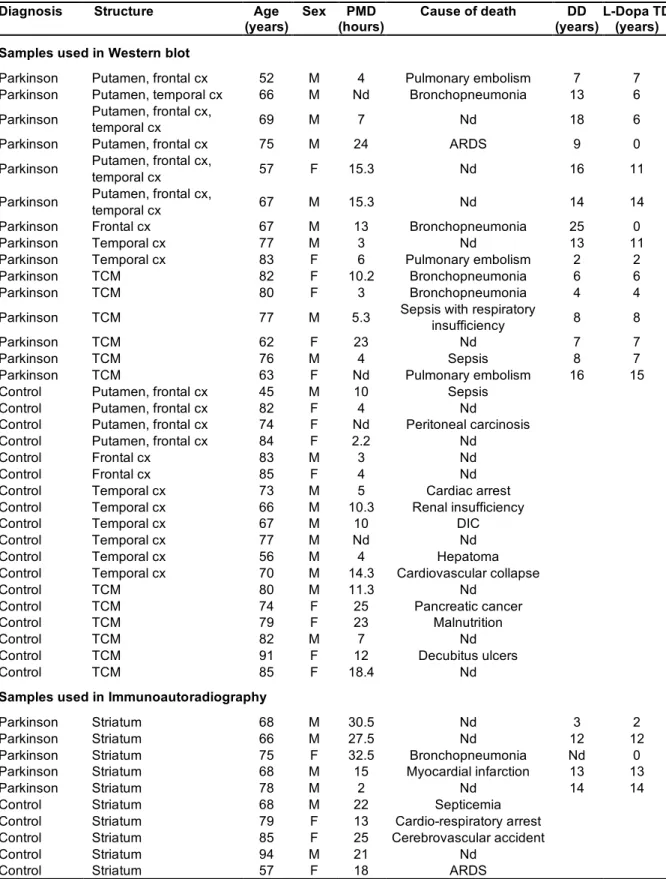
Table 1. Characteristics of patients and controls
Diagnosis Structure Age
(years) Sex PMD (hours) Cause of death DD (years) L-Dopa TD (years) Samples used in Western blot
Parkinson Putamen, frontal cx 52 M 4 Pulmonary embolism 7 7 Parkinson Putamen, temporal cx 66 M Nd Bronchopneumonia 13 6 Parkinson Putamen, frontal cx, temporal cx 69 M 7 Nd 18 6 Parkinson Putamen, frontal cx 75 M 24 ARDS 9 0 Parkinson Putamen, frontal cx, temporal cx 57 F 15.3 Nd 16 11 Parkinson Putamen, frontal cx, temporal cx 67 M 15.3 Nd 14 14 Parkinson Frontal cx 67 M 13 Bronchopneumonia 25 0
Parkinson Temporal cx 77 M 3 Nd 13 11
Parkinson Temporal cx 83 F 6 Pulmonary embolism 2 2 Parkinson TCM 82 F 10.2 Bronchopneumonia 6 6
Parkinson TCM 80 F 3 Bronchopneumonia 4 4
Parkinson TCM 77 M 5.3 Sepsis with respiratory insufficiency 8 8
Parkinson TCM 62 F 23 Nd 7 7
Parkinson TCM 76 M 4 Sepsis 8 7
Parkinson TCM 63 F Nd Pulmonary embolism 16 15 Control Putamen, frontal cx 45 M 10 Sepsis
Control Putamen, frontal cx 82 F 4 Nd
Control Putamen, frontal cx 74 F Nd Peritoneal carcinosis Control Putamen, frontal cx 84 F 2.2 Nd
Control Frontal cx 83 M 3 Nd
Control Frontal cx 85 F 4 Nd
Control Temporal cx 73 M 5 Cardiac arrest Control Temporal cx 66 M 10.3 Renal insufficiency
Control Temporal cx 67 M 10 DIC
Control Temporal cx 77 M Nd Nd
Control Temporal cx 56 M 4 Hepatoma Control Temporal cx 70 M 14.3 Cardiovascular collapse
Control TCM 80 M 11.3 Nd
Control TCM 74 F 25 Pancreatic cancer
Control TCM 79 F 23 Malnutrition
Control TCM 82 M 7 Nd
Control TCM 91 F 12 Decubitus ulcers
Control TCM 85 F 18.4 Nd
Samples used in Immunoautoradiography
Parkinson Striatum 68 M 30.5 Nd 3 2
Parkinson Striatum 66 M 27.5 Nd 12 12
Parkinson Striatum 75 F 32.5 Bronchopneumonia Nd 0 Parkinson Striatum 68 M 15 Myocardial infarction 13 13
Parkinson Striatum 78 M 2 Nd 14 14
Control Striatum 68 M 22 Septicemia Control Striatum 79 F 13 Cardio-respiratory arrest Control Striatum 85 F 25 Cerebrovascular accident
Control Striatum 94 M 21 Nd
Control Striatum 57 F 18 ARDS
Abbreviations: ARDS, adult respiratory distress syndrome; CMT, centromedial nucleus of the thalamus; cx, cortex; DD, duration of disease; DIC, disseminated intravascular coagulation; F, female; M, male; Nd, not documented; PMD, post-mortem delay; TCM, centro-medial nucleus of the thalamus; TD, therapy duration.
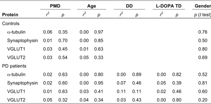
Table 2. Influence of post-mortem delay, age, disease duration, L-Dopa therapy duration and gender on protein levels in controls and patients with PD
PMD Age DD L-DOPA TD Gender
Protein r2 p r2 p r2 p r2 p p (t test) Controls α-tubulin 0.06 0.35 0.00 0.97 0.76 Synaptophysin 0.01 0.70 0.00 0.85 0.50 VGLUT1 0.03 0.45 0.01 0.63 0.80 VGLUT2 0.03 0.54 0.05 0.33 0.69 PD patients α-tubulin 0.02 0.63 0.00 0.80 0.00 0.89 0.00 0.82 0.52 Synaptophysin 0.02 0.60 0.00 0.95 0.07 0.46 0.05 0.39 0.81 VGLUT1 0.01 0.63 0.03 0.41 0.11 0.11 0.02 0.46 0.60 VGLUT2 0.05 0.32 0.04 0.34 0.03 0.43 0.00 0.80 0.20
Correlation analyses were performed using a two-tailed linear regression analysis to calculate the Pearson's coefficient (r2) and the p value for slope difference from zero. The effect of gender was evaluated with the Student's t test. Statistical analyses were performed with Western blot data obtained in the putamen of 4 controls and 6 PD patients; similar negative findings were obtained for other regions studied. Abbreviations: DD, disease duration; TD, L-Dopa therapy duration; PMD, post-mortem delay.
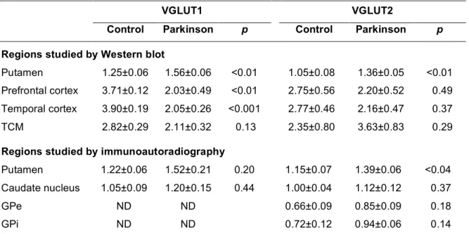
Table 3. Quantification of VGLUT1 and VGLUT2 immunoreactivity in the brain of patients with Parkinson disease and controls
VGLUT1 VGLUT2
Control Parkinson p Control Parkinson p
Regions studied by Western blot
Putamen 1.25±0.06 1.56±0.06 <0.01 1.05±0.08 1.36±0.05 <0.01 Prefrontal cortex 3.71±0.12 2.03±0.49 <0.01 2.75±0.56 2.20±0.52 0.49 Temporal cortex 3.90±0.19 2.05±0.26 <0.001 2.77±0.46 2.16±0.47 0.37
TCM 2.82±0.29 2.11±0.32 0.13 2.35±0.80 3.63±0.83 0.29
Regions studied by immunoautoradiography
Putamen 1.22±0.06 1.52±0.21 0.20 1.15±0.07 1.39±0.06 <0.04 Caudate nucleus 1.05±0.09 1.20±0.15 0.44 1.00±0.04 1.12±0.12 0.37
GPe ND ND 0.66±0.09 0.85±0.09 0.18
GPi ND ND 0.72±0.12 0.94±0.06 0.14
Results are expressed as mean ± SEM. Comparisons between groups were performed with the Student’s t test. For Western blot experiments, n=6 patients and 6 controls for all tissues except for the putamen where n=6 patients and 4 controls. For immunoautoradiography, n=5 patients and 5 controls for putamen, Gpe and Gpi; n=4 patients and 4 controls for caudate nucleus. Abbreviations: GPe, external segment of the globus pallidus; GPi, internal segment of the globus pallidus; ND, not detected; TCM, centro-medial thalamic nucleus.
Fig. 1. Distribution of VGLUT1 and VGLUT2 in human basal ganglia. Immunoblots (A) were performed using rat (lanes 1, 2, 5 and 6) or human (lanes 3, 4, 7-10) putamen extracts with the following antiserums: anti-VGLUT1 from [29] (lanes 1, 3, 4) or from Chemicon (lane 2), anti-VGLUT2 from [29] (lane 5, 7 and 8) or from Chemicon (lane 6), synaptophysin (lane 9) and α-tubulin (lane 10) both from Chemicon. Anti-VGLUT1 and anti-VGLUT2 were saturated with their respective antigen on lanes 4 and 8. Immuno-autoradiography (B-E) was performed on human brain sections taken at the level of the caudate-putamen and globus pallidus with an anti-VGLUT1 antiserum from [29] in B and D and with an anti-VGLUT2 antiserum from [29] in C and E. The saturated anti-VGLUT1 and –VGLUT2 antiserums were used in D and E, respectively. The areas that were quantified are delineated by dotted lines in C. Abbreviations: CD, caudate nucleus; GPe or GPi, external or internal segment of the globus pallidus; IC, internal capsule; PUT, putamen.
Fig. 2. VGLUT1 and VGLUT2 expression in basal ganglia from post-mortem tissues of controls and patients with Parkinson disease. A, Representative immunoblots, performed in the putamen from four control subjects and five patients (PD). The blots were probed with anti-VGLUT1, -VGLUT2, -synaptophysin or -α-tubulin. B, Representative immunoautoradiography of one control and one PD patient performed with antiVGLUT1 and -VGLUT2 antiserums on sections taken at the level of the putamen, caudate nucleus and internal/external globus pallidus. C, Scatter diagram of VGLUT1 and VGLUT2 levels quantified with Western blot (WB) or immuno-autoradiography (IAR) in the putamen, caudate nucleus, internal (GPi) or external (GPe) segment of the globus pallidus, from controls (open diamonds) and patients (black diamonds). Results are expressed as optical density (OD) in arbitrary units. Each point represents the mean of three determinations per subject (n=4-6 for Western blot and n=4-5 for immunoautoradiography) and region. Mean group values and standard error of the mean are also shown.
Fig. 3. VGLUT1 and VGLUT2 levels assessed by Western blotting in various brain areas of patients with Parkinson disease. A, Representative immunoblots of four control subjects and four patients (PD) performed with aliquots from the frontal cortex (FC), temporal cortex (TC) or the centro-median nucleus of the thalamus (CM). B, Scatter diagram of VGLUT1 and VGLUT2 levels quantified with Western blot in the same regions. Each point represents the mean of three determinations per subject (n=6) and region, expressed in arbitrary units of optical density. Mean group values and standard error of the mean are also shown.
![Fig. 1. Distribution of VGLUT1 and VGLUT2 in human basal ganglia. Immunoblots (A) were performed using rat (lanes 1, 2, 5 and 6) or human (lanes 3, 4, 7-10) putamen extracts with the following antiserums: anti-VGLUT1 from [29] (lanes 1, 3, 4) or from Ch](https://thumb-eu.123doks.com/thumbv2/123doknet/14697744.563741/18.892.112.726.113.934/distribution-ganglia-immunoblots-performed-putamen-extracts-following-antiserums.webp)