HAL Id: hal-01733958
https://hal.univ-lorraine.fr/hal-01733958
Submitted on 14 Mar 2018
HAL is a multi-disciplinary open access archive for the deposit and dissemination of sci-entific research documents, whether they are pub-lished or not. The documents may come from teaching and research institutions in France or
L’archive ouverte pluridisciplinaire HAL, est destinée au dépôt et à la diffusion de documents scientifiques de niveau recherche, publiés ou non, émanant des établissements d’enseignement et de recherche français ou étrangers, des laboratoires
Vitrectomie pour membrane épirétinienne avec acuité
visuelle supérieure ou égale 5/10
Alexandra Sayen
To cite this version:
Alexandra Sayen. Vitrectomie pour membrane épirétinienne avec acuité visuelle supérieure ou égale 5/10. Sciences du Vivant [q-bio]. 2010. �hal-01733958�
AVERTISSEMENT
Ce document est le fruit d'un long travail approuvé par le jury de
soutenance et mis à disposition de l'ensemble de la
communauté universitaire élargie.
Il est soumis à la propriété intellectuelle de l'auteur. Ceci
implique une obligation de citation et de référencement lors de
l’utilisation de ce document.
D'autre part, toute contrefaçon, plagiat, reproduction illicite
encourt une poursuite pénale.
Contact : ddoc-theses-contact@univ-lorraine.fr
LIENS
Code de la Propriété Intellectuelle. articles L 122. 4
Code de la Propriété Intellectuelle. articles L 335.2- L 335.10
http://www.cfcopies.com/V2/leg/leg_droi.php
UNIVERSITE HENRI POINCARE 1 FACULTE DE MEDECINE DE NANCY 2010
THESE
Pour obtenir le grade de
DOCTEUR EN MEDECINE
Présentée et soutenue publiquement
Dans le cadre du troisième cycle de Médecine Spécialisée
Par
Alexandra SAYEN
Le mardi 15 juin 2010
VITRECTOMIE POUR MEMBRANE EPIRETINIENNE
AVEC ACUITE VISUELLE SUPERIEURE OU EGALE
A 5/10
Jury :
M. BERROD Jean Paul Professeur Président
M. WAHL Denis Professeur Juge Mme. ANGIOI DUPREZ Karine Professeur Juge
UNIVERSITÉ HENRI POINCARÉ, NANCY 1
FACULTÉ DE MÉDECINE DE NANCY
---Président de l'Université : Professeur Jean-Pierre FINANCE Doyen de la Faculté de Médecine : Professeur Henry COUDANE
Vice Doyen Mission « sillon lorrain » : Professeur Annick BARBAUD Vice Doyen Mission « Campus » : Professeur Marie-Christine BÉNÉ
Vice Doyen Mission « Finances » : Professeur Marc BRAUN Vice Doyen Mission « Recherche » : Professeur Jean-Louis GUÉANT
Assesseurs :
- Pédagogie : Professeur Karine ANGIOÏ-DUPREZ - 1er Cycle : Professeur Bernard FOLIGUET - « Première année commune aux études de santé (PACES) et universitarisation études para-médicales » M. Christophe NÉMOS
- 2ème Cycle : Professeur Marc DEBOUVERIE - 3ème Cycle :
« DES Spécialités Médicales, Chirurgicales et Biologiques » « DES Spécialité Médecine Générale
Professeur Jean-Pierre BRONOWICKI Professeur Francis RAPHAËL
- Filières professionnalisées : M. Walter BLONDEL - Formation Continue : Professeur Hervé VESPIGNANI - Commission de Prospective : Professeur Pierre-Edouard BOLLAERT
- Recherche : Professeur Didier MAINARD
- Développement Professionnel Continu : Professeur Jean-Dominique DE KORWIN
DOYENS HONORAIRES
Professeur Adrien DUPREZ – Professeur Jean-Bernard DUREUX Professeur Jacques ROLAND – Professeur Patrick NETTER
==========
PROFESSEURS HONORAIRES
Pierre ALEXANDRE – Jean-Marie ANDRE - Daniel ANTHOINE - Alain BERTRAND - Pierre BEY - Jean BEUREY
Jacques BORRELLY - Michel BOULANGE - Jean-Claude BURDIN - Claude BURLET - Daniel BURNEL - Claude CHARDOT
Jean-Pierre CRANCE - Gérard DEBRY - Jean-Pierre DELAGOUTTE - Emile de LAVERGNE - Jean-Pierre DESCHAMPS
Michel DUC - Jean DUHEILLE - Adrien DUPREZ - Jean-Bernard DUREUX - Gabriel FAIVRE – Gérard FIEVE - Jean FLOQUET
Robert FRISCH - Alain GAUCHER - Pierre GAUCHER - Hubert GERARD - Jean-Marie GILGENKRANTZ Simone GILGENKRANTZ - Oliéro GUERCI - Pierre HARTEMANN - Claude HURIET – Christian JANOT -
Jacques LACOSTE
Henri LAMBERT - Pierre LANDES - Alain LARCAN - Marie-Claire LAXENAIRE - Michel LAXENAIRE - Jacques LECLERE
Antoine RASPILLER - Michel RENARD - Jacques ROLAND - René-Jean ROYER - Paul SADOUL - Daniel SCHMITT
Jean SOMMELET - Danièle SOMMELET - Michel STRICKER - Gilbert THIBAUT - Augusta TREHEUX - Hubert UFFHOLTZ
Gérard VAILLANT – Paul VERT - Colette VIDAILHET - Michel VIDAILHET - Michel WAYOFF - Michel WEBER
==========
PROFESSEURS DES UNIVERSITÉS PRATICIENS HOSPITALIERS
(Disciplines du Conseil National des Universités) 42ème Section : MORPHOLOGIE ET MORPHOGENÈSE
1ère sous-section : (Anatomie)
Professeur Gilles GROSDIDIER
Professeur Pierre LASCOMBES – Professeur Marc BRAUN
2ème sous-section : (Cytologie et histologie)
Professeur Bernard FOLIGUET
3ème sous-section : (Anatomie et cytologie pathologiques)
Professeur François PLENAT – Professeur Jean-Michel VIGNAUD
---43ème Section : BIOPHYSIQUE ET IMAGERIE MÉDICALE
1ère sous-section : (Biophysique et médecine nucléaire)
Professeur Gilles KARCHER – Professeur Pierre-Yves MARIE – Professeur Pierre OLIVIER
2ème sous-section : (Radiologie et imagerie médicale)
Professeur Denis REGENT – Professeur Michel CLAUDON
Professeur Serge BRACARD – Professeur Alain BLUM – Professeur Jacques FELBLINGER Professeur René ANXIONNAT
---44ème Section : BIOCHIMIE, BIOLOGIE CELLULAIRE ET MOLÉCULAIRE, PHYSIOLOGIE ET
NUTRITION
1ère sous-section : (Biochimie et biologie moléculaire)
Professeur Jean-Louis GUÉANT – Professeur Jean-Luc OLIVIER – Professeur Bernard NAMOUR
2ème sous-section : (Physiologie)
Professeur François MARCHAL – Professeur Bruno CHENUEL – Professeur Christian BEYAERT
3ème sous-section : (Biologie Cellulaire)
Professeur Ali DALLOUL
4ème sous-section : (Nutrition)
Professeur Olivier ZIEGLER – Professeur Didier QUILLIOT
---45ème Section : MICROBIOLOGIE, MALADIES TRANSMISSIBLES ET HYGIÈNE
1ère sous-section : (Bactériologie – virologie ; hygiène hospitalière)
Professeur Alain LE FAOU - Professeur Alain LOZNIEWSKI
3ème sous-section : (Maladies infectieuses ; maladies tropicales)
Professeur Thierry MAY – Professeur Christian RABAUD
---46ème Section : SANTÉ PUBLIQUE, ENVIRONNEMENT ET SOCIÉTÉ
2ème sous-section : (Médecine et santé au travail)
Professeur Christophe PARIS
3ème sous-section : (Médecine légale et droit de la santé)
Professeur Henry COUDANE
4ème sous-section : (Biostatistiques, informatique médicale et technologies de communication)
Professeur François KOHLER – Professeur Éliane ALBUISSON
---47ème Section : CANCÉROLOGIE, GÉNÉTIQUE, HÉMATOLOGIE, IMMUNOLOGIE
1ère sous-section : (Hématologie ; transfusion)
Professeur Thomas LECOMPTE – Professeur Pierre BORDIGONI Professeur Jean-François STOLTZ – Professeur Pierre FEUGIER
2ème sous-section : (Cancérologie ; radiothérapie)
Professeur François GUILLEMIN – Professeur Thierry CONROY Professeur Didier PEIFFERT – Professeur Frédéric MARCHAL
3ème sous-section : (Immunologie)
Professeur Gilbert FAURE – Professeur Marie-Christine BENE
4ème sous-section : (Génétique)
Professeur Philippe JONVEAUX – Professeur Bruno LEHEUP
---48ème Section : ANESTHÉSIOLOGIE, RÉANIMATION, MÉDECINE D’URGENCE,
PHARMACOLOGIE ET THÉRAPEUTIQUE
1ère sous-section : (Anesthésiologie et réanimation chirurgicale ; médecine d’urgence)
Professeur Claude MEISTELMAN – Professeur Hervé BOUAZIZ Professeur Paul-Michel MERTES – Professeur Gérard AUDIBERT
2ème sous-section : (Réanimation médicale ; médecine d’urgence)
Professeur Alain GERARD - Professeur Pierre-Édouard BOLLAERT Professeur Bruno LÉVY – Professeur Sébastien GIBOT
3ème sous-section : (Pharmacologie fondamentale ; pharmacologie clinique ; addictologie)
Professeur Patrick NETTER – Professeur Pierre GILLET
4ème sous-section : (Thérapeutique ; médecine d’urgence ; addictologie)
Professeur François PAILLE – Professeur Gérard GAY – Professeur Faiez ZANNAD
49ème Section : PATHOLOGIE NERVEUSE ET MUSCULAIRE, PATHOLOGIE MENTALE,
HANDICAP et RÉÉDUCATION
1ère sous-section : (Neurologie)
Professeur Gérard BARROCHE – Professeur Hervé VESPIGNANI Professeur Xavier DUCROCQ – Professeur Marc DEBOUVERIE
2ème sous-section : (Neurochirurgie)
Professeur Jean-Claude MARCHAL – Professeur Jean AUQUE Professeur Thierry CIVIT
3ème sous-section : (Psychiatrie d'adultes ; addictologie)
Professeur Jean-Pierre KAHN – Professeur Raymund SCHWAN
4ème sous-section : (Pédopsychiatrie ; addictologie)
Professeur Daniel SIBERTIN-BLANC – Professeur Bernard KABUTH
5ème sous-section : (Médecine physique et de réadaptation)
Professeur Jean PAYSANT
---50ème Section : PATHOLOGIE OSTÉO-ARTICULAIRE, DERMATOLOGIE et CHIRURGIE
Professeur François SIRVEAUX – Professeur Laurent GALOIS
3ème sous-section : (Dermato-vénéréologie)
Professeur Jean-Luc SCHMUTZ – Professeur Annick BARBAUD
4ème sous-section : (Chirurgie plastique, reconstructrice et esthétique ; brûlologie)
Professeur François DAP – Professeur Gilles DAUTEL
---51ème Section : PATHOLOGIE CARDIORESPIRATOIRE et VASCULAIRE
1ère sous-section : (Pneumologie ; addictologie)
Professeur Yves MARTINET – Professeur Jean-François CHABOT – Professeur Ari CHAOUAT
2ème sous-section : (Cardiologie)
Professeur Etienne ALIOT – Professeur Yves JUILLIERE – Professeur Nicolas SADOUL Professeur Christian de CHILLOU
3ème sous-section : (Chirurgie thoracique et cardiovasculaire)
Professeur Jean-Pierre VILLEMOT - Professeur Jean-Pierre CARTEAUX – Professeur Loïc MACÉ
4ème sous-section : (Chirurgie vasculaire ; médecine vasculaire)
Professeur Denis WAHL – Professeur Sergueï MALIKOV
---52ème Section : MALADIES DES APPAREILS DIGESTIF et URINAIRE
1ère sous-section : (Gastroentérologie ; hépatologie ; addictologie)
Professeur Marc-André BIGARD - Professeur Jean-Pierre BRONOWICKI – Professeur Laurent PEYRIN-BIROULET
2ème sous-section : (Chirurgie digestive) 3ème sous-section : (Néphrologie)
Professeur Michèle KESSLER – Professeur Dominique HESTIN – Professeur Luc FRIMAT
4ème sous-section : (Urologie)
Professeur Philippe MANGIN – Professeur Jacques HUBERT – Professeur Pascal ESCHWEGE
---53ème Section : MÉDECINE INTERNE, GÉRIATRIE et CHIRURGIE GÉNÉRALE
1ère sous-section : (Médecine interne ; gériatrie et biologie du vieillissement ; médecine générale ; addictologie)
Professeur Jean-Dominique DE KORWIN – Professeur Pierre KAMINSKY Professeur Athanase BENETOS - Professeur Gisèle KANNY
2ème sous-section : (Chirurgie générale)
Professeur Patrick BOISSEL – Professeur Laurent BRESLER Professeur Laurent BRUNAUD – Professeur Ahmet AYAV
---54ème Section : DÉVELOPPEMENT ET PATHOLOGIE DE L'ENFANT,
GYNÉCOLOGIE-OBSTÉTRIQUE,
ENDOCRINOLOGIE ET REPRODUCTION
1ère sous-section : (Pédiatrie)
Professeur Pierre MONIN - Professeur Jean-Michel HASCOET - Professeur Pascal CHASTAGNER Professeur François FEILLET - Professeur Cyril SCHWEITZER
2ème sous-section : (Chirurgie infantile)
Professeur Philippe JUDLIN – Professeur Patricia BARBARINO
4ème sous-section : (Endocrinologie, diabète et maladies métaboliques ; gynécologie médicale)
Professeur Georges WERYHA – Professeur Marc KLEIN – Professeur Bruno GUERCI
---55ème Section : PATHOLOGIE DE LA TÊTE ET DU COU
1ère sous-section : (Oto-rhino-laryngologie)
Professeur Claude SIMON – Professeur Roger JANKOWSKI
2ème sous-section : (Ophtalmologie)
Professeur Jean-Luc GEORGE – Professeur Jean-Paul BERROD – Professeur Karine ANGIOI-DUPREZ
3ème sous-section : (Chirurgie maxillo-faciale et stomatologie)
Professeur Jean-François CHASSAGNE – Professeur Etienne SIMON ==========
PROFESSEURS DES UNIVERSITÉS
64ème Section : BIOCHIMIE ET BIOLOGIE MOLÉCULAIRE
Professeur Sandrine BOSCHI-MULLER ==========
MAÎTRES DE CONFÉRENCES DES UNIVERSITÉS - PRATICIENS HOSPITALIERS
42ème Section : MORPHOLOGIE ET MORPHOGENÈSE
1ère sous-section : (Anatomie)
Docteur Bruno GRIGNON – Docteur Thierry HAUMONT
2ème sous-section : (Cytologie et histologie)
Docteur Edouard BARRAT - Docteur Françoise TOUATI – Docteur Chantal KOHLER
3ème sous-section : (Anatomie et cytologie pathologiques)
Docteur Béatrice MARIE
---43ème Section : BIOPHYSIQUE ET IMAGERIE MÉDICALE
1ère sous-section : (Biophysique et médecine nucléaire)
Docteur Marie-Hélène LAURENS – Docteur Jean-Claude MAYER
Docteur Pierre THOUVENOT – Docteur Jean-Marie ESCANYE – Docteur Amar NAOUN
2ème sous-section : (Radiologie et imagerie médicale)
Docteur Damien MANDRY
---44ème Section : BIOCHIMIE, BIOLOGIE CELLULAIRE ET MOLÉCULAIRE, PHYSIOLOGIE ET
NUTRITION
1ère sous-section : (Biochimie et biologie moléculaire)
Docteur Jean STRACZEK – Docteur Sophie FREMONT
Docteur Isabelle GASTIN – Docteur Marc MERTEN – Docteur Catherine MALAPLATE-ARMAND Docteur Shyue-Fang BATTAGLIA
2ème sous-section : (Physiologie)
Docteur Nicole LEMAU de TALANCE 3ème sous-section : (Biologie Cellulaire)
1ère sous-section : (Bactériologie – Virologie ; hygiène hospitalière)
Docteur Francine MORY – Docteur Véronique VENARD
2ème sous-section : (Parasitologie et mycologie)
Docteur Nelly CONTET-AUDONNEAU – Madame Marie MACHOUART
---46ème Section : SANTÉ PUBLIQUE, ENVIRONNEMENT ET SOCIÉTÉ
1ère sous-section : (Epidémiologie, économie de la santé et prévention)
Docteur Alexis HAUTEMANIÈRE – Docteur Frédérique CLAUDOT 3ème sous-section (Médecine légale et droit de la santé)
Docteur Laurent MARTRILLE
4ère sous-section : (Biostatistiques, informatique médicale et technologies de communication
Docteur Pierre GILLOIS – Docteur Nicolas JAY
---47ème Section : CANCÉROLOGIE, GÉNÉTIQUE, HÉMATOLOGIE, IMMUNOLOGIE
1ère sous-section : (Hématologie ; transfusion)
Docteur François SCHOONEMAN
2ème sous-section : (Cancérologie ; radiothérapie : cancérologie (type mixte : biologique)
Docteur Lina BOLOTINE
3ème sous-section : (Immunologie)
Docteur Marcelo DE CARVALHO BITTENCOURT
4ème sous-section : (Génétique)
Docteur Christophe PHILIPPE – Docteur Céline BONNET
---48ème Section : ANESTHÉSIOLOGIE, RÉANIMATION, MÉDECINE D’URGENCE,
PHARMACOLOGIE ET THÉRAPEUTIQUE
3ème sous-section : (Pharmacologie fondamentale ; pharmacologie clinique)
Docteur Françoise LAPICQUE – Docteur Marie-José ROYER-MORROT – Docteur Nicolas GAMBIER
4ème sous-section : (Thérapeutique ; médecine d’urgence ; addictologie
Docteur Patrick ROSSIGNOL
---50ème Section : RHUMATOLOGIE
1ère sous-section : (Rhumatologie)
Docteur Anne-Christine RAT
---54ème Section : DÉVELOPPEMENT ET PATHOLOGIE DE L’ENFANT,
GYNÉCOLOGIE-OBSTÉTRIQUE,
ENDOCRINOLOGIE ET REPRODUCTION
5ème sous-section : (Biologie et médecine du développement et de la reproduction ; gynécologie médicale)
Docteur Jean-Louis CORDONNIER ==========
MAÎTRES DE CONFÉRENCES
5ème section : SCIENCE ÉCONOMIE GÉNÉRALE
Monsieur Vincent LHUILLIER
---Monsieur Jean-François COLLIN
---60ème section : MÉCANIQUE, GÉNIE MÉCANIQUE ET GÉNIE CIVILE
Monsieur Alain DURAND
---61ème section : GÉNIE INFORMATIQUE, AUTOMATIQUE ET TRAITEMENT DU SIGNAL
Monsieur Jean REBSTOCK – Monsieur Walter BLONDEL 64ème section : BIOCHIMIE ET BIOLOGIE MOLÉCULAIRE
Mademoiselle Marie-Claire LANHERS
---65ème section : BIOLOGIE CELLULAIRE
Mademoiselle Françoise DREYFUSS – Monsieur Jean-Louis GELLY Madame Ketsia HESS – Monsieur Hervé MEMBRE – Monsieur Christophe NEMOS
Madame Natalia DE ISLA – Monsieur Pierre TANKOSIC
---66ème section : PHYSIOLOGIE
Monsieur Nguyen TRAN
---67ème section : BIOLOGIE DES POPULATIONS ET ÉCOLOGIE
Madame Nadine MUSSE
========
PROFESSEURS ASSOCIÉS
Médecine Générale Professeur associé Alain AUBREGE Professeur associé Francis RAPHAEL
MAÎTRES DE CONFÉRENCES ASSOCIÉS
Médecine Générale Docteur Jean-Marc BOIVIN
Docteur Jean-Louis ADAM Docteur Elisabeth STEYER
========
PROFESSEURS ÉMÉRITES
Professeur Daniel ANTHOINE - Professeur Pierre BEY - Professeur Michel BOULANGE
Professeur Jean-Pierre CRANCE - Professeur Jean FLOQUET - Professeur Jean-Marie GILGENKRANTZ Professeur Simone GILGENKRANTZ – Professeur Henri LAMBERT - Professeur Alain LARCAN
Professeur Hubert UFFHOLTZ - Professeur Paul VERT - Professeur Michel VIDAILHET
========
DOCTEURS HONORIS CAUSA
Professeur Norman SHUMWAY (1972)
Université de Stanford, Californie (U.S.A) Professeur Théodore H. SCHIEBLER (1989) Institut d'Anatomie de Würtzburg (R.F.A)
Professeur Paul MICHIELSEN (1979)
Université Catholique, Louvain (Belgique) Professeur Maria DELIVORIA-PAPADOPOULOS (1996) Université de Pennsylvanie (U.S.A)
Professeur Charles A. BERRY (1982)
Centre de Médecine Préventive, Houston (U.S.A) Professeur Mashaki KASHIWARA (1996) Research Institute for Mathematical Sciences de Kyoto (JAPON)
Professeur Pierre-Marie GALETTI (1982)
Brown University, Providence (U.S.A) Professeur Ralph GRÄSBECK (1996) Université d'Helsinki (FINLANDE)
Professeur Mamish Nisbet MUNRO (1982)
Massachusetts Institute of Technology (U.S.A) Professeur James STEICHEN (1997) Université d'Indianapolis (U.S.A)
Professeur Mildred T. STAHLMAN (1982)
Vanderbilt University, Nashville (U.S.A) Professeur Duong Quang TRUNG (1997) Centre Universitaire de Formation et de Perfectionnement des
Harry J. BUNCKE (1989)
Université de Californie, San Francisco (U.S.A) Professionnels de Santé d'Hô Chi Minh-Ville (VIÊTNAM)
Professeur Daniel G. BICHET (2001)
Université de Montréal (Canada) Professeur Marc LEVENSTON (2005) Institute of Technology, Atlanta (USA)
Professeur Brian BURCHELL (2007)
A notre Président de thèse,
Monsieur le Professeur Jean Paul BERROD,
Professeur d’Ophtalmologie
Vous nous avez fait l’honneur de nous confier ce travail et de
l’encadrer.
Nous vous témoignons toute notre gratitude pour
l’enseignement que vous nous prodiguez. A vos côtés, nous
apprenons avec humilité et rigueur ce magnifique métier.
Travailler et apprendre à vos côtés sont un honneur et une
grande fierté. Vos connaissances et votre habileté chirurgicale
sont pour nous un modèle. Nous espérons par ce travail avoir été
digne de votre confiance.
Soyez assuré de nos respectueuses considérations et de notre
profonde admiration.
A notre Juge,
Monsieur le ProfesseurDenis WAHL,
Professeur de chirurgie vasculaire et de médecine vasculaire.
Vous nous avez fait l’honneur d’accepter de juger ce travail.
Votre investissement aussi bien médical qu’universitaire est
pour nous un exemple. Votre participation à ce travail symbolise
la collaboration qui existe entre nos deux services.
Veuillez trouver dans ce travail l’expression de notre plus grand
respect.
A notre Juge,
Madame le ProfesseurKarine ANGIOI DUPREZ,
Professeur d’Ophtalmologie
Vous nous avez fait l’honneur d’accepter de juger ce travail.
Par vos connaissances, votre rigueur et votre générosité, vous
êtes un exemple pour nous tous. Vous avez accompagné nos
premiers pas dans la discipline et travailler avec vous a été une
grande fierté.
A notre Juge,
Madame le Docteur Isabelle HUBERT,
Praticien Hospitalier en Ophtalmologie
Vous nous avez fait l’honneur d’accepter de juger ce travail.
Nous vous témoignons notre plus profonde reconnaissance.
Nous vous remercions d’avoir partagé avec enthousiasme vos
connaissances. Votre dextérité chirurgicale est un exemple pour
nous tous.
Travailler à vos côtés a été un plaisir.
SERMENT
SERMENT
SERMENT
SERMENT
"Au moment d'être admise à exercer la médecine, je promets et je jure d'être fidèle aux lois de
l'honneur et de la probité. Mon premier souci sera de rétablir, de préserver ou de promouvoir
la santé dans tous ses éléments, physiques et mentaux, individuels et sociaux. Je respecterai
toutes les personnes, leur autonomie et leur volonté, sans aucune discrimination selon leur
état ou leurs convictions. J’interviendrai pour les protéger si elles sont affaiblies, vulnérables
ou menacées dans leur intégrité ou leur dignité. Même sous la contrainte, je ne ferai pas
usage de mes connaissances contre les lois de l'humanité. J'informerai les patients des
décisions envisagées, de leurs raisons et de leurs conséquences. Je ne tromperai jamais leur
confiance et n'exploiterai pas le pouvoir hérité des circonstances pour forcer les consciences.
Je donnerai mes soins à l'indigent et à quiconque me les demandera. Je ne me laisserai pas
influencer par la soif du gain ou la recherche de la gloire.
Admise dans l'intimité des personnes, je tairai les secrets qui me sont confiés. Reçue à
l'intérieur des maisons, je respecterai les secrets des foyers et ma conduite ne servira pas à
corrompre les mœurs. Je ferai tout pour soulager les souffrances. Je ne prolongerai pas
abusivement les agonies. Je ne provoquerai jamais la mort délibérément.
Je préserverai l'indépendance nécessaire à l'accomplissement de ma mission. Je
n'entreprendrai rien qui dépasse mes compétences. Je les entretiendrai et les perfectionnerai
pour assurer au mieux les services qui me seront demandés.
J'apporterai mon aide à mes confrères ainsi qu'à leurs familles dans l'adversité.
Que les hommes et mes confrères m'accordent leur estime si je suis fidèle à mes promesses ;
que je sois déshonorée et méprisée si j'y manque".
REMERCIEMENTS
A mes parents : vous avez toujours cru en moi, que ce travail soit une marque de mon amour et de toute ma reconnaissance.
A ma soeur et à ma filleule Anouk: je vous adore tout simplement A Julien, que j’aime profondément
A mes grands parents, qui ne sont plus là aujourd’hui, mais qui m’ont beaucoup apporté.
A Emilie, toujours fidèle pendant ces longues années d’études.
A la « petite » Emilie et à Sandrine, parties de la région, nos soirées « entre filles » me manquent !
A Lucie, amie formidable, qui a toujours été là pour les bons comme pour les mauvais moments.
A la famille Rangeard.
A tous mes copains lorrains d’origine, qui sont pour certains à l’autre bout du monde…
A toutes mes amies formidables rencontrées en ophtalmologie : Pascale, Rym, Malicia, Anne, Fanny.
A mes co-internes : Alexandre, Younès, Olivier, Jerôme, Ghassen, Alissa, Myriam et aux plus jeunes qui arrivent.
A Georges : vive les jaunes et bleus !
A mes premiers chefs : Charles, Angélique, Grégoire.
A tous les praticiens hospitaliers des trois différents services : Marie-Christine Bazard, Toufic Maalouf, Pierre Lesure, Jean-Luc George, Jean-Marc Perone.
A toute l’équipe paramédicale d’ophtalmologie de l’Hôpital Central, qui va me manquer.
Aux équipes d’ophtalmologie de l’Hôpital de Brabois et de Bonsecours.
SOMMAIRE ... 17
PREMIERE PARTIE ... 19
ARTICLE ... 27
INTRODUCTION ... 31
METHODS ... 33
RESULTS ... 37
DISCUSSION ... 43
CONCLUSION ... 45
BIBLIOGRAPHIE ... 46
Une membrane épirétinienne (MER) est une prolifération fibrocellulaire avasculaire se développant au niveau de l’aire maculaire à la surface de la membrane limitante interne (MLI) de la rétine. Il en résulte une distorsion de la rétine pouvant provoquer une baisse d’acuité visuelle, des métamorphopsies ou une diplopie par ectopie fovéolaire.
La composition de ces membranes a été décrite par Foos et al. en 1977 1. Elles sont constituées de cellules gliales qui ont migré au travers de la couche des fibres optiques sur la MLI et de matériel extra-cellulaire fibreux.
Des prélèvements de MER ont été obtenus après chirurgie (vitrectomie et pelage de MER) 23 et leur analyse histologique a mis en évidence la présence de plusieurs types cellulaires tels que cellules de l’épithélium pigmentaire, cellules gliales et fibroblastes 1.
Sur le plan physiopathologique, de nombreux auteurs ont suggéré l’association fréquente d’un décollement postérieur du vitré (DPV) et d’une membrane épimaculaire. L’hypothèse la plus communément admise est celle de Roth et Foos 4. Les tractions vitréo-rétiniennes, lors de la constitution du DPV, pourraient être responsables de ruptures de la limitante interne et formeraient des hiatus par lesquels les cellules gliales pourraient migrer à la face interne de la rétine et proliférer. Une seconde hypothèse est qu’ une partie du cortex vitréen postérieur pourrait rester attaché à l’aire maculaire après le DPV, ce qui jouerait un rôle important dans le développement d’une MER 5 6, 7. Cependant des arguments vont à l’encontre de ces hypothèses :
- le DPV est une évolution physiologique du vitré, sa fréquence à 80 ans est proche de 100% alors que l’incidence des membranes épimaculaires est de 5%.
- certaines membranes épimaculaires sont formées alors que le vitré n’est pas décollé.
Sur le plan épidémiologique, la membrane épirétinienne est une pathologie relativement commune chez la personne âgée. Dans 20% des cas, les MER sont secondaires à diverses pathologies oculaires, telles que déchirures rétiniennes, occlusions vasculaires, inflammations intra-oculaires. Dans 80% des cas, on ne retrouve pas de pathologies oculaires, elle est alors appelée membrane épimaculaire idiopathique.
Elles sont rarement congénitales. Elle peut toucher le sujet jeune.
Plusieurs grandes études ont donné des résultats concernant l’épidemiologie des membranes épimaculaires 8, 9 10 11 12. La prévalence des membranes épirétiniennes varie de 2.2% 13 à 18.5% 9. Dans la Beaver Dam Study10, sur une population de 4802 personnes, 11,2% présentent une membrane épirétinienne. Les membranes sont bilatérales dans 11,5% des cas.
personnes ont participé, l’incidence est de 5,3%. Les membranes sont bilatérales dans 13,5% des cas. L’incidence augmente avec l’âge : 3,7% pour les patients de moins de 60 ans, 6,6% entre 60 et 69 ans, 6,1% entre 70 et 79 ans et 1,1% chez les patients de 80 ans et plus. L’incidence est de 5,7% chez les femmes et 4,8% chez les hommes. Dans la Beijing Eye Study 13, étude réalisée sur 3265 sujets de plus de 40 ans en 2001, la prévalence est de 2.2%. Dans la Handan Eye Study 12, étude réalisée sur 4378 sujets de 2006 à 2007, la prévalence est de 3.4%.
La prévalence des membranes épimaculaires est plus élevée dans la Los Angeles Latino Eye Study 9 et dans la Beaver Dam Study. Ceci peut-être lié à la façon de grader les membranes épimaculaires. Fraser-Bell et al. ont rapporté que le taux de prévalence élevé dans la Los Angeles Latino Eye Study peut s’expliquer par la plus forte prévalence de diabète dans cette population.
Sur le plan clinique, les signes fonctionnels que l’on peut retrouver le plus fréquemment sont une baisse de l’acuité visuelle, qui s’installe le plus souvent progressivement et des métamorphopsies plus ou moins sévères. Ces métamorphopsies peuvent entraîner une gêne disproportionnée à la lecture de près, et être invalidantes malgré la conservation d’une acuité visuelle de loin encore convenable. Elles peuvent aussi entraver la vision binoculaire. Ces métamorphopsies réalisent souvent la première plainte du patient et peuvent être à elles seules une indication à une intervention chirurgicale. Il existe fréquemment des macropsies (objets vus plus grand). Dans certains cas, le patient signale aussi un scotome central relatif atténuant le contraste des lettres ou des mots lors de la lecture de près. Une diplopie peut parfois gêner le patient ; elle est liée à une ectopie fovéolaire par rétraction tangentielle de la membrane vers un point d’ancrage extra-maculaire. La deuxième image est en outre déformée et agrandie, ce qui accentue la gêne fonctionnelle.
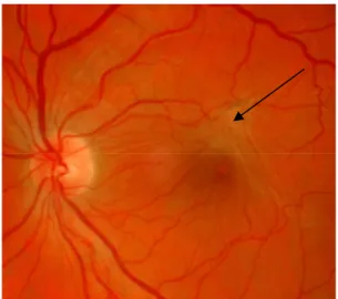
Le diagnostic de membrane épimaculaire est clinique. L’examen du fond d’œil au biomicroscope permet de visualiser le reflet brillant de la membrane épimaculaire, les déformations vasculaires, des plis rétiniens maculaires et, à l’aide d’un verre contact, un épaississement rétinien maculaire. Les signes associés peuvent être : petites hémorragies rétiniennes superficielles, interruption du flux axoplasmique, décollement séreux du neuro-épithélium, pseudotrou maculaire, trou maculaire lamellaire, ectopie maculaire.
Figure 1. Rétinophotographie : aspect d’une membrane épirétinienne (flèche)
Sur le plan paraclinique, les photos du fond d’œil en lumière verte mettent surtout en évidence la déformation vasculaire induite par la constriction de la membrane. Les photos du fond d’œil en lumière bleue mettent en évidence la membrane elle-même, qui est réfringente, et ses contours. Ces clichés montrent également la constriction, l’étirement et le déplacement du pigment xanthophylle.
L’examen qui nous paraît essentiel actuellement dans le diagnostic des MER est la tomographie par cohérence optique (OCT). Il s’agit d’une technique d’imagerie du fond d’œil, non invasive, qui permet d’obtenir des images en coupe optique de la rétine, avec une résolution de 10 à 15 µmètres. Les images permettent d’analyser la jonction vitréo-rétinienne, des modifications de la structure du tissu rétinien et de mesurer avec précision l’épaisseur rétinienne sur tous les points de la rétine. Le principe physique repose sur l’utilisation d’un faisceau lumineux infrarouge de basse cohérence de longueur d’onde de 830 nm (la cohérence du faisceau est assurée de façon répétée sur une courte longueur d’onde) et sur le principe de l’interférométrie. On réalise des coupes linéaires isolées, horizontales ou verticales, radiaires simples ou multiples, ou circulaires. Les images obtenues nous donnent une idée de l’architecture rétinienne in vivo. L’OCT permet également de mesurer l’épaisseur maculaire.
ILM : Internal Limiting Membrane GCL : Ganglion Cell Layer
INL : Internal Nuclear Layer
OPL : Outer Plexiform Layer ONL : Outer Nuclear Layer
IS/OS PR : Inner segment/Outer Segment photoreceptors junction PL : Photoreceptors Layer
RPE : Pigment Epithelium
Figure 2 : Coupe histologique de la macula
L’angiographie permet d’apprécier le retentissement sur les vaisseaux rétiniens et sur les capilllaires avec diffusion anormale du colorant. Elle met aussi en évidence l’œdème maculaire qui est parfois cystoïde. Elle est rarement réalisée en pratique courante depuis l’avènement de l’OCT qui nous donne plus d’informations, et compte tenu de son caractère invasif.
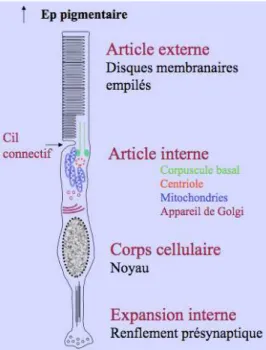
Récemment, plusieurs études ont analysé à partir d’images OCT, une ligne hyperréflective située juste au dessus la couche de l’épithélium pigmentaire 1415. Cette ligne correspond à la jonction des articles internes et des articles externes des photorécepteurs. Une ligne bien visible et continue indique un alignement normal des disques membranaires dans les articles externes des photorécepteurs et suggère un fonctionnement normal de ces derniers.
Figure 4 : schéma d’un photorécepteur.
Sur le plan thérapeutique, le pelage chirurgical des membranes épimaculaires a été publié par Machemer en 1978. Dans un premier temps réservée aux membranes secondaires, cette chirurgie s’est considérablement développée depuis lors, et s’adresse principalement aux membranes idiopathiques. L’indication est communément portée quand l’acuité visuelle est inférieure ou égale à 4/10 lorsqu’il existe une gêne visuelle ressentie par le patient. Le traitement consiste en la réalisation d’une vitrectomie postérieure centrale puis d’un pelage de la membrane épimaculaire et de la limitante interne.
Bibliographie
1. Foos RY. Vitreoretinal juncture; epiretinal membranes and vitreous. Invest Ophthalmol Vis Sci 1977;16:416-22.
2. Kampik A, Green WR, Michels RG, Nase PK. Ultrastructural features of progressive idiopathic epiretinal membrane removed by vitreous surgery. Am J Ophthalmol 1980;90:797-809.
3. Smiddy WE, Michels RG, Green WR. Morphology, pathology, and surgery of idiopathic vitreoretinal macular disorders. A review. Retina 1990;10:288-96.
4. Roth AM, Foos RY. Surface wrinkling retinopathy in eyes enucleated at autopsy. Trans Am Acad Ophthalmol Otolaryngol 1971;75:1047-58.
5. Kishi S, Shimizu K. Oval defect in detached posterior hyaloid membrane in idiopathic preretinal macular fibrosis. Am J Ophthalmol 1994;118:451-6.
6. Tadayoni. Membranes épirétiniennes maculaires. EMC 2004.
7. Jaffe GJ. Thrombospondin as a component of the extracellular matrix of epiretinal membranes: comparisons with cellular fibronectin. Surv Ophthalmol 1994;38:393-4.
8. Fraser-Bell S, Guzowski M, Rochtchina E, Wang JJ, Mitchell P. Five-year cumulative incidence and progression of epiretinal membranes: the Blue Mountains Eye Study. Ophthalmology 2003;110:34-40.
9. Fraser-Bell S, Ying-Lai M, Klein R, Varma R. Prevalence and associations of epiretinal membranes in latinos: the Los Angeles Latino Eye Study. Invest Ophthalmol Vis Sci 2004;45:1732-6.
10. Klein R, Klein BE, Wang Q, Moss SE. The epidemiology of epiretinal membranes. Trans Am Ophthalmol Soc 1994;92:403-25; discussion 425-30.
11. Pearlstone AD. The incidence of idiopathic preretinal macular gliosis. Ann Ophthalmol 1985;17:378-80.
12. Duan XR, Liang YB, Friedman DS, et al. Prevalence and associations of epiretinal membranes in a rural Chinese adult population: the Handan Eye Study. Invest Ophthalmol Vis Sci 2009;50:2018-23.
14. Suh MH, Seo JM, Park KH, Yu HG. Associations between macular findings by optical coherence tomography and visual outcomes after epiretinal membrane removal. Am J Ophthalmol 2009;147:473-480 e3.
15. Watanabe A, Arimoto S, Nishi O. Correlation between metamorphopsia and epiretinal membrane optical coherence tomography findings. Ophthalmology 2009;116:1788-93.
VITRECTOMY FOR EPIRETINAL MEMBRANE
IN EYES WITH A PREOPERATIVE VISUAL
ACUITY BETTER THAN 20/40
A.
Sayen
1, I.Hubert
1, M. Casillas-Gil
1,
ML. Erpelding
3,K. Angioi-Duprez
2,JP. Berrod
11 Department of Ophtalmology, University Hospital, Nancy, France
2 Department of Ophtalmology, University Hospital, Vandoeuvre les Nancy, France 3 Department of Biostatistics, University Hospital, Nancy, France
The authors have no financial or proprietary interest in any materials or methods mentioned.
Reprint requests to Jean-Paul Berrod, Department of Ophtalmology, University Hospital, 29
avenue du Marechal de Lattre de Tassigny, 54035 Nancy cedex, France. E-mail: jp.berrod@chu-nancy.fr
Keywords: epiretinal membrane, vitrectomy, good visual acuity, inner and outer segments of
ABSTRACT
Purpose:
To evaluate the functional and anatomical outcomes of vitrectomy for epiretinal membrane in eyes with a preoperative VA (visual acuity) ≥ 0,3 LogMAR (20/40).
Methods:
Sixty-seven of sixty-six consecutive patients that undergone surgery for an idiopathic epiretinal membrane (ERM) were studied retrospectively. A combined surgery was performed on phakic eyes (n=45). The other eyes were all pseudophakic (n=22). All patients underwent ERM and internal limiting membrane removal by using 20 (n=19) or 23-gauge (n=48) vitrectomy system between January 2002 and March 2009 by two surgeons (JPB and IH). The main studied criteria were preoperative and postoperative best corrected visual acuity (BCVA), preoperative and postoperative central macular thickness (CFT) and integrity of junction between inner and outer segments of photoreceptors (IS/OS junction) evaluated with optical coherence tomography (OCT) preoperatively and postoperatively.
Results:
The mean preoperative VA was 0.28 logMAR (range 0.15 logMAR to 0.3 logMAR). The mean VA improved to 0.19 logMAR (2 lines of vision) by the final examination at a mean of 27 months following surgery (p<0.0001). 71,6 % patients recovered visual acuity ≥ 0,1 logMAR (20/25). The mean preoperative CFT was 367 µm (range 263 µm to 496 µm) and the mean postoperative CFT was 304 µm (range 198 µm to 385 µm).
No correlation between postoperative BCVA and postoperative central macular thickness was noted (p=0.46).
The preoperative integrity of the IS/OS line correlated with postoperative BCVA (p=0.004) but not with BCVA improvement (p=0.177). We observed one retinal detachment (1.49%), one macular hole (1.49%) and no endophthalmitis.
Conclusion:
Vitrectomy for epiretinal membrane is a safe and effective procedure that provides VA ≥ 0,1 LogMAR in 71,6% eyes with relatively good preoperative VA ( ≥ 0,3 LogMAR). Besides the baseline VA, the integrity of the IS/OS line can be used to predict excellent functional outcomes after surgery.
INTRODUCTION
:ERM are commons lesions in older individuals. Their development and progression have effects on visual acuity but this progression is usually considered as low. This relative stability of epiretinal membranes and its potential surgical complications should be taken into account when considering surgery. This is for this reason that it was advised to perform vitrectomy for ERM in eyes with a visual acuity lower than 20/60.
Vitrectomy for ERM removal that has been presented by Machemer in 1978 is nowdays the most common vitreoretinal procedure in USA and also probably in European countries. Most published studies of vitrectomy for ERM consisted of eyes with visual acuities of 20/60 or worse. Eyes with better levels of visual acuity have not been reported as a separate group. A study of 264 eyes with epiretinal membranes reported by Rice and coworkers 1 included six eyes with a visual acuity of 20/50. In this subgroup the mean acuity improved to 20/40. A second report by De Bustros et al.2 of vitrectomy in 70 eyes with idiopathic epiretinal membranes included 3 eyes with visual acuity of 20/50. The visual acuity improved to 20/25 in two eyes and 20/40 in the third eye.
A two line improvement is expected for 80% of the eyes in most of the reports in which baseline visual acuity was 20/60 or worse 34, what means that we should have only a partial recovery if the patients have a baseline visual acuity worse than 20/40. Thus, the indication to operate eyes with a baseline visual acuity better than 20/40 seems interesting if we want to approach a visual acuity of 20/20 in post-operative and if we want to protect the visual function.
Optical Coherence Tomography (OCT) is increasingly used in the clinical setting to define and monitor early ERMs. It is an easy and not invasive exam to perform in case of visual acuity decrease. It is for this reason that others prognosis factors for the outcome of ERM surgery have been investigated: few studies have been performed to evaluate the influence of macular thickness on visual outcomes and have produced conflicting results. In 1996, Wilkins et al. 5 showed that preoperative mean central macular thickness correlated with visual acuity gain after ERM removal, while Massin et al. 6 in a serie of 62 eyes undergoing vitrectomy
Few trials have study the role of IS/OS junction in visual outcomes after ERM removal and have divergent results78.
For much published studies, combined and consecutive surgeries to treat ERM have equivalent functional and anatomical results 9 10. In the same way, twenty-three-gauge vitrectomy and twenty-gauge vitrectomy in epiretinal membrane surgery have comparable results 1112, 13.
The purpose of this study was to assess the functional and anatomic outcomes of vitrectomy for epiretinal membrane removal in eyes with a preoperative visual acuity better than 20/40.
METHODS
:This retrospective, interventional case series included 67 eyes of 66 consecutive patients with a visual acuity of 20/40 or better, who underwent surgery for an idiopathic ERM in Nancy University Hospital. The operations were performed by two experienced surgeons (JPB and IH) between January, 2002 and March, 2009. Patients were selected from a spreadsheet updated after every surgical procedure with visual acuity of 20/40 or better selection criteria. This file, concerning ERM surgeries, included a total of 1091 eyes operated from January 2002 till March 2010.
The indication for ERM surgery was symptomatic visual impairment, including a decreased BCVA or metamorphopsia.
Exclusion criteria were: eyes with ERM secondary to inflammatory ocular pathology, trauma, diabetic retinopathy, venous occlusion or retinal detachment. Glaucoma and age-related macular degeneration were exlusion criteria as well. Patients with an OCT signal strenght lower than 4 on OCT images were also excluded.
All patients had symptomatic visual acuity loss and remaining acuity of 20/40 or better, metamorphopsia, the visualization of the ERM in the OCT, or a combination thereof. They all consented to surgery after a discussion of the risks and benefits of the procedure and gave their written consent .
On the data of medical files and surgery reports, all per and postoperative complications were recorded and functional and anatomic results were analysed. Main outcome measures were visual acuity converted in Logmar, central macular thickness measured with OCT and the integrity of the IS/OS junction before the surgery and at the 6 months follow-up visit. Latest BCVA was obtained by contacting their referent ophthalmologist.
All patients benefited of full ophthalmological examination before and after surgery (including biomicroscopy of the fundus with contact lens). They all underwent best corrected Snellen visual acuity converted to logarithm of minimal angle of resolution units for statistical
map protocol in every visit. All OCT images were acquired through a dilated pupil.
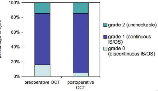
The integrity of the IS/OS junction in the OCT images at the fovea was graded retrospectively into three grades: 0, abnormal IS/OS junction (not visible or discontinuous); 1, normal (continuous) IS/OS junction, and 2, uncheckable IS/ OS junction.
Continuous IS/OS junction Uncheckable IS/OS junction
The surgical technique consisted of standard three-port pars plana central vitrectomy (Millenium Microsurgical System, Bausch & Lomb) using 20-gauge (n=19) or 23-gauge (n=48) instrumentation (Dorc).
ERM and ILM peeling were performed with microforceps from the macular area. ILM was removed systematically without dye. In phakic eyes, cataract surgery was performed by phacoemulsification with the implantation of an hydrophobic acrylic intraocular lens of 6.5 mm of diameter in the bag (AcrySof MA50BM, Alcon) after vitrectomy and ERM removal.
Procedures using 20-gauge instrumentation have been performed from January 2002 to February 2006 and surgeries using 23-gauge instruentation have been performed from February 2006 to March 2009.
Statistical Analysis
The significance of the differences in BCVA, CFT and the three IS/OS junction grades was analysed using one-factor analysis of variance (ANOVA) with post hoc comparisons tested by Fisher’s protected least significant difference (PLSD). To analyse correlations between parameters associated with VA and macular thickness, a linear regression was used.
P values of less than 0.05 were considered statistically significant.
RESULTS
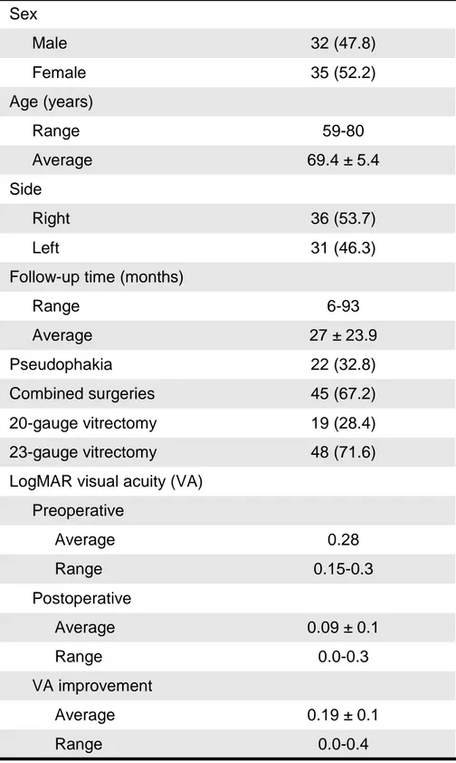
:Thirty-four patients were female and thirty-two patients were male. The mean age was 69.4 years with a range of 59 to 80 years and mean follow-up was 27 months ranged from 6 to 93 months.
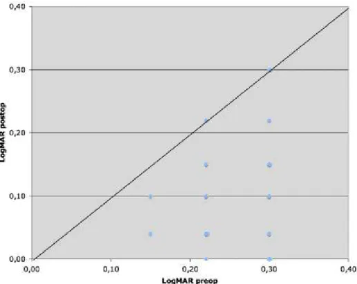
The mean preoperative visual acuity was 0.28 logMAR with a range from 0.15 logMAR to 0.3 logMAR. The mean postoperative visual acuity was 0.09 logMAR with a range from 0.0 logMAR to 0.3 logMAR.
Visual acuity improved with a mean gain of 0.19 logMAR which was statistically significant (P<0.0001). No eye presented a visual acuity decrease.
40.3% (n=27) of the eyes recovered a postoperative visual acuity better or equal to 0.04 logMAR and 71.6% (n=48) of the eyes had a postoperative visual acuity better or equal to 0.1 logMAR.
The mean preoperative CFT was 367 µm with a range from 263 µm to 496 µm and the mean postoperative CFT was 304 µm from 198 µm to 385 µm.
We found no significant correlation between preoperative foveal thickness and postoperative visual acuity (p=0.46). No correlation between postoperative foveal thickness and postoperative visual acuity was found either (p=0.19).
In the preoperative OCT scans, the IS/OS line was abnormal (grade 0) in 11 eyes (16.2%), was continuous (grade 1) in 47 eyes (70.1%) and was uncheckable (grade 2) in 10 eyes (14.7%). On the postoperative examination, we found a grade 0 in 3 eyes (4.5%), a grade 1 in 54 eyes (80.6%), and a grade 2 in 10 eyes (14.9%).
The preoperative IS/OS junction correlated significantly with the postoperative BCVA (p=0.004) but not with BCVA improvement (p=0.177). Moreover, we found a significantly correlation between the postoperative IS/OS junction and postoperative BCVA (p=0.002) and BCVA improvement (p=0.0022). BCVA improvement was statistically significant (p<0.0001) for the eyes who had discontinuous or uncheckable preoperative IS/OS line and who recovered a normal IS/OS line (10.5% of eyes).
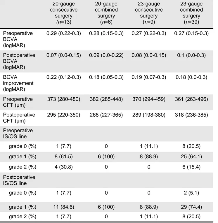
As far as the surgical techniques are concerned, we did not find any statistically significant differences between the four types of procedures: consecutive surgery with
20-surgery with 23-gauge instrumentation and combined procedure with 23-gauge instrumentation (p=0.377).
We didn’t find any predictive factor associated with a 20/20 postoperative BVCA on Snellen chart.
Of the 67 patients operated for idiopathic ERM, we observed one retinal detachment (1.49%), one macular hole (1.49%), and one extra macular hole (1.49%). All evolved favorably after specific surgical treatment.
None of the 66 patients (67 eyes) included in this study experienced postoperative complications of endopthalmitis or glaucoma.
Table 1. Characteristics for all surgical cases (n=67) Sex 32 (47.8) Male 35 (52.2) Female 59-80 Range 0.0-0.4 Range 0.0-0.3 Range 0.09 ± 0.1 Average 0.15-0.3 Range 0.28 Average 48 (71.6) 23-gauge vitrectomy 19 (28.4) 20-gauge vitrectomy 0.19 ± 0.1 45 (67.2) 22 (32.8) 27 ± 23.9 6-93 31 (46.3) 36 (53.7) 69.4 ± 5.4 No. Cases (%) Average VA improvement Postoperative Preoperative
LogMAR visual acuity (VA) Combined surgeries
Pseudophakia Average Range
Follow-up time (months) Left Right Side Average Age (years) Demographics
Table 2. Mean BCVA, CFT and IS/OS line grade according to the type of surgery
29 (74.4) 8 (88.9) 6 (100) 11 (84.6) grade 1 (%) 8 (20.5) 1 (11.1) 0 1 (7.7) grade 2 (%) 2 (5.1) 0 0 1 (7.7) grade 0 (%) Postoperative IS/OS line 6 (15.4) 0 0 4 (30.8) grade 2 (%) 25 (64.1) 8 (88.9) 6 (100) 8 (61.5) grade 1 (%) 8 (20.5) 1 (11.1) 0 1 (7.7) grade 0 (%) Preoperative IS/OS line 318 (236-385) 289 (198-380) 268 (227-365) 295 (220-350) Postoperative CFT (µm) 361 (263-496) 370 (294-459) 382 (285-448) 373 (280-480) Preoperative CFT (µm) 0.18 (0.0-0.3) 0.19 (0.07-0.3) 0.18 (0.05-0.3) 0.22 (0.12-0.3) BCVA improvement (logMAR) 0.1 (0.0-0.3) 0.08 (0.0-0.15) 0.09 (0.0-0.22) 0.07 (0.0-0.15) Postoperative BCVA (logMAR) 0.27 (0.15-0.3) 0.27 (0.22-0.3) 0.28 (0.15-0.3) 0.29 (0.22-0.3) Preoperative BCVA (logMAR) 23-gauge combined surgery (n=39) 23-gauge consecutive surgery (n=9) 20-gauge combined surgery (n=6) 20-gauge consecutive surgery (n=13)
Figure 1. Scatter plot of preoperative versus postoperative visual acuity (VA). The line indicates a stable visual acuity
DISCUSSION
:In most published studies, visual acuity improved by 2 lines or more 3 114 after ERM removal. In our serie, visual acuity improvement is 0.19 logMAR, which corresponds almost to 2 lines. However, in our study, patients had a better baseline vision.
The main preoperative prognosis factor associated with final good visual outcome was initial visual acuity 3 2 4 1. Eyes that began with vision better than 20/100 tended to have slightly better postoperative visions than eyes with poorer preoperative visual acuity. So, eyes with good preoperative visual acuity have more chance to approach a visual acuity of 20/20.
Most published reports of vitrectomy for epiretinal membranes consisted of eyes with visual acuities of 20/60 or worse. To our knowledge, only one study published serie of eyes with preoperative visual acuities of 20/60 or better 15. However, in this serie, half of the eyes were phakic, but cataract develops following vitrectomy in virtually all patients over the age of 50 years 16, 17. Cataract progression was an important factor which decreased visual acuity postoperatively.Thompson 15 found that cataract progression following vitrectomy caused decreasing visual acuities with a mean decrease 0.74 Snellen lines.
It is for this reason that we included for our study only pseudophakic patients or patients who underwent combined surgery in order to delete this skew. Cataract surgery is essential in phakic eyes to achieve long-term improvement in visual acuities in eyes with epiretinal membranes and good preoperative acuities.
As Massin et al. 6, we did not find any correlation between preoperative macular thickness and visual outcomes.
Recently, IS/OS junction integrity found to be a predictor of visual outcome after ERM removal in some studies 718 .The presence of a normal IS/OS junction was associated with good visual acuity after ERM surgery. An alignment of the discs is necessary for normal functionning of the photoreceptors, the presence of a normal IS/OS junction on OCT scans probably indicates morphological and functional recovery of the photoreceptors whereas
The thickened retina weakened the signal intensity of the outer retinal layers, making the IS/OS line uncheckable.
Mitamura et al. 8 published in 2008 a serie of 70 eyes in which the IS/OS line was normal in 47% of cases before surgery and in 75.7% of cases 6 months after surgery. In our study, the IS/OS junction was normal in 70% of eyes before surgery and in 80% of cases after surgery. We found more normal preoperative IS/OS junction in our serie (70%). However, our patients had better baseline visual acuity, what lets suppose that photoreceptors had less damages and could correspond in this physiopathological hypothesis.
In the same way, Ota et al 19 20 found a positive correlation between integrity of the IS/OS line and good visual function in eyes with cystoid macular edema associated with branch retinal vein occlusion and after macular hole surgery, respectively.
Preoperative IS/OS junction correlated significantly with postoperative BCVA but not with BCVA improvement. However, we noted a significant gain of visual acuity in patients who recovered a normal IS/OS line after ERM removal but the number of patients is small (n=7). The presence of a normal IS/OS junction may indicate anatomical and functionnal recovery of the photoreceptor after ERM surgery. That is for this reason that early membrane removals may beneficially prevent further progression of photoreceptor damage.
As Massin et al, 6 we did not find significant correlation between CFT and BCVA improvement.
In the current study, postoperative complications are serious but rare. However , we didn’t noted any decrease in visual acuity. So, the decision to operate must be based on a discussion of the risks and benefits with the patient.
This study had several limitations. First of all, most of patients had combined surgery and we could wonder if it was ERM removal or phakoemulsification which had allowed BCVA improvement. The second is that long-term postoperative follow up couldn’t be obtained in all eyes. The third limitation is that we did not use ETDRS visual acuity chart for preoperative and postoperative visual acuity.
Further studies, using spectral-domain OCT with high resolution images are necessary to determine a more precise link between the foveal photoreceptor layer and visual prognosis after ERM surgery.
CONCLUSION:
Vitrectomy for epiretinal membrane is an effective procedure with a low complications rate in eyes with relatively good preoperative VA. ERM removal must be performed early to achieve long-term visual acuity improvement and to approach a postoperative visual acuity of 20/20. The presence of a normal IS/OS junction in OCT scans was associated with better VA after ERM surgery.
1. Rice TA, De Bustros S, Michels RG, Thompson JT, Debanne SM, Rowland DY. Prognostic factors in vitrectomy for epiretinal membranes of the macula. Ophthalmology 1986;93:602-10.
2. de Bustros S, Thompson JT, Michels RG, Rice TA, Glaser BM. Vitrectomy for idiopathic epiretinal membranes causing macular pucker. Br J Ophthalmol 1988;72:692-5. 3. Gaudric A, Cohen D. [Surgery of idiopathic epimacular membranes. Prognostic factors]. J Fr Ophtalmol 1992;15:657-68.
4. Pesin SR, Olk RJ, Grand MG, et al. Vitrectomy for premacular fibroplasia. Prognostic factors, long-term follow-up, and time course of visual improvement. Ophthalmology 1991;98:1109-14.
5. Wilkins JR, Puliafito CA, Hee MR, et al. Characterization of epiretinal membranes using optical coherence tomography. Ophthalmology 1996;103:2142-51.
6. Massin. Optical Coherence Tomography of Idiopathic Macular Epiretinal Membranes Before and After Surgery . American Journal of Ophthalmology 2000;130:732-739.
7. Suh MH, Seo JM, Park KH, Yu HG. Associations between macular findings by optical coherence tomography and visual outcomes after epiretinal membrane removal. Am J Ophthalmol 2009;147:473-480 e3.
8. Mitamura Y, Hirano K, Baba T, Yamamoto S. Correlation of visual recovery with presence of photoreceptor inner/outer segment junction in optical coherence images after epiretinal membrane surgery. Br J Ophthalmol 2009;93:171-5.
9. Dugas B, Ouled-Moussa R, Lafontaine PO, et al. Idiopathic epiretinal macular membrane and cataract extraction: combined versus consecutive surgery. Am J Ophthalmol 2010;149:302-6.
10. Ando A, Nishimura T, Uyama M. Surgical outcome on combined procedures of lens extraction, intraocular lens implantation, and vitrectomy during removal of the epiretinal membrane. Ophthalmic Surg Lasers 1998;29:974-9.
11. Wimpissinger B, Kellner L, Brannath W, et al. 23-Gauge versus 20-gauge system for pars plana vitrectomy: a prospective randomised clinical trial. Br J Ophthalmol 2008;92:1483-7.
12. Hikichi T, Matsumoto N, Ohtsuka H, et al. Comparison of one-year outcomes between 23- and 20-gauge vitrectomy for preretinal membrane. Am J Ophthalmol 2009;147:639-643 e1.
14. Wong JG, Sachdev N, Beaumont PE, Chang AA. Visual outcomes following vitrectomy and peeling of epiretinal membrane. Clin Experiment Ophthalmol 2005;33:373-8. 15. Thompson JT. Epiretinal membrane removal in eyes with good visual acuities. Retina 2005;25:875-82.
16. Thompson JT. The role of patient age and intraocular gases in cataract progression following vitrectomy for macular holes and epiretinal membranes. Trans Am Ophthalmol Soc 2003;101:485-98.
17. Cherfan GM, Michels RG, de Bustros S, Enger C, Glaser BM. Nuclear sclerotic cataract after vitrectomy for idiopathic epiretinal membranes causing macular pucker. Am J Ophthalmol 1991;111:434-8.
18. Watanabe A, Arimoto S, Nishi O. Correlation between metamorphopsia and epiretinal membrane optical coherence tomography findings. Ophthalmology 2009;116:1788-93.
19. Ota M, Tsujikawa A, Murakami T, et al. Foveal photoreceptor layer in eyes with persistent cystoid macular edema associated with branch retinal vein occlusion. Am J Ophthalmol 2008;145:273-280.
20. KItaya. IRREGULARITY of photoreceptor Layer After Successful Macular Hole Surgery Prevents Visual acuity 5mprovement. American Journal of Ophthalmology 2004;138:308-310.
VU
NANCY, le 15 mai 2010 NANCY, le 18 mai 2010
Le Président de Thèse Le Doyen de la Faculté de Médecine
Par délégation
Professeur J-P. BERROD
Professeur H. COUDANEAUTORISE À SOUTENIR ET À IMPRIMER LA THÈSE 3303 NANCY, le 20 mai 2010
LE PRÉSIDENT DE L'UNIVERSITÉ DE NANCY 1 Par délégation
RESUME
Une membrane épirétinienne est une prolifération tissulaire fibro-cellulaire avasculaire se développant à la surface de la limitante interne de la rétine dans l’aire maculaire, pouvant provoquer un plissement de celle-ci et être responsable de troubles visuels à type de baisse d’ acuité visuelle, de métamorphopsies ou de diplopie. Cette pathologie a bénéficié depuis la fin des années 70 des progrès thérapeutiques de la chirurgie vitréo-rétinienne.
Nous présentons les résultats anatomiques et fonctionnels d’une série rétrospective de soixante sept yeux ayant bénéficié de chirurgie pour membrane épirétinienne dont l’acuité visuelle préopératoire était supérieure ou égale à 20/50 sur l’échelle de Snellen.
Nous avons évalué les critères suivants : meilleure acuité visuelle corrigée post-opératoire, épaisseur centrale maculaire pré et post-opératoire et aspect de la jonction articles internes et externes des photorécepteurs, évaluée par tomographie par cohérence optique.
Le gain d’acuité visuelle moyen après chirurgie était de 0.19 logMAR et était statistiquement significatif (p<0,0001). L’intégrité de la jonction articles internes et externes des photorécepteurs était statistiquement corrélée avec l’acuité visuelle post-opératoire. L’épaisseur centrale maculaire n’était pas corrélée avec l’acuité visuelle post-opératoire. Les complications post-opératoires étaient graves mais rares. Un décollement de rétine (1,49%), un trou maculaire (1,49%) et un trou extra-maculaire (1,49%) ont été déplorés. L’ indication chirurgicale doit être essentiellement posée sur la baisse d’ acuité visuelle et la gêne visuelle ressentie par le patient. La présence d’ une jonction articles internes et externes des photorécepteurs normale peut indiquer une récupération anatomique et fonctionnelle de ces cellules après pelage chirurgical de membrane épirétinienne. Une chirurgie précoce peut prévenir la progression des dommages causés sur les photorécepteurs.
TITRE EN ANGLAIS
VITRECTOMY FOR EPIRETINAL MEMBRANE IN EYES WITH A PREOPERATIVE VISUAL ACUITY BETTER THAN 20/40
THESE DE MEDECINE SPECIALISEE ANNEE 2010
MOTS CLES
MEMBRANE EPIRETINIENNE, VITRECTOMIE, BONNE ACUITE VISUELLE, ARTICLES INTERNES ET EXTERNES DES PHOTORECEPTEURS
FACULTE DE MEDECINE DE NANCY