Combating Biofilms and Antibiotic Resistance using
Synthetic Biology
by
Timothy Kuan-Ta Lu
S.B. Electrical Engineering and Computer Science M.Eng. Electrical Engineering and Computer Science
Massachusetts Institute of Technology, 2003
MASSACHUSETTS INSTI? TE OF TEGHNOLOGY
JUN 2 0 2008
LIBRARIES
M
vCIVrs
t
SUBMITTED TO THE HARVARD-MIT DIVISION OF HEALTH SCIENCES ANDTECHNOLOGY IN PARTIAL FULFILLMENT OF THE REQUIREMENTS FOR THE DEGREE OF
DOCTOR OF PHILOSOPHY IN ELECTRICAL AND BIOMEDICAL ENGINEERING AT THE
MASSACHUSETTS INSTITUTE OF TECHNOLOGY FEBRUARY 2008
© 2008 Timothy Kuan-Ta Lu. All Rights Reserved. The author hereby grants to MIT permission to reproduce
and to distribute publicly paper and electronic copies of this thesis document in whole or in part
in any medium now known or hereafte created.
'<ý
7
Signature of Author:
Harvard-MIT Division of Health Sciences and Technology December 12, 2007 Certified by:
James J. Collins, Ph.D. Professor of Biomedical Engineering Boston University Thesis Supervisor Accepted by:
- M tha L. Gray, Ph.D. Edward Hood Taplin Professor of Medical and El trical Engineering Director, Harvard-MIT Division of Health ScienCes and Technology
Combating Biofilms and Antibiotic Resistance using
Synthetic Biology
by
Timothy Kuan-Ta Lu
Submitted to the Harvard-MIT Division of Health Sciences and Technology on December 12, 2007 in Partial Fulfillment of the
Requirements for the Degree of Doctor of Philosophy in Electrical and Biomedical Engineering.
ABSTRACT
Bacterial infections represent a significant source of morbidity and mortality. Biofilms and antibiotic resistance pose challenges to our future ability to treat bacterial diseases with antibiotics (1). Bacteria frequently live in biofilms, which are surface-associated communities encased in a hydrated extracellular polymeric substances (EPS) matrix (2, 3). Biofilms are crucial in the pathogenesis of many clinically-important infections and are difficult to eradicate because they exhibit resistance to antimicrobial agents and removal by host immune systems (4). Antibiotics can even induce biofilm formation (5, 6).
The development of antibiotic-resistant bacteria is also a growing medical problem. Antibiotic-resistance genes can be acquired by horizontal gene transfer and passed vertically to later
generations (7). Antibiotic resistance can also result from persistence, a phenomena in which a subpopulation of cells can withstand antibiotic treatment without containing antibiotic-resistance genes (8). These problems, coupled with decreasing output of new antibiotics, have highlighted the need for new treatments for bacterial infections (1, 9-12).
I developed three novel strategies for attacking bacterial biofilms and antibiotic resistance using synthetic biology. To remove biofilms, I engineered bacteriophage to express a
biofilm-degrading enzyme during infection to simultaneously attack biofilm cells and the biofilm EPS matrix. These enzymatically-active bacteriophage substantially reduced biofilm cell counts by
4.5 orders of magnitude (-99.997% removal), which was about two orders of magnitude better than that of non-enzymatic phage. To address antibiotic-resistant bacteria, I targeted gene networks with synthetic bacteriophage to create antibiotic adjuvants. Suppressing the SOS network with engineered bacteriophage enhanced killing by ofloxacin, a quinolone drug, by over 2.7 and 4.5 orders of magnitude compared with control bacteriophage plus ofloxacin and
ofloxacin alone, respectively. I also built phage that targeted multiple gene networks and demonstrated their effectiveness as antibiotic adjuvants. Engineered bacteriophage reduced the number of antibiotic-resistant bacteria and performed as strong adjuvants for other bactericidal antibiotics such as aminoglycosides and p-lactams. Finally, I designed synthetic in vivo sensors for antibiotic-resistance genes that can be coupled with effector components to kill cells carrying resistance genes or to block horizontal transmission of those genes. My work demonstrates the
feasibility and benefits of using engineered bacteriophage and synthetic biology constructs to address the dual threats of bacterial biofilms and antibiotic-resistant bacteria.
Thesis Supervisor: James Collins
ACKNOWLEDGEMENTS
The process of obtaining a Ph.D. is long and tough, albeit a rewarding one. I am very appreciative for the support and encouragement of my family and many close friends. My parents, Nicky and Sue, have been supportive of my education through my entire life and for that I am ever grateful. I would like to thank Sandy, my significant other, for her understanding, encouragement, and humor throughout my Ph.D. education and for sustaining me as I complete my future medical degree. My family members, including my brother Jeff, my cousins, my aunts and uncles, and my grandparents, have been instrumental in keeping my spirits high while pursuing a higher education. Thank you so much.
I've had the good fortune and pleasure of being mentored by many great minds, including, but certainly not limited to, Prof. James Collins, Prof. Collin Stultz, Prof. Rahul Sarpeshkar, Prof. George Church, Prof. Gregory Stephanopoulos, Prof. Thomas Rocco, Prof. Joel Katz, Prof. Brian Hoffman, Prof. Peter Dallos, and Prof. James Meindl. Prof. James Collins has been
generous to give me financial and intellectual support throughout my Ph.D. Special thanks to my thesis committee, composed of Prof. James Collins, Prof. George Church, and Prof. Gregory Stephanopoulos for their time and effort. I especially appreciate the support and guidance of Prof. Collin Stultz with regards to my pursuit of an M.D. I greatly appreciate the intellectual conversations I've had with Ali Shoeb, David Nguyen, Dan Dwyer, Michael Koeris, Michael Kohanski, Lakshminarayan Srinivasan, Philip Lee, and many other colleagues and friends. I am extremely grateful for my close friends who have always kept me in good spirits despite being dispersed all over the globe and busy with their own exciting careers.
I am appreciative of comments on my work from Prof. James Collins, Nicholas Guido, Dan Dwyer, Philina Lee, Michael Koeris, Michael Kohanski, Ari Friedland, and other members of the Collins Lab and the Sarpeshkar Lab. My research has been supported by the Department of Energy and the National Science Foundation. I have also been fortunate to be funded by a Howard Hughes Medical Institute Predoctoral Fellowship and a Harvard/MIT Health Sciences and Technology Medical Engineering/Medical Physics Fellowship.
BIOGRAPHY
I was born in California and grew up in Yorktown Heights, New York until 1990, at which point I moved back to Taiwan. After completing middle school at the National Experimental High School Bilingual Department in Hsinchu, Taiwan, I attended high school at Taipei American School. I graduated valedictorian in 1999 and entered MIT as a freshman. As an undergraduate, I worked at an MIT software startup company named EyeShake Inc., the Tangible Media Group in the MIT Media Lab, Project WF at the IBM Almaden Research Center, and the Analog VLSI and Biological Systems Group in the MIT Research Laboratory of Electronics. At the same time, I obtained my S.B. and M.Eng. degrees in Electrical Engineering and Computer Science (1999-2003). I also received Minors in Biology and Biomedical
Engineering and a Concentration in Economics. My M.Eng. thesis was entitled "A Feedback Analysis of Outer Hair Cell Dynamics" and provided a solution for a twenty-year-old open
question in cochlear mechanics which asked how low-frequency outer hair cells operate as high-frequency amplifiers in the cochlea.
Subsequently, I entered the Harvard/MIT Health Sciences and Technology Medical
Engineering/Medical Physics (HST MEMP) Ph.D. program. Thanks to the flexibility of the HST MEMP program, I was free to explore research in any respected laboratory in the Boston area. I joined the laboratory of Prof. James Collins at Boston University and spent 4.5 years completing my Ph.D. program. I was able to pursue this unusual and fruitful arrangement with the generous support of the Howard Hughes Medical Institute Predoctoral Fellowship. My work has been published in journals such as the Proceedings of the National Academy of Sciences and featured in diverse publications, including Nature, PLoS Biology, Technology Review, and MSNBC.
During my graduate career, I continued working on entrepreneurial projects outside of my doctoral research. I was a team member of Asymmetrix, a 2004 MIT $50K Entrepreneurship Competition Semifinalist focused on mass-producing human red blood cells for transfusions. I produced business research for Asymmetrix and delivered the business pitch at the
Competition's Semifinalist presentation. I also worked with CellASIC Corporation, a leading-edge microfluidics startup company, to develop automation software and hardware for
proprietary microfluidic devices for laboratory and industrial applications. In addition, I am a founder and partner at Womper Inc., a technology-based startup company focused on producing useful and stimulating Internet applications.
In addition to working on doctoral research and entrepreneurial activities, I enrolled in preclinical courses at Harvard Medical School. I took these classes while I was conducting Ph.D. research and the two activities complemented each other rather well on an intellectual level. After participating in the Introduction to Clinical Medicine courses at the West Roxbury Veterans Hospital, I decided to pursue my M.D. after my Ph.D. and have applied for transfer into the Harvard M.D. program. I wish to dedicate my career to the translation of basic biomedical research to clinical practice in academic, medical, and entrepreneurial settings. Finally, any short biography of mine would be incomplete if I did not include one of the happiest events of my life, my engagement to my sweetheart, Sandy, in the winter of 2007-2008.
Journal Papers
T. K. Lu and J. J. Collins, "Engineered Bacteriophage Targeting Non-essential Gene Networks as Adjuvants for Antibiotic Therapy," submitted, 2007.
T. K. Lu and J. Collins, "Dispersing Biofilms with Engineered Enzymatic Bacteriophage," Proceedings of the National Academy of Science, vol. 104, no. 27, pp. 11197-11202, July 3, 2007. (http://www.pnas.org/cgi/content/abstract/0704624104vl). In the top 15 most read articles in July 2007.
T. K. Lu, S. Zhak, P. Dallos, and R. Sarpeshkar, "Fast cochlear amplification with slow outer hair cells," Hearing Research, vol. 214, no. 1-2, pp. 45-67, April 2006.
(http://dx.doi.org/10.1016/j.heares.2006.01.018)
R. Sarpeshkar, C. Salthouse, J.-J. Sit, M. Baker, S. Zhak, T. K.-T. Lu, L. Turicchia, and S.
Balster, "An Ultra-Low-Power Programmable Analog Bionic Ear Processor," IEEE Transactions on Biomedical Engineering, vol. 52, no. 4, pp. 711-727, April 2005.
(http://dx.doi.org/10.1109/TBME.2005.844043)
Peer-Reviewed Conference Papers
T. K. Lu, S. Zhak, P. Dallos, and R. Sarpeshkar, "A Micromechanical Model for Fast Cochlear Amplification with Slow Outer Hair Cells", Proceedings of the International Symposium on Auditory Mechanisms: Processes and Models, pp. 433-441, Portland, Oregon, July 23-28, 2005.
M.W. Baker, T. K.-T. Lu, and R. Sarpeshkar, "A Low-Power AGC with Level Independent Phase Margin," IEEE 2004 American Controls Conference, Boston, MA, vol. 1, pp. 386-389, June 30-July 2, 2004.
M.W. Baker, T. K.-T. Lu, C.D. Salthouse, J.-J. Sit, S. Zhak, and R. Sarpeshkar, "A 16-channel Analog VLSI Processor for Bionic Ears and Speech-Recognition Front Ends," invited paper, Proceedings of the IEEE Custom Integrated Circuits Conference, San Jose, CA, vol. 23, no. 4, pp. 521-526, Sep. 21-24, 2003. (http://dx.doi.org/10.1109/CICC.2003.1249452)
T. K.-T. Lu, M. Baker, C. Salthouse, J.-J. Sit, S. Zhak, and R. Sarpeshkar, "A Micropower Analog VLSI Processing Channel for Bionic Ears and Speech-Recognition Front Ends," Proceedings of the 2003 IEEE International Symposium on Circuits and Systems (ISCAS 03), Bangkok, Thailand, vol. 5, pp. 41-44, May 25-28, 2003.
(http://dx.doi.org/1 0.1109/ISCAS.2003.1206169)
Technical Reports
T. K. Lu, S. Zhak, P. Dallos, and R. Sarpeshkar, "Fast Cochlear Amplification with Slow Outer Hair Cells," Massachusetts Institute of Technology Microsystems Technology Laboratories Annual Research Report, pp. 40, September, 2006.
Honors & Awards
* Howard Hughes Medical Institute (HHMI) Predoctoral Fellowship in Biological Sciences (2003)
* Harvard/MIT Health Sciences & Technology Medical Engineering/Medical Physics Graduate Fellowship (2003)
* Siebel Scholar Award (2003), $25,000 Graduate Fellowship * Johnson & Johnson Biomedical Engineering Research Prize (2003)
* National Science Foundation Graduate Fellowship (2003), declined in favor ofHHMI fellowship
* National Defense Science and Engineering Graduate Fellowship (2003), declined in favor of HHMI fellowship
* Whitaker Foundation Graduate Fellowship in Biomedical Engineering (2003), declined in favor of HHMI fellowship
* Caltech Engineering and Applied Sciences Fellowship (2003), declined in favor of Harvard/MIT HST Program
* Stanford Graduate Fellowship (2003), declined in favor ofHarvard/MITHST Program * UC Berkeley Fellowship for Graduate Study (2003), declined in favor ofHarvard/MITHST
Program
* UC Berkeley Bioengineering Craven Fellowship (2003), declined in favor ofHarvard/MIT HST Program
* UC Berkeley $2000 Graduate Bonus for NSF Fellowship (2003), declined in favor of Harvard/MIT HST Program
* UC San Diego $2000 Graduate Bonus for NSF Fellowship (2003), declined in favor of Harvard/MIT HST Program
* Hertz Foundation Graduate Fellowship Finalist (2003) * Bioengineering Undergraduate Research Award (2002) * Winner of Analog Circuits Laboratory Project Award (2002) * U.S. National Merit Scholar (1999)
* Sigma Xi Scientific Research Society * Tau Beta Pi
* Eta Kappa Nu
TABLE OF CONTENTS
ABSTRACT ... 2
ACKNOWLEDGEMENTS ... 4
B IO G R A PH Y ... ... 5
Journal Papers ... 6
Peer-Reviewed Conference Papers ... 6
T echnical R eports ... 6
H onors & A w ards ... 7
TABLE OF CONTENTS ... ... ... . 8
LIST OF TABLES ... 10
L IST O F FIG U R E S ... 11
1 IN T R O D U C T IO N ... 16
2 ENGINEERED PHAGE THERAPY FOR BACTERIAL BIOFILMS... 20
2.1 Introduction ... ... 20
2 .2 R esults ... ... . ... 2 3 2.2.1 Design of Enzymatically-Active Bacteriophage... 23
2.2.2 Characterization of Enzymatically-Active Bacteriophage... 25
2.2.3 Time Courses and Dose-Responses for Enzymatically-Active Bacteriophage Treatment ... 27
2.3 D iscussion ... ... ... ... 31
2.4 C onclusions ... ... 34
2.5 M aterials and M ethods... ... 34
2.5.1 Bacterial strains, bacteriophage, and chemicals. ... 34
2.5.2 Construction and purification of engineered phage ... 35
2.5.3 Biofilm growth and treatment... ... 37
2.5.4 Crystal violet staining assay... 38
2.5.5 Viable cell count assay... ... 38
2.5.6 Scanning electron microscopy ... ... 39
2.5.7 Phage counts ... ... 39
2.5.8 Statistical analysis ... ... 40
3 NATURAL AND ENGINEERED BACTERIOPHAGE AS ADJUVANTS FOR A N TIB IO TIC TH ER A PY ... ... 41
3 .1 In tro du ction ... 4 1 3 .2 R esu lts... ... 4 3 3.2.1 Suppressing the SOS response with LexA3-producing Bacteriophage ... 43
3.2.2 Disrupting the Oxidative Stress Response with SoxR-producing Bacteriophage 52 3.2.3 Targeting Multiple Gene Networks with CsrA- and OmpF-producing B acterioph age ... 53
3.3 D iscussion ... ... ... ... ... 56
3.4 M aterials and M ethods... ... 59
3.4.1 Bacterial strains, bacteriophage, and chemicals. ... 59
3.4.2 Engineering M13mpl 8 bacteriophage to target genetic networks. ... 59
3.4.3 Determination of plaque forming units ... 61
3.4.4 Determination of colony forming units... ... 61
3.4.5 Flow cytometer assay of SOS induction ... 61
3.4.6 Ofloxacin killing assay. ... 62
3.4.7 Dose response assays. ... 62
3.4.8 Gentamicin and ampicillin killing assays. ... ... 63
3.4.9 Persister killing assay ... 63
3.4.10 Biofilm resistance assay ... 63
3.4.11 Antibiotic resistance assay ... 64
3.4.12 Statistical analysis ... 65
4 IN VIVO SENSORS FOR ANTIBIOTIC RESISTANCE GENES ... 66
4.1 Introduction ... 66
4.2 Design and Results... 68
4.2.1 Design 1: pZE21sl-cat ... ... 68
4.2.2 Design 2: pTAKs2-cat and pTAKs2-kan ... ... 74
4.3 Future Work ... 76
4.3.1 Design 3: pTAKs3-cat ... 76
4.3.2 Autoregulated Synthetic Gene Circuits for Suppressing Horizontal Transmission of Antibiotic Resistance ... 77
4.4 Discussion ... 78
5 CONCLUSIONS ... 80
LIST OF TABLES
Table 1. Sequence of pZE21sl-cat plasmid. PT7 antisense stem loop structure is highlighted in yellow (114). DNA coding for antisense RNA to cat is highlighted in red. DNA sequence for
LIST OF FIGURES
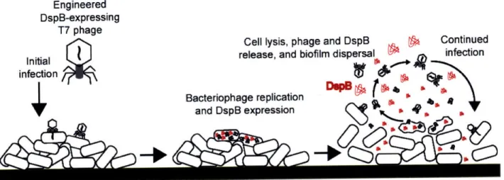
Figure 1. Two-pronged attack strategy for biofilm removal with enzymatically-active DspB-expressing T7DspB phage. Initial infection of E. coli biofilm results in rapid multiplication of phage and expression of DspB. Both phage and DspB are released upon lysis, leading to subsequent infection as well as degradation of the crucial biofilm EPS component, P-1,6-N-acetyl-D-glucosamine (49). Adapted from Ref. (42)... ... 23 Figure 2. Genomes of engineered phage used for biofilm treatment. (a) Genome of
T7select415-1 shows a unique BclI site and capsid gene lOB. (b) DspB-expressing phage T7DspB was created
by cloning T3 gene 1.2 into the unique BclI site and cloning the (pl0-dspB construct after capsid gene 10B. (c) Non-DspB-expressing control phage T7control was created by cloning T3 gene 1.2
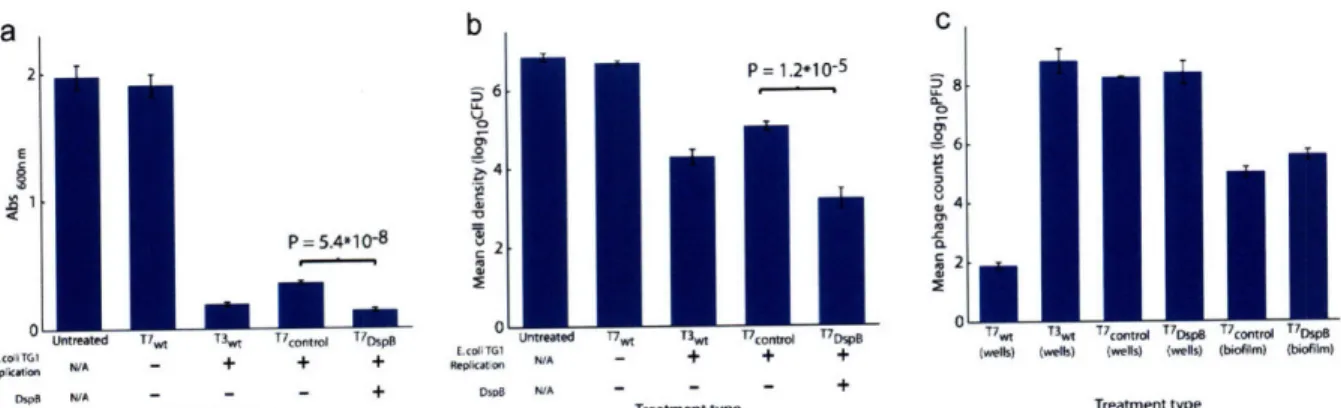
into the unique BclI site and cloning the control S-Tag insert (included in the T7select415-1 kit) as a fusion with the capsid gene 1OB. Adapted from Ref. (42). ... 24 Figure 3. Assays for E. coli TG1 biofilm levels and phage counts after 24 h with no treatment or with treatment with wild-type phage T7wt, wild-type phage T3wt, non-DspB-expressing control phage T7control, or DspB-expressing phage T7 DspB. Error bars indicate s.e.m. (a) Mean
absorbance (600 nm) for n = 16 biofilm pegs stained with 1% CV, solubilized in 33% acetic acid, and diluted 1:3 in lx PBS (62). (b) Mean cell densities (loglo(CFU/peg)) for n = 12 biofilm pegs. Pegs treated with T7DspB resulted in a 3.65 loglo(CFU/peg) reduction in viable cells recovered from E. coli biofilm compared to untreated biofilm. (c) Mean phage counts (loglo(PFU/peg)) recovered from media in n = 3 microtiter plate wells (wells) or sonication of n = 3 biofilm pegs (biofilm), as indicated, after 24 h of treatment with initial inoculations of 103 PFU/well. Both T7control and T7DspB showed evidence of replication with phage counts obtained from the microtiter plate wells or with phage counts recovered from the biofilms after sonication.
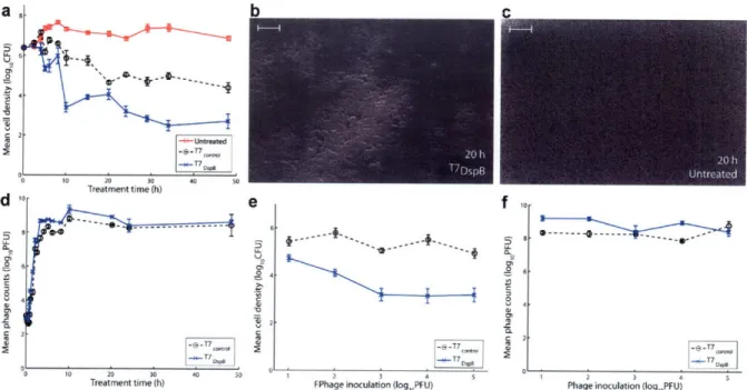
A dapted from R ef. (42) ... ... ... 26 Figure 4. Time-course curves, dosage response curves, and SEM images for engineered phage treatment targeting E. coli TG1 biofilm. Scale bars are 10 ýpm. Each data point in parts (a) and (e) represents the mean loglo-transformed cell density of n = 12 biofilm pegs. Each data point in parts (d) and (f) represents the mean loglo-transformed phage counts obtained from n = 3
microtiter plate wells. Error bars indicate s.e.m. (a) Time course (up to 48 h) of viable cell counts for no treatment (red squares), treatment with T7 ontrol (black circles), or treatment with
T7DspB (blue crosses) demonstrates that T7DspB significantly reduced biofilm levels compared
with T7control. (b) SEM image ofT7DspB-treated biofilm after 20 h shows significant disruption of the bacterial biofilm. (c) SEM image of untreated biofilm after 20 h shows a dense biofilm. (d) Time course of phage counts obtained after initial inoculation of E. coli TG1 biofilm with 103 PFU/well of T7control (black circles) or T7DspB (blue crosses). Both T7control and T7DspB began to
replicate rapidly after initial inoculation. (e) Dose response curves of mean cell densities (measured after 24 h of treatment) for T7control (black circles) and T7DspB (blue crosses). For all
initial phage inoculations, T7DspB-treated biofilm had significantly lower mean cell densities compared to T7control-treated biofilm. (f) Dose response curves of mean phage counts (measured after 24 h of treatment) for T7control (black circles) and T7DspB (blue crosses). For all initial phage
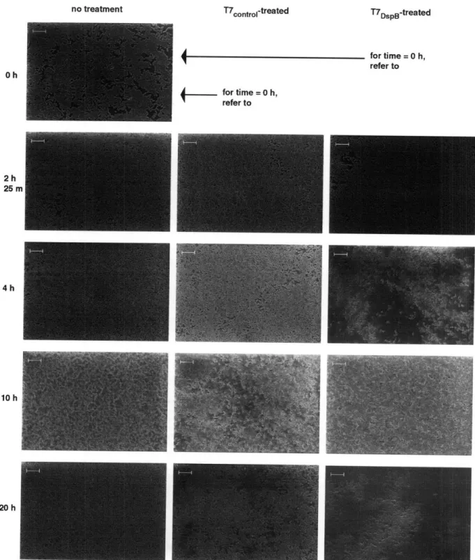
Figure 5. Scanning electron microscopy images for untreated, T7control-treated, and T7
DspB-treated biofilms. Scale bars are 10 gm. Consistent with time-course data (Figure 4a), T7
DspB-treated biofilm and T7control-DspB-treated biofilm were indistinguishable from unDspB-treated biofilm at 2 h 25 min post-infection. However, by 4 h post-infection, T7DspB-treated biofilm began to lyse and disperse significantly, while T7control-treated biofilm was still largely undisturbed. By 10 h post-infection, significant amounts of cell debris were seen in both T7control-treated and T7DspB-treated biofilms. At 20 h post-infection, T7,ýonftl-treated and T7DspB-treated biofilms had been disrupted by phage treatment, but T7DspB-treated biofilm was composed largely of cell debris and had fewer intact cells than T7conttol-treated biofilm. Adapted from Ref. (42)... 29 Figure 6. Genomes of unmodified M1 3mp 18 bacteriophage and engineered bacteriophage. Engineered bacteriophage were constructed by inserting genetic modules under the control of a synthetic promoter and ribosome-binding sequence in between SacI and PvuI restriction sites. (a) Unmodified control M13mp18 (p•on) contains lacZ to allow blue-white screening of engineered bacteriophage. (b) Engineered M13mpl8 bacteriophage expressing lexA3 (q91exA3). (C)
Engineered M13mpl 8 bacteriophage expressing soxR (9,oxR). (d) Engineered M13mpl 8
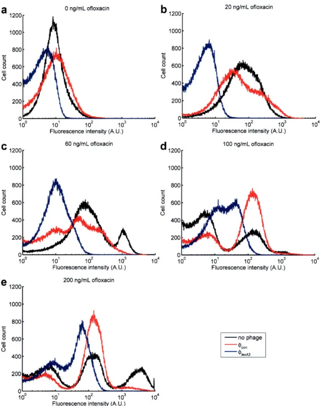
bacteriophage expressing csrA (9csrA). (e) Engineered M13mp 18 bacteriophage expressing ompF ((PompF). (f) Engineered M13mpl 8 bacteriophage expressing csrA and ompF ((PcsrA-ompF). ... 45 Figure 7. Flow cytometry of cells with an SOS-responsive GFP plasmid exposed to no phage (black lines), ,con phage (red lines), or 91exA3 phage (blue lines) for 6 hours with varying doses of ofloxacin. 10 plaque forming units per mL (PFU/mL) of phage were applied. Cells exposed to no phage or pon showed similar SOS induction profiles whereas cells with 91exA3 exhibited
significantly suppressed SOS responses. (a) 0 ng/mL ofloxacin treatment. (b) 20 ng/mL
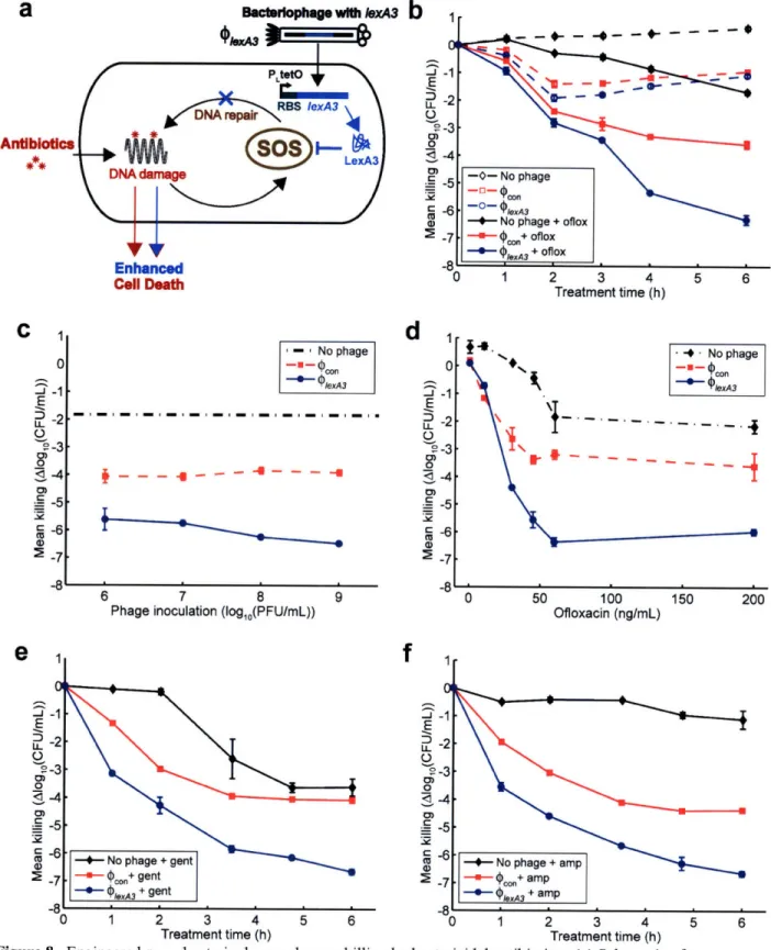
ofloxacin treatment. (c) 60 ng/mL ofloxacin treatment. (d) 100 ng/mL ofloxacin treatment. (e) 200 ng/m L ofloxacin treatm ent ... 46 Figure 8. Engineered Plex43 bacteriophage enhances killing by bactericidal antibiotics. (a)
Schematic of combination therapy with engineered bacteriophage and antibiotics. Bactericidal antibiotics induce DNA damage via hydroxyl radicals, leading to either cell death or induction of the SOS response followed by DNA repair and survival (91). Engineered phage carrying the lexA3 gene (PlexA3) under the control of the synthetic promoter PLtetO and a ribosome-binding sequence (99) acts as an antibiotic adjuvant by suppressing the SOS response due to DNA damage and increasing cell death. (b) Killing curves for no phage (black diamonds), unmodified 9pon phage (red squares), and 'PlexA3 (blue circles) without ofloxacin (dotted lines, open symbols)
or with 60 ng/mL ofloxacin [oflox] (solid lines, closed symbols). 108 PFU/mL phage was used. 9PlexA3 greatly enhanced killing by ofloxacin by 4 hours of treatment. (c) Phage dose response shows that PlexA3 (blue circles with solid line) is a strong adjuvant for ofloxacin (60 ng/mL) over a wide range of initial inoculations compared with no phage (black dash-dotted line) and
unmodified pon (red squares with dashed line). (d) Ofloxacin dose response shows that 9lexA3 (blue circles with solid line) improves killing even at low levels of drug compared with no phage (black diamonds with dash-dotted line) and unmodified pcon (red squares with dashed line). 108 PFU/mL phage was used. (e) Killing curves for no phage (black diamonds), unmodified (poon (red squares), and (PleA3 (blue circles) with 5 gLg/mL gentamicin [gent]. 109 PFU/mL phage was used. 9lexA3 phage greatly improved killing by gentamicin. (f) Killing curves for no phage (black diamonds), unmodified p(con (red squares), and (lexA3 (blue circles) with 5 pg/mL ampicillin [amp]. 109 PFU/mL phage was used. 1PexA3 phage greatly improved killing by ampicillin. ... 48
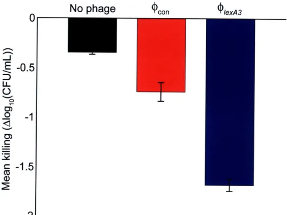
Figure 9. Persister killing assay demonstrates that engineered bacteriophage can be applied to a previously drug-treated population to increase killing of surviving persister cells. After 3 hours of 200 ng/mL ofloxacin treatment, no phage (black bar), 109 PFU/mL unmodified wcon phage (red bar), or 109 PFU/mL engineered (PlexA3 phage (blue bar) were added to the previously drug-treated cultures. Three additional hours later, viable cell counts were obtained and demonstrated that (PlexA3 was able to reduce persister cell levels better than no phage or unmodified ,,,on. ... 50
Figure 10. Mean killing with or without 60 ng/mL ofloxacin after 12 hours of treatment of E. coli biofilms pregrown for 24 hours. Where indicated, 108 PFU/mL of bacteriophage was used.
These results demonstrate that (PlexA3 and 9csrAompFmaintain their effectiveness as antibiotic
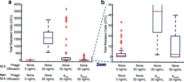
adjuvants against bacteria living in biofilms. ... ... 51 Figure 11. Box-and-whisker plot of the total number of E. coli cells in 60 observations that were resistant to 100 ng/mL ofloxacin after growth under various conditions (red bars indicate
medians, red diamonds represent outliers). (a) Cells grown with no phage and no ofloxacin for 24 hours had very low numbers of antibiotic-resistant cells. Cells grown with no phage and 30 ng/mL ofloxacin for 24 hours had high numbers of resistant cells due to growth in subinhibitory drug concentrations (101). Cells grown with no phage and 30 ng/mL ofloxacin for 12 hours followed by 109 PFU/mL unmodified 9,on and 30 ng/mL ofloxacin for 12 hours exhibited a
modest level of antibiotic-resistant bacteria. Cells grown with no phage and 30 ng/mL ofloxacin for 12 hours followed by 109 PFU/mL PlexA3 and 30 ng/mL ofloxacin for 12 hours exhibited a low level of antibiotic-resistant bacteria, close to the numbers seen with no ofloxacin and no phage. (b) Zoomed-in version of box-and-whisker plot in (a) for increased resolution around low total resistant cell counts confirms that (PlexA3 with 30 ng/mL ofloxacin treatment reduced the number
of resistant cells to levels similar to that of 0 ng/mL ofloxacin with no phage. ... 52 Figure 12. Engineered bacteriophage targeting non-SOS systems as adjuvants for ofloxacin treatment [oflox]. (a) Ofloxacin stimulates superoxide generation, which is normally countered by the oxidative stress response, coordinated by SoxR (91). Engineered bacteriophage producing
SoxR (9(oxR) enhances ofloxacin-based killing by disrupting regulation of the oxidative stress response. (b) Killing curves for no phage (black diamonds), control pcon (red squares), and psoxR
(blue downwards-facing triangles) without ofloxacin (dotted lines, open symbols) or with 60 ng/mL ofloxacin (solid lines, closed symbols). 108 PFU/mL phage was used. Killing curves for no phage and unmodified Pwonl are reproduced from Figure 8b for comparison and show that psoxR
enhances killing by ofloxacin. (c) CsrA suppresses the biofilm state in which bacterial cells tend to be more resistant to antibiotics (62). OmpF is a porin used by quinolones to enter into
bacterial cells (110). Engineered bacteriophage producing both CsrA and OmpF simultaneously
((PcsrA-ompF) represses biofilm formation and antibiotic tolerance via CsrA and enhances antibiotic
penetration via OmpF to produce an improved dual-targeting adjuvant for ofloxacin. (d) Killing
curves for PcsrA (black diamonds), 9ompF (red squares), and (PcsrA-ompF (brown upwards-facing
triangles) without ofloxacin (dotted lines, open symbols) or with 60 ng/mL ofloxacin (solid lines, closed symbols). 108 PFU/mL phage was used. Phage expressing both csrA and ompF
(pcsrA-ompF) is a better adjuvant for ofloxacin than phage expressing csrA alone (PcsrA) or ompF alone (q(ompF). (e) Phage dose response shows that both (psoxR (blue downwards-facing triangles with
solid line) and (PcsrA-ompF (brown upwards-facing triangles with solid line) are effective as adjuvants for ofloxacin (60 ng/mL) over a wide range of initial inoculations. Phage dose response curves for no phage (black dash-dotted line) and unmodified pccon (red squares with
dashed line) are reproduced from Figure 8c for comparison. (f) Ofloxacin dose response shows that both psoxR (blue downwards-facing triangles with solid line) and (PcsrA-ompF (brown upwards-facing triangles with solid line) improve killing throughout a range of drug concentrations. 108 PFU/mL phage was used. Ofloxacin dose response curves for no phage (black diamonds with dash-dotted line) and unmodified Pcon (red squares with dashed line) are reproduced from Figure
8d for comparison. ... 55 Figure 13. Autoregulated negative-feedback module with wild-type lexA repressing PLexO from Ref. (86) may increase the level of lexA expression when lexA is cleaved by recA in response to DNA damage by agents such as ofloxacin. ... 58 Figure 14. Paired-termini design from Ref. (114) in which the antisense RNA is cloned between the flanking restriction sites at the top of the stem. Reprinted from Ref. (114). ... 68 Figure 15. RNA sensor design #1 should repress the gfp fusion in the absence of a cat-containing plasmid such as pZA3, leading to low GFP output. In the presence of pZA3, cat mRNA should compete with gfp mRNA for PT7-asRNA, leading to derepression of cat-gf and thus higher GFP output. ... ... 71 Figure 16. RNA sensor design #1 (pZE21sl-cat) should detect a cat-containing plasmid such as pZA3 and not a bla-containing AmpR plasmid such as pZA1. KanR = kanamycin resistance, CmR = chloramphenicol resistance, AmpR = ampicillin resistance. ... 72 Figure 17. Testing DH5aPro + pZE21 sl-cat with 1:100 dilutions of overnight stocks in 3 mL LB + kanamycin (30 jig/mL) at 37"C with 0.1% arabinose and 30 ng/mL aTc. Co-inoculation with pZA3, a cat-expressing plasmid, yields an approximately two-fold increase in fluorescence compared with no plasmid or pZAl, a bla-expressing plasmid. These results demonstrate that pZE21sl-cat functions as a selective mRNA sensor. (a) After 4.5 hours of growth. (b) After 5.5 hours of growth. (c) After 6.25 hours of growth... ... ... 73 Figure 18. RNA sensor design #2 (pTAKs2-cat or pTAKs2-kan) should express high levels of lacd in the absence of a target-containing plasmid leading to low GFP output. In the presence of a target-containing plasmid, the target mRNA should bind to the asRNA upstream of the lacd RBS, leading to blocked translation or mRNA degradation of lacld and thus higher GFP output. 75 Figure 19. RNA sensor design #3 (pTAKs3-cat) should express high levels of lacld in the absence of a target-containing plasmid leading to low GFP output. In the presence of a target-containing plasmid, the target mRNA should bind to the PT7-asRNA, leading to mRNA degradation of lacd and thus higher GFP output. ... 77 Figure 20. Synthetic gene circuit represses horizontal transmission of antibiotic-resistance genes using an autoregulated negative-feedback loop. (a) Antibiotics can cause cleavage of repressors (such as SetR) which suppress horizontal transmission in their normal intact state. Cleavage of repressors from antibiotic-resistance operons in resistant cells results in promoter derepression and subsequent overexpression of repressor from the synthetic gene circuit. The high level of repressor results in the suppression of horizontal transmission, even in the face of antibiotics. In this circuit, the repressor protein serves as both "sensor" and "effector". (b) In the absence of
circuit and leave it dormant. (c) In the absence of antibiotic-resistance operons (in non-resistant cells), the synthetic gene circuit represses itself and therefore exhibits little activity ... 78
1
INTRODUCTION
Biofilms and antibiotic resistance pose a significant hurdle to eliminating bacterial
infections with conventional antimicrobial drugs. Patients that would have been easily cured by antibiotics in the past are now dying or remaining sick for much longer due to biofilms and antibiotic-resistant bacterial infections (10, 13, 14). The economic cost of antibiotic resistance in the United States alone is estimated to be between US $5 billion and US $24 billion per year (15). Therefore, it is imperative that new antibacterial strategies be explored (16).
Bacteria frequently live in biofilms, which are surface-associated communities enclosed in a hydrated extracellular polymeric substances (EPS) matrix composed of polysaccharides,
proteins, nucleic acids, and lipids which helps maintain a complex heterogeneous structure (2, 3). Biofilms constitute an essential and protective lifestyle for bacteria in many different natural and man-made environments, including dental plaques, water pipes, medical devices, and industrial systems (17). Bacterial biofilms have been implicated as a source of persistent infection, contamination, and biofouling due to inherent resistance to antimicrobial agents and host immune defenses (18). Thus, there exists a growing need for novel and effective treatments targeted at biofilms, particularly in light of the continually-worsening problem of antibiotic resistance and the discovery that antibiotic use can even induce biofilm formation (5, 6).
In addition to inherent bacterial resistance to antimicrobials in biofilm, antibiotic resistance can result from mutations in antibacterial targets or from acquisition of genes that encode
proteins which promote the efflux of antibiotics or bind and inactivate antibiotics (7). Gene acquisition is usually due to horizontal gene transfer via transformation, plasmids, or conjugative transposons (19, 20). For example, Enterococcusfaecalis in nosocomial settings became
completely vancomycin-resistant by 1988 (19). Co-infecting staphylococci have subsequently received vanA resistance genes from Enterococcus faecalis (19). Staphylococcus aureus acquired resistance to sulpha drugs in the 1940s, penicillin-resistance in the 1950s, methicillin-resistance in the 1980s, and vancomycin-methicillin-resistance in 2002 (19). The heavy use of antibiotics in livestock in the agricultural industry has contributed to the emergence of methicillin-resistant staphylococci and is unlikely to abate (19). Streptococcus pneumoniae and Neisseria gonorrhoeae have also obtained resistance to antibiotics (19).
Another way for bacterial cells to be resistant to antimicrobial agents is through the
phenomena of persistence (8). Persistence is believed to be a stochastic process in which certain cells in a metabolically-dormant stage are able to avoid being killed by multiple antibiotics (8). Persisters do not carry genetic mutations but instead exhibit phenotypic resistance to antibiotics (21). In E. coli, persister levels increase markedly in late-exponential and stationary phases and are important components ofbiofilm (8). Chromosomally-encoded toxins may contribute to the persister phenotype (22-24). However, the underlying mechanisms controlling the stochastic process of persistence are not well understood (8).
Proposed solutions to limit the spread of antibiotic resistance include reducing antibiotic use, preventing the spread of resistant bacteria particularly in nosocomial settings, using novel antibiotics to which pathogens are not resistant, and limiting person-to-person transmission by reducing the carrier rate in health-care workers and patients (10). These attempts to control the
spread of antibiotic resistance require great design, compliance, and effort to achieve efficacy. While these techniques may certainly help control the spread of antibiotic resistance, they may be difficult to implement successfully in real life and cannot avoid the evolutionary pressure that antibiotic use places on bacteria to select for resistance. For example, ecological models of
antibiotic cycling suggest that conventional cycling will not reduce antimicrobial resistance in hospitals (25). In many cases, bacteria with antibiotic resistance genes maintain them stably and do not fare poorly against non-resistant strains (8). Constant evolutionary pressure will ensure that antibiotic resistance bacteria will continue to grow in number. The dearth of new
antibacterial agents being developed in the last 25-30 years certainly bodes poorly for the future of the antibiotic era (1). Thus, new methods for combating bacterial infections are needed in order to prolong the antibiotic age. For example, bacteriophage therapy or synthetic antibacterial peptides have been proposed as potential solutions (16, 26).
Phage therapy has begun to be accepted in industrial and biotechnological settings. For example, the FDA recently approved the use of phage targeted at Listeria monocytogenes as a food additive (27). However, phage therapy has several challenges that must be overcome before it will be accepted in Western medicine for treating humans (28). These problems include the lack of properly designed clinical trials to date (28), development of phage resistance (11, 12, 29), phage immunogenicity in the human body and clearance by the reticuloendothelial system (RES) (11, 30), the release of toxins upon bacterial lysis (11), and phage specificity (11).
Fortunately, many of these concerns are currently being studied and addressed. For example, combination therapy with antibiotics and phage may alleviate the development of phage
resistance (11, 12, 29). Long-circulating phage can be isolated that can avoid RES clearance to increase in vivo efficacy (30). The problem of phage clearance is an important one that needs to be solved as it may make phage therapy more useful for treating transient infections rather than
chronic ones. Non-lytic and non-replicative phage have been engineered to kill bacteria while minimizing endotoxin release (31, 32). Progress is also being made in the development of
The specificity of phage for host bacteria is both an advantage and a disadvantage for
phage therapy. Specificity allows human cells as well as innocuous bacteria to be spared, potentially avoiding serious issues such as drug toxicity or Clostridium difficile overgrowth that
can arise with antibiotic use. C. difficile infection is characterized by diarrhea and colitis, and
has increased in severity in recent years (34). Antibiotic therapy is believed to alter the
microbial flora in the colon due to lack of target specificity, thus allowing C. difficile to
proliferate and cause disease (35). However, host specificity means that a well-characterized
library of phage must be maintained so that an appropriate therapy can be designed for each
individual infection (11). The diversity of bacterial infections implies that it may be difficult for
any particular engineered phage to be a therapeutic solution for a wide range of biofilms.
Indeed, phage therapy generally requires the use of phage cocktails to cover a range of target
bacteria.
To reduce biofilms, I have developed an enzymatically-active bacteriophage platform to
produce phage which express biofilm-dispersing enzymes during infection followed by cell lysis.
To attack antibiotic-resistant bacteria, I built a synthetic bacteriophage platform to target gene
networks as antibiotic adjuvants. Finally, I developed synthetic in vivo sensors to detect the
presence of antibiotic-resistant genes within individual bacterial cells. These sensors can be
connected to downstream synthetic effector components which kill bacteria that carry resistance
genes or suppress horizontal transmission of those genes. These are synthetic biology solutions
2 ENGINEERED PHAGE THERAPY FOR BACTERIAL
BIOFILMS
2.1
Introduction
Over the last few years, synthetic biology has enabled the development of many engineered biological devices and cells with interesting and well-modelled characteristics (36-38). At the same time, new technologies for more cost-effective DNA synthesis and sequencing have been reported (39). These advances allow for large-scale synthetic genomes to be designed and built with much greater ease than is currently possible with traditional molecular biology methods. Synthetic biologists have begun to address important real-world problems by modifying organisms to produce artemisin precursors (40), developing bacteria that can target cancerous cells (41), and producing new antimicrobial peptides (16), to name a few examples (37). Synthetic biology is distinguished from traditional genetic engineering through the use of modularity, abstraction, and standardization to allow generalized principles and designs to be applied to different scenarios. In this work, I engineered bacteriophage with biofilm-degrading enzymatic activity to create a synthetic biology platform for eradicating bacterial biofilms. The text used in this section to describe this work was published and therefore reproduced from Ref.
(42) in Proceedings in the National Academy of Sciences.
Bacteria frequently live in biofilms, which are surface-associated communities encased in a hydrated EPS matrix, that is composed of polysaccharides, proteins, nucleic acids, and lipids and helps maintain a complex heterogeneous structure (2, 3). Biofilms constitute an essential and protective lifestyle for bacteria in many different natural and man-made environments, including dental plaques, water pipes, medical devices, and industrial systems (17). Bacterial biofilms have been implicated as a source of persistent infection and contamination in medical, industrial, and
food processing settings due to inherent resistance to antimicrobial agents and host defenses (2, 4, 18, 43). Thus, there exists a growing need for novel and effective treatments targeted at biofilms, particularly in light of the continually-worsening problem of antibiotic resistance and the discovery that antibiotic use can even induce biofilm formation (5, 6).
Bacteriophage treatment has been proposed as one method for controlling bacterial biofilms (26). Phage have been used since the early 2 0 th century to treat bacterial infections,
especially in Eastern Europe, and have been shown to decrease biofilm formation (26, 28, 44). For example, phage T4 can infect and replicate within Escherichia coli biofilms and disrupt biofilm morphology by killing bacterial cells (45-47). Phage have also been modified to extend their natural host range. E. coli which produce the K1 polysaccharide capsule are normally resistant to infection by T7, but are susceptible to T7 that have been designed to express K1-5 endosialidase (48). Enzymatic degradation of EPS components is another useful strategy for disrupting biofilms, though bacterial cells are not killed, which may result in the release of many bacteria into the environment (2, 49, 50). For instance, enzymatic degradation of a cell-bound EPS polysaccharide adhesin known as polymeric f3-1,6-N-acetyl-D-glucosamine (PGA) by exogenously-applied dispersin B (DspB) has been demonstrated to reduce biofilms of several different species of bacteria (49, 51). DspB, an enzyme which is produced by Actinobacillus actinomycetemcomitans, hydrolyzes PGA, a crucial adhesin needed for biofilm formation and integrity in Staphylococcus and E. coli, including E. coli K-12 as well as clinical isolates (51). Reports of natural lytic phage with phage-borne polysaccharide depolymerases have shown that phage-induced lysis and EPS degradation are used in combination in natural systems to reduce bacterial biofilms (52, 53). These depolymerases appear to be carried on the surfaces of phage
However, the chance that one can isolate a natural phage that is both specific for the bacteria to
be targeted and expresses a relevant EPS-degrading enzyme is likely to be low (11).
Therefore, I propose a modular design strategy in which phage that kill bacteria in a species-specific manner are engineered to express the most effective EPS-degrading enzymes
specific to the target biofilm. This strategy should permit the development of a diverse library of biofilm-dispersing phage rather than trying to isolate such phage from the environment. By multiplying within the biofilm and hijacking the bacterial machinery, engineered enzymatically-active phage should be able to achieve high local concentrations of both enzyme and lytic phage to target multiple biofilm components, even with small initial phage inoculations. Rapid phage replication with subsequent bacterial lysis and expression ofbiofilm-degrading enzymes should render this two-pronged attack strategy an efficient, autocatalytic method for removing bacterial biofilms in environmental, industrial, and clinical settings (Figure 1). This design also removes the need to express, purify, and deliver large doses of enzyme to specific sites of infection that may be difficult to access, and should improve the efficacy of phage therapy at removing biofilms. Increasingly cost-effective genome sequencing and synthetic biology technologies, which include the refactoring of phage genomes and large-scale DNA synthesis (37, 55, 56),
should further enable the production of engineered enzymatic phage and significantly extend the limited repertoire of biofilm-degrading phage that have been isolated from the environment.
Engineered DspB-expressing
T7 phage
N. C~II Ivpif nhanA srne flcnR NGA flnntinmlld
Figure 1. Two-pronged attack strategy for biofilm removal with enzymatically-active DspB-expressing T7DspB
phage. Initial infection of E. coli biofilm results in rapid multiplication of phage and expression of DspB. Both phage and DspB are released upon lysis, leading to subsequent infection as well as degradation of the crucial biofilm EPS component, f-1,6-N-acetyl-D-glucosamine (49). Adapted from Ref. (42).
2.2 Results
2.2.1 Design of Enzymatically-Active Bacteriophage.
As a proof-of-principle design of artificial biofilm-degrading bacteriophage, I engineered T7, an E. coli-specific phage (57, 58), to express DspB intracellularly during infection so DspB would be released into the extracellular environment upon cell lysis (Figure 1). I employed a modified T7 strain (Novagen T7select415-1) with several deletions of nonessential genes (Figure 2a). I cloned the gene coding for DspB (dspB) under the control of the strong T7 cp 10 promoter so dspB would be strongly transcribed by T7 RNA polymerase during infection (Figure 2b). As a control, I cloned an S-Tag insert into the T7 genome so that no DspB would be produced (Figure 2c).
a T7select415-1
genome
Bcil
site
010
promoter
T4 terminator
b DspB-expressing
T7DspB genomeBcil
site
Bcil site
010
promoter
010
promoter
T4 terminator
T3 gene
1.2
_
I--j~--
I
dspB
//
c
non-DspB-expressing
T7controlgenome
BcII
site
Bcll site
410 promoter
T4 terminator
T3 gene 1.2
1OB-S.Tagff
Figure 2. Genomes of engineered phage used for biofilm treatment. (a) Genome of T7select415-1 shows a unique BclI site and capsid gene lOB. (b) DspB-expressing phage T7DspB was created by cloning T3 gene 1.2 into the
unique BclI site and cloning the 0p l-dspB construct after capsid gene lOB. (c) Non-DspB-expressing control phage
T7control was created by cloning T3 gene 1.2 into the unique BclI site and cloning the control S-Tag insert (included in the T7select415-1 kit) as a fusion with the capsid gene 1OB. Adapted from Ref. (42).
To test the effectiveness of our engineered phage against pre-grown biofilm, I cultivated E. coli TG1(lacl::kan) biofilms in LB media on plastic pegs using the standardized MBEC
biofilm cultivation system. I used E. coli TG1 as the target biofilm strain since TG1 forms a thick, mature biofilm and contains the F plasmid (59). The F plasmid enhances biofilm maturation along with other biofilm-promoting factors in E. coli, including PGA, flagellum, cellulose, curli, antigen 43, and other conjugative pili and cell surface adhesins (59, 60). Because T7 is unable to replicate efficiently in F-plasmid-containing E. coli, gene 1.2 from T3 phage was also cloned into the unique Bcll site in our engineered T7 phage and control T7 phage to
circumvent F-plasmid-mediated exclusion and extend the phage host range (Figure 2b and Figure 2c) (61). The control phage and engineered phage were named T7control and T7DspB, respectively
2.2.2 Characterization of Enzymatically-Active Bacteriophage
To determine whether the T7DspB phage was more effective than the T7ontrol phage, I first employed a crystal violet (CV) assay to assess the amount of biofilm on the pegs after phage treatment. Pre-grown TGl(lacI::kan) biofilm was inoculated with only LB media or infected with 103 plaque forming units per peg (PFU/peg) of T7ontrol or T7DspB phage (Figure 3a). To
assess whether our engineered enzymatic phage was more efficacious than wild-type phage at attacking biofilm despite being made with a modified T7 phage, I also treated biofilm with wild-type T7 (T7wt) or wild-wild-type T3 (T3wt) (Figure 3a). After 24 h of treatment, CV staining of untreated biofilm had a 600 nm absorbance (A600) approximately equal to that for T7wt-treated
biofilm (Figure 3a). Both T3wt-treated biofilm and T7cont~i-treated biofilm were much reduced compared with the untreated biofilm: the former had an A600 that was lower than that of
untreated biofilm by a factor of 10.3, while the latter had an A600 that was lower than that of untreated biofilm by a factor of 5.6 (Figure 3a). The amount ofbiofilm left on the T7DspB-treated pegs was the least of all the treatment types, with an A600 which was less by a factor of 14.5 than
that of untreated biofilm and less by a factor of 2.6 than that of T7control-treated biofilm (P = 5.4*10-8). These findings demonstrate that DspB expression in T7 DspB is crucial to elevating its biofilm-removing efficacy over that of wild-type phage and non-enzymatic T7control phage
n h r P 1.2*10-5 56 8 4. 8 ee T2
LMrceated T7wz T3wt WCanod TIDSP T7w, T3wt T7c0mra T7om* WTcontrl V0soB
Re Ttion A - + + + N/A - + + + (wells) (wells) (wells) (wells) (bloflm) (bNeilm) DspR N/A - - - + DwB N/A - - - +
Treatment type Treatment type Treatment type
Figure 3. Assays for E. coli TG1 biofilm levels and phage counts after 24 h with no treatment or with treatment with
wild-type phage T7wt, wild-type phage T3wt, non-DspB-expressing control phage T7contol, or DspB-expressing phage T7DspB. Error bars indicate s.e.m. (a) Mean absorbance (600 nm) for n = 16 biofilm pegs stained with 1% CV, solubilized in 33% acetic acid, and diluted 1:3 in lx PBS (62). (b) Mean cell densities (loglo(CFU/peg)) for n = 12 biofilm pegs. Pegs treated with T7DspB resulted in a 3.65 loglo(CFU/peg) reduction in viable cells recovered from E.
coli biofilm compared to untreated biofilm. (c) Mean phage counts (loglo(PFU/peg)) recovered from media in n = 3
microtiter plate wells (wells) or sonication of n = 3 biofilm pegs (biofilm), as indicated, after 24 h of treatment with initial inoculations of 103 PFU/well. Both T7contr0o and T7DspB showed evidence of replication with phage counts
obtained from the microtiter plate wells or with phage counts recovered from the biofilms after sonication. Adapted from Ref. (42).
To confirm that the decrease in CV staining corresponded with killing ofbiofilm cells, I used sonication to obtain viable cell counts (CFU/peg) for bacteria surviving in the biofilms after phage treatment. Pre-grown TGl(lacI::kan) biofilm (prior to treatment) reached a mean cell
density of 6.4 loglo(CFU/peg) after 24 h of growth (Figure 3b). After 24 h of additional growth in new LB media with no phage treatment, the untreated biofilm had a mean cell density of 6.9 loglo(CFU/peg) (Figure 3b). T3wt-treated biofilm had a mean cell density that was less than that of T7control-treated biofilm by a factor of 5.9 and greater than that of T7DspB-treated biofilm by a factor of 12 (Figure 3b). T7control-treated biofilm had a mean cell density of 5.1 loglo(CFU/peg) while the mean cell density for T7DspB-treated biofilm was 3.2 loglo(CFU/peg), the lowest of all the treatment types (Figure 3b). The difference in viable cells recovered from T7control-treated biofilm and T7DspB-treated biofilm was statistically significant (P = 1.2*10-5). These results are
consistent with the CV staining data and demonstrate that DspB-expressing T7DspB phage are substantially more effective at killing E. coli TG1 biofilm compared with type T3wt, wild-type T7wt, and non-DspB-expressing control T7control phage.
Our two-pronged method of biofilm eradication involves expression of DspB and rapid phage replication (Figure 1). To confirm that our phage multiplied, I obtained PFU counts from media in the microtiter plate wells. By 24 h of treatment, wild-type T7 had not replicated but wild-type T3 had multiplied significantly within the biofilm (Figure 3c). To compare the amount of phage in the microtiter plate wells with phage residing in the biofilms, I also obtained PFU
counts by sonicating the biofilms. After 24 h of treatment, PFU counts for T7control and T7DspB
recovered from the microtiter plate wells were several orders of magnitude greater than PFU counts recovered by sonication of the biofilms (Figure 3c). Overall, PFU counts obtained from the wells and the biofilms were all orders of magnitude greater than the initial inoculation of 103 PFU, confirming that phage multiplication indeed took place (Figure 3c).
2.2.3 Time Courses and Dose-Responses for Enzymatically-Active
Bacteriophage Treatment
Since I determined that T7 DspB had greater biofilm-removing capability than T7control after 24 h of infection, I next sought to determine the time course of biofilm destruction. As shown in Figure 4a, by 5 h post-infection, T7DspB-treated biofilm had a mean cell density that was 0.82 logio(CFU/peg) less than T7control-treated biofilm (P = 2.0*10-4). At 10 h post-infection, T7
DspB-treated biofilm began to settle at a steady-state mean cell density between 3 to 4 loglo(CFU/peg), while T7con,,-treated biofilm flattened out at approximately 5 logio(CFU/peg) by 20 h post-infection (Figure 4a). T7DspB-treated biofilms had mean cell densities that were approximately two orders of magnitude lower than T7control-treated biofilms, up to 48 h of total treatment (Figure 4a), and importantly, T7DspB treatment reduced biofilm levels by about 99.997% (4.5 loglo(CFU/peg)) compared with untreated biofilm. I found no evidence of phage resistance developing over the long time course of treatment (Figure 4a).
b C f 10 --- I-j I.----e-T7 cmc 1 2 3 4
FPhage inoculation (loglPFU)
-o
-e-n i~n~l icn
L)lpB
1 2 3 4 5
Phage inoculation (Iog,,PFU)
Figure 4. Time-course curves, dosage response curves, and SEM images for engineered phage treatment targeting E. coli TG1 biofilm. Scale bars are 10 tm. Each data point in parts (a) and (e) represents the mean loglo0
-transformed cell density of n = 12 biofilm pegs. Each data point in parts (d) and (f) represents the mean loglo0
-transformed phage counts obtained from n = 3 microtiter plate wells. Error bars indicate s.e.m. (a) Time course (up to 48 h) of viable cell counts for no treatment (red squares), treatment with T7contlol (black circles), or treatment with
T7DspB (blue crosses) demonstrates that T7DspB significantly reduced biofilm levels compared with T7control. (b) SEM
image of T7DspB-treated biofilm after 20 h shows significant disruption of the bacterial biofilm. (c) SEM image of untreated biofilm after 20 h shows a dense biofilm. (d) Time course of phage counts obtained after initial inoculation of E. coli TG1 biofilm with 103 PFU/well of T7control (black circles) or T7DspB
(blue crosses). Both T7ontrol and T7DspB began to replicate rapidly after initial inoculation. (e) Dose response curves of mean cell
densities (measured after 24 h of treatment) for T7contol (black circles) and T7
DspB (blue crosses). For all initial phage inoculations, T7DspB-treated biofilm had significantly lower mean cell densities compared to T7control-treated biofilm. (f) Dose response curves of mean phage counts (measured after 24 h of treatment) for T7control (black circles)
and T7DspB (blue crosses). For all initial phage inoculations, both T7control and T7DspB multiplied significantly.
Adapted from Ref. (42).
I also used scanning electron microscopy (SEM) to image the biofilm pegs over the time course of phage treatment in order to directly visualize biofilm dispersal by our enzymatically-active phage (Figure 4b, Figure 4c, and Figure 5). After 20 h of treatment, T7DspB-treated biofilm (Figure 4b) was significantly disrupted compared with the untreated biofilm (Figure 4c). These results confirm that T7DspB indeed causes biofilm reduction and bacterial cell killing.
Treatment time (h)
0
· · ·
'10 h
Untrýmcd
T7control-treated T7DspB-treated for time = 0 h, refer to for time = 0 h, refer to 2h 25 rr 4h 10 h 20 h
Figure 5. Scanning electron microscopy images for untreated, T7conol1-treated, and T7DspB-treated biofilms. Scale
bars are 10 gm. Consistent with time-course data (Figure 4a), T7DspB-treated biofilm and T7control-treated biofilm were indistinguishable from untreated biofilm at 2 h 25 min post-infection. However, by 4 h post-infection, T7
DspB-treated biofilm began to lyse and disperse significantly, while T7conlroz-DspB-treated biofilm was still largely undisturbed. By 10 h post-infection, significant amounts of cell debris were seen in both T7cont.o-treated and T7DspB-treated biofilms. At 20 h post-infection, T7,ono,,l-treated and T7DpB-treated biofilms had been disrupted by phage treatment, but T7DpB-treated biofilm was composed largely of cell debris and had fewer intact cells than T7con.ol-treated
biofilm. Adapted from Ref. (42).
Oh
To verify that phage replication was occurring over time, I obtained PFU counts in the microtiter wells. As seen in Figure 4d, both T7control and T7DspB began to replicate within the
bacterial biofilm as early as 50 minutes post-infection. By about 190 minutes, T7contr. and T7DspB PFU/peg approached steady-state levels of approximately 8 to 9 loglo(PFU/peg),
indicating that phage replication had occurred (Figure 4d). T7DspB PFU/peg were generally
higher than T7control PFU/peg but not by orders of magnitude as was the case for CFU counts per peg. This is because the T7 burst size (-250 PFU per infective center) (63) multiplied by the number of the extra cells killed by T7DspB, compared with T7control, equals extra PFU/peg that are insignificant compared with the PFU levels already reached by T7control. I did not note any significant differences in burst sizes and growth rates between T7DspB and T7contro0 (data not shown).
Considering that the above experiments were carried out with initial inoculations of 103 PFU/peg, which translates to a multiplicity of infection (MOI) of about 1:10 3.4 (Figure 4a), I next
aimed to determine the effect of changing the initial MOI on biofilm removal. With low phage doses, repeated rounds of phage multiplication and DspB expression should promote biofilm dispersal and allow more bacterial cells to be accessible for subsequent phage infection. With high phage doses, initial DspB production post-infection should also be very disruptive to biofilm integrity. As shown in Figure 4e, T7DspB was more effective than T7contro at removing
biofilm at all inoculation levels tested, ranging from 101 PFU/peg to 105 PFU/peg. A dose-dependent effect of phage inoculation on biofilm destruction was observed, with larger inoculations leading to lower mean cell densities, particularly for T7DspB (Figure 4e). At inoculation levels greater than or equal to 102 PFU/peg, mean cell densities (CFU/peg) for T7DspB-treated biofilm were significantly lower than those for T7contro-treated biofilm by a factor
of 49-232 (Figure 4e). Thus, at low and high initial inoculations, DspB-expressing T7 is more efficacious at disrupting E. coli TG1 biofilm compared with non-DspB-expressing control T7. Note also that all phage dosages tested exhibited phage multiplication within the biofilm (Figure 4f). These results together suggest that DspB-expressing phage may have improved efficacy in real-world situations where the ability to deliver high levels of phage to biofilms may be limited or where sustained phage replication is less likely, e.g., in the gastrointestinal tract of cholera patients (29, 30).
2.3 Discussion
In this work, I demonstrated that engineered phage which express biofilm-degrading enzymes are more efficacious at removing bacterial biofilms than non-enzymatic phage alone. Though our results were obtained for a prototype, proof-of-principle phage, I believe that our design can be adapted to work in other phage and with other biofilm-degrading enzymes to target a wide range ofbiofilms. Thus, engineered bacteriophage treatment should be considered as an addition to the therapies available for use against bacterial biofilms in medical, industrial, and biotechnological settings (28). Future improvements to this design may include directed evolution for optimal enzyme activity, delaying cell lysis or using multiple phage promoters to allow for increased enzyme production, targeting multiple biofilm EPS components with different proteins as well as targeting multi-species biofilm with a cocktail of different species-specific engineered enzymatically-active phage, and combination therapy with antibiotics and phage to improve the efficacy of both types of treatment.
Phage therapy has begun to be accepted in industrial and biotechnological settings. For example, the FDA recently approved the use of phage targeted at Listeria monocytogenes as a food additive (27). However, phage therapy has several challenges that must be overcome
before it will be accepted in Western medicine for treating humans (28). These problems include the lack of properly designed clinical trials to date (28), development of phage resistance (11, 12, 29), phage immunogenicity in the human body and clearance by the reticuloendothelial system (RES) (11, 30), the release of toxins upon bacterial lysis (11), and phage specificity (11). Fortunately, many of these concerns are currently being studied and addressed. For example, combination therapy with antibiotics and phage may alleviate the development of phage
resistance (11, 12, 29). Long-circulating phage can be isolated that can avoid RES clearance to increase in vivo efficacy (30). The problem of phage clearance is an important one that needs to be solved as it may make phage therapy more useful for treating transient infections rather than
chronic ones. Non-lytic and non-replicative phage have been engineered to kill bacteria while minimizing endotoxin release (31, 32). Progress is also being made in the development of toxin-free phage preparations (33).
The specificity of phage for host bacteria is both an advantage and a disadvantage for phage therapy. Specificity allows human cells as well as innocuous bacteria to be spared, potentially avoiding serious issues such as drug toxicity or Clostridium difficile overgrowth that
can arise with antibiotic use. C. difficile infection is characterized by diarrhea and colitis, and has increased in severity in recent years (34). Antibiotic therapy is believed to alter the microbial flora in the colon due to lack of target specificity, thus allowing C. difficile to
proliferate and cause disease (35). Furthermore, the ability of our engineered phage to utilize the local bacterial synthetic machinery to produce biofilm-degrading enzymes means that
exogenously-applied enzymes, which could have unintended effects on off-target biofilms, are not needed. However, host specificity means that a well-characterized library of phage must be maintained so that an appropriate therapy can be designed for each individual infection (11).

![Figure 12. Engineered bacteriophage targeting non-SOS systems as adjuvants for ofloxacin treatment [oflox]](https://thumb-eu.123doks.com/thumbv2/123doknet/14373991.504808/55.918.108.814.112.968/figure-engineered-bacteriophage-targeting-systems-adjuvants-ofloxacin-treatment.webp)
