HAL Id: hal-01510338
https://hal.univ-brest.fr/hal-01510338
Submitted on 19 May 2020
HAL is a multi-disciplinary open access
archive for the deposit and dissemination of sci-entific research documents, whether they are pub-lished or not. The documents may come from teaching and research institutions in France or abroad, or from public or private research centers.
L’archive ouverte pluridisciplinaire HAL, est destinée au dépôt et à la diffusion de documents scientifiques de niveau recherche, publiés ou non, émanant des établissements d’enseignement et de recherche français ou étrangers, des laboratoires publics ou privés.
quality and maturation
Marianna Pauletto, Massimo Milan, Arnaud Huvet, Charlotte Corporeau,
Marc Suquet, Josep V. Planas, Rebeca Moreira, Antonio Figueras, Beatriz
Novoa, Tomaso Patarnello, et al.
To cite this version:
Marianna Pauletto, Massimo Milan, Arnaud Huvet, Charlotte Corporeau, Marc Suquet, et al.. Tran-scriptomic features of Pecten maximus oocyte quality and maturation. PLoS ONE, Public Library of Science, 2017, 12 (3), pp.e0172805. �10.1371/journal.pone.0172805�. �hal-01510338�
Transcriptomic features of Pecten maximus
oocyte quality and maturation
Marianna Pauletto1
*, Massimo Milan1, Arnaud Huvet2, Charlotte Corporeau2,
Marc Suquet2, Josep V. Planas3, Rebeca Moreira4, Antonio Figueras4, Beatriz Novoa4, Tomaso Patarnello1, Luca Bargelloni1
1 Department of Comparative Biomedicine and Food Science, University of Padova, Legnaro, Padova, Italy, 2 Ifremer, UMR 6539 CNRS/UBO/IRD/Ifremer, Laboratoire des sciences de l’Environnement Marin
(LEMAR), Plouzane´, France, 3 Departament de Fisiologia i Immunologia, Facultat de Biologia, Universitat de Barcelona i Institut de Biomedicina de la Universitat de Barcelona, Barcelona, Spain, 4 Instituto de
Investigaciones Marinas (IIM-CSIC), Vigo, Pontevedra, Spain
Abstract
The king scallop Pecten maximus is a high valuable species of great interest in Europe for both fishery and aquaculture. Notably, there has been an increased investment to produce seed for enhancement programmes of wild scallop populations. However, hatchery produc-tion is a relatively new industry and it is still underdeveloped. Major hurdles are spawning control and gamete quality. In the present study, a total of 14 scallops were sampled in the bay of Brest (Brittany, France) to compare transcriptomic profiles of mature oocytes col-lected by spawning induction or by stripping. To reach such a goal, a microarray analysis was performed by using a custom 8x60K oligonucleotide microarray representing 45,488 unique scallop contigs. First we identified genes that were differentially expressed depend-ing on oocyte quality, estimated as the potential to produce D-larvae. Secondly, we investi-gated the transcriptional features of both stripped and spawned oocytes. Genes coding for proteins involved in cytoskeletal dynamics, serine/threonine kinases signalling pathway, mRNA processing, response to DNA damage, apoptosis and cell-cycle appeared to be of crucial importance for both oocyte maturation and developmental competence. This study allowed us to dramatically increase the knowledge about transcriptional features of oocyte quality and maturation, as well as to propose for the first time putative molecular markers to solve a major bottleneck in scallop aquaculture.
Introduction
The king scallop,Pecten maximus (Linnaeus, 1758), is a native European species of high eco-nomic value. Global production is based on both fisheries and aquaculture with 55,726 and 38 tons in the year 2014, respectively [1]. Despite the large gap between fishery and farming production, FAO statistics underestimate aquaculture output since it does not consider the amount of hatchery-produced seed employed in restocking programs that recently increased, notably in France [2–4]. To overcome bottlenecks inP. Maximus hatchery production, to date
a1111111111 a1111111111 a1111111111 a1111111111 a1111111111 OPEN ACCESS
Citation: Pauletto M, Milan M, Huvet A, Corporeau
C, Suquet M, Planas JV, et al. (2017)
Transcriptomic features of Pecten maximus oocyte quality and maturation. PLoS ONE 12(3): e0172805. doi:10.1371/journal.pone.0172805
Editor: Gao-Feng Qiu, Shanghai Ocean University,
CHINA
Received: December 7, 2016 Accepted: February 9, 2017 Published: March 2, 2017
Copyright:© 2017 Pauletto et al. This is an open access article distributed under the terms of the
Creative Commons Attribution License, which permits unrestricted use, distribution, and reproduction in any medium, provided the original author and source are credited.
Data Availability Statement: All sequencing files
and microarray probes/data are available from the NCBI database (SRR5062040, SRR5062041, SRR1009240, SRR1009241, SRR1009242, SRR5059346) and GEO archive (GPL22720, GSE90679), respectively.
Funding: This research was supported by the EU
Project ‘‘Research to improve Production of SEED’’ (REPROSEED: FP 7-KBBE-2009-1-2-11) (http:// www.reproseed.eu/). The funders had no role in study design, data collection and analysis, decision to publish, or preparation of the manuscript.
research has mainly focused on bivalve physiology under farm-specific conditions, (e.g. [5–8]). However, hatchery production of this species is still hampered by difficulties occurring in broodstock conditioning, larval rearing, and infectious disease management. Among these major hurdles, spawning control and gamete quality are the most important issues for brood-stock conditioning and larval rearing.
In hatcheries, bivalve gametes are obtained by applying thermal shocks or by stripping mature breeders/spawners. Spawning success inP. maximus is not predictable, with frequent failures to induce gamete emission. This bottleneck cannot be overcome by stripping as scallop stripped oocytes appear unfertile due to the need for a maturation process along the genital ducts [9]. In the generaPecten and Crassostrea [10–11], spawning induces meiosis exit from prophase I and germinal vesicle breakdown (GVBD), then oocytes are further blocked at the first metaphase (metaphase I). The release from metaphase I is naturally triggered by fertiliza-tion or can be artificially induced [12]. Scallop and oyster oocytes encounter two blockages during meiosis I, yet meiotic progression differs between these species. Naturally spawned oocytes of both genera are blocked at metaphase I and wait for fertilization to re-enter meiosis. In oyster, gametes stripped from ovaries are still at prophase I but their suspension in seawater permits GVBD and progression up to metaphase I, thus allowing fertilization [13]. In contrast, stripped and hydrated scallop oocytes remain blocked at prophase prior to GVBD and cannot be fertilized [14]. InR. decussatus, gene-expression profiling demonstrated that specific biolog-ical processes like cell-cycle, calcium regulation, and WNT signaling are likely associated with stripped egg infertility [15]. Such molecular determinants of gamete maturation processes remain to be investigated in pectinids.
High variability in fish and shellfish reproductive success has been shown to be partly attributable to gamete quality, sperm–egg interaction, and differential viability of genotypes [16–17]. Therefore, in the last decade the interest in gametes quality of marine species has sub-stantially increased [18–22]. Gamete quality is influenced by both abiotic and biotic factors. Notably, food availability, nutritional quality, temperature, photoperiod and salinity are key aspects affecting oocyte development as well as pollutants and harmful microorganisms (e.g. [17,23–24]). In addition, oocyte quality can be also affected by poor husbandry practices (e.g. broodstock conditioning) [25].
Oocyte quality in fish has been defined as the potential of oocytes to produce a viable prog-eny and can be measured by embryo development yields [26], corresponding in bivalves to D-larval yields, which is considered as the best descriptor of oocyte quality in several taxa. How-ever, estimating egg quality through the assessment of developmental success is time-consum-ing and technically difficult.
Accordingly, the identification of predictive markers (i.e. oocyte features that can be quanti-tatively measured to predict the developmental rate of embryos) able to get over this issue might assume key importance [21]. Predictive markers of female gamete quality have been extensively studied in many freshwater and seawater fish species [17]. Some quality criteria are size, shape, transparency, chorion and coelomic fluid aspects, distribution and volume of lipid droplets and floatability rate [27]. In addition, recent studies demonstrated that transcriptomic and proteomic data might be associated to low or high quality eggs [19–20,28–29]. However, an effective and simple proxy of gamete quality does not exist yet and it is still very difficult to accurately assess the quality of gametes prior to fertilization [21].
Compared with fish species, only a small panel of criteria is used to assess quality in bivalves, including gonad color [30], mean eggs size [23], oocyte organic matter and lipid con-tent [23,31]. Unfortunately, all these indicators did not consistently reflect the quality of gam-etes (e.g. [22]) and reliable parameters can be assessed only after fertilization (i.e. fertilization success, D-larval yields and survival). This highlights the complexity of predicting embryo
Competing interests: The authors have declared
development in molluscs, as already suggested in fish [21]. In this context, global transcrip-tional studies might help in understanding the complex molecular mechanisms underneath oocyte maturation and quality (e.g. [19,32–33]).
In the present study, a total of 14 females were sampled in the bay of Brest (Brittany; France). For eight of them, mature oocytes were collected by spawning induction using ther-mal stress whereas oocytes from the six remaining fether-males were collected through gamete stripping. Microarray analysis was then performed by using a custom oligonucleotide microar-ray. The two main objectives of the present work were (i) to investigate transcriptional features of scallop spawned oocytes in relation to gamete quality estimated via D-larval rates and (ii) to explore gene expression profiles characterizing released oocytes (REL) compared to ovarian oocytes obtained by stripping (STR). These analyses provided relevant information on tran-scriptional profiles putatively involved in egg fertility.
Methods
Ethics statement
The great scallop is not considered as an endangered or protected species in any international species catalogue, including the CITES list (www.cites.org) and it is not included in the list of species regulated by the EC Directive 2010/63/EU. Therefore, no specific authorization is required to work on scallop samples. The experiments were monitored and carried out by authorized staff to minimise the animal’s suffering.
Biological samples and RNA isolation
At the beginning of their natural spawning period, adult scallops (mean weight±SD: 174±32g, mean length: 111±7mm) were caught from Pointe du chateau (Logonna-Daoulas, France, 48.334955, -4.317432). The scientific fishing of this species was provided by the Brittany pre-fect (authorization number 267/2014). Scallops were transferred to the experimental hatchery of Ifremer (Argenton, France) where they were conditioned for 1 month under suitable condi-tions for germ cells maturation. Briefly, scallops were placed in experimental raceways sup-plied with 1μm-filtered running seawater at 17 ± 1.0˚C and fed with a mixed diet of two microalgae (Chaetoceros gracilis and Tisochrysis lutea) at a daily ratio equal to 10 exp 9 cells of each algae species/scallop.
Released oocytes were obtained by thermal stimulation to induce spawning of females, con-sisting on exposure to alternate cycles of 18˚C (20 minutes) and 23˚C (1 hour) [34]. Once spawning was completed, the collected oocytes were filtered in a 20μm sieve, to avoid self-fer-tilization. Oocytes from eight females were rinsed with iso-osmotic ammonium formate (3% w/v) to remove salt. A total of 20,000 oocytes were homogenized in 1,5 ml of Extract-all (Euro-bio) and stored at -80˚C for further transcriptomic analyses. Fertilization was then performed as described in [19]. Trochophores movement was estimate at 24h post fertilization using a CASA device, according to [35]. Then, the D-larval yield was assessed at 48h post fertilization (number of normal D-larvae/total number of oocytes) as described in [20].
In addition, gametes (20,000 oocytes per female) from six sexually mature females were dis-sected and oocytes were collected by “gamete stripping” as reported in [36]. About 20,000 oocytes from each female were harvested and stored as described above. The remaining stripped oocytes from each female were fertilized (as described above) and D-larval rate was registered.
RNA was isolated by following the Extract-all manufacturer instructions and combining the RNeasy Mini Kit (Qiagen) for the nucleic acid purification. A DNAse treatment was also carried out (Qiagen). Samples concentration was measured in a NanoDrop1ND-1000
spectrophotometer and the RNA quality was assessed through the Bioanalyzer 2010 instru-ment (Agilent).
Microarray experiments
The 8x60K microarray platform accommodating a total of 59,824 probes has been deposited in the GEO database (http://www.ncbi.nlm.nih.gov/geo/) under accession number GPL22720. It was designed in the context of the European project REPROSEED (FP 7-KBBE-2009-1-2-11) that funded the high throughput sequencing of severalP. Maximus tissues. Details on the sequencing data, the resulting assembly and the microarray design were reported inS1 File, while the sequences of the 45,488 contigs successfully employed for theP. maximus DNA microarray platform design have been provided inS2 File.
At the time of data analysis, the annotation of each contig employed for the microarray design was performed again, by running blastx similarity searches (cut off e-value of <1.0 E-5) against the updated release of several protein databases. The best hits against UniProtKB/Swis-sProt high quality proteins (release 2016_10—November 02, 2016),Danio rerio, Drosophila melanogaster, Homo sapiens, Gasterosteus aculeatus, Nematostella vectensis, Capitella teleta, Strongylocentrotus purpuratus, Lottia gigantea and Crassostrea gigas available on Ensembl Genome Browser (release 82, September 2015) and Ensembl Metazoa (release 33, October 2016) provided at least one match for 31,579 (52.8%) out of the total amount of transcripts. The best blastx hit of each probe against all the selected protein databases is reported inS1 Table.
Probe sequences and other details on the microarray platform can be found in the GEO database (http://www.ncbi.nlm.nih.gov/geo/) under accession number GPL22720.
Microarray experiments were carried out on a total of 14 samples corresponding to stripped oocytes (n = 6) and spawned oocytes (n = 8). Sample labelling and hybridization were per-formed according to the Agilent One-Color Microarray-Based Gene Expression Analysis pro-tocol with the Low Input Quick Amp Labelling kit. Briefly, for each sample, 100 ng of total RNA was linearly amplified and labelled with Cy3-dCTP. In order to verify the technical robustness of the microarray work-flow, a mixture of 10 different viral poly-adenylated RNAs (Agilent Spike-In Mix) was added to each RNA sample before amplification and labelling. Labelled cRNA was purified through the RNAeasy Mini Kit (Qiagen), and sample concentra-tion and specific activity (pmol Cy3/mg cRNA) were measured in a NanoDropHND-1000 spectrophotometer. A total of 600 ng of labeled cRNA was prepared for fragmentation by add-ing 5 ml 10X Blockadd-ing Agent and 1 ml of pre-warmed (60˚C) 25X Fragmentation Buffer, and finally diluted by addition with 25 ml 2X GE Hybridization buffer. Forty ml of hybridization solution was then dispensed in the array (a slide contained eight arrays). Slides were incubated for 17 h at 65˚C in an Agilent hybridization oven, subsequently removed from the hybridiza-tion chamber, quickly submerged in GE Wash Buffer 1 to disassemble the slides and then washed in GE Wash Buffer 1 for approximately 1 minute followed by one additional wash in pre-warmed (37˚C) GE Wash Buffer 2.
Data acquisition, correction and normalization
Hybridized slides were scanned at 2μm resolution using an Agilent G2565BA DNA microarray scanner. Each slide was scanned two times at two different sensitivity levels: XDR Hi 100% and XDR Lo 10%. The two generated images were analysed together, data were extracted and background subtracted using the standard procedures provided in the Agilent Feature Extrac-tion Software version 10.7.3.1. To evaluate goodness and reliability of spot intensity estimates
the software returns a series of spot quality measures. All control features (positive, negative, etc.), except for Spike-in (Spike-in Viral RNAs), were excluded from subsequent analyses.
The fluorescence values were normalized by performing a quantile normalization in R sta-tistical software. Stasta-tistical analyses were performed on 35,770 out of 59,824 probes with signal higher than background in at least 6 out of 14 target samples. A log base 2 transformation was applied to all expression values and finally the parametric Combat algorithm [37] was imple-mented in R in order to adjust for the known between-experiments batch effect (i.e. different microarray slides). Normalized data were deposited in GEO archive under accession number GSE90679.
Data analysis
A T-test, implemented in TMeV, was used to identify differentially expressed probes between stripped and spawned oocytes. Only the differentially expressed probes showing a significant variation have been selected (Bonferroni-adjusted p-value <0.05; Fold Change (FC) > 1.5).
To identify the transcripts whose expression was positively or negatively associated with the D-larval rate, a PMT template matching analysis (TMeV) was carried out on log2 fluorescence values and D-larval rates of released oocytes, setting a threshold p-value of 0.05 and a mini-mum correlation value (R) of 0.7.
A more systematic, functional interpretation of significant genes was then obtained through enrichment analysis using the Database for Annotation, Visualization, and Integrated Discov-ery (DAVID) software [38]. “KEGG Pathway”, “Biological process” (BP), “Cellular compo-nent” (CC), “Molecular function” (MF) annotation categories were used by setting the gene count equal to 3 and the maximum p-value equal to 0.05. Because DAVID database contains functional annotation data for a limited number of species, it was necessary to link the scallop transcripts with sequence identifiers that could be recognized in DAVID. This process was accomplished using UniProtKB/SwissProt feature identifiers corresponding to each probe. These identifiers were used to define a “gene list” (i.e. significant probes) and a “background” (i.e. all the probes represented in the array) in the bioinformatic tool DAVID, corresponding to differentially transcribed scallop genes and to all the transcripts that were represented on the array, respectively.
Results
Hatching rates
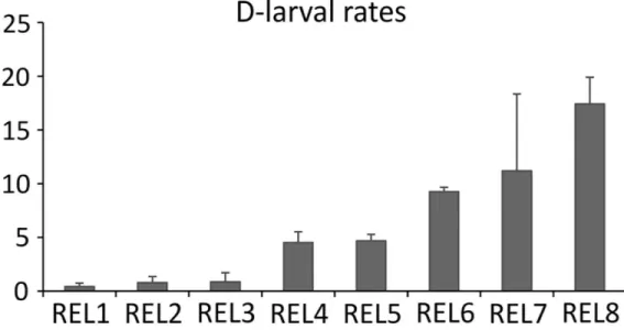
Thermal stimulation effectively induced gamete release in both males and females. At 48 hours post-fertilization, D-larval rates registered in each batch was in the expected range from 0.43% to 17.41% [39], depending on the female (Fig 1). Conversely, the fertilization of stripped oocytes did not produce any D-larvae.
Correlation between gene expression profiles and D-larval rates
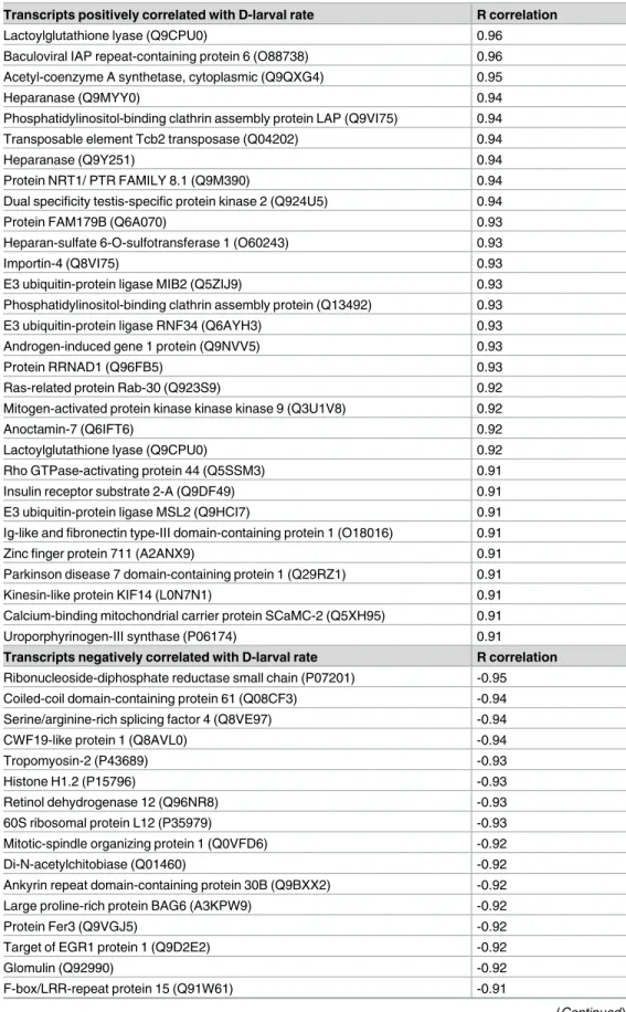
The PMT template matching analysis allowed the identification of a total of 1,904 probes (S2 Table) whose expression pattern was either positively (973) or negatively (931) correlated with D-larval rate values. Among these, a putative annotation against UniProtKB/SwissProt data-base was attained for 925 probes, corresponding to 848 unique proteins. The probes having the highest positive (R = 0.98) and negative (R = - 0.96) correlation factors did not have any match against the considered databases. The list of the putative protein identity of the most sig-nificantly correlated (R > 0.9) transcripts annotated against the protein database UniProtKB/ Uniprot has been provided inTable 1.
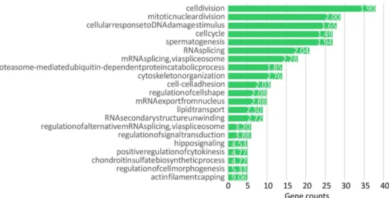
In order to investigate the main biological processes that most likely affect oocyte quality reflected by D-larval rates of each female, a functional enrichment analysis was performed by using the 811 UniProt accession numbers recognized in DAVID as gene list (S3 Table,Fig 2). Significantly enriched BP terms were “cell division” (Fold Enrichment FE: 1.89), “mitotic nuclear division” (FE: 1.99), “cytoskeleton organization” (FE: 2.75) and “RNA splicing” (FE: 2.03).
Among the significant genes participating in cell-cycle and cell division processes there wereExcision repair cross-complementation group 6-like (ERCC6L; R = 0.89), Anaphase pro-moting complex subunit 1 (APC1; R = 0.86), Lymphoid-specific helicase (LSH; R = 0.74), Regula-tor of chromosome condensation (RCC1), BTB (POZ) domain containing protein 1 (RCBTB1; R = -0.75), andSpindle and kinetochore associated complex subunit 2 (SKA2; R = -0.84). Posi-tive correlated probes encoding transcripts involved in cytoskeleton regulation included Spec-trin alpha and SpecSpec-trin beta, Kinesin family member 14 (KIF14; R = 0.91), Phosphatidylinositol-binding clathrin assembly protein LAP (PICALM; R = 0.94), Diaphanous-related formin 2 (DIAPH2; R = 0.73) andNeurabin-1 (PPP1R9A; R = 0.87). For RNA processing, transcripts positively correlated with D-larval rates wereWT1-associated protein (WTAP; R = 0.72), Tudor domain-containing protein 1 (TDRD1; R = 0.79) and Pumilio RNA-binding family member 1 (PUM1; R = 0.78), while several DEAD box proteins (DDX55, DDX19A, DDX39A, DDX39B and DEAD box protein UAP56) were negatively correlated to oocyte developmental compe-tence. Furthermore, a large number of splicing factors was either negatively or positively asso-ciated to oocyte quality (e.g. Splicing factor 4, Splicing factor 9G8, Splicing factor arginine/serine-rich 16, Splicing factor arginine/serine-arginine/serine-rich 2).
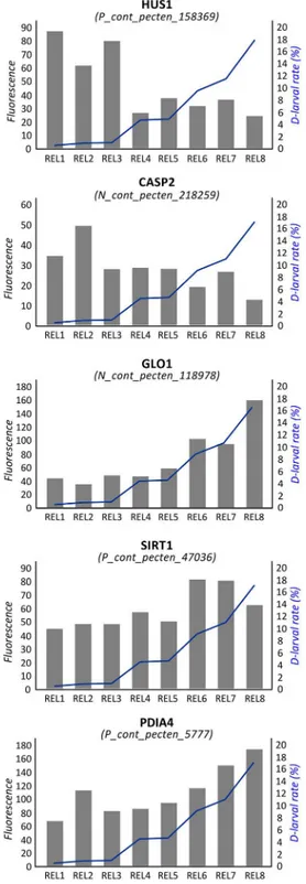
An additional enriched BP term was “cellular response to DNA damage stimulus” (FE: 1.65). In fact, several significant genes were involved in facing DNA damage and regulating apoptosis.Checkpoint protein HUS1 (seeFig 3),Caspase 2 (CASP2; seeFig 3),Growth arrest and DNA damage-inducible protein (GADD45A), and Peptidyl-tRNA hydrolase 2 (PTH2) were negatively associated to D-larval rates, whileGlyoxalase I (GLO1; seeFig 3),Regulator of telo-mere elongation helicase 1 (RTEL1), Sirtuin 1 (SIRT1; seeFig 3) and theBaculoviral IAP
repeat-Fig 1. D-larval rates. Values of D-larval rates of released oocytes (REL) expressed as percentages of
trochophore at 48 hpf on the total count of oocytes employed for the fertilization. Standard deviation refers to batch replicates (n = 3) as described in [20].
Table 1. Transcripts with the highest correlation between gene expression and D-larval rates (R>0.9). Transcripts positively correlated with D-larval rate R correlation
Lactoylglutathione lyase (Q9CPU0) 0.96
Baculoviral IAP repeat-containing protein 6 (O88738) 0.96 Acetyl-coenzyme A synthetase, cytoplasmic (Q9QXG4) 0.95
Heparanase (Q9MYY0) 0.94
Phosphatidylinositol-binding clathrin assembly protein LAP (Q9VI75) 0.94
Transposable element Tcb2 transposase (Q04202) 0.94
Heparanase (Q9Y251) 0.94
Protein NRT1/ PTR FAMILY 8.1 (Q9M390) 0.94
Dual specificity testis-specific protein kinase 2 (Q924U5) 0.94
Protein FAM179B (Q6A070) 0.93
Heparan-sulfate 6-O-sulfotransferase 1 (O60243) 0.93
Importin-4 (Q8VI75) 0.93
E3 ubiquitin-protein ligase MIB2 (Q5ZIJ9) 0.93
Phosphatidylinositol-binding clathrin assembly protein (Q13492) 0.93
E3 ubiquitin-protein ligase RNF34 (Q6AYH3) 0.93
Androgen-induced gene 1 protein (Q9NVV5) 0.93
Protein RRNAD1 (Q96FB5) 0.93
Ras-related protein Rab-30 (Q923S9) 0.92
Mitogen-activated protein kinase kinase kinase 9 (Q3U1V8) 0.92
Anoctamin-7 (Q6IFT6) 0.92
Lactoylglutathione lyase (Q9CPU0) 0.92
Rho GTPase-activating protein 44 (Q5SSM3) 0.91
Insulin receptor substrate 2-A (Q9DF49) 0.91
E3 ubiquitin-protein ligase MSL2 (Q9HCI7) 0.91
Ig-like and fibronectin type-III domain-containing protein 1 (O18016) 0.91
Zinc finger protein 711 (A2ANX9) 0.91
Parkinson disease 7 domain-containing protein 1 (Q29RZ1) 0.91
Kinesin-like protein KIF14 (L0N7N1) 0.91
Calcium-binding mitochondrial carrier protein SCaMC-2 (Q5XH95) 0.91
Uroporphyrinogen-III synthase (P06174) 0.91
Transcripts negatively correlated with D-larval rate R correlation
Ribonucleoside-diphosphate reductase small chain (P07201) -0.95 Coiled-coil domain-containing protein 61 (Q08CF3) -0.94
Serine/arginine-rich splicing factor 4 (Q8VE97) -0.94
CWF19-like protein 1 (Q8AVL0) -0.94
Tropomyosin-2 (P43689) -0.93
Histone H1.2 (P15796) -0.93
Retinol dehydrogenase 12 (Q96NR8) -0.93
60S ribosomal protein L12 (P35979) -0.93
Mitotic-spindle organizing protein 1 (Q0VFD6) -0.92
Di-N-acetylchitobiase (Q01460) -0.92
Ankyrin repeat domain-containing protein 30B (Q9BXX2) -0.92
Large proline-rich protein BAG6 (A3KPW9) -0.92
Protein Fer3 (Q9VGJ5) -0.92
Target of EGR1 protein 1 (Q9D2E2) -0.92
Glomulin (Q92990) -0.92
F-box/LRR-repeat protein 15 (Q91W61) -0.91
containing protein 6 (BIRC6) were more transcribed in oocytes with the highest developmental competence.
Probes encoding enzymes involved in energy producing processes were also highlighted: Acetyl-coenzyme A synthetase (ACECS), synthetizing Acetyl-CoA for the tricarboxylic acid (TCA) cycle, was highly correlated to oocyte quality (R = 0.95). Moreover, a similar transcrip-tional behaviour was evidenced for two enzymes driving the TCA cycle:alpha-ketoglutarate dehydrogenase-like and putative malate dehydrogenase 1B.
Full lists of significantly enriched GO terms and KEGG pathways are reported inS3 Table
(gene count>3, p-value < 0.05).
Transcriptional differences between stripped and spawned oocytes
Pairwise comparison among stripped and spawned oocytes revealed a total of 1,682 probes dif-ferentially expressed (S4 Table). Among these, 652 and 1,030 probes were more expressed in REL and STR oocytes, respectively. Putative annotation against UniProtKB/SwissProt database was attained for 597 probes, corresponding to 546 unique proteins. Enrichment analysis,
Table 1. (Continued)
E3 ubiquitin-protein ligase TRIP12 (Q14669) -0.91
RNA-directed DNA polymerase from mobile element jockey (P21329) -0.91 Peptidyl-tRNA hydrolase 2, mitochondrial (Q8R2Y8) -0.91
Probable tRNA pseudouridine synthase 2 (Q5XGG2) -0.91
Protein slowmo (Q9V3U9) -0.90
High affinity copper uptake protein 1 (Q8WNR0) -0.90
Transcript names are those retrieved from UniProtKB/SwissProt database. doi:10.1371/journal.pone.0172805.t001
Fig 2. Enrichment analysis of transcripts significantly correlated with D-larval rates. Significant enriched
BP_direct terms obtained through the enrichment analysis performed on the transcripts significantly correlated with D-larval rates. The green bars identify the number of the correlated genes belonging to the annotation term. Only terms with minimum gene counts of 5 were reported. Numbers beside the bars correspond to the Fold Enrichment reported for each term.
Fig 3. Correlation between gene expression and D-larval rates. Values of fluorescence reported for
probes encoding HUS1, CASP2, GLO1, SIRT1 and PDIA4. Samples are reported in the x axis. Expression level (principal y axis) is expressed in terms normalized fluorescence. The D-larval rate is reported in term of percentage (secondary y axis) and described by a blue line.
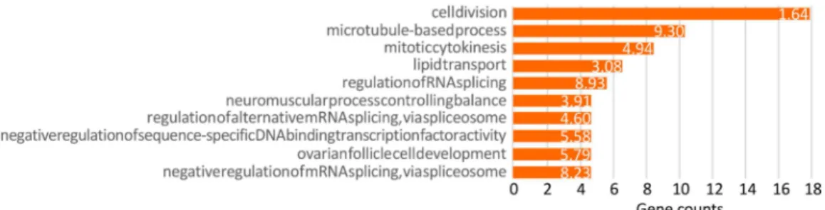
carried out by using 480 UniProtKB/SwissProt accession IDs recognized in DAVID database, evidenced the significant enrichment of 16 BP terms and 6 KEGG pathways (S5 Table,Fig 4). The most significantly enriched BP term was “microtubule-based process” (FE: 9.30), repre-sented by several tubulin alpha isoforms, more expressed in STR oocytes. “Regulation of RNA splicing” was also enriched (FE: 8.93) with genes likeCDC-like kinase 2 (CLK2) and SON DNA binding protein (SON), both found more expressed in STR compared to REL oocytes. Likewise, the term “negative regulation of mRNA splicing, via spliceosome” (FE: 8.23), represented by splicing factors SRSF12 and SFSWAP, andU2 small nuclear ribonucleoprotein auxiliary factor (U2AF), was significantly overrepresented. The BP term “Ovarian follicle cell development” was also found significantly enriched (FE: 5.79), withBeta-1,3-galactosyltransferase brn and Homeobox protein Cut genes over-expressed in REL oocytes. Finally, among enriched BP, sev-eral terms related to cell division have been detected, such as “cell division” (FE: 1.64), and “mitotic cytokinesis” (FE: 4.94). Examples of probes involved in cell division and significantly more expressed in REL oocytes were those encodingCyclin-dependent kinases regulatory sub-unit 1 (CKS1), Cyclin O (CCNO), Ubiquitin-conjugating enzyme E2-17 kDa (UBE2D1) and ERCC6L. Conversely,Cell division cycle 42 (CDC42) and Protein MIS12 homolog (MIS12) were more expressed in STR than REL oocytes. Noteworthy, additional transcripts more expressed inP. maximus STR oocytes were the egg yolk precursor Vitellogenin-4 (VTG4; FC = 238), the glycan binding Galectin-4 (GAL4, FC = 129), a Fatty acid-binding protein (FABP, FC = 90), the phosphotransferaseArginine kinase (AK, FC = 15), the detoxifying enzymes Glutathione S-transferase sigma (GSTS, FC = 46) and theta (GSTT, FC = 2.78), and the Serotonin receptor 5-hydroxytryptamine receptor 4 (HTR4; FC = 2.94). Probes expressed at higher levels in REL oocytes were those coding fornuclear protein Akirin-2 (AKIR2; FC = 9), Diacylglycerol kinase eta (DGKH; FC = 7.46) generating phosphatidic acid (PA), and mitochondrial Isocitrate dehy-drogenase [NAD] subunit beta (IDH3B; FC = 1.76).
Discussion
A key aspect deserving special attention is the generally low D-larval rates obtained from the fertilization of the eight females spawning, ranging from 0.4% to 17.4% (Fig 1). In bivalves, individual variability in oocyte quality is commonly observed [16,40] representing a key factor in hatchery-based shellfish production. Notably, previous studies carried out in the great scal-lop demonstrated that D-larval rates achieved in hatchery conditions are particularly low [41] especially if compared to what experienced in other commercial bivalves such as in the Pacific
Fig 4. Enrichment analysis of DEGs between stripped and released oocytes. Significant enriched
BP_direct terms obtained through the enrichment analysis performed on DEGs between STR and REL oocytes. The orange bars identify the number of the correlated genes belonging to the annotation term. Only terms with minimum gene counts of 5 were reported. Numbers beside the bars correspond to the Fold Enrichment reported for each term.
oysterC. gigas [39]. In our study, broadly different performances were reported across scallops (Fig 1). Because both environmental and experimental conditions were kept uniform across animals, such variability was most likely due to the intrinsic quality of oocytes of each individual.
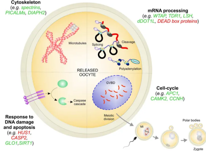
The enrichment analysis carried out on the transcripts significantly correlated with hatch-ing rates, pointhatch-ing out several biological processes that most probably regulate the quality of scallop spawned oocytes and determine their fate (S3 Table). Overall, the expression level of genes involved in cytoskeletal dynamics, mRNA processing, DNA damage, apoptosis, and cell-cycle play an important role in assuring oocyte competence and sustaining the very first larval development in scallop (Fig 5). For the sake of clarity, these biological processes will be dis-cussed separately.
Cytoskeleton
Seven probes positively correlated to oocyte quality showed a high sequence similarity with alpha and beta spectrins. Spectrins act as actin crosslinking and molecular scaffolds and a few studies suggested that they may function globally in coordinating cytoskeletal functions within epithelial tissues during early embryo development (e.g. [42–43]).
Fig 5. Main processes affecting oocyte quality. Genes positively and negatively correlated with D-larval rates were reported in green and
in red colour, respectively. doi:10.1371/journal.pone.0172805.g005
A putative member of the kinesin superfamily of microtubule-associated motors (KIF14), highly correlated to D-larval rate, was demonstrated inXenopus oocytes to be required for mitotic cytokinesis and to bind the central spindle [44]. Despite at a lower significance, addi-tional kinesins, such as KIF6 and KIF2A, appeared to be more expressed in scallop oocytes having the highest hatching rate. Oocyte kinesin stocks might play a crucial role in coordinat-ing interactions between actin and microtubule cytoskeleton of the scallop embryo, and thereby contribute to faithful cytokinesis occurring in the early developmental phases.
Furthermore, four probes encoding PICALM were positively correlated to oocytes compe-tence. PICALMs are the major proteins recruiting clathrin to cell membranes at sites of coated-pit formation and clathrin-vesicle assembly, thus mediating endocytosis of plasma membrane receptors, channels, and transporters, as well as transmembrane proteins and vari-ous soluble macromolecules [45]. Besides PICALMs, the mRNA expression of two additional molecules involved in clathrin-mediated endocytosis was positive related to D-larvae yield: the enzyme Synaptojanin-1 and the accessory protein Epsin-2 (see the review [46]). Such multiple lines of evidence suggest two hypotheses on the importance of the endocytic complex in scal-lop REL oocytes. First, a higher abundance of these transcripts could better sustain endocytic processes, which provide nutrients and mediate cellular signalling during early larval develop-ment. Secondly, we hypothesize that these transcripts inP. maximus oocytes might be trans-lated during the first life stages activating mitogenic signalling pathways of crucial importance for cell growth and differentiation.
Finally, DIAPH2 was also positively correlated to D-larval rate. DIA proteins are required for proper spindle formation, actin tubulin organization, cytokinesis and microtubule–kineto-chore attachment, thus they are expected to play a crucial role in oocyte maturation. In the starfishAsterina pectinifera inhibition of DIAPH2 activity prevented cleavage furrow closure and resulted in polar body extrusion failure [47].
mRNA processing
Once oocytes are released, meiosis resumption occurs and mRNA transcription is generally thought to cease [48]. However, translation of the stored pool of mRNAs continues throughout the final stages of meiosis [49] to synthetize proteins that are crucial to support oocyte matura-tion (meiotic maturamatura-tion), as well as the phase prior to zygote-embryonic genome activamatura-tion [50]. This evidence let us hypothesize that scallop spawned oocytes encoding the more “suit-able” pool of molecules involved in the mRNA processing dynamics are those assuring higher D-larval rates.
WTAP, positively correlated with developmental competence, is a regulatory subunit of a methyl-transferase complex that has been demonstrated to act as a mRNA splicing regulator in human and mouse [51]. The importance of this gene during the early development has been proved in zebrafish embryo, where a WTAP knockdown caused marked tissue differentiation defects and increased apoptosis [52].
TDRD1, more expressed in oocytes with the highest D-larval rate, mediates the repression of transposable elements during meiosis in mice and zebrafish by acting via piwi-interacting RNA metabolic process [53]. The tudor gene was originally discovered inD. melanogaster in a screen for maternal factors that regulate embryonic development or fertility [54].
An important role is suggested also for epigenetic processes, as demonstrated by the observed transcriptional levels of LSH and histone H3-K79 methyl-transferase (dDOT1L), both positively correlated with developmental competence. LSH is a chromatin remodelling protein acting as epigenetic regulator [55]. Interestingly, a previous study reported that in mice LSH is essential for the establishment of homologous-chromosome synapsis, thus
allowing the completion of meiosis [56]. Similarly, LSH disruption in mice caused global hypo-methylation, developmental growth retardation, and a premature aging phenotype [57]. dDOT1L is a histone methyl-transferase specific for lysine 79 of histone H3 playing important roles in meiosis progression and supposed to be associated with chromosome deacetylation of mouse oocytes [58].
Additional probes whose expression changed according to D-larval rates were those cod-ing for DEAD box proteins DDX55, DDX19A, DDX39A, DDX39B and DEAD box protein UAP56. DEAD box proteins regulate RNA secondary structure and are involved in transla-tion initiatransla-tion, ribosome assembly, RNA splicing and mRNA turnover in an ATP-depen-dent reaction. Thus, they are expected to play a central role in the oocyte, where stockpiled mRNAs are used to sustain oocyte maturation, fertilization and embryo development until the embryonic genome is activated. In the present study, DEAD box proteins were nega-tively correlated to D-larval rates. Despite the function of these proteins has been scarcely explored in bivalve oocytes (e.g. [59]), a previous study conducted in the Atlantic surf clam, Spisula solidissima, suggested that a DEAD box protein represses translation of maternal mRNA in early development [60]. Moreover, Minshall and co-workers demonstrated that the helicase activity may be attenuated during meiotic maturation, prior to cytoplasmic polyadenylation, allowing mRNA translation of key developmental proteins. Accordingly, the higher mRNA expression of DEAD box proteins in poor-quality REL oocytes might reflect stronger repression of translation and lower maturation level, compared to oocytes with higher D-larval rates.
Response to DNA damage and apoptosis
Programmed cell death may lead to DNA fragmentation and oocyte degeneration [61], mean-ing poor oocyte quality and lower fertility in mammals [62–63]. Thus, apoptosis in oocytes has been considered a marker of oocyte quality and its capacity to develop into a viable embryo. There are several evidence supporting the idea that apoptosis in the oocyte can affect embryo quality because of the presence of maternal mRNAs stored in the oocyte that regulate the apo-ptotic mechanism [64–65].
In the present study, several transcripts correlated with oocyte quality were involved in cel-lular response to DNA damage stimuli.Checkpoint protein HUS1 (Fig 3) andDNA damage-binding protein 2 (DDB2) were negatively associated to D-larval rates. HUS1 is a component of the 9-1-1 cell-cycle checkpoint response complex. InDrosophila it plays a major role in homol-ogous recombination DNA repair [66] and is essential for activation of the meiotic checkpoint [67]. Human DDB2 plays important roles in nucleotide excision repair and it is critical in deciding cell fate (apoptosis or arrest) upon DNA damage [68]. Thus, overexpression of HUS1 and DDB2 in poor quality oocytes suggests the presence of DNA damage. Conversely, RTEL1 and SIRT1 (Fig 3) were positively correlated with D-larval rates. RTEL1 is an ATP-dependent DNA helicase required to suppress inappropriate homologous recombination, thereby playing a central role in the protection of genome against instability. SIRT1 is a NAD-dependent dea-cetylase suggested to be a marker of oxidative stress and aging in mammals [69–70] and it has been demonstrated to protect oocytes against oxidative stress in mouse [71]. Similarly, in pigs, sirtuins are involved in cortical polarity and spindle organization and their inhibition adversely affects oocyte meiosis [72]. SIRT1 and GLO1 (Fig 3) have been also demonstrated to participate in the cellular pathways activated by the oocyte to counteract methylglyoxal (MG), a highly reactive dicarbonyl promoting AGE (advanced glycation end-products) accumulation and oxidative stress [73]. Here, GLO1 expression was highly correlated (R = 0.96) to scallop developmental competence (Fig 3).
Several transcripts that encode molecules regulating apoptosis were correlated to D-yield. The most interesting transcripts were those coding for Large proline-rich protein BAG6, CASP2, GADD45 alpha and BIRC6. BAG6 plays a role in protein folding and protea-somal degradation [74] and is involved in DNA damage-induced apoptosis. InXenopus egg extracts, binding of apoptosis inducer factor Reaper to BAG6 promoted cytochrome c-mediated caspase activation leading to cell death [75]. CASP2 is crucial for oocyte apopto-sis in the mouse and inX. laevis [76], since it induces apoptosis by releasing pro-apoptotic proteins from mitochondria. Evidence that BAG6 and CASP2 negatively correlated with D-larval rates (Fig 3) suggests that programmed cell death has likely a high incidence in oocytes with low developmental competence. This was also suggested inR. decussatus oocytes, showing a negative correlation between D-larval yield and Caspase 8 (CASP8) [20]. GADD45A is a stress-inducible nuclear protein involved in maintenance of genomic stabil-ity, senescence, apoptosis, DNA repair [77] and suppression of cell growth, and has a key role in active DNA demethylation that occurs inXenopus oocytes [78]. The negative correla-tion between GADD45 expression and D-yields corroborates the hypothesis that oocytes with low developmental competence might exhibit elevated apoptosis. Conversely, BIRC6 was expressed at higher extent in oocytes with higher D-larval rates. BIRC6 is believed to inhibit apoptosis by targeting key cell-death proteins, thus a higher transcription in REL oocytes might be linked to protection from programmed cell death. Similarly, BIRC6 expression is crucial for embryo survival during bovine preimplantation embryo develop-ment [79].
Cell-cycle
Several transcripts acting through cell-cycle were correlated to D-larval rates.APC1was more expressed in oocytes with the highest D-larval rates. APC is an E3 ubiquitin-ligase essential for progression through meiosis since it promotes Cyclin B destruction and meiotic exit until fer-tilization occurs. Notably, APC has been recognized in mammals as a key molecular determi-nant of oocyte quality of direct relevance to reproductive performance [80]. APC is activated by the Ca2+ signal by a meiosis-specific mechanism via calcium/calmodulin-dependent kinase II (CAMK2) [81]. A putative scallop CAMK2 alpha chain was highly correlated with oocyte competence, thus reinforcing the hypothesis that also in scallop oocytes, as in model species [82], APC Ca-dependent signaling might enable meiosis progression.
Gene expression of a putative Cyclin H (CCNH) was lower in oocytes showing poor devel-opmental competence compared to discrete-quality oocytes. Despite the functions of CCHN in meiosis have been poorly investigated, this evidence is in accordance with a previous study demonstrating that suppression of CCNH inhibits pig early meiotic resumption and matura-tion to MII [83].
Additional interesting genes significantly correlated to D-larval rates wereHeparanase (HPSE),Anoctamin-7 (ANO7), Phospholipase C-beta-1 (PLCb1) and Protein disulfide-isomerase A4 (PDIA4;Fig 3). HPSE is an endoglycosidase that cleaves heparan sulfate, thus participating in degradation and remodeling of the extracellular matrix. Notably, HPSE supplementation resulted in approximately a two-fold increase in mouse embryo implantation rate in vivo [84]. Anoctamins have been proposed to be responsible for Ca2+-activated Cl− currents and in Xenopus oocytes these channels play a role in the fast block to polyspermy [85]. PLCb1 is a phosphoinositide-specific phospholipase having a role in resumption of meiosis in the mouse oocyte [86–87]. PDIs are chaperone molecules advantageous for the viability and immune pro-tection of eggs and early embryos of Pacific oyster, and it is up-accumulated in good quality oyster oocytes [19]. Previous studies reported that PDIs are involved in oocyte development
[88] and sperm–egg fusion at fertilization [89–90], suggesting that also sperm–egg interaction are crucial determinants in oocytes quality and developmental success.
P. maximus oocyte maturation
Gene expression analysis and evaluation of DEGs between oocytes before and after spawn-ing provides a first overview on transcriptome changes that are most likely correlated with scallop stripped oocyte infertility. Key biological processes affecting oocyte maturation were those regulating cell division, with several genes being more expressed in REL oocytes. Two interesting examples are CKS1 and CCNO. CKS1 encodes a protein that binds to cyclin-dependent kinases and regulates cell-cycle progression [91]. InC. elegans, CKSs were dem-onstrated to have an essential role in meiosis M phase exit [92]; mice lacking CKS2, a mam-malian homolog of yeast CKS1, were viable but sterile due to failure of female germ cells to progress past the first meiotic metaphase [93]. Likewise, CCNO is a cyclin acting as upstream regulator of MPF and demonstrated to play an important role in mouse oocytes since the CCNO knockdown blocked meiosis resumption [94]. Thus, higher expression of transcripts encoding CKS1 and CCNO in REL oocytes, might suggest that higher synthesis of cell-cycle regulators is necessary for scallop oocyte maturation. Conversely, we found a few cell-cycle genes that were more expressed in immature oocytes compared to REL oocytes. This is the case of CDC42, a member of the Rho family of small guanosine triphos-phatase proteins, playing pivotal roles in the establishment of mouse oocyte cellular polarity [95]. In mouse, CDC42 high-expression levels have been revealed in Germinal Vesicle (GV) stage oocytes (prophase I stage) and the expression decreased up to the 2-cell (2C) stage embryo [96].
Noteworthy, a few genes included in the enriched term “ovarian follicle cell development were more expressed in REL oocytes. The transcript encoding BRAINIAC protein (Beta-1,3-galactosyltransferase brn) was an interesting example.D. melanogaster BRAINIAC is a secreted protein produced by oocytes and its activity is needed in the germ line for proper organization of the follicle [97].
The probe showing the higher FC was the one encodingVitellogenin (Vg-4), being more expressed in STR oocytes. Vitellogenins are large phospholipoglycoprotein precursors that are cleaved to generate yolk storage proteins traditionally regarded as the energy reserve for nour-ishment of the developing embryo [98]. Higher Vg expression in STR compared to REL oocytes likely reflects maturation stage. In fact, intra-gonadal oocytes arrested in prophase I, before being released and fertilized, undergo a period of vitellogenesis [99] that requires exten-sive Vg synthesis. InM. galloprovincialis, vitellogenesis seems to stop before spawning, since full-grown intra-gonadal oocytes do not express Vg mRNA [100].
Nutrients and energy reserves are key factors in supporting oocyte development. Accord-ingly, aFatty acid-binding protein and a putative AK were more expressed in scallop STR oocytes. In zebrafish a FA-binding protein (FABP3) has been demonstrated to be most abun-dant immediately prior to and during the vitellogenic stage of oocyte development and to decrease during the oocyte growth phase, being nearly undetectable in matured oocytes [101]. Likewise, the ovarian expression of AK, related to provision of energy, was higher in a penaeid shrimp during pre-vitellogenic stages and decreases in mature oocytes [102].
A similar expression pattern also for aGlutathione peroxidase (GPx), which is expressed at a higher extent in STR oocytes compared to REL group. GPx may function in protecting matu-rating oocytes against peroxidation [102]. Besides GPx, two glutathione-dependent enzymes showed higher expression in STR oocytes: GSTS and GSTT. The relation between GST and oocyte maturation has never been studied in bivalves, however in humans a negative
correlation between GSTT mRNA and cumulus-oocytes complexes maturity has been demon-strated [103].
Finally, an interesting gene showing a differential expression between STR and REL oocytes was the excitatorySerotonin receptor HTR4 (more expressed in STR group). Serotonin is a major neurotransmitter that triggers spawning and oocyte germinal vesicle breakdown (GVBD) in bivalve molluscs [104–106]. Expression levels of HTR4 might be low in released oocytes because spawning was already occurred, and high in stripped oocytes, since HT recep-tors are still engaged in sustaining meiosis progression prior to spawning.
Conclusions
In this study, a new species-specific microarray platform for great scallop has been employed to identify differentially expressed genes in relation to oocyte quality and to investigate the transcriptional features of both stripped and spawned scallop oocytes.
To date, most of the studies on egg quality and development have been conducted in model species, therefore it seems too early to hypothesize specific functions of the majority of tran-scripts expressed in scallop oocytes. The lack of scallop genome and, in general, of high quality bivalve genomes also hinders a full comprehension of transcriptomic data. However, sequence similarity searches against model species allowed us to infer putative functions of expressed transcripts, thus allowing the identification of candidate transcriptomic markers of oocytes quality, such as CASP2 and PDIA4 (Fig 3), both positively associated to female gametes quality also in other bivalve species [19–20]. The identification of generic mechanisms shared by evo-lutionary distant species is of special interest, since in-depth investigation of these pathways in a single species could lead to important knowledge applicable to aquaculture practices of a large number of species [21]. At the same time, cross-species conserved pathways might pro-vide more reliable markers, which could be used to develop cost-effective tools for rapid assess-ment of oocyte quality. In this perspective, the validation of these biomarkers in additional hatchery-based productions might be of crucial interest.
Supporting information
S1 File. Pecten maximus transcriptome assembly. (DOCX)
S2 File. Sequence of the contigs employed for the DNA microarray platform. (FASTA)
S1 Table. Blastx best hit of each probe represented in the microarray platform. (XLSX)
S2 Table. Correlation between gene expression and D-larval rate. (XLSX)
S3 Table. Enrichment analysis of transcripts correlated with D-larval rate. (XLSX)
S4 Table. DEGs between stripped and spawned oocytes. (XLSX)
S5 Table. Enrichment analysis of DEGs between stripped and spawned oocytes. (XLSX)
Acknowledgments
We thank Professor Thorolf Magnesen and the staff of the commercial bivalve hatchery Scal-pro (Norway) for Scal-providing scallop adult tissues and larval stages employed in the sequencing experiments.
Author Contributions
Conceptualization: MP AH LB. Formal analysis: MP MM AH CC MS. Funding acquisition: AH AF BN LB. Investigation: MP AH CC MS LB. Methodology: MP MM AH CC MS RM. Project administration: LB. Resources: AH JVP AF BN TP LB. Supervision: AH TP LB.Writing – original draft: MP LB.
Writing – review & editing: MM AH CC MS RM.
References
1. FAO (2016) Pecten maximus: species fact sheet. [Internet].http://www.fao.org/fishery/species/3516/ en(accessed 14 October 2016).
2. Andersen S, Christophersen G, Magnesen T. Spat production of the great scallop (Pecten maximus): a roller coaster. 2011 Can J Zool; 89: 579–598.
3. Hold N, Murray LG, Kaiser MJ, Hinz H, Beaumont AR, Taylor MI. Potential effects of stock enhance-ment with hatchery-reared seed on genetic diversity and effective population size. Can J Fish Aquat Sci. 2012; 70: 330–338.
4. Morvezen R, Boudry P, Laroche J, Charrier G. Stock enhancement or sea ranching? Insights from monitoring the genetic diversity, relatedness and effective population size in a seeded great scallop population (Pecten maximus). Heredity (Edinb). 2016 Sep; 117(3):142–8.
5. Sandaa RA, Magnesen T, Torkildsen L, Bergh O. Characterisation of the bacterial community associ-ated with early stages of Great Scallop (Pecten maximus), using denaturing gradient gel electrophore-sis (DGGE). Syst Appl Microbiol. 2003 Jun; 26(2):302–11. doi:10.1078/072320203322346164PMID: 12866858
6. Pauletto M, Milan M, Moreira R, Novoa B, Figueras A, Babbucci M, et al. Deep transcriptome sequenc-ing of Pecten maximus hemocytes: a genomic resource for bivalve immunology. Fish Shellfish Immu-nol. 2014 Mar; 37(1):154–65. doi:10.1016/j.fsi.2014.01.017PMID:24486903
7. Robson AA, Halsey LG, Chauvaud L. Feet, heat and scallops: what is the cost of anthropogenic distur-bance in bivalve aquaculture? R Soc Open Sci. 2016 Mar 9; 3(3):150679. doi:10.1098/rsos.150679 PMID:27069659
8. Suquet M, Gourtay C, Donval A, Le Goïc N, Quere C, Malo F, et al. The quality of great scallop (Pecten maximus) sperm after thawing. Gen Comp Endocrinol. 2016 Apr 1; 229:127–31. doi:10.1016/j.ygcen. 2016.02.023PMID:26944486
9. Widowati I, Cochard JC, Dorange G, Le Pennec M. E´ tude expe´rimentale de la maturation ovocytaire chez Pecten maximus et Crassostrea gigas (Mollusca Bivalvia). Aspects re´cents de la biologie des mollusques, Ifremer, Actes Coll., 13 (1992), pp. 47–56.
10. Osanai K Kuraishi R. Response of oocytes to meiosis-inducing agents in pelecypods. Bull Mar Biol Stn Asamushi 1998; 182:45–56.
11. Developments in Aquaculture and Fisheries Science Volume 40, Pages 1–1196 (2016) Scallops Biol-ogy, EcolBiol-ogy, Aquaculture, and Fisheries Edited by Sandra E. Shumway and Parsons G. Jay.
12. Aji LP. Review: Spawning Induction in Bivalve. J Penelit Sains. 2011; 14: 33–36.
13. Osanai K. In vitro induction of germinal vesicle breakdown in oyster oocytes. Bull Mar Biol Stn Asa-mushi 1985; 18:1–9.
14. Beaumont AR, Budd MD. Effects of self fertilization and other factors on the early development of the scallop Pecten maximus. Marine Biol 1983, 76: 285–289.
15. Pauletto M, Milan M, de Sousa JT, Huvet A, Joaquim S, Matias D, et al. Insights into molecular fea-tures of Venerupis decussata oocytes: a microarray-based study. PLoS One. 2014 Dec; 9(12): e113925. doi:10.1371/journal.pone.0113925PMID:25470487
16. Boudry P, Collet B, Cornette F, Hervouet V, Bonhomme F. High variance in reproductive success of the Pacific oyster (Crassotrea gigas; Thunberg) revealed by microsatellite—based parentage analysis of multifactorial crosses. Aquaculture. 2002; 204:283–96.
17. Bobe J, Labbe´ C. Egg and sperm quality in fish. Gen Comp Endocrinol. 2010 Feb 1; 165(3):535–48. doi:10.1016/j.ygcen.2009.02.011PMID:19272390
18. Suquet M, Labbe C, Brizard R, Donval A, Le Coz JR, Quere C, et al. Changes in motility, ATP content, morphology and fertilisation capacity during the movement phase of tetraploid Pacific oyster (Crassos-trea gigas) sperm. Theriogenology. 2010 Jul 1; 74(1):111–7. doi:10.1016/j.theriogenology.2010.01. 021PMID:20189635
19. Corporeau C, Vanderplancke G, Boulais M, Suquet M, Que´re´ C, Boudry P, et al. Proteomic identifica-tion of quality factors for oocytes in the Pacific oyster Crassostrea gigas. J Proteomics. 2012 Oct 22; 75(18):5554–63. doi:10.1016/j.jprot.2012.07.040PMID:22878033
20. de Sousa JT, Milan M, Pauletto M, Bargelloni L, Joaquim S, Matias D, et al. A microarray-based analy-sis of oocyte quality in the European clam Ruditapes decussatus. Aquaculture. 2015 Sept 1; 446:17– 24.http://dx.doi.org/10.1016/j.aquaculture.2015.04.018.
21. Bobe J. Egg quality in fish: Present and future challenges. Animal Frontiers. 2015; 5(1):66–72.
22. Boulais M, Corporeau C, Huvet A, Bernard I, Quere C, Quillien V, et al. Assessment of oocyte and trochophore quality in Pacific oyster, Crassostrea gigas. Aquaculture. 2015; 437,201–207.
23. Cannuel R, Beninger PG. Is oyster broodstock feeding always necessary? A study using oocyte qual-ity predictors and validators in Crassostrea gigas. Aquat Living Resour.2005; 18, 35–43.
24. Migaud H, Bell G, Cabrita E, McAndrew B,Davie A, Bobe J, et al. Gamete quality and broodstock man-agement in temperate fish. Rev Aquac. 2013; 5:S194–S223.
25. Joaquim F, Matias D, Matias AM, Gonc¸alves R, Vera C, Chı´charo L, et al. Relationships between broodstock condition, oocyte quality, and 24 h D-larval survival during the spawning season of the pul-let carpet shell Venerupis corrugata (Gmelin, 1791). Int J Inver Rep Dev. 2016.
26. Kjorsvik E, Mangor-Jensen A, Holmefjord I. Egg quality in fishes. Adv Mar Biol. 1990; 26:71–113.
27. Valdebenito II, Gallegos PC, Effer BR. Gamete quality in fish: evaluation parameters and determining factors. Zygote. 2015 Apr; 23(2):177–97. Epub 2013 Nov 15. Review. doi:10.1017/
S0967199413000506PMID:24229714
28. Castets MD, Schaerlinger B, Silvestre F, Gardeur JN, Dieu M, Corbier C,Kestemont P, Fontaine P. Combined analysis of Perca fluviatilis reproductiveperformance and oocyte proteomic profile. Therio-genology. 2012 Jul15; 78(2):432–42, 442.e1–13. doi:10.1016/j.theriogenology.2012.02.023PMID: 22578620
29. Sullivan CV, Chapman RW, Reading BJ, Anderson PE. Transcriptomics of mRNA and egg quality in farmed fish: Some recent developments and future directions. Gen Comp Endocrinol. 2015 Sep 15; 221:23–30. doi:10.1016/j.ygcen.2015.02.012PMID:25725305
30. Mason J. The breeding of the scallop, Pecten maximus (L.), in Manx waters. J Mar Biol Assoc UK. 1958; 37:653–671.
31. Massapina C, Joaquim S, Matias D, Devauchelle N. Oocyte and embryo quality in Crassostrea gigas (Portuguese strain) during a spawning period in Algarve, South Portugal. Aquat Living Resour. 1999; 12:327–333.
32. Mommens M, Fernandes JM, Tollefsen KE, Johnston IA, Babiak I. Profiling of the embryonic Atlantic halibut (Hippoglossus hippoglossus L.) transcriptome reveals maternal transcripts as potential mark-ers of embryo quality. BMC Genomics. 2014 Sep 30; 15:829. doi:10.1186/1471-2164-15-829PMID: 25269745
33. Ma H, Weber GM, Hostuttler MA, Wei H, Wang L, Yao J. MicroRNA expression profiles from eggs of different qualities associated with post-ovulatory ageing in rainbow trout (Oncorhynchus mykiss). BMC Genomics. 2015 Mar 17; 16:201. doi:10.1186/s12864-015-1400-0PMID:25885637
34. Gruffydd LD, Beaumont AR. Determination of the optimum concentration of eggs and spermatozoa for the production of normal larvae inPecten maximus (Mollusca, Lamellibranchia). Helgolander Wiss. Meeresunters. 1970; 20:486.
35. Suquet M, Le Mercier A, Rimond F, Mingant C, Haffray P, Labbe C. Setting tools for the early assess-ment of the quality of thawed Pacific oyster (Crassostrea gigas) D-larvae. Theriogenology. 2012 Jul 15; 78(2):462–7. doi:10.1016/j.theriogenology.2012.02.014PMID:22538008
36. Song YP, Suquet M, Queau I, Lebrun L. Setting of a procedure for experimental fertilisation of Pacific oyster (Crassostrea gigas) oocytes. Aquaculture. 2009; 287(3–4):311–314.
37. Johnson WE, Rabinovic A, Li C. Adjusting batch effects in microarray expression data using Empirical Bayes methods. Biostatistics. 2007; 8:118–127. doi:10.1093/biostatistics/kxj037PMID:16632515
38. Huang DW, Sherman BT, Lempicki RA. Systematic and integrative analysis of large gene lists using DAVID bioinformatics resources. Nat Protoc. 2009; 4:44–57. doi:10.1038/nprot.2008.211PMID: 19131956
39. Robert R, Ge´rard A. Bivalve hatchery techniques: current situation for the oyster Crassostrea gigas and the scallop Pecten maximus. Aquat Living Resour. 1999; 12:121–130.
40. Le Pennec M, Robert R, Avendano M.The importance of gonadal development on larval production in pectinids. J Shellfish Res.1998; 17(1):97–101.
41. Le Pennec M, Paugam A, Le Pennec G. The pelagic life of the Pecten maximus—a review. ICES J Mar Sci. 2003; 60:211–223.
42. Zarnescu DC, Thomas GH. Apical spectrin is essential for epithelial morphogenesis but not apicobasal polarity in Drosophila. J Cell Biol. 1999; 146:1075–1086. PMID:10477760
43. Norman KR, Moerman DG. Alpha spectrin is essential for morphogenesis and body wall muscle for-mation in Caenorhabditis elegans. J Cell Biol. 2002 May 13; 157(4):665–77. doi:10.1083/jcb. 200111051PMID:11994313
44. Samwer M, Dehne HJ, Spira F, Kollmar M, Gerlich DW, Urlaub H, et al. The nuclear F-actin interac-tome of Xenopus oocytes reveals an actin-bundling kinesin that is essential for meiotic cytokinesis. EMBO J. 2013 Jul 3; 32(13):1886–902. doi:10.1038/emboj.2013.108PMID:23727888
45. Takei K, Haucke V. Clathrin-mediated endocytosis: membrane factors pull the trigger. Trends Cell Biol. 2001 Sep; 11(9):385–91. PMID:11514193
46. McPherson PS, Kay BK, Hussain NK. Signaling on the endocytic pathway.Traffic. 2001 Jun; 2 (6):375–84. Review. PMID:11389765
47. Ucar H, Tachibana K, Kishimoto T. The Mos-MAPK pathway regulates Diaphanous-related formin activity to drive cleavage furrow closure during polar body extrusion in starfish oocytes. J Cell Sci. 2013 Nov 15; 126(Pt 22):5153–65. doi:10.1242/jcs.130476PMID:24046444
48. Heikinheimo O, Gibbons WE. The molecular mechanisms of oocyte maturation and early embryonic development are unveiling new insights into reproductive medicine. Mol Hum Reprod.1998; 4:745–56. PMID:9733431
49. Wassarman PM, Liu C, Litscher ES.Constructing the mammalian egg zona pellucida: some new pieces of an old puzzle. J Cell Sci.1996; 109:2001–2004. PMID:8856495
50. Song JL, Wessel GM. How to make an egg: transcriptional regulation in oocytes. Differentiation. 2005; 73:1–17. doi:10.1111/j.1432-0436.2005.07301005.xPMID:15733063
51. Dominissini D, Moshitch-Moshkovitz S, Schwartz S, Salmon-Divon M, Ungar L, Osenberg S, et al. Topology of the human and mouse m6A RNA methylomes revealed by m6A-seq. Nature. 2012 Apr 29; 485(7397):201–6. doi:10.1038/nature11112PMID:22575960
52. Ping XL, Sun BF, Wang L, Xiao W, Yang X, Wang WJ, et al. Mammalian WTAP is a regulatory subunit of the RNA N6-methyladenosine methyltransferase. Cell Res. 2014 Feb; 24(2):177–89. doi:10.1038/ cr.2014.3PMID:24407421
53. Huang H, Gao Q, Peng X, Choi SY, Sarma K, Ren H, et al. piRNA-associated germline nuage forma-tion and spermatogenesis require MitoPLD profusogenic mitochondrial-surface lipid signaling. Dev Cell. 2011 Mar 15; 20(3):376–87. doi:10.1016/j.devcel.2011.01.004PMID:21397848
54. Boswell RE, Mahowald AP. tudor, a gene required for assembly of the germ plasm in Drosophila mela-nogaster. Cell. 1985 Nov; 43(1):97–104. PMID:3935320
55. Jarvis CD, Geiman T, Vila-Storm MP, Osipovich O, Akella U, Candeias S, et al. A novel putative heli-case produced in early murine lymphocytes. Gene. 1996 Mar 9; 169(2):203–7. PMID:8647447
56. De La Fuente R, Baumann C, Fan T, Schmidtmann A, Dobrinski I, Muegge K. Lsh is required for mei-otic chromosome synapsis and retrotransposon silencing in female germ cells. Nat Cell Biol. 2006 Dec; 8(12):1448–54. doi:10.1038/ncb1513PMID:17115026
57. Sun LQ, Lee DW, Zhang Q, Xiao W, Raabe EH, Meeker A, et al. Growth retardation and premature aging phenotypes in mice with disruption of the SNF2-like gene, PASG. Genes Dev. 2004 May 1; 18(9):1035–46. doi:10.1101/gad.1176104PMID:15105378
58. Wang Q, He M. Molecular characterization and analysis of a putative 5-HT receptor involved in repro-duction process of the pearl oyster Pinctada fucata. Gen Comp Endocrinol. 2014 Aug 1; 204:71–9. doi:10.1016/j.ygcen.2014.05.010PMID:24852353
59. Fabioux C, Pouvreau S, Le Roux F, Huvet A. The oyster vasa-like gene: a specific marker of the germ-line in Crassostrea gigas. Biochem Biophys Res Commun. 2004 Mar 19; 315(4):897–904. doi:10. 1016/j.bbrc.2004.01.145PMID:14985097
60. Minshall N, Thom G, Standart N. A conserved role of a DEAD box helicase in mRNA masking.RNA. 2001 Dec; 7(12):1728–42. PMID:11780630
61. Takase K, Ishikawa M, Hoshiai H. Apoptosis in the degeneration process of unfertilized mouse ova. Tohoku J Exp Med. 1995 Jan; 175(1):69–76. PMID:7610461
62. Wu J, Zhang L, Wang X. Maturation and apoptosis of human oocytes in vitro are age-related. Fertil Steril. 2000 Dec; 74(6):1137–41. PMID:11119740
63. Fujino Y, Ozaki K, Yamamasu S, Ito F, Matsuoka I, Hayashi E, Nakamura H, Ogita S, Sato E, Inoue M. DNA fragmentation of oocytes in aged mice. Hum Reprod. 1996 Jul; 11(7):1480–3. PMID:8671488
64. Jurisicova A, Latham KE, Casper RF, Casper RF, Varmuza SL. Expression and regulation of genes associated with cell death during murine preimplantation embryo development. Mol Reprod Dev. 1998 Nov; 51(3):243–53. doi:10.1002/(SICI)1098-2795(199811)51:3<243::AID-MRD3>3.0.CO;2-PPMID: 9771644
65. Metcalfe AD, Hunter HR, Bloor DJ, Lieberman BA, Picton HM, Leese HJ, et al. Expression of 11 mem-bers of the BCL-2 family of apoptosis regulatory molecules during human preimplantation embryo development and fragmentation. Mol Reprod Dev. 2004 May; 68(1):35–50. doi:10.1002/mrd.20055 PMID:15039946
66. Peretz G, Arie LG, Bakhrat A, Abdu U. The Drosophila hus1 gene is required for homologous recombi-nation repair during meiosis. Mech Dev. 2009 Aug-Sep; 126(8–9):677–86. doi:10.1016/j.mod.2009. 05.004PMID:19501158
67. Abdu U, Klovstad M, Butin-Israeli V, Bakhrat A, Schu¨pbach T. An essential role for Drosophila hus1 in somatic and meiotic DNA damage responses. J Cell Sci. 2007 Mar 15; 120(Pt 6):1042–9. doi:10. 1242/jcs.03414PMID:17327271
68. Stoyanova T, Roy N, Kopanja D, Bagchi S, Raychaudhuri P. DDB2 decides cell fate following DNA damage. Proc Natl Acad Sci U S A. 2009 Jun 30; 106(26):10690–5. doi:10.1073/pnas.0812254106 PMID:19541625
69. Tatone C, Di Emidio G, Vitti M, Di Carlo M, Santini S Jr, D’Alessandro AM, et al. Sirtuin Functions in Female Fertility: Possible Role in Oxidative Stress and Aging. Oxid Med Cell Longev. 2015:659687. doi:10.1155/2015/659687PMID:26075037
70. Zhang J, Fang L, Lu Z, Xiong J, Wu M, Shi L, et al. Are sirtuins markers of ovarian aging? Gene. 2016 Jan 10; 575(2 Pt 3):680–6. doi:10.1016/j.gene.2015.09.043PMID:26403315
71. Di Emidio G, Falone S, Vitti M, D’Alessandro AM, Vento M, Di Pietro C, et al. SIRT1 signalling protects mouse oocytes against oxidative stress and is deregulated during aging. Hum Reprod. 2014 Sep; 29(9):2006–17. doi:10.1093/humrep/deu160PMID:24963165
72. Zhang L, Ma R, Hu J, Ding X, Xu Y. Sirtuin Inhibition Adversely Affects Porcine Oocyte Meiosis. PLoS One. 2015 Jul 15; 10(7):e0132941. doi:10.1371/journal.pone.0132941PMID:26176547
73. Tatone C, Eichenlaub-Ritter U, Amicarelli F. Dicarbonyl stress and glyoxalases in ovarian function. Biochem Soc Trans. 2014 Apr; 42(2):433–8. Review. doi:10.1042/BST20140023PMID:24646256
74. Doong H, Vrailas A, Kohn EC. What’s in the ’BAG’? A functional domain analysis of the BAG-family proteins. Cancer Lett. 2002 Dec 15; 188(1–2):25–32. PMID:12406544
75. Thress K, Evans EK, Kornbluth S. Reaper-induced dissociation of a Scythe-sequestered cytochrome c-releasing activity. EMBO J. 1999 Oct 15; 18(20):5486–93. doi:10.1093/emboj/18.20.5486PMID: 10523293
76. Fava LL, Bock FJ, Geley S, Villunger A. Caspase-2 at a glance. J Cell Sci. 2012 Dec 15; 125(Pt 24):5911–5. doi:10.1242/jcs.115105PMID:23447670
77. Milan M, Matozzo V, Pauletto M, Di Camillo B, Giacomazzo M, Boffo L, et al. Can ecological history influence response to pollutants? Transcriptomic analysis of Manila clam collected in different Venice lagoon areas and exposed to heavy metal. Aquat Toxicol. 2016 May; 174:123–33. doi:10.1016/j. aquatox.2016.02.024PMID:26945539
78. Barreto G, Scha¨fer A, Marhold J, Stach D, Swaminathan SK, Handa V, et al. Gadd45a promotes epi-genetic gene activation by repair-mediated DNA demethylation. Nature. 2007 Feb 8; 445(7128):671– 5. doi:10.1038/nature05515PMID:17268471
79. Salilew-Wondim D, Ho¨lker M, Rings F, Phatsara C, Mohammadi-Sangcheshmeh A, Tholen E, et al. Depletion of BIRC6 leads to retarded bovine early embryonic development and blastocyst formation in vitro. Reprod Fertil Dev. 2010; 22(3):564–79. doi:10.1071/RD09112PMID:20188030
80. Homer H. The APC/C in female mammalian meiosis I. Reproduction. 2013 Jun 27; 146(2):R61–71. doi:10.1530/REP-13-0163PMID:23687281
81. Rauh NR, Schmidt A, Bormann J, Nigg EA, Mayer TU. Calcium triggers exit from meiosis II by target-ing the APC/C inhibitor XErp1 for degradation. Nature. 2005 Oct 13; 437(7061):1048–52. doi:10. 1038/nature04093PMID:16127448
82. Von Stetina JR, Orr-Weaver TL. Developmental control of oocyte maturation and egg activation in metazoan models. Cold Spring Harb Perspect Biol. 2011 Oct 1; 3(10):a005553. Review. doi:10.1101/ cshperspect.a005553PMID:21709181
83. Fujii W, Nishimura T, Kano K, Sugiura K, Naito K. CDK7 and CCNH are components of CDK-activating kinase and are required for meiotic progression of pig oocytes. Biol Reprod. 2011 Dec; 85(6):1124–32. doi:10.1095/biolreprod.111.091801PMID:21778139
84. Revel A, Helman A, Koler M, Shushan A, Goldshmidt O, Zcharia E, et al. Heparanase improves mouse embryo implantation. Fertil Steril. 2005 Mar; 83(3):580–6. doi:10.1016/j.fertnstert.2004.11.008 PMID:15749484
85. Hartzell HC, Yu K, Xiao Q, Chien LT, Qu Z. Anoctamin/TMEM16 family members are Ca2+—activated Cl-channels. J Physiol. 2009 May 15; 587(Pt 10):2127–39. doi:10.1113/jphysiol.2008.163709PMID: 19015192
86. Avazeri N, Courtot AM, Pesty A, Duquenne C, Lefèvre B. Cytoplasmic and nuclear phospholipase C-beta 1 relocation: role in resumption of meiosis in the mouse oocyte. Mol Biol Cell. 2000 Dec; 11 (12):4369–80. PMID:11102530
87. Lefèvre B, Pesty A, Courtot AM, Martins CV, Broca O, Denys A, et al. The phosphoinositide-phospholi-pase C (PI-PLC) pathway in the mouse oocyte. Crit Rev Eukaryot Gene Expr. 2007; 17(4):259–69. Review. PMID:17725492
88. Ohashi Y, Hoshino Y, Tanemura K, Sato E. Distribution of protein disulfide isomerase during matura-tion of pig oocytes. Anim Sci J. 2013 Jan; 84(1):15–22. doi:10.1111/j.1740-0929.2012.01030.xPMID: 23302077
89. Calvert ME, Digilio LC, Herr JC, Coonrod SA. Oolemmal proteomics—identification of highly abundant heat shock proteins and molecular chaperones in the mature mouse egg and their localization on the plasma membrane. Reprod Biol Endocrinol. 2003 Feb 14; 1:27. doi:10.1186/1477-7827-1-27PMID: 12646049
90. Ellerman DA, Myles DG, Primakoff P. A role for sperm surface protein disulfide isomerase activity in gamete fusion: evidence for the participation of ERp57. Dev Cell. 2006 Jun; 10(6):831–7. doi:10. 1016/j.devcel.2006.03.011PMID:16740484
91. Patra D, Dunphy WG. Xe-p9, a Xenopus Suc1/Cks protein, is essential for the Cdc2-dependent phos-phorylation of the anaphase- promoting complex at mitosis. Genes Dev. 1998 Aug 15; 12(16):2549– 59. PMID:9716407
92. Polinko ES, Strome S. Depletion of a Cks homolog in C. elegans embryos uncovers a post-metaphase role in both meiosis and mitosis. Curr Biol. 2000 Nov 16; 10(22):1471–4. PMID:11102813
93. Spruck CH, de Miguel MP, Smith AP, Ryan A, Stein P, Schultz RM, et al. Requirement of Cks2 for the first metaphase/anaphase transition of mammalian meiosis. Science. 2003 Apr 25; 300(5619):647– 50. doi:10.1126/science.1084149PMID:12714746
94. Ma JY, Ou-Yang YC, Luo YB, Wang ZB, Hou Y, Han ZM, et al. Cyclin O regulates germinal vesicle breakdown in mouse oocytes. Biol Reprod. 2013 May 2; 88(5):110. PMID:23515676
95. Wang ZB, Jiang ZZ, Zhang QH, Hu MW, Huang L, Ou XH, et al. Specific deletion of Cdc42 does not affect meiotic spindle organization/migration and homologous chromosome segregation but disrupts polarity establishment and cytokinesis in mouse oocytes. Mol Biol Cell. 2013 Dec; 24(24):3832–41. doi:10.1091/mbc.E13-03-0123PMID:24131996
96. Cui XS, Li XY, Kim NH. Cdc42 is implicated in polarity during meiotic resumption and blastocyst forma-tion in the mouse. Mol Reprod Dev. 2007 Jun; 74(6):785–94. doi:10.1002/mrd.20571PMID:
17154294
97. Goode S, Morgan M, Liang YP, Mahowald AP. Brainiac encodes a novel, putative secreted protein that cooperates with Grk TGF alpha in the genesis of the follicular epithelium. Dev Biol. 1996 Aug 25; 178(1):35–50. doi:10.1006/dbio.1996.0196PMID:8812107