HAL Id: hal-02143557
https://hal-amu.archives-ouvertes.fr/hal-02143557
Submitted on 17 Oct 2019
HAL is a multi-disciplinary open access archive for the deposit and dissemination of sci-entific research documents, whether they are pub-lished or not. The documents may come from teaching and research institutions in France or abroad, or from public or private research centers.
L’archive ouverte pluridisciplinaire HAL, est destinée au dépôt et à la diffusion de documents scientifiques de niveau recherche, publiés ou non, émanant des établissements d’enseignement et de recherche français ou étrangers, des laboratoires publics ou privés.
Rates of immune cell infiltration in patients with
triple-negative breast cancer by molecular subtype
Kenichi Harano, Ying Wang, Bora Lim, Robert S. Seitz, Stephan W. Morris,
Daniel B. Bailey, David R. Hout, Rachel L. Skelton, Brian Z. Ring, Hiroko
Masuda, et al.
To cite this version:
Kenichi Harano, Ying Wang, Bora Lim, Robert S. Seitz, Stephan W. Morris, et al.. Rates of immune cell infiltration in patients with triple-negative breast cancer by molecular subtype. PLoS ONE, Public Library of Science, 2018, 13 (10), pp.e0204513. �10.1371/journal.pone.0204513�. �hal-02143557�
Rates of immune cell infiltration in patients
with triple-negative breast cancer by
molecular subtype
Kenichi Harano1,2,3, Ying Wang4, Bora Lim1,2, Robert S. Seitz5, Stephan W. Morris5, Daniel B. Bailey5, David R. Hout5, Rachel L. Skelton5, Brian Z. Ring5,6, Hiroko Masuda7, Arvind U. K. Rao4,8, Steven Van Laere9, Francois Bertucci10, Wendy A. Woodward1,8, James M. Reuben1,11, Savitri Krishnamurthy1,12*, Naoto T. UenoID1,2*
1 Morgan Welch Inflammatory Breast Cancer Research Program and Clinic, The University of Texas MD
Anderson Cancer Center, Houston, Texas, United States of America, 2 Department of Breast Medical Oncology, The University of Texas MD Anderson Cancer Center, Houston, Texas, United States of America,
3 Department of Pulmonology Medicine and Oncology, Graduate School of Medicine, Nippon Medical School,
Bunkyo-ku, Tokyo, Japan, 4 Department of Bioinformatics and Computational Biology, The University of Texas MD Anderson Cancer Center, Houston, Texas, United States of America, 5 Insight Genetics, Inc., Nashville, Tennessee, United States of America, 6 College of Life Science, Huazhong University of Science and Technology, Wuhan, China, 7 Department of Breast Surgical Oncology, Showa University, Shinagawa-ku, Tokyo, Japan, 8 Department of Radiation Oncology, The University of Texas MD Anderson Cancer Center, Houston, Texas, United States of America, 9 Center for Oncological Research, Faculty of Medicine and Health Sciences, University of Antwerp, Antwerp, Belgium, 10 Predictive Oncology team, CRCM, Institut Paoli-Calmettes, Marseille, France, 11 Department of Hematopathology, The University of Texas MD Anderson Cancer Center, Houston, Texas, United States of America, 12 Department of Pathology, The University of Texas MD Anderson Cancer Center, Houston, Texas, United States of America
*skrishna@mdanderson.org(SK);nueno@mdanderson.org(NTU)
Abstract
In patients with triple-negative breast cancer (TNBC), tumor-infiltrating lymphocytes (TILs) are associated with improved survival. Lehmann et al. identified 4 molecular subtypes of TNBC [basal-like (BL) 1, BL2, mesenchymal (M), and luminal androgen receptor (LAR)], and an immunomodulatory (IM) gene expression signature indicates the presence of TILs and modifies these subtypes. The association between TNBC subtype and TILs is not known. Also, the association between inflammatory breast cancer (IBC) and the presence of TILs is not known. Therefore, we studied the IM subtype distribution among different TNBC subtypes. We retrospectively analyzed patients with TNBC from the World IBC Con-sortium dataset. The molecular subtype and the IM signature [positive (IM+) or negative (IM-)] were analyzed. Fisher’s exact test was used to analyze the distribution of positivity for the IM signature according to the TNBC molecular subtype and IBC status. There were 88 patients with TNBC in the dataset, and among them 39 patients (44%) had IBC and 49 (56%) had non-IBC. The frequency of IM+ cases differed by TNBC subtype (p = 0.001). The frequency of IM+ cases by subtype was as follows: BL1, 48% (14/29); BL2, 30% (3/10); LAR, 18% (3/17); and M, 0% (0/21) (in 11 patients, the subtype could not be determined). The frequency of IM+ cases did not differ between patients with IBC and non-IBC (23% and 33%, respectively; p = 0.35). In conclusion, the IM signature representing the underlying molecular correlate of TILs in the tumor may differ by TNBC subtype but not by IBC status.
a1111111111 a1111111111 a1111111111 a1111111111 a1111111111 OPEN ACCESS
Citation: Harano K, Wang Y, Lim B, Seitz RS,
Morris SW, Bailey DB, et al. (2018) Rates of immune cell infiltration in patients with triple-negative breast cancer by molecular subtype. PLoS ONE 13(10): e0204513.https://doi.org/10.1371/ journal.pone.0204513
Editor: Aamir Ahmad, University of South Alabama
Mitchell Cancer Institute, UNITED STATES
Received: April 29, 2018 Accepted: September 10, 2018 Published: October 12, 2018
Copyright:© 2018 Harano et al. This is an open access article distributed under the terms of the
Creative Commons Attribution License, which permits unrestricted use, distribution, and reproduction in any medium, provided the original author and source are credited.
Data Availability Statement: All relevant data are
within the paper and its Supporting Information files.
Funding: KH received the Japan Cancer Society
"My Oncology Dream award 2014". The funder, Insight Genetics, Inc., provided support in the form of stock and salaries for authors RSS, SWM, DBB, DRH, RLS, and BZR. Insight Genetics, Inc., performed the analysis of gene expression profile data and played a role in the decision to publish.
Introduction
Triple-negative breast cancer (TNBC) accounts for 10% to 20% of breast cancers. TNBC is an aggressive tumor, and patients with TNBC have a higher risk of both local and distant recur-rence compared to patients with other types of breast cancer [1]. Patients with TNBC have higher rates of pathological complete response (pCR) following neoadjuvant chemotherapy than patients with other types of breast cancer, but TNBC patients without a pCR have a markedly worse prognosis than TNBC patients with a pCR [2].
Several studies have shown that in patients with TNBC, there is a linear relationship between the number of tumor-infiltrating lymphocytes (TILs), mononuclear immune cells that infiltrate tumor, and recurrence-free survival [3–5]. It has also been reported that in patients with breast cancer, the presence of TILs is associated with increased rates of pCR fol-lowing neoadjuvant chemotherapy [6,7]. Thus, TILs are a prognostic factor and predictor of response to cytotoxic chemotherapy in patients with TNBC.
TNBCs are heterogeneous. Lehmann et al. reported in 2011 that TNBCs could be grouped into 6 molecular subtypes: basal-like (BL) 1, BL2, mesenchymal (M), mesenchymal stem-like (MSL), immunomodulatory (IM), and luminal androgen receptor (LAR) [8]. They suggested that the subtypes exhibit the following characteristics: BL1, increased expression of genes asso-ciated with the cell cycle and DNA damage response; BL2, growth factor signaling; M,
increased expression of epithelial-mesenchymal transition (EMT) and growth factor pathways; MSL, increased expression of EMT and growth factor pathways and decreased expression of genes involved in proliferation; IM, expression of genes encoding immune antigens and cyto-kines; and LAR, androgen receptor signaling. We previously reported that the TNBC subtype is a predictor of pCR after neoadjuvant chemotherapy [9]: the BL1 subtype was associated with the highest pCR rate (52%), and the BL2 and LAR subtypes had the lowest pCR rates (0% and 10%, respectively).
Recently, using laser capture microdissection and histopathological quantification, Leh-mann et al. found that transcripts in the previously defined IM and MSL subtypes came from TILs and tumor-associated stromal cells, respectively, and they reduced the number of TNBC molecular subtypes to 4: BL1, BL2, M, and LAR [10]. Further, they showed that the IM gene expression signature is an indicator of the presence of TILs and incorporated the IM signature into TNBC subtyping as a modifier of the other subtypes rather than a separate subtype.
The association between TNBC subtype and the presence of TILs is not known. On the basis of the above-noted clinical and molecular data, we hypothesized that the BL1 subtype has a high rate of IM signature and that the BL2 and LAR subtypes have low rates of IM signature, which reflects immune infiltration.
In addition to being characterized by TNBC subtype, TNBC can be classified according to whether it represents inflammatory breast cancer (IBC) or non-inflammatory breast cancer (non-IBC). IBC is a relatively rare and aggressive cancer that presents with rapid onset of red-ness and swelling of the breast [11]. Several inflammatory signaling pathways, including NF-κB, COX-2, JAK/STAT, IL-6, tumor necrosis factor alpha, and interferon gamma, have been sug-gested to contribute to the tumorigenesis of IBC [12]. We previously reported that the TNBC molecular subtypes are expressed in both inflammatory and non-inflammatory TNBC and that we found no unique IBC-specific TNBC subtypes by mRNA gene expression profiling [13]. However, the association between IBC and the presence of TILs is not known. We hypothesized that IM signature is more frequent in patients with IBC than in those with non-IBC.
To test our hypotheses, we studied gene expression in patients with TNBC with IBC and non-IBC and evaluated the relationship between TNBC molecular subtype and IM signature and also the relationship between IBC status and IM signature.
Immune cell infiltration in TNBC
Competing interests: Insight Genetics, Inc.,
provided support in the form of stock and salaries for authors RSS, SWM, DBB, DRH, RLS, and BZR. This does not alter our adherence to PLOS ONE policies on sharing data and materials.
Methods
Patients
We retrospectively analyzed gene expression profiles and clinical data of patients with TNBC with known IBC status from the World IBC Consortium dataset [14]. Three institutions con-tributed to this dataset: The University of Texas MD Anderson Cancer Center, Houston, TX; General Hospital Sint-Augustinus, Antwerp, Wilrijk, Belgium; and Institut Paoli-Calmettes, Marseille, France. We obtained clinical data (patient age, stage, histologic subtype, grade, and treatment) and gene expression profiles from the 3 institutes. For the World IBC Consortium database study, patients at each site gave informed consent for voluntary participation, and the study was approved by the institutional review boards of the 3 participating centers. IBC was identified according to the consensus diagnostic criteria [11,14,15]. At each of the 3 centers, the diagnostic criteria for the diagnosis of IBC were as follows: 1) rapid onset of breast ery-thema and/or peau d’orange and/or warm breast with or without an underlying palpable mass, 2) duration of clinical symptoms/signs no more than 6 months, 3) erythema occupying at least one-third of the breast, and 4) pathological confirmation of invasive carcinoma. TNBC was diagnosed according to gene expression profiling. The original gene expression profiling and identification of TNBC cases has already been reported in our previous paper [13]. For gene expression profiling, all samples were run on the Affymetrix HGU133 platform.
TNBCtype (Insight Genetics, Inc., Nashville, TN, USA) was used to assign TNBC subtypes. TNBCtype is a new algorithm for TNBC subtyping that reduces gene signatures from the origi-nal 2188 genes described by Lehmann et al. [8] to 101 genes including control housekeeping genes in order to optimize gene expression profiles [16,17]. The optimized TNBCtype algo-rithm is designed to classify TNBC according to 1 of the 4 intrinsic subtypes and then define TNBC tumors as positive (IM+) or negative (IM-) for the IM gene expression signature. It has been shown that IM gene expression signature is correlated with the level of TILs in the tumor specimen [10]. TNBC tumors classified as IM+ are highly enriched for genes involved in immune cell processes, including immune cell signaling, cytokine signaling, antigen process-ing and presentation, and signalprocess-ing through core immune signal transduction pathways. Using TNBCtype, we grouped patients by the 4 subtypes (BL1, BL2, M, and LAR), then identi-fied whether each case of TNBC was positive or negative for the IM signature.
The study reported herein was approved by the Institutional Review Board of MD Ander-son Cancer Center (protocol number PA15-0954). The Institutional Review Board waived the requirement for informed consent because this study was a retrospective data review that involved no diagnostic or therapeutic intervention and no direct patient contact.
Statistical analysis
We constructed 4×2 (molecular subtype versus positive or negative for IM signature) or 2×2 (IBC statusversus positive or negative for IM signature) contingency tables and compared the
distributions using Fisher’s exact test. A p value of <0.05 was considered to indicate a signifi-cant association.
Results
In total, 88 TNBC patients (39 with IBC and 49 with non-IBC) were included in this study. The patient characteristics are summarized inTable 1. Of the 88 TNBC patients in this study, 55 (63%) had stage III or IV disease, and 39 (44%) had IBC. All patients received multidisci-plinary treatment according to treatment guidelines of each hospital. Among the 71 patients with stage I-III disease, 21 (78%) of the 27 IBC patients and 29 (66%) of the 44 non-IBC
patients received neoadjuvant chemotherapy. The neoadjuvant chemotherapy regimen con-sisted of an anthracycline-based plus taxane-based chemotherapy.
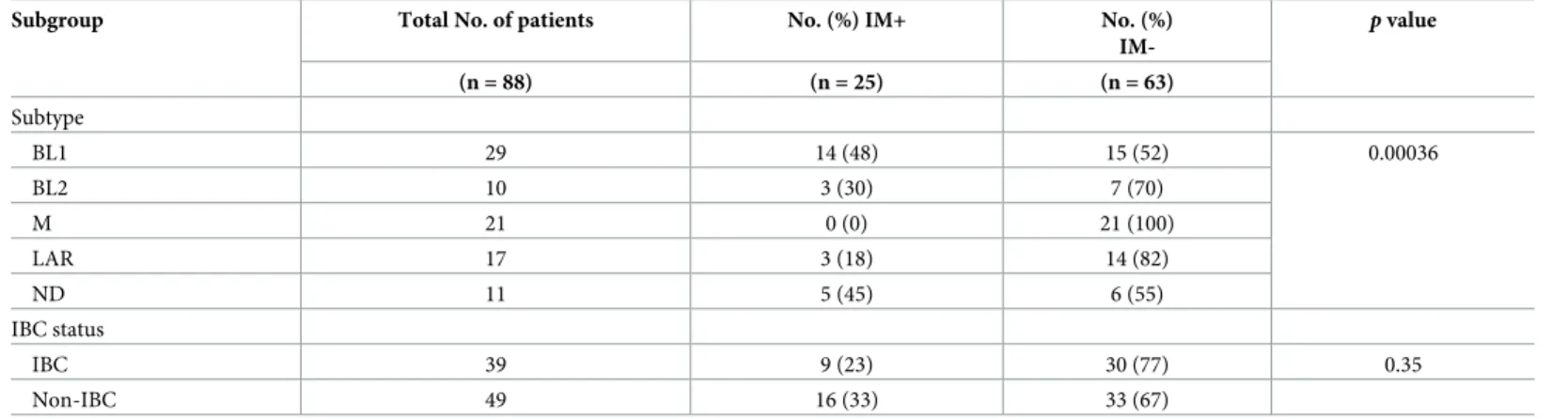
The distribution of IM signature by intrinsic subtype is shown inTable 2. Among the 88 patients with TNBC, 29 had BL1, 10 had BL2, 21 had M, and 17 had LAR subtype, while 11 did not have a clear subtype. In total, 25 patients (28%) had IM+ and 63 (72%) had IM- TNBC. The frequency of IM+ cases differed significantly by molecular subtype (p = 0.00036). The fre-quency of IM+ cases was higher in the BL1 subtype (14 of 29, 48%) than in other subtypes. No IM+ cases were observed in the M subtype, and only 3 of 17 (18%) cases of LAR subtype were IM+.
The distribution of IM signature in patients with IBC and non-IBC is also shown in
Table 2. IM+ cases occurred at roughly the same frequency in patients with IBC (23%) and non-IBC (33%) (p = 0.35).
Discussion
We found that TNBC molecular subtypes differed with respect to the proportion of cases with the IM signature. Forty-eight percent of the BL1-subtype TNBCs were positive for the IM sig-nature, whereas the signature was not observed in TNBCs of M subtype and was found in only 18% of TNBCS of LAR subtype. The distribution of the IM signature did not differ between patients with IBC and non-IBC.
Table 1. Patient characteristics.
Variable N %
Total number of patients 88 Age, median (range), years 52 (26–78) Stage I 9 10 II 17 19 III 45 51 IV 10 12 Unknown 7 8 IBC status Non-IBC 49 56 IBC 39 44 Histology
Invasive ductal carcinoma 80 91 Invasive lobular carcinoma 4 5
Other 4 5 Nuclear grade I 3 3 II 14 16 III 68 77 Unknown 3 3
Neoadjuvant chemotherapy (for stage I-III disease, 71 patients)
Received 50 70
Not received 20 28
Unknown 1 2
IBC, inflammatory breast cancer.
https://doi.org/10.1371/journal.pone.0204513.t001
Recent studies have shown significant correlation between TILs and clinical outcomes in TNBC. Several studies showed that a higher number of TILs was associated with improved recurrence-free survival [3–5]. Further, the presence of TILs has been associated with greater benefit from neoadjuvant chemotherapy in patients with breast cancer. For example, in the GeparDuo and GeparTrio clinical trials of neoadjuvant chemotherapy with an anthracycline and a taxane, the pCR rate was significantly higher in patients with tumors in which TILs accounted for at least 60% of the tumor stromal area than in patients with tumors in which TILs accounted for less than 60% [6].
To our knowledge, the association between TNBC subtype and the presence of TILs has previously been examined only in a single study performed by Lehmann et al. [10]. We previ-ously reported that TNBC molecular subtype predicts pCR status. The BL1 subtype had the highest pCR rate (52%), while the BL2 and LAR subtypes had the lowest pCR rates (0% and 10%, respectively) [9]. However, it has not been known why the pCR rate differs by TNBC sub-type. In the current study, the BL1 subtype had a high rate of the IM+ signature (48%), while the BL2 (30%) and LAR (18%) subtypes had lower rates of the IM+ signature. These results, which are consistent with those reported by Lehmann et al. [10], suggest that the degree of immune cell infiltration and the IM signature status of tumors, which reflects this infiltration, are influenced by subtype and that immune infiltration affects the response to chemotherapy, which may partially explain our previous finding that pCR rates differ by TNBC subtypes.
In this study, we found that no patient with TNBC M subtype had IM+ signature. The M subtype is associated with increased expression of EMT and growth factor pathways. The expression of EMT genes in the tumor microenvironment has been associated with immune suppression [18]. Dongre et al. recently reported that breast tumors that have mesenchymal features express low levels of MHC-I and high levels of PD-L1 and contain immunosuppres-sive cells such as regulatory T-cells, M2 macrophages, and exhausted CD8+ T-cells within their stroma [19]. Our study is consistent with this observation and further suggests that TNBC of M subtype that expresses an activation of EMT is associated with immunosuppres-sion. One potential discrepancy concerns the relatively high chemosensitivity our previous study showed for TNBC M-subtype tumors, which had a pCR rate of 31% following neoadju-vant chemotherapy [9]. Tumors that have an immunosuppressive environment and contain EMT features are generally considered to be refractory to cytotoxic chemotherapy [20,21]; this discordance remains unsolved.
Table 2. Distribution of IM signature in patients with TNBC by molecular subtype and IBC status.
Subgroup Total No. of patients No. (%) IM+ No. (%)
IM-p value (n = 88) (n = 25) (n = 63) Subtype BL1 29 14 (48) 15 (52) 0.00036 BL2 10 3 (30) 7 (70) M 21 0 (0) 21 (100) LAR 17 3 (18) 14 (82) ND 11 5 (45) 6 (55) IBC status IBC 39 9 (23) 30 (77) 0.35 Non-IBC 49 16 (33) 33 (67)
IM, immunomodulatory; TNBC, triple-negative breast cancer; IBC, inflammatory breast cancer; BL, basal-like; M, mesenchymal; LAR, luminal androgen receptor; ND, not determined.
In our analysis, the frequency of IM+ tumors did not differ significantly between TNBC patients with IBC and those with non-IBC. We initially hypothesized that the proportion of IM+ tumors would be higher in IBC than in non-IBC because several inflammatory signaling pathways have been shown to be active in IBC. The reason why our results did not support this hypothesis is unclear; however, our results are consistent with our previous report that no unique IBC-specific signature was identified by mRNA gene expression analysis [13]. There may be other, non-inflammatory molecular mechanisms that lead to the tumorigenesis of IBC. For example, it has been reported that the aggressive phenotype of IBC is associated with an enrichment of cancer stem cells [22]. The function of cancer stem cells is modulated by many signaling pathways, including IL-6/STAT3, hedgehog, WNT, and Notch. Syndecan-1
(CD138), a cell-surface heparan sulfate proteoglycan, modulates cell proliferation and growth, and it has been reported that syndecan-1 may regulate expression of the IL-6/STAT3, Notch, and EGFR signaling pathways in inflammatory TNBC [23]. IBC is characterized by the clinical appearance of inflammation; however, IBC may be characterized molecularly not by inflam-matory immune cells but rather by cancer stem cells.
This study has several limitations. First, the number of patients was limited, and we could not analyze the survival outcome according to intrinsic subtypes and IM signature status. Only 10 patients (11%) in this study had BL2-subtype disease. We found a similar result in our pre-vious study in which the same dataset was used but TNBC subtyping was done by a different algorithm; in that study, only 5 patients (5.6%) had BL2-subtype disease [13]. Second, we used the World IBC Consortium dataset, which includes many patients with advanced breast tumors. This dataset may not be representative of the general population of patients with TNBC. Third, 11 patients (12.5%) had an unclassifiable molecular subtype. This may reflect the existence of hybrid TNBCs comprising more than one subtype or be due to other unknown factors. Fourth, we only analyzed gene expression profile data and did not perform histopatho-logical confirmation of TILs. On the basis of previously reported findings, we considered the IM gene expression signature to be an indicator of the presence of TILs [10]; however, this molecular definition is not in widespread use.
In conclusion, our study demonstrated that the rate of IM+ subtype differs according to TNBC subtype, with the highest percentage of IM+ cases seen among BL1-subtype tumors and no IM+ cases seen among M-subtype tumors. This leads us to speculate that the rate of immune infiltration differs by TNBC molecular subtype. The findings also suggest that the TNBC subtype, because of its association with IM subtype, may influence the response to chemotherapy.
Supporting information
S1 Data. TNBC IM study data.
(XLSX)
Author Contributions
Conceptualization: Kenichi Harano, Bora Lim, Hiroko Masuda, Arvind U. K. Rao, Steven
Van Laere, Francois Bertucci, Wendy A. Woodward, James M. Reuben, Savitri Krishna-murthy, Naoto T. Ueno.
Data curation: Kenichi Harano, Robert S. Seitz, Stephan W. Morris, Daniel B. Bailey, David
R. Hout, Rachel L. Skelton.
Formal analysis: Ying Wang, Brian Z. Ring, Arvind U. K. Rao.
Investigation: Kenichi Harano, Robert S. Seitz, Stephan W. Morris, Daniel B. Bailey, David R.
Hout, Rachel L. Skelton, Brian Z. Ring.
Methodology: Kenichi Harano.
Supervision: Bora Lim, Hiroko Masuda, Steven Van Laere, Francois Bertucci, Wendy A.
Woodward, James M. Reuben, Savitri Krishnamurthy, Naoto T. Ueno.
Writing – original draft: Kenichi Harano, Ying Wang.
Writing – review & editing: Ying Wang, Bora Lim, Robert S. Seitz, Stephan W. Morris, Daniel
B. Bailey, David R. Hout, Rachel L. Skelton, Brian Z. Ring, Hiroko Masuda, Arvind U. K. Rao, Steven Van Laere, Francois Bertucci, Wendy A. Woodward, James M. Reuben, Savitri Krishnamurthy, Naoto T. Ueno.
References
1. Dent R, Trudeau M, Pritchard KI, Hanna WM, Kahn HK, Sawka CA, et al. Triple-negative breast cancer: clinical features and patterns of recurrence. Clin Cancer Res. 2007; 13: 4429–4434.https://doi.org/10. 1158/1078-0432.CCR-06-3045PMID:17671126
2. Liedtke C, Mazouni C, Hess KR, Andre´ F, Tordai A, Mejia JA, et al. Response to neoadjuvant therapy and long-term survival in patients with triple-negative breast cancer. J Clin Oncol. 2008; 26: 1275–1281.
https://doi.org/10.1200/JCO.2007.14.4147PMID:18250347
3. Loi S, Sirtaine N, Piette F, Salgado R, Viale G, Van Eenoo F, et al. Prognostic and predictive value of tumor-infiltrating lymphocytes in a phase III randomized adjuvant breast cancer trial in node-positive breast cancer comparing the addition of docetaxel to doxorubicin with doxorubicin-based chemother-apy: BIG 02–98. J Clin Oncol. 2013; 31: 860–867.https://doi.org/10.1200/JCO.2011.41.0902PMID:
23341518
4. Loi S, Michiels S, Salgado R, Sirtaine N, Jose V, Fumagalli D, et al. Tumor infiltrating lymphocytes are prognostic in triple negative breast cancer and predictive for trastuzumab benefit in early breast cancer: results from the FinHER trial. Ann Oncol. 2014; 25: 1544–1550.https://doi.org/10.1093/annonc/ mdu112PMID:24608200
5. Adams S, Gray RJ, Demaria S, Goldstein L, Perez EA, Shulman LN, et al. Prognostic value of tumor-infiltrating lymphocytes in triple-negative breast cancers from two phase III randomized adjuvant breast cancer trials: ECOG 2197 and ECOG 1199. J Clin Oncol. 2014; 32: 2959–2966.https://doi.org/10. 1200/JCO.2013.55.0491PMID:25071121
6. Denkert C, Loibl S, Noske A, Roller M, Mu¨ller BM, Komor M, et al. Tumor-associated lymphocytes as an independent predictor of response to neoadjuvant chemotherapy in breast cancer. J Clin Oncol. 2010; 28: 105–113.https://doi.org/10.1200/JCO.2009.23.7370PMID:19917869
7. Issa-Nummer Y, Darb-Esfahani S, Loibl S, Kunz G, Nekljudova V, Schrader I, et al. Prospective valida-tion of immunological infiltrate for predicvalida-tion of response to neoadjuvant chemotherapy in HER2-nega-tive breast cancer—a substudy of the neoadjuvant GeparQuinto trial. PLoS One. 2013; 8: e79775.
https://doi.org/10.1371/journal.pone.0079775PMID:24312450
8. Lehmann BD, Bauer JA, Chen X, Sanders ME, Chakravarthy AB, Shyr Y, et al. Identification of human triple-negative breast cancer subtypes and preclinical models for selection of targeted therapies. J Clin Invest. 2011; 121: 2750–2767.https://doi.org/10.1172/JCI45014PMID:21633166
9. Masuda H, Baggerly KA, Wang Y, Zhang Y, Gonzalez-Angulo AM, Meric-Bernstam F, et al. Differential response to neoadjuvant chemotherapy among 7 triple-negative breast cancer molecular subtypes. Clin Cancer Res. 2013; 19: 5533–5540.https://doi.org/10.1158/1078-0432.CCR-13-0799PMID:
23948975
10. Lehmann BD, Jovanovic B, Chen X, Estrada MV, Johnson KN, Shyr Y, et al. Refinement of triple-nega-tive breast cancer molecular subtypes: implications for neoadjuvant chemotherapy selection. PLoS One. 2016; 11: e0157368.https://doi.org/10.1371/journal.pone.0157368PMID:27310713
11. Dawood S, Merajver SD, Viens P, Vermeulen PB, Swain SM, Buchholz TA, et al. International expert panel on inflammatory breast cancer: consensus statement for standardized diagnosis and treatment. Ann Oncol. 2011; 22: 515–523.https://doi.org/10.1093/annonc/mdq345PMID:20603440
12. Fouad TM, Kogawa T, Reuben JM, Ueno NT. The role of inflammation in inflammatory breast cancer. Adv Exp Med Biol. 2014; 816: 53–73.https://doi.org/10.1007/978-3-0348-0837-8_3PMID:
13. Masuda H, Baggerly KA, Wang Y, Iwamoto T, Brewer T, Pusztai L, et al. Comparison of molecular sub-type distribution in triple-negative inflammatory and non-inflammatory breast cancers. Breast Cancer Res. 2013; 15: R112.https://doi.org/10.1186/bcr3579PMID:24274653
14. Van Laere SJ, Ueno NT, Finetti P, Vermeulen P, Lucci A, Robertson FM, et al. Uncovering the molecu-lar secrets of inflammatory breast cancer biology: an integrated analysis of three distinct affymetrix gene expression datasets. Clin Cancer Res. 2013; 19: 4685–4696.https://doi.org/10.1158/1078-0432. CCR-12-2549PMID:23396049
15. Dawood S, Ueno NT, Valero V, Woodward WA, Buchholz TA, Hortobagyi GN, et al. Differences in sur-vival among women with stage III inflammatory and noninflammatory locally advanced breast cancer appear early: a large population-based study. Cancer. 2011; 117: 1819–1826.https://doi.org/10.1002/ cncr.25682PMID:21509759
16. Chen X, Li J, Gray WH, Lehmann BD, Bauer JA, Shyr Y, et al. TNBCtype: a subtyping tool for triple-neg-ative breast cancer. Cancer Inform. 2012; 11: 147–156.https://doi.org/10.4137/CIN.S9983PMID:
22872785
17. Ring BZ, Hout DR, Morris SW, Lawrence K, Schweitzer BL, Bailey DB, et al. Generation of an algorithm based on minimal gene sets to clinically subtype triple negative breast cancer patients. BMC Cancer. 2016; 16: 143.https://doi.org/10.1186/s12885-016-2198-0PMID:26908167
18. Alessandro Poggi MG. Mesenchymal stromal cells can regulate the immune response in the tumor microenvironment. Vaccines 2016; 4: 41.
19. Dongre A, Rashidian M, Reinhardt F, Bagnato A, Keckesova Z, Ploegh HL, et al. Epithelial-to-mesen-chymal transition contributes to immunosuppression in breast carcinomas. Cancer Res. 2017; 77: 3982–3989.https://doi.org/10.1158/0008-5472.CAN-16-3292PMID:28428275
20. Hennessy BT, Gonzalez-Angulo AM, Stemke-Hale K, Gilcrease MZ, Krishnamurthy S, Lee JS, et al. Characterization of a naturally occurring breast cancer subset enriched in epithelial-to-mesenchymal transition and stem cell characteristics. Cancer Res. 2009; 69: 4116–4124.https://doi.org/10.1158/ 0008-5472.CAN-08-3441PMID:19435916
21. Miyashita M, Sasano H, Tamaki K, Chan M, Hirakawa H, Suzuki A, et al. Tumor-infiltrating CD8+ and FOXP3+ lymphocytes in triple-negative breast cancer: its correlation with pathological complete response to neoadjuvant chemotherapy. Breast Cancer Res Treat. 2014; 148: 525–534.https://doi.org/ 10.1007/s10549-014-3197-yPMID:25395319
22. Charafe-Jauffret E, Ginestier C, Iovino F, Tarpin C, Diebel M, Esterni B, et al. Aldehyde dehydrogenase 1-positive cancer stem cells mediate metastasis and poor clinical outcome in inflammatory breast can-cer. Clin Cancer Res. 2010; 16: 45–55.https://doi.org/10.1158/1078-0432.CCR-09-1630PMID:
20028757
23. Ibrahim SA, Gadalla R, El-Ghonaimy EA Samir O, Mohamed HT, Hassan H, et al. Syndecan-1 is a novel molecular marker for triple negative inflammatory breast cancer and modulates the cancer stem cell phenotype via the IL-6/STAT3, Notch and EGFR signaling pathways. Mol Cancer. 2017; 16: 57.
https://doi.org/10.1186/s12943-017-0621-zPMID:28270211