HAL Id: inserm-00391608
https://www.hal.inserm.fr/inserm-00391608
Submitted on 17 Nov 2009
HAL is a multi-disciplinary open access
archive for the deposit and dissemination of
sci-entific research documents, whether they are
pub-lished or not. The documents may come from
teaching and research institutions in France or
abroad, or from public or private research centers.
L’archive ouverte pluridisciplinaire HAL, est
destinée au dépôt et à la diffusion de documents
scientifiques de niveau recherche, publiés ou non,
émanant des établissements d’enseignement et de
recherche français ou étrangers, des laboratoires
publics ou privés.
Subthalamic stimulation-induced forelimb dyskinesias
are linked to an increase in glutamate levels in the
substantia nigra pars reticulata.
Sabrina Boulet, Emilie Lacombe, Carole Carcenac, Claude Feuerstein,
Véronique Sgambato-Faure, Annie Poupard, Marc Savasta
To cite this version:
Sabrina Boulet, Emilie Lacombe, Carole Carcenac, Claude Feuerstein, Véronique Sgambato-Faure, et
al.. Subthalamic stimulation-induced forelimb dyskinesias are linked to an increase in glutamate levels
in the substantia nigra pars reticulata.. Journal of Neuroscience, Society for Neuroscience, 2006, 26
(42), pp.10768-76. �10.1523/JNEUROSCI.3065-06.2006�. �inserm-00391608�
Neurobiology of Disease
Subthalamic Stimulation-Induced Forelimb Dyskinesias Are
Linked to an Increase in Glutamate Levels in the Substantia
Nigra Pars Reticulata
Sabrina Boulet,
1,2Emilie Lacombe,
1,2Carole Carcenac,
1,2Claude Feuerstein,
1,2Ve´ronique Sgambato-Faure,
1,2Annie Poupard,
1,2and Marc Savasta
1,21Dynamique des Re´seaux Neuronaux, Institut National de la Sante´ et de la Recherche Me´dicale, Unite´ 704, F-38041 Grenoble, France, and2Universite´ Joseph Fourier, F-38041 Grenoble, France
The neurobiological mechanisms by which high-frequency stimulation of the subthalamic nucleus (STN–HFS) alleviates the motor
symptoms of Parkinson’s disease (PD) remain unclear. In this study, we analyzed the effects of STN–HFS on motor behavior in intact or
hemiparkinsonian rats (6-hydroxydopamine lesion of the substantia nigra pars compacta) and investigated the correlation between
these effects and extracellular glutamate (Glu) and GABA levels, assessed by intracerebral microdialysis in the substantia nigra pars
reticulata (SNr). STN–HFS at an intensity corresponding to the threshold inducing contralateral forelimb dyskinesia, increased Glu levels
in the SNr of both intact and hemiparkinsonian rats. In contrast, STN–HFS at half this intensity did not affect Glu levels in the SNr in intact
or hemiparkinsonian rats but increased GABA levels in hemiparkinsonian rats only. STN–HFS-induced forelimb dyskinesia was blocked
by microinjection of the Glu receptor antagonist kynurenate into the SNr and facilitated by microinjection of a mixture of the Glu receptor
agonists AMPA and NMDA into the SNr. These new neurochemical data suggest that STN–HFS-induced forelimb dyskinesia is mediated
by glutamate, probably via the direct activation of STN axons, shedding light on the mechanisms of STN–HFS in PD.
Key words: subthalamic nucleus; substantia nigra pars reticulata; glutamate; GABA; high-frequency deep brain stimulation; Parkinson’s
disease; dyskinesia
Introduction
The subthalamic nucleus (STN) plays a key role in controlling the
output nuclei of the basal ganglia (BG), the substantia nigra pars
reticulata (SNr), and the internal segment of the globus pallidus
(GPi). It therefore plays an important role in controlling
move-ment (Kitai and Deniau, 1981; Kita and Kitai, 1987; Smith et al.,
1998; Nambu, 2004). It receives its main inputs from the cortex,
thalamus, and brainstem (Kita, 1994) and, via its glutamatergic
projections, excites the GABAergic neurons of the SNr and GPi,
thereby reinforcing the inhibitory effects of the BG on the
tha-lamic and brainstem premotor networks (Albin et al., 1989;
Al-exander and Crutcher, 1990; Chevalier and Deniau, 1990).
Pathological or experimental lesions of the STN result in
dyski-nesia or hemiballism (Whittier and Mettler, 1949; Carpenter et
al., 1950; Hammond et al., 1979; Dewey and Jankovic, 1989).
STN neurons have higher firing rates and burst activity in
Par-kinson’s disease (PD) patients and animal models of PD than in
normal subjects (Miller and DeLong, 1987; Bergman et al., 1994;
Hassani et al., 1996; Hutchison et al., 1998; Bevan et al., 2002;
Levy et al., 2002). The STN thus constitutes a strategic target for
the neurosurgical treatment of PD (Bergman et al., 1990,
Limou-sin et al., 1998; Benabid, 2003).
High-frequency stimulation (HFS) of the STN is a powerful
approach for treatment of parkinsonian motor syndrome. It has
been reported to result in clinical improvement in both PD
pa-tients and experimental animal models, further reducing
levo-dopa requirements and therefore levolevo-dopa-induced dyskinesia
(Benazzouz et al., 1993; Limousin et al., 1995; Krack et al., 2003).
However, the mechanisms underlying the effects of STN–HFS
remain unclear (Benabid et al., 2002; Dostrovsky and Lozano,
2002; McIntyre et al., 2004a). Because STN–HFS effects have
been shown to be functionally equivalent to those of STN lesions
(Bergman et al., 1990; Aziz et al., 1991; Guridi et al., 1996; Alvarez
et al., 2005), it has been suggested that STN–HFS silences STN
neurons (Benazzouz et al., 1993). However, recent modeling and
in vivo studies have shown that additional mechanisms, such as
activation of the STN afferent and efferent pathways, may be
involved, leading to distant synaptic inhibitory and excitatory
effects in BG output nuclei (Windels et al., 2000, 2003, 2005; Salin
et al., 2002; Hashimoto et al., 2003; Maurice et al., 2003; McIntyre
et al., 2004b; Kita et al., 2005).
We investigated the mechanisms involved further by studying
Received May 15, 2006; revised Sept. 1, 2006; accepted Sept. 4, 2006.
This work was supported by the Institut National de la Sante´ et de la Recherche Me´dicale, Ministe`re de la Recherche et des Nouvelles Technologies (Agence Nationale pour la Recherche Grant ANR-05-NEUR-013-01), Re´gion Rhoˆne-Alpes (Cluster 11), and the Association France Parkinson. We thank Drs. M. Albrieux, G. Chouvet, and P. Krack for critical reading of this manuscript.
Correspondence should be addressed to Dr. Marc Savasta, Dynamique des Re´seaux Neuronaux, Institut National de la Sante´ et de la Recherche Me´dicale, Unite´ 704, Universite´ Joseph Fourier, Unite´ de Formation par la Recherche en Biologie, Baˆtiment B, Domaine Universitaire, 2280 rue de la Piscine, Boıˆte Postale 53, 38041 Grenoble Cedex 09, France. E-mail: marc.savasta@ujf-grenoble.fr.
DOI:10.1523/JNEUROSCI.3065-06.2006
STN stimulation in rats in conditions comparable with those
used in clinical practice: parameters, monopolar stimulation, and
awake animals. We also analyzed the effects of various HFS
pa-rameters on motor behavior in freely moving intact and
hemipar-kinsonian rats. We then used intracerebral microdialysis to assess
variations of extracellular glutamate (Glu) and GABA levels in
the SNr during STN–HFS, according to whether the intensity of
stimulation used did or did not evoke forelimb dyskinesia. We
also investigated the effects of local injections of active Glu
com-pounds during STN–HFS in hemiparkinsonian animals to
deter-mine whether SNr Glu transmission mediated
STN–HFS-induced forelimb dyskinesia.
Materials and Methods
Animals. We used 69 adult male Sprague Dawley rats (Janvier, Le Genest
St. Isle, France), weighing 280 to 350 g, housed under standard laboratory conditions (12 h light/dark cycle) with food and water provided ad
libi-tum. Protocols conformed to the National Institutes of Health Guide for the Care and Use of Laboratory Animals (publication 865-23) and French
Ministry of Agriculture regulations (authorization number 38-R 1001).
Lesion procedure. For substantia nigra pars compacta (SNc) lesioning,
all animals were anesthetized with chloral hydrate (400 mg/kg, i.p.) and secured in a Kopf stereotaxic apparatus (Phymep, Paris, France). We treated 43 animals with desipramine (25 mg/kg, s.c.), to protect norad-renergic neurons, and then injected 9g of 6-hydroxydopamine (6-OHDA) (Sigma, St. Quentin-Fallavier, France) dissolved in 3l of sterile 0.9% NaCl and 0.2% ascorbic acid into the left SNc of these animals, at a flow rate of 0.5l/min. The stereotaxic coordinates of the injection site relative to the bregma were as follows: anteroposterior (AP),⫺5.3 mm; lateral (L),⫹2.35 mm; and dorsoventral (DV), 7.5 mm, with the incisor bar at 3.3 mm below the interaural plane. All stereotaxic coordinates cited here are according to the stereotaxic atlas of Paxinos and Watson (1982). Animals were kept warm after the injections and allowed to re-cover from anesthesia. They were returned to the animal facility for 3 weeks to allow the degeneration of DA neurons induced by the neuro-toxin to stabilize and were then processed for microdialysis experiments.
Implantation of guide cannula and microdialysis probe, injection can-nula, and stimulation electrode. For microdialysis experiments, normal
(n⫽ 20) and 6-OHDA-lesioned (n ⫽ 20) rats were anesthetized by inhalation (1 L/min) of a 5% halothane/air mixture (22% O2, 78% N2)
and mounted in a stereotaxic frame (David Kopf Instruments, Tujunga, CA). Anesthesia was maintained with an inhaled 1% halothane/air mix-ture (1 L/min). The dorsal skull was exposed, and holes were drilled to facilitate the unilateral (left side, i.e., ipsilateral to 6-OHDA injection) implantation of the guide cannula, through which the dialysis probe was lowered into the SNr, and of the stimulation electrode in the STN. The guide cannula, which consisted of a stainless steel tube (outer diameter, 0.60 mm; inner diameter, 0.48 mm) was inserted obliquely (17° from the vertical) such that its tip was located at the edge of the SNr. The stereo-taxic coordinates used were (expressed relative to bregma): (1) guide cannula: AP,⫺5.3 mm; L, 5 mm; V, ⫺7.3 mm; (2) microdialysis probe (into SNr): AP,⫺5.3 mm; L, 5 mm; V, ⫺8.5 mm; (3) stimulation elec-trode (into STN): AP,⫺3.7 mm; L, 2.4 mm; V, ⫺7.8 mm.
For the local injection of drugs, stainless steel cannulas (outer diame-ter, 0.28 mm; inner diamediame-ter, 0.18 mm) were inserted bilaterally into the SNr in 6-OHDA-lesioned rats only (n⫽ 17). The stereotaxic coordinates of the injection site relative to bregma were as follows: AP,⫺5.3 mm; L, 2.4 mm; DV, 8.2 mm. The guide and injection cannulas, together with the stimulation electrode, were fixed onto the skull with dental cement (Methax, Perrigot, France). During surgery, body temperature was maintained at 37°C with a feedback-controlled heating pad (Harvard Apparatus, Edenbridge, UK). Animals were allowed to recover from surgery for 3 d before microdi-alysis experiments, which were performed in awake animals.
Electrical stimulation. Monopolar electrodes consisting of platinum–
iridium wire insulated with Teflon but with an exposed end (wire diam-eter, 110m insulated, 76 m bare) (Phymep) were implanted unilat-erally into 43 6-OHDA-lesioned rats and 26 intact animals. The electrode was implanted such that the exposed end (length, 400m) was inserted
into the sensorimotor part of the STN. The exposed end of the electrode served as the negative stimulation pole, with a screw fixed on the skull used as the positive pole. Stimuli were delivered with a World Precision Instruments (Stevenage, UK) acupulser and a stimulus isolation unit generating rectangular pulses.
For behavioral studies, two series of stimulation experiments were performed in intact (n⫽ 6) and 6-OHDA-injected (n ⫽ 6) rats. In the first series, frequency and pulse width were set at 130 Hz and 60s, respectively, for the entire stimulation period, for all stimulated animals. The behavioral effects of increasing the stimulation intensity from 0 to 350A were analyzed in each animal during the first 2 min of stimula-tion. In particular, we determined whether the STN–HFS applied trig-gered dyskinetic movements similar to those induced after levodopa treatment, i.e., orofacial, axial, or forelimb dyskinesia (Winkler et al., 2002), as well as rotational behavior.
In the second series, STN stimulations were performed with various frequencies (10, 60, 130, 150, and 200 Hz) and pulse widths (20, 40, 60, 80, or 100s). Pulse width was set at 60 s for experiments involving frequency variation, whereas the frequency was set at 130 Hz for experi-ments involving pulse width variation. In both cases, the STN was stim-ulated electrically by gradually increasing the intensity of stimulation until dyskinetic movement of the contralateral forelimb was observed, as described by Salin et al. (2002).
For microdialysis and microinjection experiments, frequency and pulse width values were kept constant throughout the stimulation period (1 h) for all stimulated animals and were similar to those used routinely in clinical practice (130 Hz and 60s, respectively). Two different intensities were tested: I1, corresponding to the threshold value inducing contralateral
fore-limb dyskinesia, ranging from 60 to 200A, depending on the animal con-sidered; I2, a value below this threshold value, arbitrarily fixed at half I1,
varying with the individual and often corresponding to values⬍60A. At the end of each experiment, an electrical lesion was created in the STN so that the position of the electrode could be checked postmortem.
Microdialysis experiments. Homemade microdialysis probes were
pre-pared and used as described previously (Windels et al., 2000; Bruet et al., 2003). They consisted of a concentric arrangement of stainless steel tube (outer diameter, 0.4 mm) (Phymep), and polyethylene tubing (outer diameter, 1.09 mm; inner diameter, 0.38 mm) (Phymep) into which we inserted a piece of silica tubing (outer diameter, 150m; inner diameter, 75m) (Phymep). The silica tubing extended beyond the distal end of the steel tube and was covered with a cuprophan tubular dialysis mem-brane (Hospal, Lyon, France), sealed at the bottom with epoxy glue. The length of the dialysis membrane was adapted for the rat brain nucleus studied (1 mm for the SNr).
The perfusion liquid flowed out of the distal end of the steel tube, passing proximally between the tube and the membrane (Tossman and Ungerstedt, 1986). The probes were perfused with artificial CSF (in mM:
149 NaCl, 2.8 KCl, 1.2 MgCl2, 1.2 CaCl2, and 5.4 glucose, pH 7.3) at a
flow rate of 1l/min. Before implantation, each probe was tested in vitro in a standard amino acid solution, to determine amino acid recovery (Tossman and Ungerstedt, 1986).
The dialysis probe was implanted via the guide cannula and dialysates were collected at 15 min intervals, for 5 h, in awake animals. The first eight fractions were discarded to prevent effects caused by parenchymal disturbance and to obtain an approximate steady state. The next eight fractions (120 min) were collected for basal value determination, and four fractions were then collected during STN–HFS. An additional eight fractions were collected to estimate poststimulation effects over a 2 h period. Dialysates were collected automatically with a refrigerated au-tosampler (Univentor, Zejton, Malta) and stored at⫺80°C until analysis.
Glutamate and GABA assays. Glu and GABA concentrations in the
dialysates were determined by HPLC, with laser-induced fluorescence detection. Briefly, 2 l of sample or standard was derivatized with naphthalene-2,3-dicarboxaldehyde. The resulting mixture was automat-ically loaded onto a Symmetry Shield-C18 reverse-phase column (100⫻ 2.1 mm, 3.5m particle size; Waters, Milford, MA), using a refrigerated Triathlon autoinjector (Polymer Laboratories, Marseille, France). The mobile phase consisted of 0.04MNaH2PO4, pH 6, in a 3–50% acetonitrile
gradient. The flow rate was 0.35 ml/min, maintained with two Shimadzu Boulet et al.• SNr Glutamate and STN–HFS-Induced Dyskinesia J. Neurosci., October 18, 2006•26(42):10768 –10776 • 10769
(Kyoto, Japan) LC 10AT pumps. Amino acid peaks were identified based on retention time. Extracellular amino acid concentrations were estimated by rationing peak areas of each amino acid and their respective external standard (an-alytical software class LC10; Shimadzu). The running time for each determination was 12 min.
Local drug injections. We applied Glu and
GABA agonists or antagonists bilaterally to the SNr of 17 freely moving 6-OHDA-lesioned rats through a stainless steel cannula, implanted as described above. Bilateral injections were used to prevent crossed effects of drugs. The cannula was connected to a 10l syringe (Hamilton, Bonaduz, Switzerland) via a piece of polyethyl-ene tubing (outer diameter, 1.09 mm; inner di-ameter, 0.38 mm) (Phymep). The syringes con-tained a mixture of Glu agonists (AMPA and NMDA), a Glu antagonist (kynurenic acid), or a GABAA agonist (muscimol). These drugs
were purchased from Sigma and dissolved in saline. We injected 0.5l of solution into both sides of the SNr, at a rate of 0.25l/min, using a micropump (CMA/100; Phymep).
Ten, 20, and 30 min after local drug injection, STN–HFS with an intensity of I1or I2(as
deter-mined for each animal as described above), was applied for 2 min, and effects on motor behav-ior were analyzed. If an intensity of I1was used
for STN–HFS, kynurenic acid (n⫽ 5) or mus-cimol (n⫽ 5) was injected. If an intensity of I2
was used, only the AMPA/NMDA mixture was injected (n⫽ 5). We tested the effects of two or three doses of each drug for each application to
the SNr: 1 or 2 nmol AMPA/NMDA; 0.5 or 1 nmol kynurenic acid; and 0.8, 1.5, or 3 nmol muscimol. Higher concentrations of Glu agonists were not used to prevent depolarization block phenomena attributable to exces-sive excitatory stimulation. Each animal always received the same drug, and injections of different concentrations were administered 48 h apart.
Histology. At the end of the microdialysis or microinjection
experi-ments, unlesioned rats (n⫽ 26) were killed by decapitation under deep anesthesia; their brains were quickly removed from the skull and frozen in isopentane at⫺30°C. Lesioned animals (n ⫽ 43) were perfused tran-scardially, under chloral hydrate anesthesia, with 200 ml of 0.9% NaCl, pH 7.2, followed by 300 ml of 4% paraformaldehyde in 0.1MPBS, pH 7.4
(in mM: 2.6 KCl, 1.4 KH2PO4, 136 NaCl, and 83 NaH2PO4). Brains were
quickly removed and immersed overnight in 20% sucrose in 0.1 phos-phate buffer, pH 7.4, frozen in cooled (⫺40°C) isopentane, and stored at ⫺30°C. Serial frontal sections (20m) were cut on a cryostat (Microm HM 500; Microm, Francheville, France). The correct locations of the microdialysis probe, injection cannula, and stimulation electrode were checked by collecting several nigral and subthalamic tissue sections (Paxinos and Watson, 1982) and counterstained with cresyl violet. These histological controls were systematically performed for all of the animals in each experimental group. All animals presenting internal bleeding around the microdialysis probes or electrodes were excluded to prevent microdialysate contamination (n⫽ 2), as were animals with incorrectly positioned microdialysis probes (n⫽ 4), injection cannulas (n ⫽ 2), or stimulation electrodes (n⫽ 5). We assessed the dopaminergic denerva-tion induced by nigral 6-OHDA injecdenerva-tion by tyrosine hydroxylase (TH) immunostaining of striatal and nigral sections from the fixed brains of lesioned animals. Free-floating sections were thoroughly washed with TBS and incubated for 1 h in 0.3% Triton X-100 in TBS (TBST) and 3% normal goat serum (Sigma). They were then incubated with primary antisera diluted in TBST containing 1% normal goat serum for 24 h, with shaking, at 4°C. The antiserum was diluted 1:500 for TH staining (mouse monoclonal antibody; Chemicon, Temecula, CA). Antibody binding was detected with the avidin– biotin–peroxidase conjugate (Vectastain ABC
Elite; Vector Laboratories, Burlingame, CA) using 3,3⬘-diaminobenzidine as chromogen. It was applied to the sections for 2–5 min, as described previ-ously (Guesdon et al., 1979). Sections were dehydrated through graded eth-anol solutions, cleared in xylene, mounted in DPX (DBH Laboratory Sup-plies, Poole, UK), and covered with a coverslip for microscopy.
Data and statistical analysis. For behavioral and microinjection
exper-iments, results are expressed as the mean⫾ SEM. Mann–Whitney and Wilcoxon’s tests were used to analyze data from behavioral and local injection experiments, respectively.
For microdialysis experiments, the basal levels of the measured sub-stances are expressed as concentrations in dialysates. Basal amino acid levels in the SNr were analyzed, for the various experimental groups, with a Mann–Whitney U test. For data expressed as a percentage of the con-trol, the mean concentration of the four samples preceding STN–HFS was set at 100%. The effects of STN–HFS on extracellular Glu and GABA levels were analyzed by one-way repeated-measures ANOVA over time (Windels et al., 2005). Dunnett’s or Games–Howell test was used for comparisons with prestimulation baseline levels. Values of p⬍ 0.05 were considered statistically significant.
Results
Control of electrode and microdialysis probe location and of
the extent of the dopamine lesion
Three weeks after the unilateral injection of 6-OHDA (n
⫽ 41), a
massive loss of TH immunolabeling was observed in the SNc (Fig.
1 A) and in the striatum (caudate–putamen nucleus) (Fig. 1 B) on
the lesioned side. Dense TH immunolabeling was detected
throughout the SNc, the ventral tegmental area, the striatum, the
nucleus accumbens, and the olfactory tubercles, on the intact
side, in 6-OHDA-injected rats (Fig. 1 A, B). Figure 1 illustrates the
correct implantation of the microdialysis probe in the
paren-chyma of the SNr (Fig. 1 D) and of the stimulation electrode in the
STN (Fig. 1 F). Figure 1G shows, at a higher magnification, the
small electrical lesion created at the end of the experiment.
Figure 1. A, B, D, F, G, Photographs of TH-immunostained coronal rat brain sections at the nigral (A) and striatal (B) levels and of cresyl violet-stained coronal rat brain sections at the nigral (D) and subthalamic (F, G) levels in 6-OHDA-lesioned rats. C, E, Schematic diagrams adapted from the stereotaxic atlas of Paxinos and Watson (1982). A, B, Note on the lesioned side (left) the loss of dopaminergic cells in the SNc (A) and the loss of dopaminergic terminals in the striatum (B). D, F, G, Note also the correct implantation of the microdialysis probe in the SNr (D) and of the stimulation electrode within the STN (F, G). F, The asterisk indicates the point stimulated. Scale bar, 1 mm. G, Coronal section, at a higher magnification, through the subthalamus (corre-sponding to the area limited by a square in F ) in SNc-lesioned animals on the lesioned side. Scale bars, 0.1 mm. CPu, Caudate– putamen; Hi, hippocampus; LV, lateral ventricle; VTA, ventral tegmental area.
Basal extracellular Glu and GABA levels in the SNr are higher
in awake hemiparkinsonian rats
Lesioning the SNc with 6-OHDA dramatically increased the
con-centration of Glu in the SNr (Table 1) ipsilaterally to the lesion. In
the SNr of SNc-lesioned rats, mean Glu concentration was 1.88
⫾
0.7
M(n
⫽ 17), 20 times ( p ⬍ 0.05)
higher than basal levels in intact control
animals (0.09
⫾ 0.01
M; n
⫽ 14).
Treat-ment with 6-OHDA also tripled GABA
levels in the SNr, from the basal level of
0.006
⫾ 0.0007
Min intact animals (n
⫽
14) to 0.02
⫾ 0.004
Min SNc-lesioned
animals (n
⫽ 17; p ⬍ 0.05) (Table 1).
Motor behavior observations
Dyskinesia is induced by STN–HFS in an
intensity-dependent manner
A gradual increase in stimulation intensity
(from 0 to 350
A) led to the induction of
a motor sequence that appeared similar in
freely moving intact and
6-OHDA-lesioned animals (Fig. 2 A). This motor
be-havioral sequence was characterized by (1)
orofacial dyskinesia followed by (2) axial
dyskinesia, consisting of contralateral
po-sitioning of the head and trunk, (3)
con-tralateral forelimb dyskinesia and a strong
contralateral bias in head position, and
fi-nally, (4) contralateral rotation. The
stim-ulation intensity required to produce
these sequential motor effects was similar
in intact and lesioned animals. Figure 2 A
illustrates the range of stimulation
inten-sity threshold values corresponding to
each behavioral effect observed when
fre-quency and pulse width were set at 130 Hz
and 60
s, respectively: orofacial
dyskine-sias, 15–50
A, corresponding to a
fre-quency of 69
⫾ 12 movements/min; axial
dyskinesias, 65–110
A, with a
continu-ous effect; contralateral forelimb
dyskine-sias, 60 –200
A corresponding to 87 ⫾ 15
movements/min; contralateral rotations,
ⱖ190
A with a frequency of 6 ⫾ 0.5
ro-tations/min. I
1, the stimulation intensity
used in the first set of microdialysis
exper-iments, corresponded to the intensity
threshold generating contralateral
fore-limb dyskinesia.
Influence of frequency or pulse width on
behavioral effects
The intensity required to induce
con-tralateral forelimb dyskinesia depended
on the frequency (Fig. 2 B) and pulse width
(Fig. 2C). The stimulation intensity required to produce these
effects was higher for fixed frequencies of 10 or 60 Hz or fixed
pulse widths of 20 or 40
s than for fixed frequencies 130 or 200
Hz or fixed pulse widths of 60 or 100
s. Stimulation intensity
Table 1. Extracellular levels of glutamate and GABA (expressed as a micromolar concentration) measured in the SNr (nⴝ 5–6) on the stimulated side of intact and SNc-lesioned rats in basal conditions and evoked by STN high (I1)- or low (I2)-intensity stimulation
Intact SNc-lesioned
Basal STN–HFS I1 STN–HFS I2 Basal STN–HFS I1 STN–HFS I2
Glu (M) 0.09⫾ 0.014 0.13⫾ 0.008 0.09⫾ 0.004 1.88⫾ 0.708 3.18⫾ 0.225 1.54⫾ 0.034
(⫹44%; p ⬍ 0.001) (No effect) (⫹69%; p ⬍ 0.001) (No effect)
GABA (M) 0.006⫾ 0.0007 0.009⫾ 0.0006 0.006⫾ 0.0002 0.020⫾ 0.004 0.019⫾ 0.002 0.028⫾ 0.002
(⫹50%; p ⬍ 0.001) (No effect) (No effect) (⫹40%; p ⬍ 0.001)
Figure 2. Graphic representation of the motor behavior observed in intact and 6-OHDA-lesioned rats in response to electrical stimulation of the STN. A, STN–HFS-induced movements (frequency, 130 Hz; pulse width, 60s; intensity, 0–350 A) in intact (n⫽ 5) or 6-OHDA-lesioned (n ⫽ 5) rats. Each symbol indicates the threshold value for the stimulation intensity triggering dyskinesia. The horizontal bar indicates the mean intensity value inducing each specific movement, as observed for all animals (intact and hemiparkinsonian) considered together (n⫽ 10). B, C, Determination of the threshold value for stimulation intensity inducing contralateral forelimb dyskinesia under STN stimulation at various frequencies and constant pulse width (60s) (B) or with various pulse width values and constant frequency (130 Hz) (C). Note that the threshold intensity value was higher, in both normal and 6-OHDA-lesioned rats, only at low frequency (10 and 60 Hz) or at short pulse width (20 and 40s). For short pulse width, the intensity threshold values were higher for 6-OHDA-lesioned rats than for normal rats. For frequency values over 130 Hz (B) or pulse width values over 60s(C),thisthresholdwassimilarinbothgroups,at⬃90–100A.*p⬍0.05versusnormalrats.
was also significantly higher ( p
⬍ 0.05) in
lesioned than in intact stimulated rats,
in-dicating a lower sensitivity to the
stimula-tion. However, this did not hold for
fre-quency values of 130 Hz and over (Fig. 2 B)
or for pulse width values
⬎60
s (Fig. 2C),
for which an intensity of
⬃90–100
A was
required for dyskinetic movements.
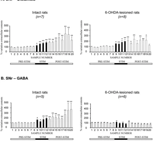
STN–HFS-induced forelimb dyskinesia
in hemiparkinsonian rats is linked to an
increase in Glu levels in the SNr, with no
change in GABA levels
In intact (n
⫽ 7) and 6-OHDA-treated
(n
⫽ 8) rats, STN–HFS at intensity I
1(60 –
200
A, the intensity threshold value
trig-gering contralateral forelimb dyskinesia)
triggered a gradual increase in
extracellu-lar Glu level in the ipsilateral SNr during
the 1 h stimulation period (Fig. 3A,
frac-tions 9 –12). This increase was maximal in
the last 15 min of this period:
⫹80 ⫾ 24%
for intact rats ( p
⬍ 0.01; n ⫽ 7) and
⫹97 ⫾ 37% for hemiparkinsonian
ani-mals ( p
⬍ 0.01; n ⫽ 8). In both intact and
6-OHDA-treated rats, Glu levels remained
high during the poststimulation period
(fractions 13–19), decreasing in the last
fraction collected (Fig. 3A). However,
changes in Glu levels were observed
smaller in hemiparkinsonian than in intact
control rats during this period, possibly
because Glu levels had already been
strongly increased by the dopaminergic
le-sion. Interestingly, forelimb dyskinesias
induced by STN stimulation were always
paralleled by increases in extracellular Glu
levels in the SNr.
In contrast, STN–HFS had a very
dif-ferent effect on GABA levels. STN–HFS did not affect
extracellu-lar GABA levels in the ipsilateral SNr of 6-OHDA-treated rats
during the 1 h stimulation period (Fig. 3B, fractions 9 –12).
Con-versely, in intact rats, STN–HFS increased extracellular GABA
levels in the ipsilateral SNr. This increase was maximal in the last
fraction collected (⫹90 ⫾ 20%; p ⬍ 0.01; fraction 12; n ⫽ 5) and
remained high throughout most of the poststimulation period.
This effect of stimulation is consistent with previous reports
(Windels et al., 2000) but was of lower amplitude in awake than in
anesthetized intact rats.
STN–HFS at an intensity below the threshold value triggering
forelimb dyskinesia increases GABA levels, but not Glu levels,
in the SNr of hemiparkinsonian rats
STN–HFS at intensity I
2(below the threshold value evoking
con-tralateral forelimb dyskinesia) did not affect extracellular Glu
level in the ipsilateral SNr in intact (n
⫽ 6) or 6-OHDA-treated
rats (n
⫽ 9). Extracellular Glu levels remained stable around
baseline throughout the microdialysis experiment (Fig. 4 A).
In contrast to what was observed for Glu levels, STN–HFS at
intensity I
2increased extracellular GABA levels in the ipsilateral
SNr during the 1 h stimulation period (Fig. 4B, fractions 9 –12) in
hemiparkinsonian rats only. This increase reached
⫹69⫾21%( p⬍
0.01; fraction 10; n
⫽ 8) during the stimulation period and
re-mained high throughout the poststimulation period, peaking at
⫹108 ⫾ 25% ( p ⬍ 0.05; fraction 16; n ⫽ 8) (Fig. 4B).
Modulation of STN–HFS-induced forelimb dyskinesia by the
nigral injection of Glu antagonists or agonist in
hemiparkinsonian rats
We investigated the possible involvement of increases in
extracel-lular Glu levels in the SNr of hemiparkinsonian rats in the
induc-tion of forelimb dyskinesia by high-intensity STN–HFS by
ana-lyzing the effects of bilateral SNr injections of kynurenic acid. All
animals subjected to high-intensity STN–HFS (I
1) presented
contralateral forelimb dyskinesia. These motor effects were
to-tally prevented by the bilateral nigral injection of kynurenate at a
dose of 1 nmol, 10 min before STN–HFS (n
⫽ 5) (Fig. 5A). A
similar effect was obtained with a dose of 0.5 nmol. This effect of
kynurenate persisted over 30 min under STN–HFS. No motor
effects were detected when these doses of kynurenate were
in-jected alone into the SNr of 6-OHDA-lesioned animals, without
STN stimulation.
When STN–HFS was applied at an intensity of I
2, eliciting no
forelimb dyskinesia, the bilateral injection of a mixture of Glu
agonists (NMDA plus AMPA) into the SNr facilitated dyskinetic
movements (n
⫽ 5) similar to those observed under STN–HFS at
an intensity of I
1, whatever the dose used (1 or 2 nmol) (Fig. 5B).
Figure 3. Extracellular glutamate (A) and GABA (B) levels, determined at 15 min intervals, in the SNr ipsilateral to the stimulation in intact and 6-OHDA-lesioned rats under basal conditions and during 1 h of STN–HFS with an intensity inducing forelimb dyskinesia (I1,⬎60A). The prestimulation period (PRE-STIM) corresponds to fractions 1–8 of the dialysates; the
stimulation period (STIM), indicated by the black horizontal bar, corresponds to fractions 9 –12 (4 fractions) and the poststimu-lation period (POST-STIM) corresponds to fractions 13–20 (8 fractions). The mean⫾ SEM of the eight PRE-STIM dialysates, collected before STN–HFS, was used to determine baseline levels. Results are expressed as a percentage of variation of this baseline value. Each percentage corresponds to the mean⫾ SEM variations calculated for five to eight animals. Note that Glu levels increased in both intact and 6-OHDA-lesioned rats, whereas GABA levels increased only in intact animals. *p⬍0.05,**p⬍ 0.01 versus baseline values. Error bars correspond to the SEM.
However, 2 nmol doses of these compounds led to slight paralysis
of the hindquarters and akinesia of the forelimbs. As in
experi-ments with kynurenate, the injection of these substances (NMDA
plus AMPA) alone did not induce dyskinesia in
hemiparkinso-nian rats in the absence of stimulation.
In contrast, dyskinetic movements induced by STN–HFS at
intensity I
1were not blocked by the intranigral injection of
mus-cimol (a GABA
Areceptor agonist), whatever the dose
adminis-tered (0.8, 1.5, or 3 nmol) (Fig. 5C).
Discussion
The mechanisms by which STN–HFS affects the BG output
nu-clei remain unclear. We show here that unilateral STN–HFS
duces stimulation intensity-dependent dyskinesias in awake
in-tact or hemiparkinsonian rats. High-intensity STN stimulation
provokes contralateral forelimb dyskinesia and increases Glu
lev-els in the SNr, probably attributable to subthalamonigral
path-way activation, increases of Glu levels being detectable from the
first sample collected during stimulation. Low-intensity
stimula-tion did not trigger forelimb dyskinesia but increased GABA
lev-els in the SNr. STN–HFS-induced
fore-limb dyskinesia was blocked by intranigral
microinjection of a Glu antagonist in
hemiparkinsonian rats. Subthalamonigral
pathway activation, modulating SNr
activ-ity, may therefore be involved in forelimb
dyskinesia, providing insight into the
mechanisms of STN–HFS in PD.
Changes in Glu and GABA levels in
awake rats after SNc lesion
The large increase in basal Glu levels in the
SNr in awake hemiparkinsonian rats is
consistent with electrophysiological data
showing that SNc lesion or neuroleptic
treatment increases STN neurons
dis-charge (Bergman et al., 1994; Hassani et
al., 1996; Degos et al., 2005). This
extracel-lular Glu may come from STN axon
termi-nals, glial cells, or both (Danbolt, 2001;
Parpura et al., 2004). However, STN
le-sions strongly decrease basal Glu levels in
the SNr (Rosales et al., 1997) and prevent
their increase under STN–HFS (M.
Savasta and F. Windels, unpublished
ob-servations). Most of the extracellular Glu
in the SNr is therefore probably linked to
STN neuron activity, confirming the
hy-peractivity of the subthalamonigral
path-way after dopamine lesioning in freely
moving 6-OHDA-lesioned rats and
con-sistent with data for anesthetized animals
(Windels et al., 2005). SNr GABA levels
also increased hemiparkinsonian rats but
less so than Glu levels. Some of this nigral
GABA may result from an increase in
glo-bus pallidus (GP) neuron activity
attribut-able to STN hyperactivity, itself resulting
from dopaminergic denervation (Hassani
et al., 1996). However, additional GABA
sources, such as neuron collaterals from
the SNr or striatum, may also be involved.
The increase in basal nigral Glu and GABA
levels after SNc lesioning was larger in
awake than in anesthetized animals (Windels et al., 2005),
con-sistent with anesthetic effects on amino acid levels (Rozza et al.,
2000; Windels and Kiyatkin, 2006).
Effects of STN stimulation parameters on motor behavior
STN–HFS in normal monkeys induces contralateral dyskinesia
(Beurrier et al., 1997) resembling human and primate
hemibal-lism after STN lesioning (Hamada and DeLong, 1992; Lee
and Marsden, 1994). We found that unilateral STN–HFS in
awake intact or hemiparkinsonian rats induced contralateral
forelimb dyskinesia and even contralateral rotation, depending
on stimulation parameters and intensity, as observed after STN
lesioning (Henderson et al., 1999). Rotational effects were
ob-served at intensities exceeding 190
A and were preceded by
orofacial, axial, and forelimb dyskinesias resembling
L-3,4-dihydroxyphenylalanine (
L-DOPA)-induced dyskinesias in
hemiparkinsonian rats (Winkler et al., 2002; Sgambato-Faure et
al., 2005). Higher-intensity stimulation was required to elicit
dys-kinesias in SNc-lesioned than in intact rats, but only for
frequen-Figure 4. Extracellular glutamate (A) and GABA (B) levels determined, at 15 min intervals, in the SNr ipsilateral to the stimulation in intact and 6-OHDA-lesioned rats, under basal conditions and during 1 h of STN–HFS with an intensity below the threshold for forelimb dyskinesia (I2). The prestimulation period (PRE-STIM) corresponds to fractions 1– 8 of the dialysates;
the stimulation period (STIM), indicated by the black horizontal bar, corresponds to fractions 9 –12 (4 fractions), and the post-stimulation period (POST-STIM) corresponds to fractions 13–20 (8 fractions). The mean⫾ SEM of the eight PRE-STIM dialysates, collected before STN–HFS, was used to determine baseline levels. Results are expressed as a percentage of variation of this baseline value. Each percentage corresponds to the mean⫾SEMvariationscalculatedforsixtonineanimals.NotethatGlulevels in the SNr were not significantly affected by STN–HFS in the SNr in either intact or 6-OHDA-lesioned rats, whereas GABA levels increased only in 6-OHDA-lesioned animals. *p⬍ 0.05, **p ⬍ 0.01 versus baseline values. Error bars indicate the SEM.
cies
⬍130 Hz or pulse widths ⬍60
s. Sensitivity to STN–HFS
therefore differed between the parkinsonian and normal states, as
predicted after STN lesioning (Guridi and Obeso, 2001) and
con-sistent with previous studies (Salin et al., 2002). No differential
effect was observed, in any group, if STN–HFS parameters were
set so as to induce dyskinesia (Chang et al., 2003). We determined
the threshold value for forelimb dyskinesia, for investigation of
the neurochemical changes induced by STN stimulation in
nor-mal and hemiparkinsonian rats at two different intensities, only
one of which induced forelimb dyskinesia.
Changes in nigral Glu and GABA levels induced by high
(I
1)- or low (I
2)-intensity STN stimulation
The neurochemical changes in the SNr depended on STN–HFS
intensity. In normal and hemiparkinsonian rats, high-intensity
STN–HFS increased Glu concentration in the SNr, whereas
lower-intensity stimulation not inducing forelimb dyskinesia
in-creased GABA levels in 6-OHDA-lesioned rats only. The
excita-tory responses to high-intensity STN stimulation in the SNr
ob-served in electrophysiological studies probably result from
specific activation of the glutamatergic subthalamonigral
path-way (Hammond et al., 1978; Maurice et al., 2003), because the
latency of the excitatory responses evoked in nigral cells
corre-sponds to the conduction time for this pathway (Kitai and
De-niau, 1981). STN–HFS at higher frequencies systematically led to
forelimb dyskinesia in both normal and hemiparkinsonian rats,
probably by increasing the rate of SNr cell discharge through
glutamatergic transmission (Hashimoto et al., 2003; Kita et al.,
2005). These findings are consistent with reports of hemiballism
in PD patients during STN–HFS at voltages higher than used for
chronic treatment (Limousin et al., 1996; Moro et al., 2002). This
seems to conflict with data reporting a direct relationship
be-tween hemiballism and STN lesions in humans and experimental
models (Whittier and Mettler, 1949; Carpenter et al., 1950;
Ham-mond et al., 1979; Hamada and DeLong, 1992; Guridi and Obeso,
2001). Indeed, even discrete STN lesions in primates, probably
linked to a decrease in the Glu outflow, have been reported to
produce hemiballism. Our data linking forelimb dyskinesia and
an increase in Glu outflow under STN–HFS seem to conflict with
these previous observations. However, pharmacological STN
ac-tivation has also been reported to produce hemiballism
(Cross-man et al., 1980, 1984; Perier et al., 2002), so the mechanisms
involved remain unclear and are still under debate. The STN thus
forms part of a complex network. The impact of STN–HFS
should therefore not be considered as a simple “functional
le-sion” of the STN, because it may have multiple effects on synapses
(Shen et al., 2003) and neurotransmitter release.
The forelimb dyskinesia induced by high-intensity STN
stim-ulation in hemiparkinsonian rats was prevented by nigral
injec-tion of the broad-spectrum Glu antagonist kynurenic acid,
dem-onstrating that the hyperactive Glu transmission induced in the
SNr by STN–HFS probably causes the observed forelimb
dyski-nesia. Glu transmission and forelimb dyskinesia are clearly linked
because direct infusion of NMDA plus AMPA into the SNr
trig-gered forelimb dyskinesia in hemiparkinsonian rats under
low-intensity STN–HFS. Neither low-low-intensity STN–HFS nor local
Glu agonist injection without STN–HFS caused forelimb
dyski-nesia, suggesting synergistic action. The lack of forelimb
dyskine-sia after Glu agonist injection alone suggests that STN–HFS may
induce forelimb dyskinesia by activating/modulating several sites
simultaneously and that SNr activation alone may be insufficient.
Furthermore, the doses of Glu agonists used here may have been
too low and/or may not have reached the effective synaptic sites
within the SNr neuronal network. The GABA
Aagonist
(musci-mol) may have been unable to prevent dyskinetic movements for
the same reason. The action of muscimol on presynaptic and
postsynaptic GABA
Areceptors in the SNr may also account for
complex opposite cellular interactions within the nigral network
and related structures. However, regardless of the functional
ef-fects of high- or low-intensity STN–HFS, our data suggest that
Glu transmission is involved in generating of forelimb
dyskine-sias after STN stimulation. It would be interesting to identify the
subtypes of Glu receptors involved in generating
STN–HFS-induced forelimb dyskinesia, because recent studies in animal
models of
L-DOPA-induced dyskinesia have implicated the
NMDA Glu receptor and/or the metabotropic Glu receptor 5
(Papa and Chase, 1996; Nash et al., 2004; Dekundy et al., 2006).
Figure 5. Effects of bilateral nigral injections of kynurenic acid (A), NMDA plus AMPA (B), and muscimol (C) on STN–HFS-induced contralateral forelimb dyskinesia in 6-OHDA-lesioned rats. STN stimulation was applied before and after drug injection. Note that, in A, kynurenate (1 nmol/side) alone did not provoke dyskinesia but, at the same dose, did prevent those induced by STN–HFS at intensity I1(n⫽5).*p⬍0.05versus6-OHDA-lesionedratsplusSTN–HFSatI1.
In B, STN stimulation was applied at intensity I2, resulting in dyskinesia only if NMDA plus AMPA
(1 nmol/side) was injected (n⫽ 5) during STN–HFS. *p ⬍ 0.05 versus 6-OHDA-lesioned rats plus NMDA/AMPA or versus 6-OHDA-lesioned rats plus STN–HFS at I2. Note in C that muscimol
However, it is unclear whether
L-DOPA therapy and
high-intensity STN–HFS induced forelimb dyskinesia in similar ways.
Orofacial dyskinesia may also be induced by low-intensity
STN–HFS but with no Glu concentration increase in the SNr. We
cannot exclude the possibility that this type of dyskinesia results
from preferential Glu release in an unsampled part of the SNr or
from Glu release in quantities too small for detection by the
method used. The mechanisms underlying orofacial dyskinesia
may therefore differ from those underlying forelimb dyskinesia
under STN–HFS. This is consistent with data showing that
L
-DOPA-induced dyskinesias involve different BG structures/
pathways (Sharp et al., 1987; Winkler et al., 2002).
GABA release in the SNr after high-intensity STN stimulation
in intact rats or low-intensity stimulation in hemiparkinsonian
rats probably inhibits SNr cell activity, consistent with
electro-physiological data showing that SNr inhibition is abolished by the
iontophoretic application of bicuculline, a GABA
Areceptor
an-tagonist (Maurice et al., 2003). This inhibitory effect results
prin-cipally from pallidonigral fiber activation through axon
collater-als, because GABAergic pallidosubthalamic neurons send an
axon collateral to the SNr (Kita and Kitai, 1994). We also
dem-onstrated that, in anesthetized hemiparkinsonian animals, GP
lesions abolish the increase in SNr GABA levels induced by STN–
HFS (Windels et al., 2005). However, SNr inhibition may also
result from activation of the intranigral axon collateral network
of GABAergic SNr cells (Mailly et al., 2003). According to the
“classical” model, direct striatonigral GABAergic pathway
activa-tion inhibits the tonically active GABAergic neurons of the SNr,
abolishing target nucleus inhibition in premotor structures in the
thalamus and brainstem (Chevalier and Deniau, 1990; DeLong,
1990). This process is crucial in BG physiology (Mink and Thach,
1993). In PD patients and experimental models, dopaminergic
neuron degeneration leads to STN neuron hyperactivity
increas-ing the tonic discharge of BG GABAergic output structures
(Bergman et al., 1994; Levy et al., 2002) and to imbalance between
the direct and indirect striatonigral pathways, inducing motor
disturbances (Chesselet and Delfs, 1996; Obeso et al., 1997). Our
neurochemical data show that motor function recovery in PD
patients probably requires low-intensity STN–HFS (1) to abolish
the excitatory influence of the indirect striatonigral pathway on
SNr cells and other BG output structures (Vitek, 2002; McIntyre
et al., 2004a,b) and/or (2) to modify the spatiotemporal pattern of
neuronal discharge in STN and BG output nuclei (Hashimoto et
al., 2003).
In conclusion, STN–HFS-induced forelimb dyskinesia is
me-diated by Glu, probably through STN axon activation, in awake
hemiparkinsonian rats. During STN–HFS at intensities below the
threshold for forelimb dyskinesia, GABA may be involved in the
functional disinhibition by which the BG organize normal
move-ment and in the mechanisms of STN–HFS in PD.
References
Albin RL, Young AB, Penney JB (1989) The functional anatomy of basal ganglia disorders. Trends Neurosci 12:366 –375.
Alexander GE, Crutcher MD (1990) Functional architecture of basal ganglia circuits: neural substrates of parallel processing. Trends Neurosci 13:266 –271.
Alvarez L, Macias R, Lopez G, Alvarez E, Pavon N, Rodriguez-Oroz MC, Juncos JL, Maragoto C, Guridi J, Litvan I, Tolosa ES, Koller W, Vitek J, DeLong MR, Obeso JA (2005) Bilateral subthalamotomy in Parkinson’s disease: initial and long-term response. Brain 128:570 –583.
Aziz TZ, Peggs D, Sambrook MA, Crossman AR (1991) Lesion of the sub-thalamic nucleus for the alleviation of 1-methyl-4-phenyl-1,2,3,6-tetrahydropyridine (MPTP)-induced parkinsonism in the primate. Mov Disord 6:288 –292.
Benabid AL (2003) Deep brain stimulation for Parkinson’s disease. Curr Opin Neurobiol 13:696 –706.
Benabid AL, Benazzouz AL, Pollak P (2002) Mechanisms of deep brain stimulation. Mov Disord 3:S73–S74.
Benazzouz A, Gross C, Fe´ger J, Boraud T, Bioulac B (1993) Reversal of ri-gidity and improvement in motor performance by subthalamic high-frequency stimulation in MPTP-treated monkey. Eur J Neurosci 5:382–389.
Bergman H, Wichmann T, DeLong MR (1990) Reversal of experimental parkinsonism by lesions of the subthalamic nucleus. Science 249:1436 –1438.
Bergman H, Wichmann T, Karmon B, DeLong MR (1994) The primate subthalamic nucleus. II. Neuronal activity in the MPTP model of parkin-sonism. J Neurophysiol 72:507–520.
Beurrier C, Bezard E, Bioulac B, Gross C (1997) Subthalamic stimulation elicits hemiballismus in normal monkey. NeuroReport 8:1625–1629. Bevan MD, Magill PJ, Terman D, Bolam JP, Wilson CJ (2002) Move to the
rhythm: oscillations in the subthalamic nucleus-external globus pallidus network. Trends Neurosci 25:525–531.
Bruet N, Windels F, Carcenac C, Feuerstein C, Bertrand A, Poupard A, Savasta M (2003) Neurochemical mechanisms induced by high fre-quency stimulation of the subthalamic nucleus: increase of extracellular striatal glutamate and GABA in normal and hemiparkinsonian rats. J Neuropathol Exp Neurol 62:1228 –1240.
Carpenter MB, Whittier JR, Mettler FA (1950) Analysis of choreoid hy-perkinesia in the rhesus monkey: surgical and pharmacological analysis of hyperkinesia resulting from lesions in the subthalamic nucleus of Luys. J Comp Neurol 92:293–332.
Chang JY, Shi LH, Luo F, Woodward DJ (2003) High frequency stimulation of the subthalamic nucleus improves treadmill locomotion in unilateral 6-hydroxydopamine lesioned rats. Brain Res 983:174 –184.
Chesselet MF, Delfs JM (1996) Basal ganglia and movement disorders: an update. Trends Neurosci 19:417– 422.
Chevalier G, Deniau JM (1990) Disinhibition as a basic process in the ex-pression of striatal functions. Trends Neurosci 13:277–280.
Crossman AR, Sambrook MA, Jackson A (1980) Experimental hemiballis-mus in the baboon produced by injection of a gamma-aminobutyric acid antagonist into the basal ganglia. Neurosci Lett 20:369 –372.
Crossman AR, Sambrook MA, Jackson A (1984) Experimental hemichorea/ hemiballismus in the monkey. Studies on the intracerebral site of action in a drug-induced dyskinesia. Brain 107:579 –596.
Danbolt NC (2001) Glutamate uptake. Prog Neurobiol 65:1–105. Degos B, Deniau JM, Thierry AM, Glowinski J, Pezard L, Maurice N (2005)
Neuroleptic-induced catalepsy: electrophysiological mechanisms of func-tional recovery induced by high-frequency stimulation of the subthalamic nucleus. J Neurosci 25:7687–7696.
Dekundy A, Malgorzata P, Schaefer D, Cenci MA, Danysz W (2006) Effects of group I metabotropic glutamate receptors blockade in experimental models of Parkinson’s disease. Brain Res Bull 69:318 –326.
DeLong MR (1990) Primate models of movement disorders of basal ganglia origin. Trends Neurosci 13:281–285.
Dewey RB, Jankovic J (1989) Hemiballism-hemichorea. Clinical and phar-macologic findings in 21 patients. Arch Neurol 46:862– 867.
Dostrovsky JO, Lozano AM (2002) Mechanisms of deep brain stimulation. Mov Disord 17:S63–S68.
Guesdon JL, Ternynck T, Avrameas S (1979) The use of avidin-biotin inter-action in immunoenzymatic techniques. J Histochem Cytochem 27:1131–1139.
Guridi J, Herrero MT, Luquin MR, Guillen J, Ruberg M, Laguna J, Vila M, Javoy-Agid F, Agid Y, Hirsch E, Obeso JA (1996) Subthalamotomy in parkinsonian monkeys. Behavioral and biochemical analysis. Brain 119:1717–1727.
Guridi J, Obeso JA (2001) The subthalamic nucleus, hemiballismus and Parkinson’s disease: reappraisal of a neurosurgical dogma. Brain 124:5–19.
Hamada I, DeLong MR (1992) Excitotoxic acid lesions of the primate sub-thalamic nucleus result in transient dyskinesias of the contralateral limbs. J Neurophysiol 68:1850 –1858.
Hammond C, Deniau JM, Rizk A, Fe´ger J (1978) Electrophysiological dem-onstration of an excitatory subthalamonigral pathway in the rat. Brain Res 151:235–244.
Hammond C, Fe´ger J, Bioulac B, Souteyrand JP (1979) Experimental hemi-Boulet et al.• SNr Glutamate and STN–HFS-Induced Dyskinesia J. Neurosci., October 18, 2006•26(42):10768 –10776 • 10775
ballism in the monkey produced by unilateral kainic acid lesion in corpus Luysii. Brain Res 171:577–580.
Hashimoto T, Elder C, Okun MS, Patrick SK, Vitek JL (2003) Stimulation of the subthalamic nucleus changes the firing pattern of pallidal neurons. J Neurosci 23:1916 –1923.
Hassani OK, Mouroux M, Fe´ger J (1996) Increased subthalamic neuronal activity after nigral dopaminergic lesion independent of disinhibition via the globus pallidus. Neuroscience 72:105–115.
Henderson JM, Annett LE, Ryan LJ, Chiang W, Hidaka S, Torres EM, Dun-nett SB (1999) Subthalamic nucleus lesions induce deficits as well as benefits in the hemiparkinsonian rat. Eur J Neurosci 11:2749 –2757. Hutchison WD, Allan RJ, Opitz H, Levy R, Dostrovsky JO, Lang AE, Lozano
AM (1998) Neurophysiological identification of the subthalamic nu-cleus in surgery for Parkinson’s disease. Ann Neurol 44:622– 628. Kita H (1994) Physiology of two disynaptic pathways from the
sensorimo-tor cortex to the basal ganglia output nuclei. In: The basal ganglia IV (Percheron G, McKenzie JS, Fe´ger J, eds), pp 263–276. New York: Plenum.
Kita H, Kitai ST (1987) Efferent projections of the subthalamic nucleus in the rat: light and electron microscopic analysis with the PHA-L method. J Comp Neurol 260:435– 452.
Kita H, Kitai ST (1994) The morphology of globus pallidus projection neu-rons in the rat: an intracellular staining study. Brain Res 636:308 –319. Kita H, Tachibana Y, Nambu A, Chicken S (2005) Balance of monosynaptic
excitatory and disynaptic inhibitory responses of the globus pallidus in-duced after stimulation of the subthalamic nucleus in the monkey. J Neu-rosci 25:8611– 8619.
Kitai ST, Deniau JM (1981) Cortical inputs to the subthalamus: intracellular analysis. Brain Res 214:411– 415.
Krack P, Batir A, Van Blercom N, Chabardes S, Fraix V, Ardouin C, Koudsie A, Dowsey Limousin P, Benazzouz A, Lebas JF, Benabid AL, Pollak P (2003) Five-year follow-up bilateral stimulation of the subthalamic nu-cleus in advanced Parkinson’s disease. N Eng J Med 349:1925–1934. Lee MS, Marsden CD (1994) Movement disorders following lesions of the
thalamus or subthalamic region. Mov Disord 9:493–507.
Levy R, Ashby P, Hutchinson WD, Lang AE, Lozano AM, Dostrovsky JO (2002) Dependence of subthalamic nucleus oscillations on movement and dopamine in Parkinson’s disease. Brain 125:1196 –1209.
Limousin P, Pollak P, Benazzouz A, Hoffman D, Broussolle E, Perret JE, Benaabid (1995) Bilateral subthalamic nucleus stimulation for severe Parkinson’s disease. Mov Disord 10:672– 674.
Limousin P, Pollak P, Hoffmann D, Benazzouz A, Perret JE, Benabid AL (1996) Abnormal involuntary movements induced by subthalamic stim-ulation in parkinsonian patients. Mov Disord 11:231–235.
Limousin P, Krack P, Pollak P, Benazzouz A, Ardouin C, Hoffmann D, Benabid AL (1998) Electrical stimulation of the subthalamic nucleus in advanced Parkinson’s disease. N Engl J Med 339:1105–1111.
Mailly P, Charpier S, Menetrey A, Deniau JM (2003) Three-dimensional organization of the recurrent axon collateral network of the substantia nigra pars reticulata neurons in the rat. J Neurosci 23:5247–5257. Maurice N, Thierry AM, Glowinski J, Deniau JM (2003) Spontaneous and
evoked activity of substantia nigra pars reticulata neuron during high-frequency stimulation of the subthalamic nucleus. J Neurosci 23:9929 –9936.
McIntyre CC, Savasta M, Kerkerian-Legoff L, Vitek JL (2004a) Uncovering the mechanism(s) of action of deep brain stimulation: activation, inhibi-tion, or both. Clin Neurophysiol 115:1239 –1248.
McIntyre CC, Savasta M, Walter B, Vitek JL (2004b) How does deep brain stimulation work? Present understanding and future questions. J Clin Neurophysiol 21:1–11.
Miller WC, DeLong MR (1987) Altered tonic activity in the globus pallidus and subthalamic nucleus in the primate MPTP model parkinsonian. In: The basal ganglia II. Structure and function: current concepts (Carpenter MB, Jayaraman A, eds), pp 415– 427. New York: Plenum.
Mink JW, Thach WT (1993) Basal ganglia intrinsic circuits and their role in behavior. Curr Opin Neurobiol 3:950 –957.
Moro E, Esselink RJA, Xie J, Hommel M, Benabid AL, Pollak P (2002) The impact on Parkinson’s disease of electrical parameter settings in STN stimulation. Neurology 59:1–7.
Nambu A (2004) A new dynamic model of the cortico-basal ganglia loop. Prog Brain Res 143:461– 466.
Nash JE, Ravenscroft P, McGuire S, Crossman AR, Menniti FS, Brotchie JM (2004) The NR2B-selective NMDA receptor antagonist CP-101,606 ex-acerbatesL-DOPA-induced dyskinesia and provides mild potentiation of anti-parkinsonian effects ofL-DOPA in the MPTP-lesioned marmoset model of Parkinson’s disease. Exp Neurol 188:471– 479.
Obeso JA, Rodriguez MC, DeLong MR (1997) Basal ganglia pathophysiol-ogy. A critical review. Adv Neurol 74:3–18.
Papa SM, Chase TN (1996) Levodopa induced dyskinesias improved by a glutamate antagonist in parkinsonian monkeys. Ann Neurol 39:574 –578. Parpura V, Scemes E, Spray DC (2004) Mechanisms of glutamate release from astrocytes: gap junction “hemichannels,” purinergic receptors and exocytotic release. Neurochem Int 45:259 –264.
Paxinos G, Watson C (1982) The rat brain in stereotaxic coordinates, Ed 4. San Diego: Academic.
Perier C, Tremblay L, Feger J, Hirsch EC (2002) Behavioral consequences of bicuculline injection in the subthalamic nucleus and the zona incerta in rat. J Neurosci 22:8711– 8719.
Rosales MG, Martinez-Fong D, Morales R, Nunez A, Flores G, Gongora-Alfaro JL, Floran B, Aceves J (1997) Reciprocal interaction between glu-tamate and dopamine in the pars reticulata of the rat substantia nigra: a microdialysis study. Neuroscience 80:803– 810.
Rozza A, Masoero E, Favalli L, Lanza E, Govoni S, Rizzo V, Montalbetti L (2000) Influence of different anaesthetics on extracellular aminoacids in rat brain. J Neurosci Methods 101:165–169.
Salin P, Manrique C, Forni C, Kerkerian-Le Goff L (2002) High-frequency stimulation of the subthalamic nucleus selectively reverses dopamine de-nervation induced cellular defects in the output structures of the basal ganglia in the rat. J Neurosci 22:5137–5148.
Sgambato-Faure V, Buggia V, Gilbert F, Le´vesque D, Benabid AL, Berger F (2005) Coordinated and spatial up regulation of Arc in striato-nigral neurons correlates withL-Dopa-induced-behavioral sensitization in dys-kinetic rats. J Neuropathol Exp Neurol 64:936 –947.
Sharp T, Zetterstrom T, Ljunberg T, Ungerstedt U (1987) A direct compar-ison of amphetamine-induced behaviours and regional brain dopamine release in the rat using intracerebral dialysis. Brain Res 401:322–330. Shen K, Zhu ZT, Munhall A, Johnson SW (2003) Synaptic plasticity in rat
subthalamic induced by high frequency stimulation. Synapse 50: 314 –319.
Smith Y, Shink E, Sidibe M (1998) Neuronal circuitry and synaptic connec-tivity of the basal ganglia. Neurosurg Clin N Am 9:203–222.
Tossman U, Ungerstedt U (1986) Microdialysis in the study of extracellular levels of amino acids in the rat brain. Acta Physiol Scand 128:9 –14. Vitek JL (2002) Mechanisms of deep brain stimulation: excitation or
inhi-bition. Mov Disord 17:S69 –S72.
Whittier JR, Mettler FA (1949) Studies of the subthalamus of the rhesus monkey. II. Hyperkinesia and other physiologic effects of subthalamic lesions with special references to the subthalamic nucleus of Luys. J Comp Neurol 90:319 –372.
Windels F, Kiyatkin EA (2006) General anesthesia as a factor affecting im-pulse activity and neuronal responses to putative neurotransmitters. Brain Res 1086:104 –116.
Windels F, Bruet N, Poupard A, Urbain N, Chouvet G, Feuerstein C, Savasta M (2000) Effects of high frequency stimulation of subthalamic nucleus on extracellular glutamate and GABA in substantia nigra and globus pal-lidus in the normal rat. Eur J Neurosci 12:4141– 4146.
Windels F, Bruet N, Poupard A, Feuerstein C, Bertrand A, Savasta M (2003) Influence of the frequency parameter on extracellular glutamate and gamma-aminobutyric acid in substantia nigra and globus pallidus during electrical stimulation of subthalamic nucleus in rats. J Neurosci Res 72:259 –267.
Windels F, Carcenac C, Poupard A, Savasta M (2005) Pallidal origin of GABA release within the substantia nigra pars reticulata during high fre-quency stimulation of the subthalamic nucleus. J Neurosci 25:5079 –5086. Winkler C, Kirik D, Bjorklund A, Cenci MA (2002) L-DOPA induced dys-kinesia in the intrastriatal 6-hydroxydopamine model of Parkinson’s dis-ease: relation to motor and cellular parameters of nigrostriatal function. Neurobiol Dis 10:165–186.