HAL Id: tel-02918162
https://tel.archives-ouvertes.fr/tel-02918162
Submitted on 20 Aug 2020HAL is a multi-disciplinary open access archive for the deposit and dissemination of sci-entific research documents, whether they are pub-lished or not. The documents may come from teaching and research institutions in France or abroad, or from public or private research centers.
L’archive ouverte pluridisciplinaire HAL, est destinée au dépôt et à la diffusion de documents scientifiques de niveau recherche, publiés ou non, émanant des établissements d’enseignement et de recherche français ou étrangers, des laboratoires publics ou privés.
Yousra Ben Zouari
To cite this version:
Yousra Ben Zouari. The functional and spatial organization of chromatin during Thymocyte de-velopment. Cellular Biology. Université de Strasbourg, 2018. English. �NNT : 2018STRAJ025�. �tel-02918162�
The functional and spatial organization of
chromatin during Thymocyte development
UNIVERSITÉ DE STRASBOURG
ÉCOLE DOCTORALE DES SCIENCES DE LA VIE ET DE LA SANTÉ
IGBMC - CNRS UMR 7104 - Inserm U 1258
THÉSE
présentée par:Yousra BEN ZOUARI
Soutenue le : 03 May 2018
pour obtenir le grade de :
Docteur de l’université de Strasbourg
Discipline/ Spécialité:
Aspects moléculaires et cellulaires de la biologieTHÈSE dirigée par :
M. SEXTON Thomas CR, IGBMC, Illkirch, France
RAPPORTEURS :
Mme SACCANI Simona DR, IRCAN, Nice, France
M. MANKE Thomas Professeur, Max Planck, Freiburg, Allemagne
AUTRES MEMBRES DU JURY :
Mme BOEVA Valentina CR, Institut Cochin, France M. KASTNER Philipe MCF, IGBMC, Illkirch, France Mme SOUTOGLOU Evi DR, IGBMC, Illkirch, France
Philippe Kastner, Professeur Thomas Manke, Dr. Valentina Boeva and Dr. Evi Soutoglou for accepting to be members of my thesis committee. I am sure your expertise will provide great insight and help defining future directions of the project.
Afterwards, I want to a give special thanks to my supervisor Tom Sexton who made these almost 4 years of PhD an exciting experience. I have been extremely lucky to have a supervisor who cared so much about my work, and who responded to my questions and queries so promptly. Thank you for being supportive, trustful and encouraging. I am very grateful for everything you did for me.
I would like to thank both past and current members of Sexton laboratory. Anne, that started the journey with me, thank you for the laughs and fun moments and for your support in the hard times. I need more than these lines to tell you how much I'm counting on you my best friend. Dominique, you are the wisdom and the caring of the lab. Thank you for being helpful and supportive. I am grateful to every advice you gave me. Audrey, thank you for the joy and fun we had together. I was very pleased to share with you the same space. Sunjay, thank you for the Indian food you made for every special moments. Nezih, thank you for driving me all the time and for the fun moments we had in the car with Turkish music without forgetting the good food of your mum. Manon, thank you for your pure smile and your optimism in the hard times. Natalia, thank you for your smile and spoiling us with chocolates. Angeleki, thank you for being so pretty and elegant. You are the sunshine of our lab.
I would like also to thank other people from IGBMC who became very close friends during these past 4 years. Rana and Naima, thank you for your kindness and the joyful moments I had with you. It was a great pleasure for me to spend time with you.
3
I also have to thank everyone that, one way or another, had an important technical and/or scientific input on my work. I want to thank everyone from the IGBMC Informatic servicesn namely Serge Uge for the help with server problems.
Je souhaite également remercier ma famille pour leur soutien et leur amour. Maman et papa, Fathia et Mokhtar, je ne trouve pas de mots pour vous dire à quel point je vous suis reconnaissante pour tout ce que vous avez et continuez à faire pour moi. San nul doute, c’est grâce à vous que je suis la personne que je suis aujourd’hui. Merci pour votre amour à toute épreuve et pour avoir toujours fait en sorte que je ne manque de rien. Mes sœurs et mon frère, Intissar, safa et yassine, je ne pouvais pas rêver de meilleur frère et sœur. J’ai beaucoup de chance d’avoir partagé mon enfance avec vous.
Je remercie Sameh, mon meilleur ami pour avoir toujours été là. Aussi loin que je m’en souvienne, tu as toujours su trouver les mots pour me remonter le moral pour calmer mes crises de panique. Il me faudrait bien plus que ces quelques lignes pour te dire à quel point je compte sur toi.
Je remercie également mes amis Afef et Takoi pour toutes les soirées, journées, sorties etc … Vous êtes des amis comme tout le monde en rêverait ! Je sais que je peux compter sur vous aussi bien pour faire la fête qu’en cas de besoin !
Finally, I would like to thank also Badr, for being supportive, encouraging and caring. You have been a part of this dream which came true. Thank you for being a part of this journey and pushing me forward in the hard times.
Introduction
DNA, the genetic code of nearly all living organisms, is associated with proteins, predominantly histones in eukaryotes to facilitate their folding into chromatin within the cell nucleus. Chromatin needs to be densely compacted, while still allowing access of genes and regulatory elements for control of biological processes such as transcription. Chromosome folding takes place at different hierarchical levels, with various topologies correlated with control of gene expression. At the kilobase-to-megabase scale, chromatin forms loops which bring gene promoters and their distal regulatory elements, such as transcriptionally activating enhancers, into direct physical proximity. These topologies have been proposed to form an “active chromatin hub”, whereby the combination of regulatory factors bound to the promoter and interacting enhancers generates an environment permissive to transcriptional activation. At a higher level, groups of chromatin loops are confined within larger, megabase-scale structures termed “topologically associated domains” (TADs). These can in turn be organized into transcriptionally active (“A”) or repressed (“B”) compartments. TAD borders may play an important functional role in preventing aberrant contacts between enhancers and non-target neighboring genes.
Despite the large number of recent studies describing chromatin topologies and their correlations with gene activity, many questions remain, in particular how these topologies are formed and maintained. Interestingly, the epigenetic state (as determined by histone modifications) of enhancers varies much more widely across cell types than at promoters, suggesting that most cell fate potential is actually encoded at a distance from developmental genes. Studies of chromatin loops between developmental genes and their enhancers give different views on the role of chromatin topology in gene control. Some indicate that
5
chromatin loops form concomitantly with transcriptional activation, concluding that the topology is directly responsible for gene expression. However other studies show that the chromatin loop can precede transcriptional activation, suggesting that the topology represents an earlier “priming” event, allowing the gene to be regulated by downstream acute signals. Furthermore, it is not clear how and which epigenetic marks on enhancers may be instructive for chromatin loop formation. Analogously, questions remain as to what extent TAD structures are developmentally plastic or stable. It is thus very important to understand better the link between epigenetic marks, chromatin topology and transcriptional control.
Most studies of chromatin topology are based on the chromosome conformation capture (3C) method, whereby formaldehyde crosslinked chromatin regions is digested and re-ligated to create chimeric DNA products between restriction fragments which were physically proximal in vivo. Specific target products can be amplified and measured by quantitative PCR; the Hi-C method employs high-throughput sequencing to measure such chromatin interactions on a genome-wide scale. However, the strength of this latter approach is also its disadvantage: the number of potential interactions which can be detected is far greater than current sequencing capacities, limiting the resolution of the technique. To overcome this limitation, we and others have introduced an oligonucleotide capture step into the Hi-C procedure (Cap-C) to study interactions within subsets of genomic regions at high resolution. In one experimental setup, we have designed capture probes to each of the ~22,000 mouse promoters to systematically characterize their chromatin looping interactions. In another, we have designed a tiling strategy across selected ~600 kb regions, targeting studies of chromatin interactions to those flanking TAD borders close to differentially expressed developmental genes.
In the lab, we performed Cap-C experiments to study topological changes during thymocyte development, specifically at the transition between Double Negative (DN) and
epigenomic changes during this transition, we aim to address the following questions: 1. Is chromatin structure stable or dynamic during development?
2. How are chromatin topologies (loops and TADs) established and maintained?
Methods
During my PhD, I performed the computational analyses of the lab’s Cap-C datasets. Although these datasets have a superior resolution to most Hi-C datasets, the previously developed bioinformatics tools for Hi-C analysis were inappropriate for the unique challenges presented by Cap-C data. To determine the most reliable interactions between promoters and distal regulatory elements, I developed a computational method (PromoMaxima) more robust and specific than previously published approaches. I then performed integrative analyses of these called interactions (and called TADs/borders from the other Cap-C strategy) with published ChIP-seq and RNA-seq data, to obtain the following hypotheses and conclusions about the interplay of genome structure and function in thymocyte development.
Results
I. Identification of a complex network of dynamic chromatin loops during thymopoesis
With PromoMaxima, I identified thousands of chromatin looping interactions in thymocytes. Whereas many were developmentally stable, hundreds were nevertheless much stronger in one cell type or another, often linked to a transcriptional change of the interacting gene. As was previously reported, a large number of chromatin loops were detected between promoters and distal regions containing CTCF binding sites and/or the histone signatures of enhancers. Although many epigenomic studies have distinguished the histone modifications
7
of “poised” and “active” enhancer elements, such marks do not seem to be predictive of looping state: depending on the genomic context, poised or active enhancers appear just as likely to interact with their target genes. However, unlike the previous studies, which focused on enhancers, we also identified hundreds of interactions with distal elements which correlated with repression of the target gene, indicating an extensive network of distal silencers. These regulatory elements have been described in specific case studies decades ago, but to date no study has characterized them on a genome-wide scale, nor identified a signature epigenomic mark. The putative silencers I identified are also enriched for CTCF sites, but are depleted of active histone modifications, and enriched in LINE repetitive elements. Other members of the lab have already functionally validated a number of the putative silencers that I identified, and are currently performing experiments to characterize them in greater depth in
vivo.
II. TADs are predominantly developmentally stable, with notable remodeling at specific borders.
In contrast with our findings for chromatin loops, TADs appear very robust to developmental changes, with the structures largely maintained despite large transcriptional changes in the underlying genes. However a minority of TADs were remodeled during the DN-to-DP transition, in each case linked to transcriptional induction of the component genes. We observed: 1) The formation of new “sub-TADs” containing the body of the induced gene; 2) a shift in the border of an existing TAD, so that the enhancer is in the same TAD as the entire transcribed gene, and not just the poised promoter. Artificial transcriptional induction of these genes by the dCas9-VP64 system showed that transcription was sufficient to remodel TADs in some cases but not others.
analytical method I developed, allows efficient and sensitive detection of looping chromatin interactions. We have uncovered extensive chromatin topology dynamics during thymocyte development, much of which is correlated with transcriptional regulation. In particular, we uncovered networks of interactions with putative regulatory elements, both activating enhancers and repressing silencers, the latter at a previously underappreciated scale. Previous studies have noted an enrichment of SINE repetitive elements at enhancers, and have hypothesized that these and long terminal repeat retroviral activating elements could have been co-opted during evolution to activate endogenous genes. Based on our finding of LINE enrichment at putative silencers, it is interesting to speculate that these regions, normally shut down by host defense mechanisms against ancestral parasitic elements, may also be co-opted as developmental repressive elements. Future experiments in the lab will explore this possibility, and their potential interplay with the CTCF sites with which they are juxtaposed.
Very recent studies have given conflicting information on whether transcription directly instructs TAD formation or remodeling. We have shown that the majority of TADs are robust to transcriptional changes during development, but that a subset are reorganized around induced genes, in some cases directly. Future experiments of the lab will examine mechanisms other than transcription which may influence chromatin architecture, such as differential binding of CTCF, and how these may interplay with transcriptional control and chromatin architecture.
9
Résumé
Introduction
L’ADN constitue le patrimoine génétique de la plupart des organismes vivants. Il est associé à des protéines dont majoritairement des histones pour former la composante principale du noyau, la chromatine. Celle-ci est fortement condensée pour tenir dans le noyau, une organisation génomique complexe qui toutefois permet l’accessibilité de l’ADN aux différentes activités nucléaires. Ainsi, le contrôle de la transcription survient dans un contexte de repliement chromosomique avec différents niveaux hiérarchiques. A l’échelle de plusieurs centaines de kilobases, la chromatine forme des boucles qui permettent les contacts physiques à distance entre les amplificateurs de transcription « enhancers » et les promoteurs de leurs gènes cibles. Ces structures de chromatine forment ainsi des « active chromatin hubs » qui amènent les facteurs de transcription à se lier aux promoteurs et aux éléments enhancers formant un environnement de régulation plus permissif que celui des promoteurs isolés. Le second niveau hiérarchique est constitué d’un ensemble de boucles chromatiniennes confinées dans des structures de l’ordre du mégabase appelées « domaines topologiques ». Selon l’activité des gènes inclus, les domaines topologiques constituent ensemble un de compartiments actifs « A » ou inactifs « B ». Les frontières de ces domaines topologiques jouent le rôle de barrière en empêchant les contacts aberrants entre des éléments régulateurs et des gènes voisins.
Malgré les vastes études démontrant le rôle de la conformation génomique dans le contrôle transcriptionnel, de nombreuses questions restent en suspens, et en particulier, comment ces structures chromatiniennes sont formées et maintenues. De manière intéressante, l’état de la chromatine au niveau des séquences enhancers varie bien plus d’un type cellulaire à l’autre que celui des promoteurs de gènes, suggérant que le potentiel de régulation
séquences enhancers avec les régions promotrices des gènes. Certains indiquent que la boucle de chromatine se forme de manière concomitante à l’activation de la transcription et concluent que les interactions enhancer-promoteur stimulent directement l’expression des gènes. D’autres montrent que la boucle de chromatine en réalité précède la transcription suggérant que les structures formées sont des événements épigénétiques déjà présents rendant le locus compétent pour une expression efficace en réponse à des signaux de développement tardif. De plus, il n’est pas clair si les profils épigénétiques différents au niveau des enhancers affectent la capacité de former des interactions avec les gènes cibles. Des études précédentes proposent également des points de vue conflictuels à propos de la maintenance des domaines topologiques durant la différenciation cellulaire. Certains montrent que les domaines topologiques sont des structures stables en se basant sur une étude exhaustive de la conformation génomique de différent type cellulaire. D’autres les décrivent comme des structures dynamiques. Il est donc primordial de mieux comprendre les liens entre l’état de la
chromatine au niveau des éléments régulateurs, la topologie de la chromatine et la régulation de la transcription.
L’étude de l’organisation spatiale des chromosomes est basée sur une approche de capture de la conformation chromosomique (3C). Cette technique permet de lier entre elles, grâce au formaldéhyde, les zones chromosomiques proches. Les étapes de digestion / ligation permettront finalement de révéler les rapprochements qui seront détectés par PCR quantitative. Quant au séquençage à haut débit, il donnera accès aux repliements chromosomiques à l’échelle du génome, on parle alors de Hi-C. Cependant, la force de cette approche dans l’accessibilité à toutes les interactions possibles est également sa faiblesse : le nombre d’interactions qui devrait être détecté est bien supérieur à la capacité actuelle de
11
séquençage, conduisant à une perte d’information. Pour contourner ces limitations, nous avons introduit une étape supplémentaire de capture de séquences dans la procédure du Hi-C pour augmenter la résolution à un sous-ensemble de régions du génome (Cap-C). Ainsi, nous avons utilisé des sondes de capture pour les 22 000 promoteurs des gènes de la souris, afin de caractériser systématiquement les interactions chromosomiques entre tous les promoteurs de gènes et leurs enhancers. Dans une deuxième série d’expérience, nous avons ciblés quelques frontières des domaines topologiques contenant des variations d’expression génique essentielles au cours de processus de développement.
Ces expériences de capture de la conformation chromosomique ont été réalisées pour le processus de différenciation des thymocytes en tenant compte uniquement des stades développementaux critiques : Double Négatif (DN) et Double positif (DP). Nous espérons mettre en évidence les liens entre la conformation de la chromatine avec le contrôle de l’expression génique tout en répondant aux questions suivantes :
1.1 La structure chromatinienne, est-elle stable ou dynamique durant la différenciation cellulaire ?
1.2 Comment les structures chromatiniennes (domaines topologiques et boucles chromatiniennes) sont-elles formées et maintenues ?
Méthodologie
Durant ma thèse, j’ai été en charge de l’analyse des données issues des expériences de Cap-C. Les Cap-Cs ont montré une résolution bien supérieure à celles des HiC, cependant les outils d’analyse bio-informatique disponibles se sont avérés inappropriés. Afin de déterminer les interactions significatives entre les promoteurs et les éléments régulateurs, j’ai donc développé une méthode d’analyse plus robuste et efficace que les approches déjà publiées. Par ailleurs, j’ai analysé et intégré les données de Chip-Seq et RNA-seq avec les données de
Résultat
I. Identification d’un large éventail de boucles chromatiniennes au cours du développement des thymocytes
Grâce à notre nouvelle approche, j’ai identifié des milliers de boucles chromatiniennes. Nous avons pu observer que la majorité de ces boucles sont stables au cours du processus de développement des thymocytes. Un certain nombre d’entre elles présente néanmoins un profil dynamique, souvent liées avec une réponse transcriptionelle du gène cible. Comme il a également été publié, un grand nombre de ces boucles ont été répertoriées entre les promoteurs et les régions régulatrices qui portent la signature chromatiniennes des enhancers ainsi que des sites de liaison de CTCF. Bien que de nombreuses études en épigénomique ont identifiées des marques distinctes d’histones entre les enhancers actifs et « poised » pour l’activation des gènes à différentes étapes du développement, ces marques épigénétiques ne sont pas prédictives de la formation des boucles de chromatine. En effet, selon le contexte génomique, tant les enhancers actifs que les enhancers « poised » participent à la formation des boucles chromatiniennes en liant les promoteurs cibles. Contrairement aux études antérieurs qui se sont focalisées sur les enhancers, nous avons pu déterminer des nouveaux éléments régulateurs impliqués dans la répression de l’expression (les « silencers »). Cette classe d’élément régulateur a été décrite il y a quelques décennies, mais aucune étude n’a pu les caractériser à l’échelle du génome jusqu’à présent. Le profil épigénétique des silencers se distingue par une absence de marqueurs d’histone active et es enrichi par la présence d’éléments répétitifs de la classe des LINEs. L’équipe a déjà réalisé un certain nombre de
13
validation de nouveaux silencers identifié par mes soins, et a présente tente de les caractériser plus en profondeur in vivo.
II. Les domaines topologiques sont des structures stables avec quelques changements potentiels au niveau de leurs frontières
En revanche, les domaines topologiques semblent être des structures robustes sur le plan développemental, avec très peu de changements observés entre les deux types cellulaires. Une minorité de domaines ont été remodelés au cours du développement, liés à l'induction transcriptionelle des gènes. Nous avons observé : 1) la formation des nouveaux domaines sur des gènes transcrit. 2) un shift de frontière d’un domaine topologique afin d’inclure la boucle en chromatine du promoteur et son enhancer. L'induction artificielle de ces gènes a montré que certains changements de TAD peuvent être liés à la transcription, tandis que d'autres ne le sont pas.
Conclusion et Discussion
La technique des Cap -C est récente ce qui explique que le laboratoire a dû mettre au point des outils d’analyses complémentaires afin de déterminer de manière fiable les interactions chromatinienne au niveau des régions ciblées. La méthode d’analyse, que j’ai établie, a démontré une bonne efficacité et sensibilité pour la détection de ces interactions chromatiniennes et permettra donc de répondre de manière plus précise aux questions biologiques posées. Nous avons ainsi pu décrypter la structure chromatinienne associée à la différenciation des thymocytes et mettre en évidence des mécanismes de contrôle transcriptionnel de certains gènes. Nous avons identifiés différents éléments régulateurs dont les enhancers et les silencers. Par ailleurs, des études déjà publiées ont montré une corrélation de la présence d’éléments de répétitions SINEs à proximité des enhancers. Dans notre
s’interroger sur les éléments répétitifs du génome. En effet, ils sont considérés comme des « éléments parasitaires ancestraux » qui peuvent être utilisés au cours de l’évolution pour le contrôle des gènes développementaux. Ainsi, il a été proposé que des enhancers rétroviraux ancestraux participent à l’activation de gènes et que d'autres classes d'éléments répétitifs, qui sont naturellement réduites au silence dans le cadre de la défense du génome hôte contre la transposition, puissent aussi être co-optées pour la répression des gènes.
Des études très récentes ont montré des conclusions contradictoires sur la question de savoir si la transcription est directement liée à la formation de domaines topologiques. Nous avons démontré que la plupart des domaines sont robustes aux changements de la transcription, mais qu’il y a certains domaines topologiques qui peuvent être réorganisés directement suite à l’induction des gènes. Les expériences futures de l’équipe vont consister à examiner les facteurs (hors transcription) qui peuvent influencer l'architecture de la chromatine, comme la liaison différentielle des CTCF, et comment ces facteurs peuvent être coordonnés par le contrôle de transcription.
15
Table of contents
Acknowledgments ... 2
Summary ... 4
Résumé ... 9
List of figures ... 18
List of tables ... 20
Introduction ... 22
I: Nuclear and genome architecture ... 23
1. An overview of nuclear organization… ... 23
1.1 The nuclear periphery………..24
1.2 The nuclear pore complex… ... 25
1.3 The nucleolus and other nuclear foci ... 26
2. Chromosome organization in the nuclear space ... 27
2.1 Chromatin loops and gene regulation… ... 28
2.1.1 Cis-regulatory elements: enhancers, silencers and insulators ... 28
2.1.2 Chromatin looping with enhancers ... 30
2.1.3 Chromatin looping NOT just transcription; the role of CTCF and cohesin ... 30
2.1.4 Chromatin loops –stable and/or dynamic structures? ... 32
2.2 Topological associated domains (TADs)- units of genome folding… ... 34
2.2.1 How are TADs formed and maintained? ... 36
16
II: Assessing chromatin interactions ... 44
1. Microscopic approaches ... 44
2. Chromosome conformation capture and its variants ... 48
2.1 3C ... 48
2.2 4C and 5C ... 49
2.3 Hi-C ... 50
2.4 ChIA-PET and HiChIP……….53
2.5 Capture Hi-C (CHi-C) ... ………53
III: Thymocyte development ... 56
1. Thymopoesis ... 56
2. Transcription factors during Thymocyte differentiation… ... 57
Aims ... 60
Results ... 62
I. Hi-C and CHi-C quality control ... 63
II. PromoMaxima: a pipeline for detection and visualization of cis-DNA looping in Capture Hi-C ... 76
III. Developmentally dynamic gene promoter interactions in transcriptional activation and repression… ... 98
IV. TADs caller benchmarking ... 119
V. Transcription directly remodels a small subset of topologically associated domains ... 124
17
1. How are chromatin configurations altered during transcriptional changes accompanying
development? ... 141
1.1 A mixture of stable and dynamic loops during development ... 142
1.1.1 Enhancer-promoter communication: when to loop? ... 143
1.1.2 A LINE to transcriptional silencing? ... 144
1.1.3 Looping beyond transcriptional control? ... 145
1.2 TADs: an architectural buffer? ... ……….146
2. Is chromatin topology important in controlling cell differentiation and development? .... 147
General material and methods ……….150
Abbreviations ... 165
19
List of figures
INTRODUCTION
Figure 1: Chromatin loops stable or dynamic structures. Figure 2: TADs may define gene regulatory zone. Figure 3: The loop extrusion model.
Figure 4: Inferring chromatin architectures from Hi-C contact maps.
RESULTS
I. Hi-C and CHi-C quality control
Figure 1: Hi-C library quality controls. Figure 2: Capture efficiency control.II. PromoMaxima: a pipeline for detection and visualization of cis-DNA
looping in Capture Hi-C
Figure 1: Called interactions by PromoMaxima for Nxt1 gene in mESCs.
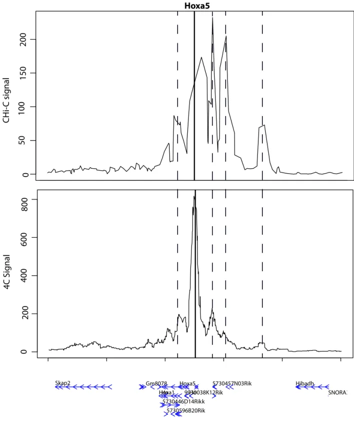
Figure 2: Example of DNA loops, called with PromoMaxima for the gene Hoxa5, and
validated by 4C in mESC.
Figure 3: Benchmark of PromoMaxima, GOTHIC and CHiCAGO. Figure 4: PromoMaxima Browser.
Figure 5: Jaccard index of called interactions maintained in biological replicates, using
PromoMaxima and CHiCAGO on three different promoter CHi-C experiments.
Figure 6: ROC curve of window size and span parameters for local maxima calling in
PromoMaxima.
Figure 7: Distance between interaction peaks in biological replicates of CHi-C data from
mEScs (bp).
III. Developmentally dynamic gene promoter interactions in transcriptional
activation and repression
Figure 1: The mouse thymocyte promoter interactome.
Figure 2: Stable and dynamic promoter interactions linked to transcriptional activation and
repression.
Figure 3: Thymocyte-specific and dynamic enhancers. Figure 4: Distal silencers may regulate contacted genes.
Figure 8: Stable and dynamic promoter interactions linked to transcriptional activation and
repression.
Figure 9: Following up the link between LINEs and putative silencers.
IV. TADs caller benchmarking
Figure 1: Comparative results of methods for the identification of TADs.
V. Transcription directly remodels a small subset of topologically associated
domains
Figure 1: Conservation of TAD structure across thymocyte development. Figure 2: Transcription-coupled sub-TAD formation at the Nfatc3 gene. Figure 3: Transcription-coupled sub-TAD formation at the Tmem131 gene. Figure 4: Transcription directly remodels the Nfatc3 sub-TAD.
Figure 5: TAD border remodeling around the Bcl6 gene during thymocyte maturation. Figure 6: CHi-C enhances resolution of interaction maps.
Figure 7: High reproducibility across biological replicates. Figure 8: Potential differential CTCF binding at the Bcl6 locus.
Figure 9: Transcriptional induction does not remodel the Bcl6 TAD in ES cells. Figure 10: TAD remodeling events uncovered in Hi-C.
21
List of tables
RESULTS
I. Hi-C and CHi-C quality control
Table 1: Total Sequencing reads.Table 2: Captured reads after filtering step in CHi-C (Promoters).
II. PromoMaxima: a pipeline for detection and visualization of cis-DNA
looping in Capture Hi-C
Table 1: GEO datasets.
Table 2: Comparison of CHiCAGO, GOTHIC and PromoMaxima detected interactions in
mESCs.
Table 3: Overlap of called interactions by different tools and called Enhancers based on their
epigenetic features.
III. Developmentally dynamic gene promoter interactions in transcriptional
activation and repression
Table 1: CHi-C interactions with DN3, DP and ES promoters. Table 2: Source of epigenomic datasets used in this analysis.
V. Transcription directly remodels a small subset of topologically associated
domains
22
Introduction
Introduction
The DNA is the genetic material that encodes for all the information essential for life (Dahm, 2005). In eukaryotes, it is wrapped around structural proteins called histones to construct strings of nucleosomes that can be further compacted into a three-dimensional (3D) organization within cell nuclei. At the most extreme, during metaphase, the chromatin fiber folds to the 0.7 µm thick chromatid. The process underlying this compaction remains unclear, although condensins and topoisomerase IIα are implicated in this process (Swedlow and Hirano, 2003;Gibcus et al., 2018). Even at interphase, the spatial arrangement of the chromatin in the nucleus is highly organized at different levels, and can have a direct impact on genomic activity, such as transcription, by regulating DNA accessibility to the genomic machinery. For example, histones can impede the access of many regulatory proteins to their binding motifs, and hinder the movement of polymerases along the DNA fiber. The post- translational modification of histone tails, and/or chromatin remodeling on binding of sequence-specific transcription factors, can facilitate access to DNA, in turn activating some genomic elements (Berger, 2007). Different studies recently demonstrated a further correlation between chromatin topology and underlying gene activity (Cavalli & Misteli, 2013). For example, it was revealed that chromatin looping events can facilitate transcription by bringing distal regulatory elements such as enhancers in direct physical proximity with gene promoters (Palstra et al., 2003). Developmental fate decisions are underpinned by the combinatorial action of tissue-specific enhancers (Osterwalder et al., 2018); it is therefore likely that promoter-enhancer interactions need to be highly regulated to prevent aberrant gene responses. At the megabase scale, the genome folds into discrete 3D structures that tend to favor internal rather than external interactions. These structures have been termed
“topologically associating domains” (TADs) and they are largely conserved among different cell types in animals (Sexton & Cavalli, 2015). At the chromosome level, each chromosome occupies different nuclear regions termed chromosome territories, which are radially organized such that gene-poor chromosomes are placed at the nuclear periphery and the gene- rich chromosomes occupy more central positions (Cremer & Cremer, 2001). Over the past decades many different technologies have been developed in order to assess genome organization, the principles underlying its folding and its relationship with its activity. However it is still unclear whether chromosome folding is a cause or a consequence of genomic functions. In this Introduction, I will describe our current understanding of the link between gene position or chromosome folding and the potential for transcriptional regulation, before giving a technical appraisal of the different methods that have allowed us to interrogate chromosome folding. As our group uses thymocyte differentiation as a model system for studying developmental dynamics of chromatin topology, I will then give a description of this process, and then highlight the Research Aims of my thesis in the following section.
I: Nuclear and genome architecture
1. An overview of nuclear organization
Since early microscopy studies identified the partitioning of chromatin into densely-packed
heterochromatin and lighter-staining euchromatin, it has been appreciated that the nucleus is a
highly heterogeneous organelle, likely linked to regulation of the underlying genes. In this
section, I discuss nuclear substructures which have been implicated in transcriptional
Introduction
Nuclear and genome architecture
1.1 The nuclear periphery
With rare exceptions (Solovei et al., 2009), heterochromatin is predominantly located at the
periphery of the nucleus, which is proposed to form a repressive environment due to restricted
access of transcription factors and polymerase to DNA sequences. In support of this, gene-
poor chromosomes preferentially occupy more peripheral radial locations in the nucleus
(Cremer & Cremer, 2001), and specific genes can relocate from the periphery to the nuclear
interior on transcriptional induction (Chuang et al., 2006; Kosak et al., 2002). One factor
implicated in gene repression at the periphery is the nuclear lamina, an architectural support
for the internal nuclear membrane. It is composed of intermediate filament proteins (nuclear
lamins). The lamins interact with different repressive chromatin proteins, in particular
heterochromatin protein 1 (HP1) (Ye, Callebaut, Pezhman, Courvalin, & Worman, 1997) and
histone deacetylases (Somech et al., 2005). Genome-wide approaches have identified large
genomic regions (lamin-associated domains; LADs) which associate with the lamina (Peric-
Hupkes et al., 2010). In general, LADs are associated with repressed transcription, which may
be directly caused by lamin interactions and/or attachment of the chromatin to the nuclear
periphery. We distinguish two types of LADs: cell type specific LADs and conserved LADs
(Meuleman et al., 2013). The conserved LADs usually span gene poor genomic regions with
low GC content, whereas cell type-specific LADs span genomic regions that enclose tissue-
specific genes. It is currently unclear if such facultative lamina attachment is a direct cause of
of genes to the lamina did not always result in transcriptional silencing (Finlan et al., 2008;
Reddy, Zullo, Bertolino, & Singh, 2008).
1.2 The nuclear pore complex
Not all regions of the nuclear periphery are necessarily repressive. The nuclear pore complex is an evolutionarily conserved structure regulating all transport of protein and mRNA between the nucleus and the cytoplasm, but it also appears to play a role in cell division and transcriptional activation (Ptak, Aitchison, & Wozniak, 2014). Electron microscopy studies in yeast demonstrated the presence of transcriptionally active regions (euchromatin) around the nuclear pore complex while the heterochromatin regions were adjacent to the nuclear lamina (Rodrı́guez-Navarro et al., 2004). This suggests that the nuclear pore complex may be involved in the activation of transcription, and/or facilitates efficient export of nascent mRNA to the cytosol for translation (Capelson et al., 2010). Most evidence for the role of the nuclear pore complex in transcriptional control has been obtained in yeast; it is unclear whether similar mechanisms are conserved in species with much larger nuclei, where chromatin access to the periphery may be more limited. In Drosophila, nuclear pore components (nucleoporins) have been implicated in dosage compensation (Mendjan et al., 2006), and mammalian nucleoporins have been shown to be involved in diverse activities, including gene activation (Ptak et al., 2014), but it is unclear whether such activities occur at genuine nuclear pores or different nucleoplasmic protein complexes containing nucleoporins.
Introduction
Nuclear and genome architecture
1.3 The nucleolus and other nuclear foci
The nucleolus is a ribosome production “factory” where the rRNA is transcribed and the ribosomal subunits are assembled. It is usually organized around the genomic regions that contain rRNA genes and transcribed by RNA polymerase I (PolI) (Németh et al., 2010). Curiously, this highly active nuclear landmark is frequently surrounded by perinucleolar
heterochromatin. Recent studies have identified DNA sequences bound to biochemically isolated nucleoli (nucleolus-associated domains; NADs) (van Koningsbruggen et al., 2010). They comprise large domains interspersed across all the chromosomes, including those lacking rDNA loci (van Koningsbruggen et al., 2010). Generally, NADs are AT-rich and gene-poor, covering about 4% of the human genome which includes tissue-specific repressed regions, transposable elements and repetitive sequences (Thomson, Gilchrist, Bickmore, & Chubb, 2004). Some genes found to associate with the periphery of the nucleus (namely, LADs) were shown also to associate at the nucleolus, such as olfactory receptor genes (Clowney et al., 2012). Since nucleoli are not found at the periphery, this implies a heterogeneous nuclear organization within cell populations, whereby many loci can be repressed equally well at either the perinucleolar environment or the lamina.
In addition to rRNA, mRNA transcription also appears to be highly compartmentalized in the nucleus. Labeling of RNA polymerase II or nascent RNA revealed that virtually all gene transcription takes place in a relatively limited number of foci or “transcription factories” (Jackson & Cook, 1985; Osborne et al., 2004). Active genes have
been shown to colocalize at factories, presumably for their efficient co-regulation. In support of this, there is evidence that genes sharing common transcription factors may preferentially co-occupy “specialized factories” enriched in these factors(Papantonis et al., 2012; Schoenfelder et al., 2010). However, recent super-resolution microscopy and live imaging experiments raise questions as to how ubiquitous and/or stable such factories may be (Cisse et al., 2013; Conic et al., 2018; Zhao et al., 2006).
In Drosophila, co-regulated gene clustering has additionally been described for repressed genes, which are recruited to foci of Polycomb group protein repressors (Bantignies et al., 2011), implying the existence of silent spatial gene networks as well as active ones. Although the existence of such “Polycomb bodies” is contentious in mammals (Saurin et al., 1998), a growing body of evidence in mouse embryonic stem (ES) cells suggests that many Polycomb-regulated genes spatially co-associate in networks distinct from those linked to pluripotency transcription factors (Denholtz et al., 2013; Schoenfelder, Sugar, et al., 2015).
2. Chromosome organization in the nuclear space
The nucleus carries many structural features, some of which have been observed in microscopy studies since the early twentieth century. However, with the advent of the chromosome conformation capture (3C) technology and its derivatives (see section II - 2.3 for details), the structural organization of the genome itself is now beginning to be appreciated.
Introduction
Nuclear and genome architecture
Chromosomes appear to be hierarchically built up, with architectural features at each scale correlated with transcriptional control (Sexton & Cavalli, 2015).
2.1 Chromatin loops and gene regulation
2.1.1 Cis-regulatory elements: enhancers, silencers and insulators
Since early transgenic studies, it is appreciated that promoters alone are incapable of fully and sufficiently activating genes, particularly those implicated in cell development (Talbot et al., 1989) . For efficient gene transcription, some regulatory DNA regions that are distant from promoters are implicated, the best studied class of which is enhancers, which stimulate transcription. Most metazoan genes are under the control of these enhancers, which can act over megabase distances, and even from within introns of unregulated genes (Amano et al., 2009). The first enhancer identified was a 72 bp element of the SV40 virus genome which was capable of activating the transcription of a reporter gene in HeLa cells (cancer cell line) by several hundred-fold (E. May, Omilli, Ernoult-Lange, Zenke, & Chambon, 1987) . Since then, transgenic experiments and reporter assays genetically identified many enhancers as short DNA motifs that act as binding sites for specific transcription factors, which activate transcription independently of the distance and orientation of their target gene. Recently, elegant genome-wide versions of such reporter assays, such as STARR-seq (Arnold et al., 2013), allow identification of enhancer elements within specific mammalian cell types (Muerdter et al., 2017; Vanhille et al., 2015). A large body of epigenomic profiling studies have correlated enhancers with signature chromatin features, such as histone lysine 4 monomethylation (H3K4me1), H3K27 acetylation (H3K27ac), hypersensitivity to DNaseI digestion, and the production of short bidirectional transcripts (eRNAs) (Creyghton et al., 2010; Heintzman et al., 2009; Kim et al., 2010; Koch et al., 2011; Rada-Iglesias et al., 2011).
Enhancers lacking these extra features, and sometimes even encompassing repressive marks, such as H3K27 trimethylation (H3K27me3), are proposed to be “poised” enhancers, which may become activated at later developmental stage, or “decommissioned” enhancers, which were active in prior stages.
Silencers are the functional opposite of enhancers, defined as genetic elements which negatively regulate gene transcription in a position-independent fashion, and were first described more than three decades ago (Kadesch, Zervos, & Ruezinsky, 1986). Since then, several silencers have been discovered to control the expression of key developmental and immunological model genes (e.g. (Sawada, Scarborough, Killeen, & Littman, 1994)). However unlike for enhancers, no genome-wide identification of silencers has been made to date, and it is currently unknown how extensive they are, nor if they carry a signature epigenetic mark. Notably, very recent studies aimed at dissecting functional subsequences within selected enhancers have revealed that some can be bound by a spectrum of activating and repressing transcription factors, depending on the cellular context (Rajagopal et al., 2016). Thus, it is possible that some enhancers and silencers may comprise an overlapping set of genetic elements, which exhibit divergent behaviours under different biological conditions.
The third class of cis-regulatory element, insulators, does not directly activate or repress
genes. Instead, they prevent communication between different genetic regions, defined by
“enhancer-blocker” (preventing enhancer activation of a gene when placed in between them) or “barrier” (preventing spreading of heterochromatin) activities in genetic assays (West & Fraser, 2005). The predominant insulator in mammals is the binding motif for the factor
CTCF (Phillips & Corces, 2009), although tRNA genes have also been described to have
Introduction
Nuclear and genome architecture
2.1.2 Chromatin looping with enhancers
Until the advent of 3C, it was unclear how distal regulatory elements were able to exert their
effects on target genes. Seminal studies of the beta-globin locus revealed that enhancers come
into direct physical proximity with their target promoter by looping out the intervening
chromatin (Palstra et al., 2003). The resulting “active chromatin hub” containing the regulatory factors at both the enhancer and the promoter is proposed to form a permissive
environment for transcriptional firing. Numerous enhancer-promoter interactions have
subsequently been identified in many different species and cell types. Notably, attempts to
systematically identify all promoter-enhancer interactions within a given cell type (e.g.
(Sanyal, Lajoie, Jain, & Dekker, 2012; Schoenfelder, Furlan-Magaril, et al., 2015a)) revealed
that many enhancers do not contact (and presumably regulate) the genes that are closest on
the linear chromosome fiber. However, it remains largely unknown exactly how enhancers
find their cognate genes. One likely aspect dictating looping specificity is protein-protein
interactions between compatible transcription factors bound to enhancer and promoter
sequences. Initial studies in the beta-globin locus identified various erythrocyte-specific
transcription factors, such as GATA-1, whose expression correlated with establishment of the
enhancer-promoter loop (Drissen et al., 2004; Vakoc et al., 2005). Transcription factor
exchange during development has also been associated with a rewiring of chromatin loops
(Jing et al., 2008). Recent elegant experiments have even demonstrated that such protein-
protein interactions can induce chromatin loops in certain contexts, which can even be
causally linked to transcriptional induction (Deng et al., 2014; Morgan et al., 2017).
2.1.3 Chromatin looping NOT just transcription: the role of CTCF and cohesin
In addition to transcription factors, insulator proteins such as CTCF have been reported to be implicated in chromatin loop formation (Phillips & Corces, 2009), which are often stronger or
more stable than promoter-enhancer contacts (Rao et al., 2014a). Most of these CTCF mediated loops appear to be constitutive and associated with a more general architectural role, such as might be expected for a classical insulator preventing aberrant enhancer-promoter interactions (Phillips-Cremins et al., 2013a). However, the depletion of specific CTCF sites located right next to enhancers can actually perturb enhancer-promoter contacts and increase transcriptional noise. It thus appears that in these genomic contexts, CTCF-CTCF interactions are reinforcing enhancer-promoter interactions to confer robust expression control (Ren et al., 2017). Interestingly, the orientation of CTCF sites seems to be very important for loop formation. In fact, CTCF loops are almost exclusively between CTCF sites in convergent orientation (Rao et al., 2014a; Vietri Rudan et al., 2015). The disruption of CTCF orientation binding sites by inversion severely altered chromatin loops but it did not affect the CTCF binding (de Wit et al., 2015a; Guo et al., 2015). However, the inverted sites did not engage in
de novo loops with compatible CTCF orientation, suggesting that other mechanisms dictate
CTCF looping specificity.
Another major factor implicated in both transcriptional and architectural chromatin loops is cohesin, a multi-subunit protein complex initially recognized for its role in sister chromatid adherence, mitotic and meiotic chromosome segregation and DNA repair (Kim Nasmyth & Haering, 2009). Like CTCF, cohesin was also found to bind thousands sites of interphase chromatin but in a more tissue-specific manner (Parelho et al., 2008). In addition to that, cohesin was demonstrated to co-localize with the transcriptional co-activator Mediator and CTCF (Kagey et al., 2010), thus potentially facilitating enhancer-promoter and architectural looping. In fact, many of the original CTCF loops were later found associated to cohesin, and cohesin degradation severely disrupts all chromatin looping events (Rao et al., 2017). However, CTCF does not exclusively co-localize with cohesin and vice versa. Cohesin complexes have been shown to form rings to physically tether sister chromatids after DNA
Introduction
Nuclear and genome architecture
replication (Kim Nasmyth & Haering, 2009); it is interesting to speculate that similar rings physically stabilize enhancer-promoter interactions, but this has yet to be demonstrated.
2.1.4 Chromatin loops - stable and/or dynamic structures?
In the beta-globin locus, only the expressed gene forms interactions with the enhancer, and only specifically in erythrocyte cells (Palstra et al., 2003), implying an instructive model (Fig
1) where chromatin looping is concomitant with, and necessary and sufficient for
transcriptional induction. Subsequent studies made similar conclusions for many other genes (Sanyal et al., 2012; Schoenfelder, Furlan-Magaril, et al., 2015); for example, establishment of the promoter-enhancer loop at the endogenous OCT4 locus distinguished reprogrammed from unresponsive cells during human induced pluripotent stem cell production (H. Zhang et al., 2013). However, other studies have identified pre-formed chromatin loops which can arise cell cycles before the target gene is transcribed, implying a permissive model where chromatin looping may be necessary but not sufficient for transcriptional firing (Fig 1). Examples of this instance include Drosophila mesoderm enhancers (Ghavi-Helm et al., 2014), and TNF-α responsive genes in human fibroblasts (Jin et al., 2013). This configuration has been proposed to allow rapid transcriptional induction of genes in response to acute stimuli, which is supported by the finding of paused RNA polymerase at many promoters participating in these “poised” interactions (Ghavi-Helm et al., 2014). The most recent systematic assessments of promoter-enhancer interactions during development actually found a high prevalence of both instructive and permissive loops (Freire-Pritchett et al., 2017; Rubin et al., 2017), but it is unclear what epigenetic factors distinguish these two classes. A case study of epidermal differentiation found that cohesin was enriched at “stable” chromatin interactions (Rubin et al., 2017), but it is still unknown what factors cause the preferential loading (or removal) of cohesin at different sites.
Target gene
Enhancer
Development
Fig 1. Chromatin loops as stable or dynamic structures.
Two models of chromatin loop dynamics during cell development. Left: target gene promoter is brought into proximity with enhancer at onset of transcriptional activation. Right: chromatin loop precedes transcriptional activation. The factors promoting transcription (green) may be brought concomitantly (left) or after (right) chromatin looping.
Introduction
Nuclear and genome architecture
2.2 Topological associated domains (TADs) - units of genome folding
At the kilobase-to-megabase scale, genome-wide 3C (Hi-C; see section II - 2.4) studies have
revealed that metazoan genomes are organized into discretely folded modules, termed
topologically associated domains (TADs), whereby genomic interactions are strong within the
domain but are sharply reduced on crossing a boundary between two TADs (Dixon et al.,
2012a; Sexton et al., 2012a). TAD organization correlates well with histone modifications,
coordinated gene expression, lamina association, and DNA replication timing, and their
borders are enriched with binding sites for insulator proteins (Dixon et al., 2012a; Le Dily et
al., 2014; Nora et al., 2012a; Pope et al., 2014), suggesting that they may represent
functionally autonomous units of the genome. In support of this, TADs appear to delimit the
functional range of enhancer activity (Symmons et al., 2014); naturally occurring TAD border
deletions have been shown to permit aberrant enhancer-promoter contacts with concomitant
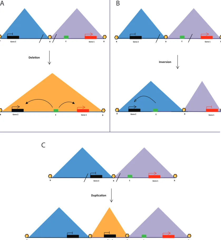
developmental defects (Fig 2) (Lupiáñez et al., 2015). Further, pathological genomic
duplications have been shown to not cause a phenotype if the duplicated region is insulated
Duplica�on Dele�on Β Β Gene 2 E Gene 1 E Gene 1 Β Β Gene 2 Β E Gene 1 Β Β Gene 2 Β E Gene 1 Β Β Gene 2 Β Gene 2 Β Gene 1 Gene 2 Β Β Β E E Gene 1 Β Β Gene 2 Β Inversion
C
Fig 2: TADs may define gene regulatory zones
A) TAD border deletion leads to aberrant enhancer-promoter contacts.
B) TAD inversion disrupts certain enhancer-promoter contacts and leads to aberrant contacts with other genes.
C) TAD border duplication creates new TADs which are functionally separate from neighboring regions.
Introduction
Nuclear and genome architecture
2.2.1 How are TADs formed and maintained?
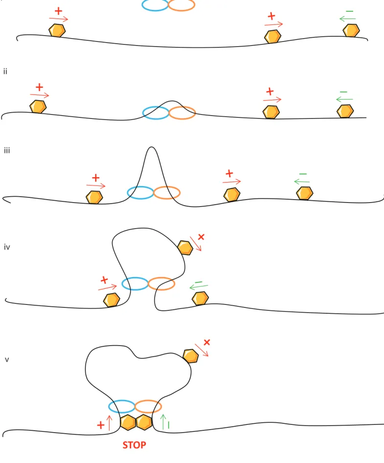
Although TADs have been recently well studied, it remains unclear how they are formed or maintained. In fact, TAD borders are enriched in active genes and active histone marks such as RNA polymerase and H3K4me3 as well as “architectural” proteins cohesin and CTCF (Dixon et al., 2012b; Sexton et al., 2012b). As these factors are also enriched in chromatin loops, TADs could be a consequence of very strong interactions between TAD borders (Rao et al., 2014a). However, many TAD borders do not contain CTCF or cohesin and importantly the large majority of binding sites are not TAD borders. Deletions of single CTCF sites cause mild effects on the overall TAD structure but they may have important functional consequences by aberrant enhancer promoter communications (V. Narendra et al., 2015; Varun Narendra, Bulajić, Dekker, Mazzoni, & Reinberg, 2016). Interestingly, a very recent study with a complete ablation of CTCF in pluripotent cells caused a severe disruption of TADs (~80% of TADs disappeared) with a genome wide misregulation of gene transcription (Nora et al., 2017). Further, a total and systematic TAD loss has been observed with a complete ablation of cohesin (Rao et al., 2017; Wutz et al., 2017). Therefore, cohesin plays an essential role for TAD formation and maintenance, whereas CTCF is complementary to TAD stabilization. To date the best model to explain TADs and these phenotypes is the loop extrusion model, which gives a rationale for the observations of relatively uniform intra-TAD interactions, and the prevalence for convergent CTCF elements at their borders (Alipour & Marko, 2012; Fudenberg et al., 2016; Sanborn et al., 2015). Outlined in Fig 3, the model entails (i.) binding of an extrusion factor (or factors) at random positions in the genome; (ii.) physical extrusion of a chromatin loop, starting from this bound site, by two components of the extrusion factor translocating in opposite directions; (iii.) growing of the extruded loop, with a physical equilibrium between extrusion and disassociation of the extruding factor; (iv.) barriers to extrusion at specific regions within the genome, such as TAD borders. As
extrusion occurs by bidirectional translocation of the chromatin fiber, asymmetric barrier elements would need to be in a convergent orientation to function as TAD borders. CTCF sites thus fit in the model as candidate barrier elements to loop extrusion. Cohesin is the primary candidate for the extrusion factor, based on what is known about how the ring structure can organize tethered sister chromatids (K Nasmyth, 2001). The frequent co- occupancy of CTCF and cohesin at TAD borders could thus be interpreted as stalled loop extrusion complexes, which are more stable than actively translocating regions and are thus more frequently detected in chromatin immunoprecipitation studies. A prediction of the loop extrusion model is that the residence time of the extruding factor would determine the loop/domain size. In further support for cohesin playing this role, deletions of the cohesin loading factors SCC2 (Nipbl)/SCC4 or release factor, WAPL, in a human haploid cell line reduce or increase the average chromatin loop size, respectively (Haarhuis et al., 2017). Similar findings with depleted cohesin unloaders have been independently reported (Wutz et al., 2017). Interestingly, TAD structures were weakened but not completely destroyed in these studies, in contrast to the extreme effects of deleting the Nipbl cohesin loader in mouse liver (Schwarzer et al., 2017), suggesting that cohesin may sometimes be inefficiently loaded and unloaded from interphase chromosomes in the absence of these factors, and/or that extruding factors other than cohesin can also be present. It is currently unclear where cohesin-mediated enhancer-promoter interactions fit into this model. Large transcription complexes with RNA polymerase and its co-activators could reasonably be a barrier to loop extrusion, potentially explaining why active genes are frequently found at TAD borders (Dixon et al., 2012a; Nora et al., 2012a). Enhancer-promoter loops could thus conceivably be a metastable loop extrusion intermediate. However, cohesin binding is not detected at many chromatin interactions (e.g. (Rubin et al., 2017)), so loop extrusion may be mediated by other factors or not required at loops stabilized by multiple protein-protein interactions.
STOP
Fig 3: The loop extrusion model for TAD formation.
An extrusion factor (blue and orange ovals), often proposed to be cohesin, binds to chromatin and extrudes a growing loop by translocating in opposite directions. The growing loop is stalled when the base contains two barrier elements (yellow hexagons) in convergent orientation, proposed to be CTCF sites. The equilibrium of growing, stalled and disassembled extruded loops may explain TAD organization and the dependence of these structures on cohesin and CTCF.
i ii iii iv v 39
2.2.2 TAD dynamics during development
Hi-C studies in disparate cell lines and tissues found that most TADs are invariant with cell type (Dixon et al., 2012a, 2015). This structural conservation is further supported by DNA fluorescent in situ hybridization (FISH) experiments coupled to super-resolution microscopy (Fabre, Benke, Manley, & Duboule, 2015), and even applies to syntenic chromosomal regions within different species (Pope et al., 2014; Vietri Rudan et al., 2015). Considering that TADs correlate so well with epigenetic marks, which are themselves extremely developmentally plastic (Roadmap Epigenomics Consortium et al., 2015), this observation was initially surprising. Higher-resolution studies at selected loci during pluripotent cells differentiation identified remodeled enhancer-promoter interactions as well as “sub-TADs” within larger conserved domains (Phillips-Cremins et al., 2013a). It was thus proposed that finer scale chromatin topology dynamics accompany cell reprogramming within a stable larger-scale architecture defined by TAD organization.
Original studies dedicated to investigate the spatial and temporal collinearity of mouse Hox gene expression, have demonstrated that the Hox loci form distinct topological domains with the active domain expanding and the silent domain shrinking according to collinear gene activation (Noordermeer et al., 2011). In fact, the mouse Hox genes are sequentially activated during development and according to anterior-posterior body position, in the order of the genes along the chromosome fiber. Hox gene expression is accompanied by a loss of H3K27me3-coated chromatin and concomitant gain of active histone marks(Soshnikova & Duboule, 2009). Developmentally dynamic “sub-TADs” within larger, stable domains have also been reported (Phillips-Cremins et al., 2013a). Current Hi-C data can support two conflicting models of TAD dynamics. First, TADs represent “ground state” chromosomal folding, on which other regulatory mechanisms (e.g. histone modifications, specific regulatory chromatin loops) are overlaid. Alternatively, TAD organization is responsive to
Introduction
Nuclear and genome architecture
underlying chromatin state, but the limited resolution of current Hi-C studies overlooked subtle tissue-specific differences in limited resolution of current Hi-C studies overlooked subtle tissue-specific differences in chromosome conformation. The most high-resolution Hi- C experiment to date, performed on mouse ES cells during neuronal differentiation, has blurred these two models, reporting many developmentally stable and dynamic TADs (Bonev et al., 2017). As for promoter-enhancer interactions, it is currently unclear what features distinguish a remodeled TAD from a stable one; remodeled TADs are associated with transcriptional induction, but many stable TADs present similar expression differences, and ectopic induction of otherwise remodeled genes is insufficient to alter their TAD architecture (Bonev et al., 2017).
In summary, TADs are mostly evolutionarily and developmentally stable structures, organizing seemingly autonomous regulatory domains in pluripotent and differentiated cells. We have made much progress in understanding their basic architectural principles, which in turn may ensure specific and efficient homing of genes to their regulatory elements. However, we have much to learn about the “fine print” of these architectural “blueprints”, in particular if and how specific intra-TAD interactions are set up, and whether they contribute to TAD stability (Giorgetti et al., 2014). Furthermore, much remains to be learned on the interplay between “stable” bulk TAD organization and “dynamic” chromatin ultrastructure during the large-scale transcriptional changes accompanying development.
2.3 Genomic compartments
Individual chromosomes are spatially organized into large compartments. These were first inferred from the “plaid” patterns of Hi-C contact maps, suggesting that multi-megabase regions are organized into one of two categories, “A” or “B,” whereby preferential
interactions occur between regions belonging to the same category, with very little mixing of the resulting A and B compartments (Lieberman-Aiden et al., 2009a). Epigenomic profiling of these compartments revealed that “A” chromatin is generally “open” and transcribed, whereas “B” chromatin carries repressive histone modifications and is more gene-poor. More refined analyses on higher-resolution Hi-C datasets are able to further split the A and B compartments into subcategories of preferentially interacting regions, based on location relative to the centromere(Yaffe & Tanay, 2011) or more specific histone modifications (Rao et al., 2014b). This compartmentalized organization could be a general result of preferential homotypic interactions between genomic elements sharing the same functions and chromatin states, as has been observed in the clustering of co-transcribed genes (Schoenfelder et al., 2010) or genes repressed by Polycomb (Bantignies et al., 2011). Self-organization models propose that this chromatin compartmentalization allows robust genomic control by ensuring that co- expressed genes share access to the same regulatory factors (Sexton & Cavalli, 2015). Such a model is difficult to experimentally assess, although abrogation of one gene has been shown to perturb expression of distal interacting genes (Bantignies et al., 2011; Fanucchi, Shibayama, Burd, Weinberg, & Mhlanga, 2013).
Until recently, it was unclear whether compartments were essentially larger-scale TADs, subject to the same organizational principles, considering that TADs form sub- domains (Phillips-Cremins et al., 2013b) and the patterns of inter-TAD interactions in Drosophila essentially follow genome compartments (Sexton et al., 2012b). However, the