1040
Clinical Significance of Extraintestinal
Hafnia alvei
Isolates from 61 Patients and
Review of the Literature
Huldrych Giinthard and Andreas Pennekamp From the Department of Internal Medicine, Stadtspital Triemli, and the Department of Medical Microbiology of the University of Zurich, Zurich, Switzerland Hafnia alveiisa gram-negative bacterium that is rarely isolated from human specimens and is
rarely considered to be pathogenic.Ithas been associated with gastroenteritis, meningitis, bacter-emia, pneumonia, nosocomial wound infections, endophthalmitis, and a buttock abscess. We studied 80H. alveiisolates recovered from 61 patients within a period of 30 months.H. alveiwas cultured from sites that included the respiratory tract (n
=
38), the gastrointestinal tract (n=
16), and the urogenital tract (n = 12); the organism was found in blood cultures (n = 8), on central venous catheters (n=
3), and on the skin (n=
3). Only 25% ofH. alveiisolates were recovered in pure cultures. Fifty-seven (93.4%) of the patients had an underlying illness. H. alveiproved to be the etiologic agent in two episodes of septicemia and in one episode of peritonitis and was probably responsible for septicemia in two other patients and pneumonia in one. All six of these patients recovered after receiving antibiotic treatment and/or standard surgical treatment, when needed. Three of these infections were nosocomial, and three were community acquired. Of the strains ofH. alveitested in our study, 100% were susceptible to netilmicin, ciprofloxacin, and imipenem; 92% were susceptible to piperacillin; 90% were susceptible to co-trimoxazole; and 88% were susceptible to ceftriaxone and ceftazidime.Inthis study, we foundH. alveito be a rare but significant etiologic agent of nosocomial and community-acquired infections.
Hafnia alvei is a gram-negative facultative rod-shaped anaer-obe that belongs to the Enterobacteriaceae; the organism was formerly named Enterobacter hafniae. H. alvei is rarely consid-ered to be pathogenic; rather, H alvei has been found to be related to the enteropathogenic Escherichia coli on the basis of a single virulence factor. Electron microscopy has demon-strated inflammation and mucosal invasion by H alvei in rabbit bowels, and an attachment-effacement gene like that detected in enteropathogenicE.coli was found in H. alvei by hybridiza-tion [1, 2]. No other distinct virulence factors have been noted in this organism so far [3]. Stool specimens are generally not examined for the presence of H alvei.
Cases of diarrhea due to H alvei have occurred mainly in children. H. alvei was reported to have caused acute gastroen-teritis in children from Bangladesh and Spain [1-2]. In one study, 16% of Finnish tourists with acute gastroenteritis and diarrhea who were returning from Morocco excreted H alvei in their stools [4]. Seven strains isolated from children with diarrhea expressed the attachment-effacement gene [2]. In two case reports, H alvei was mentioned as a cause of nonbloody diarrhea. One of these cases was presumably associated with reactive arthritis [5, 6]; however, cultures of joint aspirates
Received 3 October 1995; revised 16 January 1996.
Reprints or correspondence: Dr. Andreas Pennekamp, Department of Medi-cal Microbiology of the University of Zurich, Gloriastrasse 32, CH-S028 Zu-rich, Switzerland.
Clinical Infectious Diseases 1996;22:1040-5 © 1996 by The University of Chicago. All rights reserved. 1058-4838/96/2206-0021$02.00
were negative. A single case of H alvei meningitis in a 1-year-old girl [7] and a case of necrotizing H alvei enterocolitis with septicemia in a 20-day-old boy [8] have also been de-scribed. In adults, H alvei has caused bacteremia [9, 10], pneu-monia, and nosocomial wound infections [11-13]. H. alvei was recovered with Salmonella arizonae from a patient with endogenous endophthalmitis who was receiving steroids [14]. Recently, H alvei was isolated from a buttock abscess that resulted from skin puncture with carpet nails in an otherwise healthy middle-aged man [15].
Until now, there have been no data published about the true rate of isolation of H. alvei from clinical specimens, and the clinical significance of this bacterium remains to be defined. Therefore, we performed a study on the frequency of isolation of H alvei from clinical specimens (except stools) and corre-lated the microbiological findings with the clinical data.
Materials and Methods
Patient evaluation. Patients from whom isolates of H. alvei were recovered during routine diagnostic testing at the Depart-ment of Medical Microbiology, University of Zurich (Zurich) and at the Microbiology Laboratory of the Stadtspital Triemli (Zurich) between January 1992 and May 1995 were identified by reviewing the laboratories' records. The patients' charts were reviewed for data on isolation sites, underlying illnesses, cocultivation of other bacteria, and discrimination between nosocomial infections or community-acquired infections.
Isolation and identification. H. alvei was isolated from nor-mally sterile body fluids such as blood, ascitic fluid, and
ab-em
1996;22(June) Extraintestinal H alvei Infection 1041 dominal aspirates as well as from postoperative indwellingcatheters, central venous lines, and urine collected from cathe-ters. Respiratory tract isolates were cultured from sputum and tracheal and bronchial aspirates. Samples other than blood were plated on selective and differential media, and colonies of En-terobacteriaceae were detected on MacConkey agar (Becton Dickinson, Basel, Switzerland); blood was cultured with use of the BacT/Alert blood culture system (Organon Teknika, Basel).
The identification of Enterobacteriaceae was based on bio-chemical data obtained by means of commercial identification systems (API20E and RapID 32E; bioMerieux, Geneva, Swit-zerland) and on lack of lactose fermentation on MacConkey agar. Biochemical reactions for which H. alvei was positive included lysine decarboxylase, ornithine decarboxylase, and mannitol fermentation, whereas it was negative for fermenta-tion of sorbitol, inositol, sucrose, and melibiose; these negative reactions differentiated H. alvei from Enterobacter aerogenes. Strains with T values of <0.50 and probabilities of identifica-tion of <90% in one of the commercial systems were excluded from the study.
Antimicrobial susceptibility testing. Bacteria were tested for susceptibility by the disk diffusion method according to the National Committee for Clinical Laboratory Standards (NCCLS) guidelines. Strains were interpreted as susceptible, intermediately susceptible, or resistant on the basis of the NCCLS criteria. Susceptibilities were compared with use of the
X2
test. Strains that were susceptible to ampicillin, amoxicillin/ clavulanic acid, and cephalothin are most likely not part of the genus Hafnia [16] and thus were excluded from the study. We excluded strains of Salmonella and Enterobacter (one each), which were misidentified as H alvei by the commercial sys-tems, by means of these procedures.
We conducted a MEDLINE search with use of the terms Hafnia alveiand Enterobacter hafnia. All reports indexed from December 1966 to August 1995 were evaluated if they pre-sented clinically important information.
Results
Within the 30-month study period H. alvei was isolated from 80 samples collected from 61 patients. Twenty of these patients were female, and 41 were male. The mean age for all patients was 55.4 years (range, 13-81 years); the mean age for women was 61.8 years, and that for men was 53.5 years. H alvei was cultured from the respiratory tract 38 times for 35 patients (57.4%), from the gastrointestinal tract 16 times for nine pa-tients (14.8%), and from the urogenital tract 12 times for seven patients (11.5%). The organism was isolated from eight blood cultures of seven patients (11.5%), from three central venous catheters of three patients (3.8%), and from the skin of three patients (3.8%). Table 1 shows the sites of isolation of H alvei, which was isolated in pure culture of only 20 samples (25%). Organisms cultured concomitantly with H alvei were
Entero-bacteriaceae, staphylococci, streptococci, and yeasts. None of these microorganisms showed a predilection for association with H. alvei (data not shown).
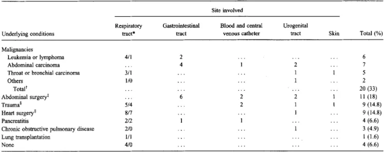
H. alveiwas isolated from 57 patients (93.4%) with underly-ing illnesses. Twenty (33%) of these patients had malignancies: six had leukemia or lymphoma, seven had carcinomas of the abdomen, five had carcinomas of the throat and bronchi, one had a sarcoma, and one had an endometrial carcinoma. Abdom-inal surgery was performed on 11 (18%) of the 61 patients; nine (14.8%) were hospitalized because of trauma; nine (14.8%) underwent heart surgery; four (6.6%) had acute or chronic pancreatitis; three (4.9%) had chronic obstructive pulmonary disease; and one (1.6%) underwent lung transplantation. An underlying disease was not detected in four patients (6.6%). Table 2 shows the isolation sites of H. alvei among patients with underlying illnesses. Eighteen of 35 respiratory isolates were from intubated patients; almost all respiratory isolates from patients who had undergone cardiac surgery (seven of eight isolates) or who were hospitalized because of trauma (four of five isolates) were recovered from intubated patients. None of the patients who had undergone abdominal surgery had isolates of H alvei recovered from respiratory specimens. Overall, 40 patients (66%) infected with H. alvei were intu-bated, and 19 of them had H. alvei isolated from the respiratory tract.
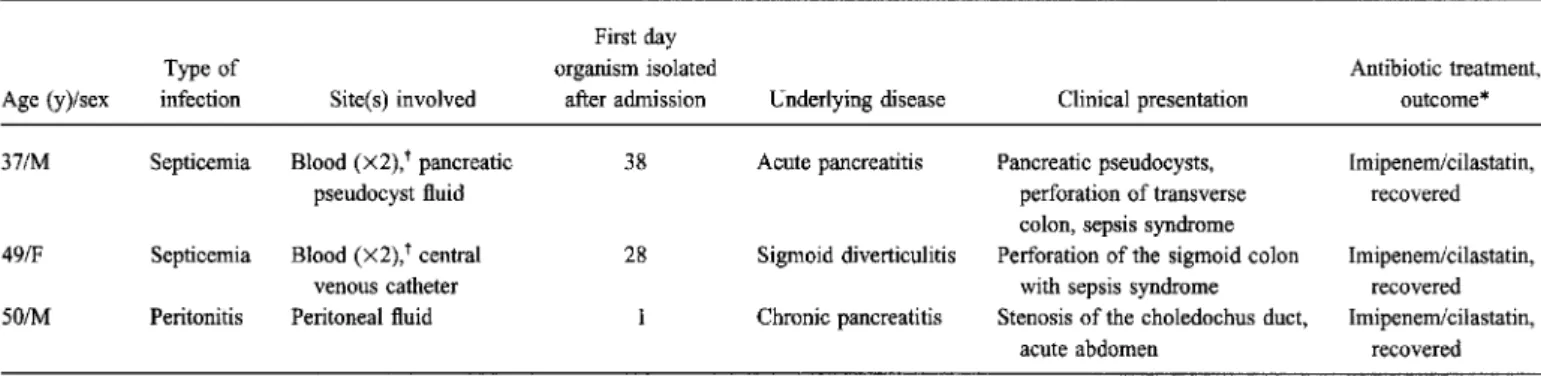
H. alveiwas found to be the sole etiologic agent of invasive disease in three (4.9%) of the 61 patients; two had septicemia due to this organism, and one had peritonitis. All three patients recovered after receiving treatment with imipenem/cilastatin. The clinical characteristics of these three cases are displayed in table 3. H. alvei, together with other organisms, was possibly responsible for two other cases of septicemia and a case of pneumonia.
Ten additional patients in our review received adequate anti-biotic coverage for H. alvei, but it was not possible to prove in retrospect that the organism had caused clinically significant infection.
A comparison of the results of antimicrobial susceptibility testing of our strains and those from patients described pre-viously is shown in table 4. The H alvei strains tested in our study were all susceptible to netilmicin and ciprofloxacin; 99% were susceptible to imipenem and tobramycin; 92% to pipera-cillin; 90% to co-trimoxazole; 89% to piperacillin/tazobactam; and 88% each to ceftriaxone and ceftazidime.
Discussion
H alvei is rarely considered to be a pathogenic organism. In recent years, case reports of various infections due to H. alveihave been published [1-15, 17, 18]. In our retrospec-tive 30-month study, H. alvei was recovered in 80 specimens from 61 patients. The majority of these patients (93.4%) had a severe underlying condition; this finding supports other re-ports that H alvei is primarily isolated from patients with
1042 Glinthard and Pennekamp
Table1. Isolation of Hafnia alvei in pure or mixed cultures of specimens from different body sites of 61 patients. CID 1996; 22 (June) Site involved Respiratory tract* Gastrointestinal tractt Blood
Central venous catheter Urogenital tractt Skin
No. of isolates recovered in pure cultures 8 5 3 1 3
o
No. of isolates recovered in mixed cultures 30 11 5 2 9 3
Total no. (%) from indicated site(n= 80) 38 (47.5) 16 (20) 8(10) 3 (3.75) 12(15) 3 (3.75) NOTE. Twenty (25%) of the isolates were recovered in pure cultures, and 60 (75%) of the isolates were recovered in mixed cultures.
* The following specimens were obtained: tracheal aspirates (16), pharyngeal smears (4), bronchoalveolar lavage fluid (2), nasal smears (1), bronchial aspirates (7), and sputum (8).
tThe following specimens were obtained: ascitic fluid aspirates (2), gallbladder (1), abscesses and deep abdominal wounds (10), and pancreatic pseudocysts (3); no stool samples were included.
tThe following specimens were obtained: urine collected from indwelling catheters (6), clean-catch urine (3), scrotal smears (2), and episiotomy wound (1).
underlying illnesses [11-12], except when it is an enteric pathogen [1,4-6, 8, 17].
Thirty-eightH alvei isolates were recovered from respira-tory specimens, 16 were recovered from the gastrointestinal tract, 12 were recovered from the urogenital tract, eight were recovered from blood, three were recovered from intravenous catheters, and three were recovered from the skin. Only 25% of all H. alvei isolates grew in pure culture. H. alvei was
cultured with a variety of other bacteria and fungi in the respira-tory and gastrointestinal specimens.
H. alvei was the only identified pathogen causing two cases of septicemia and one case of acute peritonitis. The peritonitis occurred after a patient with stenosis of the choledochus duct underwent endoscopic retrograde cholangiopancreatography, andH. alvei was isolated from his blood and peritoneal fluid. This circumstance indicates that the bacterium's portal of entry
Table 2. Isolation of Hafnia alvei from different body sites of 61 patients with and without underlying conditions. Site involved
Underlying conditions Malignancies
Leukemia or lymphoma Abdominal carcinoma Throat or bronchial carcinoma Others Totalt Abdominal surgeryt Trauma§ Heart surgeryII Pancreatitis
Chronic obstructive pulmonary disease Lung transplantation None Respiratory tract* 4/1 3/1 1/0 5/4 8/7 2/2 2/0 1/1 4/0 Gastrointestinal tract 2 4 6
Blood and central venous catheter 2 2 Urogenital tract 2 1 1 2 1 1 Skin Total(%) 6 7 5 2 20 (33) 11(18) 9(14.8) 9 (14.8) 4(6.6) 3 (4.9) 1 (1.6) 4 (6.6) NOTE. Only one isolate per patient was taken into account. Of the 61 patients, 35 (18 of whom were intubated) had isolates recovered from the respiratory tract; nine, from the gastrointestinal tract; seven, from blood and central venous catheters; seven, from the urogenital tract; and three, from the skin.
* Total no. of isolates/no. of isolates from intubated patients.
tIncludes carcinoma (13 patients), acute myelocytic leukemia (3), plasmocytoma (1), non-Hodgkin's lymphoma (1), sarcoma (1), and aplastic anemia of unknown etiology (1).
tIncludes incarcerated hernia (3 patients), perforation of the sigmoid colon (2), small bowel ileus (2), infrarenal aortic aneurysm (2), intra-abdominal abscess (2), delivery of infant by vacuum extraction (1), and endoscopic retrograde cholangiopancreatography (1).
§ Includes subarachnoidal bleeding (3 patients), polytrauma (2), knife injuries (2), bums (1), femur fracture (1). IIIncludes aortocoronary bypass surgery (8 patients) and aortic valve replacement (1).
cm1996;22 (June) ExtraintestinalH alveiInfection 1043
Table3. Clinical characteristics of patients withHafnia alveiinfection.
First day
Type of organism isolated Antibiotic treatment,
Age (y)/sex infection Site(s) involved after admission Underlying disease Clinical presentation outcome* 371M Septicemia Blood(X2),tpancreatic 38 Acute pancreatitis Pancreatic pseudocysts, Imipenemlcilastatin,
pseudocyst fluid perforation of transverse recovered
colon, sepsis syndrome
49/F Septicemia Blood(X2),tcentral 28 Sigmoid diverticulitis Perforation of the sigmoid colon Imipenemlcilastatin,
venous catheter with sepsis syndrome recovered
501M Peritonitis Peritoneal fluid Chronic pancreatitis Stenosis of the choledochus duct, Imipenemlcilastatin, acute abdomen recovered * The surgical treatment in these cases is not included because standard procedures were performed; no surgical interventions were primarily done for infection with H. alvei.
t Organism isolated in two separate blood cultures.
might have been the gastrointestinal tract, where it is consid-ered to be a commensal [16]. Earlier reports have also men-tioned a correlation between abdominal wounds or abdominal surgery and the isolation ofH alvei in blood [10]. H alvei was isolated along with other pathogens from three other pa-tients who had septicemia, pneumonia, and cholangitis.
Overall, 11.5% of the patients in our study population had proven (or at least probable) infections caused byH alvei, and 57 of them presented with an underlying illness. Of six patients withH. alvei infections, three had nosocomial infections [19], and three had community-acquired infections. The number of
infections might even have been higher, but the retrospective nature of our study did not allow correlation of the other H alvei isolates with clinically significant infections. However, 10 additional patients from whomH. alvei was isolated were treated with broad-spectrum antibiotics in the absence of a clear diagnosis of infection. This circumstance supports the hypothesis that there was a higher rate of significant infections in our study population. In all six of our patients with proven or probableH. alvei infections, the infections were successfully treated with antibiotics chosen according to the results of sus-ceptibility testing.
Table 4. Antimicrobial susceptibility test results forHafnia alveiisolates.
Present report Literature reviewt No. of strains Percent No. of strains Percent
Antibiotic tested susceptible* tested susceptible
Amoxicillinlclavulanic acid 77 1 NA NA Ampicillin 76 9 23 9 Cephalothin 77 a 23 17 Cefamandole 71 79 NA NA Ceftriaxone 73 88 NA NA Ceftazidime 73 88 12 42 Cefuroxime 69 77 NA NA Gentamicin NA NA 23 100 Amikacin NA NA 13 100 Netilmicin 75 100 NA NA Tobramycin 73 99 NA NA Ciprofloxacin 75 100 7 100 Piperacillin 64 92 13 85 Tetracycline NA NA 5 84 Piperacillinltazobactam 19 89 NA NA Trimethoprim-sulfamethoxazole 76 90 13 92 Imipenemlcilastatin 73 99 8 100 Colistin 72 75 NA NA
NOTE. NA= not available.
* Susceptibility testing was performed according to the National Committee for Clinical Laboratory Standards (NCCLS) for the disk diffusion method and interpreted according to NCCLS guidelines.
1044 Giinthard and Pennekamp cm 1996;22 (June)
As shown in table 1, 38 (47.5%) of all isolates recovered from 35 patients were of respiratory origin. Eighteen of the patients were intubated at the time that samples were obtained, and 17 were not intubated during their hospital courses. Intu-bated patients who are immunosuppressed as a result of cardiac surgery or trauma seem to harborH. alvei in their respiratory tracts more often than do patients who have previously under-gone abdominal surgery. Whether the oropharyngeal-tracheal regions of these patients had been colonized previously or whether they became colonized as a result of hospital proce-dures (e.g., intubation) cannot be answered sufficiently. We do not know the prevalence ofH. alvei colonization of the oropharyngeal-tracheal regions and other bodily sites in healthy persons or patients with underlying diseases. In large studies [20-21], this bacterium was rarely recovered from any site, making nosocomial infection improbable and emphasizing the likelihood of endogenous colonization in most patients before admission to the hospital.
On the other hand, only eight of 30 respiratory tract isolates were recovered from pure cultures, compared with 22 isolates that were recovered from mixed cultures; these mixed cultures mainly consisted of other gram-negative bacteria that are known to colonize the oropharyngeal-tracheal tract in severely
illpatients [22]. The presence ofH. alvei in feces reflects the fact that the organism is part of the normal bowel flora. Case reports ofH. alvei as the etiologic agent of diarrhea must be evaluated critically because no toxin production or toxic muco-sal changes in humans have been detected so far. However, it is worth mentioning thatH. alvei might cause diarrhea in immunocompromised patients, as case reports of neonates and malnourished children with diarrhea have shown [1, 7, 17].
In comparing the susceptibilities of H. alvei isolates, we observed a higher rate of susceptibility to ampicillinJamoxicil-lin and cephalothin among the strains described in the literature. In conducting this study we used susceptibility to amoxicillin and first-generation cephalosporins as selection criteria; there-fore, these data are not comparable to those from previous reports.
In our study the most active antimicrobials were netilmicin (100% of isolates were susceptible), ciprofloxacin (100% sus-ceptible), and imipenem (99% susceptible). These results are similar to the scant results reported in the literature (only a few strains have been tested), where 100% of the H. alvei strains were found to be susceptible to amikacin, gentamicin, ciprofloxacin, and imipenem [23 - 26].
The susceptibility to ceftazidime observed in our study was significantly different from that reported in the literature(P
<
.001). This difference cannot be explained but is probably not caused by the larger number of strains tested in our study because the results were otherwise almost congruent. Another slight difference was observed when susceptibilities to pipera-cillin were compared: 92% of our strains (64 tested) were susceptible, while 85% of strains in other reports (13 tested) were susceptible. However, the fact that susceptibility results
from other reports as well as from our study were comparable for most antimicrobials and that the biochemical features were similar indicates that H. alvei was correctly identified as the pathogen.
In summary, this study shows thatH. alvei is a rare human pathogen; however, the organism may be responsible for seri-ous nosocomial and community-acquired infections. Infections mainly occur in patients with underlying illnesses, andH.alvei is often isolated in coculture with different gram-negative rods, which may be due to endogenous colonization of the bowel. Treatment ofH. alvei infection on the basis of antimicrobial susceptibility testing results is effective. In severe cases, treat-ment with imipenem or a third-generation cephalosporin in combination with an aminoglycoside is recommended. More data on the role of H. alvei and its pathogenicity are needed. In addition, the colonizing rate for this organismin different sites in healthy adults must be determined.
Acknowledgments
The authors are indebted to ProfessorA. von Graevenitz and Dr.J.Gubler for their helpful comments and careful review of the manuscript.
References
1. Albert MJ, Alam K, Islam M, et al.Hafnia alvei, a probable cause of diarrhea in humans. Infect Immun 1991;59:1507-13.
2. Albert MJ, Faruque SM, Ansaruzzaman M, et al. Sharing of virulence associated properties at the phenotypic and genetic levels between en-teropathogenic Escherichia coli and Hafnia alvei. J Med Microbiol 1992;37:310-4.
3. RidellJ,Siitonen A, Paulin L, Lindroos0,Korkeala H, Albert1. Character-ization ofHafnia alveiby biochemical tests, random amplified polymor-phic DNA PCR, and partial sequencing of 16S rRNA gene. J Clin Microbiol 1995;33:2372-6.
4. Ridell J, Siitonen A, Paulin L, Mattila L, Korkeala H, Albert MJ.Hafnia alveiin stool specimens from patients with diarrhea and healthy controls. J Clin Microbiol 1994; 32:2335-7.
5. Westblom TU, Milligan TW. Acute bacterial gastroenteritis caused by Hafnia alvei[letter]. Clin Infect Dis 1992; 14:1271-2.
6. Newmark 11, Hobbs WN, Wilson BE. Reactive arthritis associated with Hafnia alveienteritis. Arthritis Rheum 1994;37:960.
7. Mojtabaee A, SiadatiA.Enterobacter hafniameningitis. J Pediatr 1978; 93:1062-3.
8. Ginsberg HG, GoldsmithJP.Hafnia alveisepticemia in an infant with necrotizing enterocolitis. J Perinatol 1988; 8: 122-3.
9. Englund GW. Persistent septicemia due toHafnia alvei.Report of a case.
AmJ Clin PathoI1969;51:717-9.
10. Jennis F, Mccarthy SW. Hafnia: an unusual cause of postoperative gram-negative bacteriae. Med J Aust 1967; 1:286-7.
11. Klapholz A, Lessnau KD, Huang B, Talavera W, Boyle JF.Hafnia alvei. Respiratory tract isolates in a community hospital over a three-year period and a literature review. Chest 1994; 105:1098-1100: 12. Frick T, KUDZ M, Vogt M, Turina M. Typical nosocomial infection with
an unusual cause:Hafnia alvei.Report of 2 cases and literature review. Schweiz Rundsch Med Prax 1990;79:1092-4.
13. Berger SA, Edberg SC, Klein RS.Enterobacter hafnaieinfection: report of two cases and review of the literature.AmJ Med Sci 1977;273: 101-4.
em1996;22 (June) ExtraintestinalH. alveiInfection 1045
14. Caravalho J Jr, McMillan VM, Ellis RE, Betancourt A. Endogenous en-dophthalmitis due to Salmonella arizonae and Hafnia alvei. South Med J 1990;83:325-7.
15. Agustin ET, Cunha BA. Buttock abscess due to Hafnia alvei [letter]. Clin Infect Dis 1995;20:1426.
16. SakazakiR. Genus IX.Hafnia.In:Holt JG, ed. Bergey's manual of systematic bacteriology. 7thed.Baltimore~ Williams and Wilkins, 1986:484-6. 17. Reina J, Hervas J, Borrell N. Acute gastroenteritis caused by Hafnia alvei
in children [letter]. Clin Infect Dis 1993; 16:443.
18. Eisenstein BI. Enterobacteriaceae.In:Mandell GL, BennettJE, Dolin R, eds. Mandell, Douglas and Bennett's principles and practice ofinfectious diseases. 4th ed. New York: Churchill Livingstone, 1995:1964-80.
19. Martone WJ, Gamer JS, Duma1. Preventing nosocomial infections-progress in the 1980s; plans for the 1990s. In: Martone WJ, Gamer JS, eds. Proceedings of the Third Decennial International Conference on Nosocomial Infections. Am J Med 1991;913B-lS.
20. Weber DJ, Rutala WA, Samsa GP, Wilson MB, Hoffmann KK. Relative frequency of nosocomial pathogens at a university hospital during the decade 1980 to 1989. Am J Infect Control 1992:192-7.
21. Carrel T, Schmid ER, von Segesser L, Vogt M, Turina M. Preoperative assessment of the likelihood of infection of the lower respiratory tract after cardiac surgery. Thorac Cardiovasc Surg 1991;39:85-8. 22. Palmer LB, Donelan SV, Fox G, Bellemore E, Greene WHo Gastric flora
in chronically mechanically ventilated patients. Relationship to upper and lower airway colonization. Am J Respir Crit Care Med 1995; 151: 1063-7.
23. Qadri SM, Belobraydic KA. In vitro activity of aztreonam against gram negative bacteria from clinical specimens and its comparison with other commonly used antibiotics. Methods Find Exp Clin Pharmacol 1986; 8:223-6.
24. Thomson KS, Sanders CC, Washington JA II. Ceftazidime resistance in Hafnia alvei.Antimicrob Agents Chemother 1993;37:1375-6. 25. Belobraydic KA, Qadri SM. Antimicrobial activity of imipenem against
1386 clinical isolates. Methods Find Exp Clin Pharmacol 1986; 8: 675-8.
26. Washington JA II, BirkRJ, Ritts RE Jr. Bacteriologic and epidemiologic characteristics of Enterobacter hafniae and Enterobacter liquefaciens. J Infect Dis 1971; 124:379-86.