Lichenologist 17(3): 273-279 (1985)
THE HYPHOMYCETOUS ANAMORPH OF
CONIOCYBE FURFURACEA
Rosmarie HONEGGER*
Abstract: Under favourable climatic conditions the mycobiont of Coniocybe furfuracea bears masses of conidia in chains on macronematous conidiophores. The same type of conidia were also formed by axenically grown mycobionts which had been isolated from single ascospores. Germinating conidia were found on the thalli. Coniocybe furfuracea is one of the very few lichen mycobionts so far known with a teleomorph and hyphomycetous anamorph.
Introduction
About 8000 species of lichenized fungi form conidia in pycnidial conidiomata
(Vobis & Hawksworth 1981), some of which represent the coelomycetous
anamorph, others spermatial stages or, with high probability, functionless
relics of spermatial stages (Vobis 1977, Vobis & Hawksworth 1981, Honegger
1984a, b). Various lichenized Hyphomycetes have been described (reviewed
by Vobis & Hawksworth 1981), but hyphomycetous anamorphs seem to be
extremely rare among lichen-forming fungi with known teleomorphs. The
mycobiont of Vezdaea aestivalis forms macronematous conidiophores in the
symbiotic state and very soon after germination in pure cultures of ascospores
(Tschermak-Woess & Poelt 1976). Conidial formation was also observed in
cultured, but not in free-living symbiotic states of the mycobiont of Pertusaria
pertusa (Lallemant 1977a, b). Other reports on hyphomycetous structures in
cultured lichen mycobionts (Hale 1957, Ahmadjian 1963) were either not
substantiated in subsequent studies (Ahmadjian 1965) or require further
examination. Hyphomycete-like structures embedded in the gelatinous
thallus which bear spermatia were observed in some Collema species
(Bachmann 1912, Degelius 1954), and very peculiar synnematous structures
termed hyphophores have been detected in the foliicolous Asterothyriaceae
(reviewed by Vobis & Hawsksworth 1981).
In the course of a study on the mycobiont-phycobiont relationship in
Coniocybe furfuracea, many macronematous conidiophores were noted. The
relationship of these structures to the teleomorph of Coniocybe furfuracea
(Fig. 1) was investigated by means of culture techniques and scanning
electron microscopy.
Materials and Methods
Fresh material was collected in the cold season (November to April 1982/83 and 1983/84) in dif-ferent locations of central Switzerland. Only very well developed, fertile thalli were used. (1)
•Cytology, Institute of Plant Biology, University of Zurich, CH-8008 Zurich, Switzerland. 0024-2829/85/030273 + 07 $03.00/0 © 1985 British Lichen Society
274 THELICHENOLOGIST Vol.17
Merliwald W Giswil, NW, 1300 m, November 1982. (2) Ratenpass E Aegeri, SZ, 1000 m, November 1982, March 1983, November 1983. (3) Murgtal S Walensee, SG, 750 m, April 1984, E. Urmi.Herbarium specimens (Herbarium Institute of Systematic Botany, University of Zurich, Zurich) examined: Switzerland: (1) Hongg bei Felsenegg, ZH, 1900, Hegetschweiler. (2) Saleve bei Genf, GE, Schweiz, Bernet [Krypt. exsicc. no. 814]. (3) Hiitten bei Zurich, ZH, 1975, K. Urmi. (4) Silenen, UR, 1974, K. Urmi. Germany: (1) Wombrunn, im Griinewalder Park bei Munchen, 1892, Arnold [Lich. Monacenses exs. no. 225]. (2) Kreuzberger Alpe bei Schliersee, bayr. Alpen, 1882, Arnold.
Isolation of the mycobiont and culture techniques. Mature ascomata were broken off the thallus with the aid of fine, sterile forceps. The surface of a sterile non-nutrient agar (Bold's basal medium, BBM, according to Deason & Bold 1960) was touched repeatedly with the mazaedium. At each contact of the mazaedium with the agar surface a group of ascospores remained on the agar. The first five groups of each ascoma were discarded in order to eliminate possible con-taminants. Dispersion of the ascospores was obtained by spreading the groups of ascospores over the agar surface with the aid of a sterile needle and using a dissecting microscope. The Petri dishes were incubated at 17°C at a 14:10h light:dark interval. Germination of ascospores occurred within 1 week and was studied with a Reichert Biovert conversion microscope.
Single spore isolates were transferred either to a nutrient medium containing BBM with 3-5°0
malt extract, 0-5°0 glucose, 005°o peptone, 0025°o casamino acids, and 0025°o yeast extract
(agar: T5°(l), or to fresh non-nutrient agar (BBM) and covered with the Stichococcus bacillaris
phycobiont which had been cultured axenically on the same non-nutrient medium. All strains are kept in our laboratory.
Transmission electron microscopy. See Honegger (1984b).
Scanning electron microscopy (SEM). Lichen thalli or agar blocks with cultured symbionts were exposed to the vapour of a 4% solution of osmium tetroxide at room temperature overnight, dehydrated in a graded series of acetone, critical point dryed and mounted on specimen stubs with conductive silver print paint (GC Electronics). Mounted specimens were sputter-coated with an alloy of gold and palladium (80°o/20°o) and examined in a Cambridge Stereoscan S-4.
Results
In the fluffy thalli of freshly collected and herbarium samples of Coniocybe
furfuracea the mycobiont either tightly adheres to or overgrows the chain-like
colonies of the ulotrichalean Stichococcus phycobiont, or forms erect hyphae
(Fig. 2A-C). In most of the samples examined no special structures could be
seen at the tip of the erect hyphae (Fig. 2B). In material collected in late winter
(March-April), when the temperature was not too cold and the humidity high
due to melting snow around the tree trunks, these erect hyphae produced a
mass of conidia, often in chains (Fig. 2D-E) and so represent conidiophores.
As the loose thalli of Coniocybe furfuracea are not clearly delimited and yeasts
and other conidial fungi can occasionally be seen developing within the tufts,
it was not clear whether the abundantly formed conidiophores of the fungus
contacting the phycobiont cells were produced by the mycobiont or by
another fungus living in the same habitat. Culture experiments became
necessary.
Single ascospore isolates of Coniocybe furfuracea were easily obtained (see
above). In material collected in the winter months the germination rate was
above 90%, and the contamination rate was surprisingly low. When
trans-ferred to nutrient agar the mycobiont formed cartilaginous, thallus-like
structures with filamentous hyphal growth at the periphery (Fig. 3A), but
almost pseudoparenchymatous structures in the older parts. This is a common
feature among lichen mycobionts (reviewed by Lallemant 1977b), and the
1985
Hyphomycetous anamorph of C. furfur acea—Honegger
275
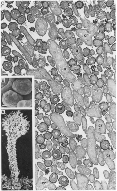
FIG. 1. The teleomorph of Coniocybe furfuracea. A, SEM preparation of a young, not fully developed ascoma, x 300. B, SEM preparation of two fully differentiated ascospores with charac-teristically structured exospore, x 5000. C, TEM preparation of a cross-section of part of the mazaedium with different developmental stages of asci (a) and ascospores (as), cr, croziers,
276
THE LICHENOLOGIST
Vol. 17
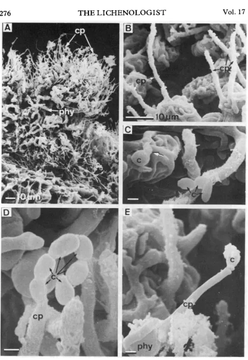
FIG. 2. SEM of thallus morphology and conidiophore structure in the symbiotic (freeliving) state of Coniocyte furfuracea. A, Thalline tuft with chain-like colonies of the ulotrichalean Stichococcus bacillaris phycobiont (phy) which are held together by the closely contacting mycobiont, and numerous erect hyphae of the mycobiont (cp), x 460. B, Phycobiont cells overgrown by the mycobiont, and erect hyphae which represent dormant conidiophores (cp), x 1300. C, A germinating (arrow) conidium (c) and a chain of conidia (c) on the thallus surface, x 4700. D, Tip of a conidiophore (cp) producing a chain of conidia (c), x 6700. E, Conidiophore producing a
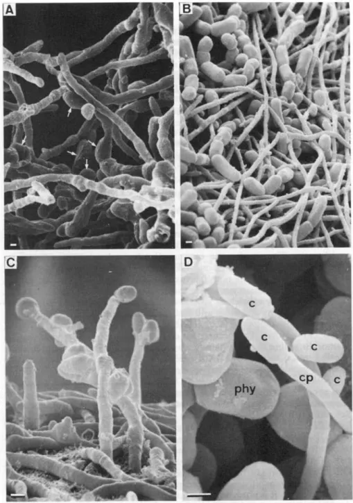
FIG. 3. SEM of growth pattern and conidium formation in axenically cultured Coniocybe furfur-acea. A, The growing edge of a thallus-like structure of the mycobiont on nutrient agar with numerous swollen cells (arrows), x 2000. B, Filamentous growth of the mycobiont on non-nutrient agar with chain-like colonies of Stichococcus bacillaris, the cultured phycobiont, x 1900. C, Conidiophores and conidia developing on non-nutrient agar without phycobiont cells, x 4700. D, Conidium (c) producing conidiophore (cp) developing in between phycobiont cells (phy) on
278 THE LICHENOLOGIST Vol.17
same is true for the many swollen cells rich in lipid droplets (Fig. 3A) which
are produced on nutrient media. On non-nutrient media, filamentous hyphal
growth was observed (Fig. 3B-C). No conidiophores were formed on the
nutrient medium, but a few developed on the non-nutrient medium (Fig. 3D).
A limited number of conidiophores appeared on the non-nutrient medium
after addition of the Stichococcus phycobiont (Fig. 3D), although the
mycobiont-phycobiont relationship was nowhere as close as in the free-living
symbiotic state (Fig. 2B—C). Development from ascospore germination to
conidium production took 7.\ months. The culture experiments were carried
out in triplicate, with lichens collected at three different sites.
Conidial germination was also observed on the surface of free-living thalli
(Fig. 2B). The conidia can be easily distinguished from ascospores (Figs
1B-C) by their different size, shape, and surface structure.
Discussion
The present data demonstrate the teleomorph-anamorph relationship in
Coniocybe furfuracea, a situation also observed in other calicialean
myco-bionts. Vobis (1977) reported conidium germination under axenic conditions
in Calicium adspersum but there the conidia are formed in pycnidial
conidiomata. This is also the case in all other calicialean conidia so far
reported (e.g. Gallee 1972, Henssen & Jahns 1973, Poelt 1974, Tibell 1978),
with the exception of the non-lichenized Mycocalicium schefflerae which has
a Phialophora-\ike macronematous anamorph (Samuels & Buchanan 1983).
However, in many calicialean species no data on conidia are available, since
ascomatal ontogeny and ascus and ascospore structures are the main
charac-ters used in the delimitation of species and genera (e.g. Schmidt 1970a, b,
Tibell 1971, 1978). The occurrence of coelomycetous and hyphomycetous
anamorphs within the same family may at first seem anomalous, but in some
non-lichenized ascomycetes both types of conidium-bearing structures are
reported within closely related species or even in different developmental
stages of a single species (reviewed by Miiller 1981). Sporodochial, acervular,
and pycnidial conidiomata were detected in the lichen genus Micarea
(Coppins 1983). On the basis of studies on ascomatal and ascal ontogeny,
Schmidt (1970b) concluded first, that the Caliciaceae (and even more so the
Caliciales) are a very heterogeneous group, and secondly that some species
of Coniocybe (including C. furfuracea) and Chaenotheca differ considerably
from other Caliciaceae in that ascus formation occurs in chains. Whether the
type of conidial formation might provide a further taxonomic criterion in the
Caliciaceae requires further investigation.
The mechanism of conidial formation in Coniocybe furfuracea requires
further study as it is not possible to interpret the type of conidiogenesis from
the SEM pictures available. The conidiophores might represent either true or
false chain phialides in the sense of Minter et al. (1982, 1983a, b) or even
other types of conidial-bearing structures. Due to the very small size of the
conidiogenous cells, only transmission electron microscopic studies will
enable a more detailed interpretation to be made. Without these data it is
not possible to make comparisons between the conidiophores of Coniocybe
1985 Hyphomycetous anamorph of C. furfuracea—Honegger 279
furfuracea and non-lichenized fungi, although a certain morphological
similarity to saprophytes of the genera Humicola and Botryotrichum may be
noted.
My sincere thanks are due to Professor E. Miiller for stimulating discussions; to Drs Katharina and E. Urmi for the loan of herbarium specimens and collecting fresh material; and to Professor D. L. Hawksworth for valuable comments and for improving the style of this manuscript.
REFERENCES
Ahmadjian, V. (1963) The fungi of lichens. Sci. Am. 208: 122-132. Ahmadjian, V. (1965) Lichens. A. Rev. Microbiol. 19: 1-20.
Bachmann, F. M. (1912) A new type of spermogonium and fertilization in Collema. Ann. Bot. London 26: 747-760.
Coppins, B. J. (1983) A taxonomic study of the lichen genus Micarea in Europe. Bull. Br. Mus. not. Hist. (Bot.) 11: 7-214.
Deason, T. R. & Bold, H. C. (1960) Phycological studies. I. Exploratory studies of Texas soil algae. Univ. Texas Publ. 6022.
Degelius, G. (1954) The lichen genus Collema in Europe. Symb. Bot. Upsal. 13(2): 1-429. Gallee, O. (1972) Natural history of the Danish lichens. Vol. 10 (M. S. Christiansen, ed.).
Copenhagen: E. Galloe.
Hale, M. E. (1957) Conidial stage of the lichen fungus Buellia stillingiana and its relation to Sporidesmium folliculatum. Mycologia 49: 417-419.
Henssen, A. & Jahns, H. M. (1973) ['1974'] Lichenes. Eine Einfuhrung in die Flechtenkunde. Stuttgart: Thieme.
Honegger, R. (1984a) Scanning electron microscopy of the contact site of conidia and trichogynes in Cladonia furcata. Lichenologist 16: 11-19.
Honegger, R. (1984b) Ultrastructural studies on conidiomata, conidiophores, and conidiogenous cells in six lichen-forming ascomycetes. Can. J. Bot. 62: 2081-2093.
Lallemant, R. (1977a) La morphogenese du mycobionte du Penusaria pertusa (L.) Tuck, en cultures pures in vitro. C. R. Acad. Sc. Paris, ser. D, 284: 923-926.
Lallemant, R. (1977b) Recherches sur le developpement en cultures pures in vitro du mycobionte du discolichen Pertusaria pertusa (L.) Tuck. Rev. Bryol. Lichenol. 43: 225-282.
Minter, D. W., Kirk, P. M. & Sutton, B. C. (1982) Holoblastic phialides. Trans. Br. mycol. Soc. 79: 75-93.
Minter, D. W., Kirk, P. M. & Sutton, B. C. (1983a) Thallic phialides. Trans. Br. mycol. Soc. 80: 39-66.
Minter, D. W., Sutton, B. C. & Brady, B. L. (1983b) What are phialides anyway? Trans. Br. mycol. Soc. 81: 109-120.
Miiller, E. (1981) Relations between conidial anamorphs and their teleomorphs. In Biology of Conidial Fungi (G. T. Cole & B. Kendrick, eds) Vol. 1: 145-169. New York etc.: Academic Press.
Poelt, J. (1974) ['1973'] Classification. In The Lichens (V. Ahmadjian & M. E. Hale, eds): 599-632. New York: Academic Press.
Samuels, G. J. & Buchanan, D. E. (1983) Ascomycetes of New Zealand 5. Mycocalicium schefflerae sp. nov., its ascal ultrastructure and Phialophora anamorph. N. Z. Jl Bot. 21:
163-169.
Schmidt, A. (1970a) Anatomisch-taxonomische Untersuchungen an europaischen Arten der Flechtenfamilie Caliciaceae. Mitt. Staalsinst. Allg. Bot. Hamburg 13:111-166.
Schmidt, A. (1970b) Ascustypen in der Familie Caliciaceae (Ordnung Caliciales). Ber. dtsch. bot. G«.,N.F. 4: 127-137.
Tibell, L. (1971) The genus Cyphelium in Europe. Svensk. bot. Tidskr. 65: 138-164. Tibell, L. (1978) The genus Microcalicium. Bot. Not. 131: 229-246.
Tschermak-Woess, E. & Poelt, J. (1976) Vezdaea, a peculiar lichen genus, and its phycobiont. In Lichenology: Progress and Problems (D. H. Brown, D. L. Hawksworth & R. H. Bailey, eds): 89-105. London: Academic Press.
Vobis, G. (1977) Studies on the germination of lichen conidia. Lichenologist 9: 131-136.
Vobis, G. & Hawksworth, D. L. (1981) Conidial lichen-forming fungi. In Biology of Conidial Fungi (G. T. Cole & B. Kendrick, eds) 1: 245-273. New York: Academic Press.