Original Article
Antitumor properties of a new non-anticoagulant
heparin analog from the mollusk Nodipecten
nodosus: Effect on P-selectin, heparanase,
metastasis and cellular recruitment
Angélica Maciel Gomes
2, Eliene Oliveira Kozlowski
2, Lubor Borsig
3,
Felipe C O B Teixeira
2, Israel Vlodavsky
4, and Mauro S G Pavão
1,22
Laboratório de Bioquímica e Biologia Celular de Glicoconjugados, Programa de Glicobiologia, Instituto de
Bioquí-mica Médica Leopoldo de Meis and Hospital Universitário Clementino Fraga Filho, Universidade Federal do Rio de
Janeiro, Rio de Janeiro 21941 913, Brazil,
3Institute of Physiology, University of Zurich and Zurich Center for Integrative
Human Physiology, Zurich 8057, Switzerland, and
4Cancer and Vascular Biology Research Center, Rappaport Faculty
of Medicine, Haifa 31096, Israel
1To whom correspondence should be addressed: Tel: +55-21-2562-2093; e-mail: mpavao@hucff.ufrj.br
Received 5 March 2014; Revised 27 October 2014; Accepted 27 October 2014
Abstract
In
flammation and cancer are related pathologies acting synergistically to promote tumor
progres-sion. In both, hematogenous metastasis and in
flammation, P-selectin participates in interactions
involving tumor cells, platelets, leukocytes and endothelium. Heparin has been shown to inhibit
P-selectin and as a consequence it blunts metastasis and in
flammation. Some heparin analogs
obtained from marine invertebrates are P-selectin inhibitors and do not induce bleeding effects.
The present work focuses on the P-selectin blocking activity of a unique heparan sulfate (HS) from
the bivalve mollusk Nodipecten nodosus. Initially, we showed that the mollusk HS inhibited LS180
colon carcinoma cell adhesion to immobilized P-selectin in a dose-dependent manner. In addition,
we demonstrated that this glycan attenuates leukocyte rolling on activated endothelium and in
flam-matory cell recruitment in thioglycollate-induced peritonitis in mice. Biochemical analysis indicated
that the invertebrate glycan also inhibits heparanase, a key player in cell invasion and metastasis.
Experimental metastasis of Lewis lung carcinoma cells was drastically attenuated by the mollusk
HS through a mechanism involving inhibition of platelet
–tumor-cell complex formation in blood
vessels. These data suggest that the mollusk HS is a potential alternative to heparin for inhibiting
P-selectin-mediated events such as metastasis and in
flammatory cell recruitment.
Key words: anti-cancer activity, heparanase, mollusk heparan sulfate, P-selectin
Introduction
The relationship between cancer and inflammation has been increasing-ly reported during the last decade (Coussens and Werb 2002). However, the connection between these two pathologies wasfirst suggested ∼150 years ago (Balkwill and Mantovani 2001). Whereas acute inflammation is part of the organism defense response, chronic inflammation can lead
to cancer. Indeed, patients with ulcerative colitis and Crohn’s disease, have a higher risk to develop colorectal cancer (Itzkowitz and Yio 2004). On the other hand, during hematogenous metastasis, natural killer cells can attack tumor cells, decreasing the metastatic rate. Thus, the function of leukocytes in tumor biology is complex (Lanca and Silva-Santos 2012) and represents an attractive research area.
doi: 10.1093/glycob/cwu119 Advance Access Publication Date: 3 November 2014 Original Article
Hematogenous metastasis and inflammatory cell recruitment strongly depend on selectin function. P-selectin is a family member of glycan-recognizing adhesion molecules. In endothelial cells, P-selectin is stored in Weibel–Palade bodies, whereas in platelets it oc-curs inα-granules. P-selectin is readily exposed at the surface of plate-lets upon activation, mediating its interaction with leukocytes and endothelial cells (Kansas 1996). Tumor cells are characterized by aberrant glycosylation patterns (Altevogt et al. 1983;Kim et al. 1996), including the over-expression of highly branched or sialylated oligosaccharides, especially fucosylated glycans, such as sialyl-LewisX
and sialyl-Lewisa. These glycans are ligands for selectins and their presence is related to poor prognosis due to increased metastatic dis-ease (Fukuoka et al. 1998;Tatsumi et al. 1998).
The metastatic process is comprised of several steps that include degradation of basement membrane, entry of cancer cell into the bloodstream, evasion of innate immune surveillance, adhesion to the vascular endothelium of secondary sites with subsequent extravasation and colonization. Once in the bloodstream, cancer cells are covered by platelets, in a P-selectin-mediated process, form-ing a natural barrier against immune system cells (Kim et al. 1998). On the other hand, leukocytes recruitment during inflammation is mediated by P- and L-selectins. These adhesion molecules are in-volved in thefirst steps of cellular recruitment by reducing the rolling velocity of leukocytes, contributing to their adhesion and arrest at sites of inflammation. In fact, P-selectin null mice exhibit reduced platelet aggregation, delayed leukocyte recruitment and attenuated metastasis, suggesting that P-selectin might be a common therapeut-ic target to treat cancer-related inflammation (Mayadas et al. 1993;
Kim et al. 1998;Ludwig et al. 2007). The anticoagulant glycosami-noglycan heparin is a potent inhibitor of P- and L-selectin binding to their natural ligands (Nelson et al. 1993). Accordingly, heparin treatment has been shown to reduce leukocyte recruitment and metastasis through inhibition of P-selectin (Nelson et al. 1993;
Borsig et al. 2001).
Cancer patients have high-risk of developing thromboembolic disease and therefore, heparin has been used as a prophylactic thera-peutic agent. Retrospective analysis of patients under heparin therapy revealed a better prognosis of the malignant disease, which has not been associated with the anticoagulant effect of the drug (Kragh et al. 2005; Stevenson et al. 2007). Several studies aimed to investigate the anti-cancer effect of heparin revealed that it attenuates experimental metastasis in animals (Borsig et al. 2001), mainly by binding to P-selectin and decreasing the interaction between tumor cells and platelets. Additionally, heparin can also attenuate metastasis by inhibition of heparanase (Vlodavsky et al. 1994), the only known mammalian endoglycosidase that cleaves heparan sulfate (HS) and is over-expressed in essentially all human tumors. In fact, heparanase over-expression correlates with poor prognosis in a variety of cancers (Vlodavsky et al. 2012;Gomes et al. 2013).
We have previously shown that heparin analogues obtained from marine invertebrate bind to P-selectin, attenuating metastasis, in flam-mation and thrombosis (Borsig et al. 2007;Kozlowski et al. 2011). In a recent study, we described a unique HS isolated from the bivalve mollusk N. nodosus (Figure1). This sulfated polysaccharide is formed by glucuronic acid and Glucosamine and can also contain a rare sulfa-tion pattern on carbon 2 or 3 of the glucuronic acid units. We also de-scribed that this compound was able to inhibit thrombus growth without inducing bleeding effect (Gomes et al. 2010).
Although increasing evidence points to a beneficial therapeutic ac-tion of heparin in cancer patients, its bleeding effect still limits its use. Here, we addressed the ability of HS from N. nodosus to attenuate
leukocyte recruitment and cancer metastasis. Our results revealed that the mollusk HS effectively inhibits P-selectin activity, decreasing the binding of carcinoma cells to P-selectin in vitro and its interaction with platelets in vivo. The mollusk glycan also inhibits heparanase en-zymatic activity in vitro. Moreover, we have demonstrated that this glycan attenuates hematogenous metastasis and polymorphonuclear cells recruitment in vivo.
Results
Nodipecten nodosus HS attenuates tumor cell binding
to P-selectin
Because P-selectin is a crucial mediator of cell–cell interactions during inflammation and cancer, we evaluated whether HS from N. nodosus could inhibit P-selectin. For this purpose, we analyzed the ability of this compound to impair adhesion of LS180 cells to immobilized P-selectin. It is known from previous work that this colon cancer cell line expresses high content of selectin ligands (Mannori et al. 1995). Nodipecten nodosus HS decreased tumor cell binding to P-selectin in a dose-dependent manner, with an IC50of 38.1 µg/mL. Heparin was
more efficient in reducing tumor cell binding to P-selectin, showing an IC50of 24.5 µg/mL (Figure2). It is worth mentioning that, because
heparin presents lower molecular weight (∼15 kDa) than N. nodosus HS (∼30 kDa), the molar concentration of heparin is higher than that of the mollusk HS. As expected, chondroitin sulfate did not inhibit P-selectin binding to tumor cells, even at high concentrations.
Nodipecten nodosus HS reduces leukocyte rolling
and cell recruitment after in
flammatory stimulus
While the involvement of inflammatory cells during several steps of cancer progression is well documented, little is known about the underlying mechanism. Because leukocyte rolling on activated endo-thelium during inflammatory response is mediated by P-selectin, we investigated how leukocyte rolling is affected by N. nodosus HS. Pre-vious studies have shown that lipopolysaccharide (LPS) treatment in-duces P-selectin upregulation in endothelial cells (Gotsch et al. 1994). Based on that, we analyzed leukocyte rolling on LPS-activated endo-thelium by intravital microscopy, before or after N. nodosus HS ad-ministration. It was observed that treatment with the mollusk HS inhibited LPS-induced leukocyte rolling to the basal level (Figure3A and Supplementary data). Because leukocyte rolling is thefirst step for cell recruitment, we further evaluated the effect N. nodosus HS on a thioglycollate-induced peritonitis model in mice. After 3 h of in flam-matory stimulus the peritoneal lavage was harvested and differential
Fig. 1. Nodipecten nodosus HS structure. Major disaccharide unit of the N. nodosus HS.
counting of leukocytes was performed. Figure3B shows that thioglycol-late promoted a significant increase in the recruitment of polymorpho-nuclear cells to the peritoneal cavity, compared with control. Whereas treatment with N. nodosus HS resulted in a 50% reduction in leukocyte recruitment mammalian heparin induced 60% of inhibition. This dif-ference is not statistically significant. Interestingly, treatment of P-sel−/−
mice with N. nodosus HS led to lower leukocyte recruitment in comparison with both HS-treated wild-type or untreated P-sel−/− mice (Figure3C). Thesefindings suggest an additional mechanism in-volved in the inhibition of leukocyte recruitment by N. nodosus HS besides P-selectin.
Nodipecten nodosus HS inhibits association between
tumor cells and platelets in vivo
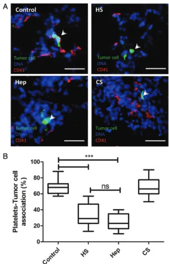
As mentioned earlier in the introduction, P-selectin-dependent leuko-cyte recruitment and intercellular interactions are crucial during in-flammation as well as during tumor metastasis. P-selectin-mediated platelet association with tumor cells contributes to the success of me-tastasis. In previous studies, we showed that glycosaminoglycans from marine invertebrates could disrupt this association. In order to analyze the ability of HS from N. nodusus to inhibit tumor cell–platelet inter-actions in vivo, HS, heparin and chondroitin sulfate (8 mg/kg of each glycan) was injected via tail vein 5–10 min before injection of Lewis lung carcinoma (LLC) cells. The lungs were analyzed by immuno fluor-escence 30 min after tumor cell (calcein labeled) injection. While in the control group,∼70% of tumor cells were associated with platelets (Figure4), tumor cell–platelet complex was reduced to 30% after N. nodosus HS administration. Mammalian heparin attenuates tumor cell–platelet association to a similar extent as observed in mice treated with mollusk HS. Chondroitin sulfate did not inhibit tumor cell–plate-let association.
Mollusk HS inhibits heparanase and attenuates
experimental metastasis
LLC cells express high amounts of heparanase (Takahashi et al. 2005), which plays a relevant role in tumor invasion and metastasis (Ilan et al. 2006). Because heparanase has been pointed as a molecular target of
heparin in cancer, we wondered whether N. nodosus HS inhibits he-paranase enzymatic activity. For this purpose, we used a naturally pro-duced sulfate-labeled extracellular matrix (ECM) as substrate and measured the release of HS degradation fragments upon incubation with heparanase (Vlodavsky et al. 1983; Vlodavsky 2001). As demonstrated in Figure5, heparanase enzymatic activity was signi fi-cantly inhibited by the mollusk HS. Because metastasis efficiency de-pends on P-selectin and heparanase, and heparin treatment is known to decrease tumor metastasis, we sought to assess the antimetastatic activity of N. nodosus HS (Kim et al. 1998;Borsig et al. 2001). In order to investigate that, we performed an experimental metastasis model in mice, using LLC cells that express P-selectin ligands and he-paranase. This experiment involved administration of N. nodosus HS, mammalian heparin and chondroitin sulfate (8 mg/kg of each glycan) followed by injection (tail vein) of LLC carcinoma cells. Figure6
shows the effect of the mollusk HS treatment on metastasis. Whereas the number of metastatic foci was high in control lungs, HS-treated animals presented just few metastatic foci. In other words, in control animals we observed around 10 foci per lung while in mollusk HS-treated animals an average of 1 focus per lung was detected. We also noticed that the tumor burden (size) in control lungs was markedly higher than in the treated animals. As expected, heparin attenuates experimental metastasis, however at the same potency observed in mice treated with mollusk HS. Injection of chondroitin sulfate did not affect metastasis. Overall, these results indicate that the mollusk HS may be an attractive therapeutic drug to block both P-selectin-mediated interactions and heparanase activity, blunting metastasis and inflammation without inducing bleeding effects.
Discussion
Cell interactions among leukocytes, platelets and endothelium are mediated by selectins and contribute to the pathophysiology of in flam-mation and metastasis. Several studies have shown that heparin can block these interactions and therefore attenuate hematogenous metas-tasis and inflammatory cell recruitment. In 2010, we described a un-ique HS obtained from the bivalve mollusk N. nodosus, which presents 2 or 3 O-sulfation on glucuronic acid (Figure1). This com-pound is absent in the adductor muscle, and extracted from organs commonly discarded during preparation for commercialization. The mollusk glycan significantly reduces experimental thrombus growth in vivo without inducing any bleeding effect (Gomes et al. 2010). Therefore, we decided to investigate if this glycan could inhibit P-selectin. In order to evaluate this inhibitory potential we performed in vitro and in vivo experiments and showed that the mollusk HS inhibits P-selectin interaction with colon carcinoma cell line (LS180), decreases cell rolling and inflammatory cell recruitment. Additionally it attenuates platelets–tumor cell association and hepar-anase enzymatic activity, thereby blunting metastasis.
Leukocyte function in tumor biology has deserved attention in cancer research (Mantovani et al. 2008;Alderton and Bordon 2012). It has been proposed that, in some types of cancer, the in-flammatory cascade is already activated before the tumor initiates (Itzkowitz and Yio 2004). Conversely, in other situations the malig-nant microenvironment promotes inflammatory cells recruitment, which in turn can induce tumor growth (Pollard 2004;Sica et al. 2006) or lysis of tumor cells. Inflammatory leukocyte rolling on acti-vated endothelium is a P-selectin mediated event. Applying intravital microscopy, we showed that mollusk HS decreases leukocyte rolling after endothelial cell activation (Figure3A). We speculate that as a
Fig. 2. Nodipecten nodosus HS attenuates P-selectin binding to LS180 tumor cells. Human colon carcinoma cells (LS180) adhesion to immobilized P-selectin chimera was measured in the presence of increasing concentrations of HS from N. nodosus (squares), unfractionated heparin (circles) or chondroitin sulfate (triangles). The curves are representative of three independent experiments. See the“Materials and methods” section for experimental details.
consequence of this inhibition, polymorphonuclear cells recruitment to the peritoneal cavity was attenuated (Figure3B). We also observed a decrease in leukocyte recruitment in P-selectin deficient mice (Figure3C). Because some glycosaminoglycans also bind to L-selectin, inhibition of leukocyte recruitment through L-selectin interac-tion could also be expected. Therefore, we suggest that HS from N. nodosus might also inhibit L-selectin.
Another set of P-selectin-mediated interactions occurs during dissemination of metastatic cancer cells. Along this process, into the bloodstream, tumor cells are often covered with platelets in a
P-selectin-dependent manner. This interaction confers tumor cells with physical shielding mediated by platelets, avoiding natural killer cell-mediated tumor cell lysis. We found that the mollusk glycan sig-nificantly inhibited tumor-cell platelet association, as early as 30 min after tumor cells injection (Figure5). We suggest that without this interaction tumor cells would be more vulnerable to immune surveil-lance and, as a result, the metastasis rate would decrease. Using LLC cells, which are carcinoma cells that express selectin ligands at the cell surface (Brown et al. 2006), we demonstrated that the mollusk glycan drastically attenuates seeding of tumor cells to the lungs. An elegant
Fig. 3. Nodipecten nodosus HS inhibitis leukocyte rolling and polymorphonuclear cell recruitment. (A) Leukocyte rolling (cells/min) along the endothelium of postcapillary venules in distal ileum was measured by use of intravital microscopy. Four hours before counting, rats were intravenously injected with LPS (0.5 mg/kg) or saline. In rats treated with LPS the leukocyte rolling was measured before and after intravenous administration of HS from N. nodusus (2 mg/kg). (B) Wild-type (WT) mice were injected intraperitoneally with thioglycollate, followed by intravenous injection of mollusk HS, unfractionated heparin, chondroitin sulfate (20 mg/kg of each glycan) or PBS 5 min later. Peritoneal lavage was harvested 3 h later. Cells were stained with hematoxylin and eosin and differentially counted for mononuclear and polymorphonuclear cells. (C) Counting of polymorphonuclear cells in WT or P-sel−/−mice injected intraperitoneally with thioglycollate, followed by injection of mollusk heparan sulfate. Scale bar represents 20 mm. The statistical significance was determined by one-way analysis of variance (ANOVA) or t-test (***P < 0.05).
work of Labelle et al. (2011) showed that apart of protection from leu-kocytes, platelets is also able to induce epithelial-mesenchymal transi-tion in tumor cells via transforming growth factor beta (TGF-β) mediated-mechanism and thereby promotes metastasis. Similarly, through inhibition of platelet–tumor cell interaction the mollusk gly-can may exert antimetastatic effect by suppressing the epithelial-mesenchymal transition induced by platelets.
When tumor cells exit the bloodstream they need to degrade the sub-endothelial basement membrane, which is rich in HS, in order to colonize a secondary site (Liotta et al. 1980). Previous studies have shown that heparin-mimicking compounds attenuate metastasis by acting as heparanase inhibitors (Borsig et al. 2011). Therefore, we suggest that the heparanase inhibitory activity of the mollusk glycan contributes to the antimetastatic effect observed in our experiments.
Overall, the present work identifies a new P-selectin inhibitor that does not induce hemorrhagic effects observed in mammalian heparin. We also showed that this compound attenuates thrombosis, inflamma-tion and metastasis. Since cancer disease is usually associated with thrombotic events we suggest that the mollusk HS is an attractive can-didate to be a therapeutic drug for cancer-associated thrombosis and cancer-related inflammation.
Materials and methods
Cell lines and reagents
Human colon carcinoma cells (LS180; ATCC, Manassas, VA) were grown in minimum essential medium-α (MEM-α) (Invitrogen, Carlsbad, CA) supplemented with 10% fetal bovine serum (FBS) (Invitrogen). Mouse LLC cells were grown in Dulbecco’s modified Eagle’s medium (Vitrocell) supplemented with 10% FBS. All reagents were from Sigma (St. Louis, MO), unless otherwise stated. Heparin (Liquemine) was obtained from Roche Pharma (Reinach, Switzerland).
Isolation of HS from N. nodosus
Adult specimens of the bivalve mollusk N. nodosus (Linnaeus 1778) were collected from Baia da Ilha Grande, Angra dos Reis, Rio de Janeiro, Brazil. The polysaccharides were extracted by protease diges-tion and purified as described previously (Gomes et al. 2010).
Inhibition of tumor cell binding to immobilized P-selectin
The ability of glycosaminoglycans to inhibit the adhesion of calcein acetoxymethyl (AM)-labeled LS180 cells to immobilized P-selectin chimeras was investigated as described previously ( Hostet-tler et al. 2007). The glycans were tested in triplicate wells at each concentration.
Intravital microscopy
Intravital microscopy was performed according to (Fortes et al. 1991) with slight modifications. Leukocyte rolling in the mesenteric venules was analyzed after 4 h of LPS (Sigma; 0.5 mg/kg) or phosphate-buffered saline (PBS) intravenous injection. Adult male and female Wistar rats (250 g body weight) were anesthetized with an intramus-cular injection of 100 mg kg−1of ketamine (Cristália, São Paulo, Brazil) and 16 mg kg−1of xylazine (Bayer AS, São Paulo, Brazil). An abdominal midline incision (∼1.5 cm) was performed and the mesentery was exposed for analysis. After positioning under the microscope, a 30 min equilibration period preceded quantitative mea-surements. The microscope used was a Zeiss Axio ImagerA1. Leuko-cyte rolling was counted for 10 min. In the group that received
Fig. 4. Nodipecten nodosus HS inhibits platelet adhesion to tumor cells in vivo. LLC cells labeled with calcein AM were intravenously injected via tail vein. (A) Representative images of platelet–tumor cell association in the lungs of mice injected with N. nodosus HS, unfractionated heparin, chondroitin sulfate (8 mg/kg of each glycan) or PBS and euthanized 30 min later. Scale bars represent 20 µm; (B) Numbers of platelet–tumor cell aggregates are presented as percentages (in columns) of all counted tumor cells. The statistical significance of tumor cell–platelet association was determined by ANOVA (***P < 0.05).
Fig. 5. Nodipecten nodosus HS is a heparanase inhibitor. The ability of the mollusk HS to inhibit recombinant heparanase enzymatic activity was determined as described in the“Materials and methods” section. Data are representative of three independent experiments.
LPS, leukocyte rolling was evaluated before and after circulation of N. nodosus HS for 5 min (2 mg/kg—intravenous injection). Videos are available at Supplementary data. All animal work was performed ac-cording to institutional guidelines for the use and care of experimental animal, approved by the protocol number IBqM 014. Representative videos of experimental groups are available as Supplementary data.
Thioglycollate-induced peritoneal in
flammation
Mice were injected intraperitoneally with 4% thioglycollate (1 mL). After 5 min, N. nodosus HS was administrated via tail vein injection. Mice were sacrificed after 3 h and peritoneal lavage was collected using 4 mL ice-cold PBS, containing 3 mM Ethylenediamine tetraacetic acid (EDTA) to prevent clotting. The peritonealfluid, 200 µL, was analyzed
Fig. 6. Experimental lung metastasis of LLC carcinoma cells is attenuated by mollusk HS treatment. Mice were intravenously injected with of PBS, mollusk HS, heparin or chondroitin sulfate (8 mg/kg of each glycan) 10 min before injection of 106LLC cells and terminated 21 days later. Metastatic foci in the lungs were evaluated macroscopically. (A) Representative images of lungs harvested from mice injected with mollusk HS versus control. (B) Counting of metastatic foci. The statistical significance was determined by ANOVA.
after cytospin preparation by hematoxylin and eosin staining. Then, dif-ferential counting was performed to evaluate the amount of polymorpho-nuclear cells present in the peritoneal cavity. P-sel−/−and wild-type mice were used in this experiment.
Tumor cell
–platelets association in vivo
The formation of tumor cell–platelet complex was performed as described before (Borsig et al. 2001). Briefly, LLC cells were harvested with 2 mM EDTA in PBS, labeled with calcein AM, and injected intravenously into mice with or without previous intravenous application of 200 µg of N. nodosus HS. After 30 min, the lungs were harvested for analysis. Lung sections were stained with goat anti-integrinαIIb (CD41) (Santa Cruz biotechnology-sc6602), followed by goat Cy3-conjugated anti-body (Sigma), and analyzed by immunofluorescence microscopy. The ex-tent of platelet association with tumor cells was quantified by evaluating calcein-labeled cells present in 40fields of lung sections.
Heparanase enzymatic activity assay
Preparation of sulfate-labeled extracellular matrix (ECM)-coated dishes and determination of heparanase enzymatic activity were per-formed as previously described (Vlodavsky et al. 1983;Vlodavsky 2001). Briefly, sulfate-labeled ECM coating the surface of 35-mm
cul-ture dishes was incubated (4 h, 37°C, pH 6.0) with constitutively ac-tive recombinant human heparanase (120 ng/mL) in the absence or presence of 5μg/mL of mollusk HS, as described [18]. The incubation medium containing sulfate-labeled degradation fragments was sub-jected to gel filtration on a Sepharose CL-6B column. Fractions (0.2 mL) were eluted with PBS and their radioactivity counted in a β-scintillation counter. HS degradation fragments were eluted at 0.5 < Kav< 0.8 ( peak II, fractions 10–25).
Experimental metastasis model
Mice (8–10 weeks old) were intravenously injected with 106LLC cells
via the tail vein. Nodipecten nodosus HS, heparin or chondroitin (8 mg/kg of each glycan) administration was performed 10 min prior to cell injection. Mice were sacrificed after 21 days. The lungs were macroscopically evaluated for the number of metastatic foci.
Supplementary data
Supplementary data for this article is available online at http://glycob. oxfordjournals.org/.
Funding
This work was supported by Conselho Nacional de Desenvolvimento Científico e Tecnológico (CNPq), Fundação de Amparo à Pesquisa do Estado do Rio de Janeiro (FAPERJ), Fundação do Cancer. MSGP is a research fellow from CNPq and FAPERJ. AMG is a graduate student supported by CNPq.
Con
flict of interest
None declared.
Abbreviations
AM, acetoxymethyl; ANOVA, analysis of variance; ECM, extracellu-lar matrix; EDTA, Ethylenediamine tetraacetic acid; FBS, fetal bovine serum; HS, heparan sulfate; Hep, unfractionated heparin; LLC, Lewis
lung carcinoma; LPS, lipopolysaccharide; MEM-α, minimum essential medium-α; N. nodosus, Nodipecten nodosus; P-sel−/−, P-selectin-null
mice; PBS, phosphate-buffered saline; TGF-β, transforming growth factor beta; WT, wild-type.
References
Alderton GK, Bordon Y. 2012. Tumour immunotherapy–leukocytes take up the fight. Nat Rev Immunol. 12(4):237.
Altevogt P, Fogel M, Cheingsong-Popov R, Dennis J, Robinson P, Schirrmacher V. 1983. Different patterns of lectin binding and cell surface sialylation detected on related high- and low-metastatic tumor lines. Cancer Res. 43(11):5138–5144.
Balkwill F, Mantovani A. 2001. Inflammation and cancer: back to Virchow? Lancet. 357(9255):539–545.
Borsig L, Vlodavsky I, Ishai-Michaeli R, Torri G, Vismara E. 2011. Sulfated hexasaccharides attenuate metastasis by inhibition of P-selectin and hepar-anase. Neoplasia. 13(5):445–452.
Borsig L, Wang L, Cavalcante MC, Cardilo-Reis L, Ferreira PL, Mourão PA, Esko JD, Pavão MS. 2001. Heparin and cancer revisited: mechanistic connections involving platelets, P-selectin, carcinoma mucins, and tumor metastasis. Proc Natl Acad Sci U S A. 98(6):3352–3357.
Borsig L, Wong R, Feramisco J, Nadeau DR, Varki NM, Varki A. 2007. Selectin blocking activity of a fucosylated chondroitin sulfate glycosaminoglycan from sea cucumber. Effect on tumor metastasis and neutrophil recruitment. J Biol Chem. 282(20):14984–14991.
Brown JR, Fuster MM, Li R, Varki N, Glass CA, Esko JD. 2006. A disaccharide-based inhibitor of glycosylation attenuates metastatic tumor cell dissemin-ation. Clin Cancer Res. 12(9):2894–2901.
Coussens LM, Werb Z. 2002. Inflammation and cancer. Nature. 420 (6917):860–867.
Fortes ZB, Farsky SP, Oliveira MA, Garcia-Leme J. 1991. Direct vital micro-scopic study of defective leukocyte-endothelial interaction in diabetes melli-tus. Diabetes. 40(10):1267–1273.
Fukuoka K, Narita N, Saijo N. 1998. Increased expression of sialyl Lewis(x) antigen is associated with distant metastasis in lung cancer patients: immu-nohistochemical study on bronchofiberscopic biopsy specimens. Lung Cancer. 20(2):109–116.
Gomes AM, Kozlowski EO, Pomin VH, de Barros CM, Zaganeli JL, Pavão MS. 2010. Unique extracellular matrix heparan sulfate from the bivalve Nodi-pecten nodosus (Linnaeus, 1758) safely inhibits arterial thrombosis after photochemically induced endothelial lesion. J Biol Chem. 285 (10):7312–7323.
Gomes AM, Stelling MP, Pavão MS. 2013. Heparan sulfate and heparanase as modulators of breast cancer progression. Biomed Res Int. 2013:852093. Gotsch U, Jäger U, Dominis M, Vestweber D. 1994. Expression of P-selectin on
endothelial cells is upregulated by LPS and TNF-alpha in vivo. Cell Adhes Commun. 2(1):7–14.
Hostettler N, Naggi A, Torri G, Ishai-Michaeli R, Casu B, Vlodavsky I, Borsig L. 2007. P-selectin- and heparanase-dependent antimetastatic activ-ity of non-anticoagulant heparins. FASEB J. 21(13):3562–3572. Ilan N, Elkin M, Vlodavsky I. 2006. Regulation, function and clinical signi
fi-cance of heparanase in fi-cancer metastasis and angiogenesis. Int J Biochem Cell Biol. 38(12):2018–2039.
Itzkowitz SH, Yio X. 2004. Inflammation and cancer IV. Colorectal cancer in inflammatory bowel disease: the role of inflammation. Am J Physiol Gastrointest Liver Physiol. 287(1):G7–17.
Kansas GS. 1996. Selectins and their ligands: current concepts and controver-sies. Blood. 88(9):3259–3287.
Kim YJ, Borsig L, Varki NM, Varki A. 1998. P-selectin deficiency attenuates tumor growth and metastasis. Proc Natl Acad Sci USA. 95(16):9325–9330. Kim YS, Gum J Jr, Brockhausen I. 1996. Mucin glycoproteins in neoplasia.
Glycoconj J. 13(5):693–707.
Kozlowski EO, Pavão MS, Borsig L. 2011. Ascidian dermatan sulfates attenuate metastasis, inflammation and thrombosis by inhibition of P-selectin. J Thromb Haemost. 9(9):1807–1815.
Kragh M, Binderup L, Vig Hjarnaa PJ, Bramm E, Johansen KB, Frimundt Petersen C. 2005. Non-anti-coagulant heparin inhibits metastasis but not primary tumor growth. Oncol Rep. 14(1):99–104.
Labelle M, Begum S, Hynes RO. (2011). Direct signaling between platelets and cancer cells induces an epithelial-mesenchymal-like transition and promotes metastasis. Cancer Cell 20(5): 576–590.
Lança T, Silva-Santos B. 2012. The split nature of tumor-infiltrating leukocytes: Implications for cancer surveillance and immunotherapy. Oncoimmunology. 1(5):717–725.
Liotta LA, Tryggvason K, Garbisa S, Hart I, Foltz CM, Shafie S. 1980. Meta-static potential correlates with enzymatic degradation of basement mem-brane collagen. Nature. 284(5751):67–68.
Ludwig RJ, Schön MP, Boehncke WH. 2007. P-selectin: a common therapeutic target for cardiovascular disorders, inflammation and tumour metastasis. Expert Opin Ther Targets. 11(8):1103–1117.
Mannori G, Crottet P, Cecconi O, Hanasaki K, Aruffo A, Nelson RM, Varki A, Bevilacqua MP. 1995. Differential colon cancer cell adhesion to E-, P-, and L-selectin: role of mucin-type glycoproteins. Cancer Res. 55(19):4425–4431. Mantovani A, Allavena P, Sica A, Balkwill F. 2008. Cancer-related in
flamma-tion. Nature. 454(7203):436–444.
Mayadas TN, Johnson RC, Rayburn H, Hynes RO, Wagner DD. 1993. Leuko-cyte rolling and extravasation are severely compromised in P selectin-deficient mice. Cell. 74(3):541–554.
Nelson RM, Cecconi O, Roberts WG, Aruffo A, Linhardt RJ, Bevilacqua MP. 1993. Heparin oligosaccharides bind L- and P-selectin and inhibit acute in-flammation. Blood. 82(11):3253–3258.
Pollard JW. 2004. Tumour-educated macrophages promote tumour progression and metastasis. Nat Rev Cancer. 4(1):71–78.
Sica A, Schioppa T, Mantovani A, Allavena P. 2006. Tumour-associated macrophages are a distinct M2 polarised population promoting tumour progression: potential targets of anti-cancer therapy. Eur J Cancer. 42 (6):717–727.
Stevenson JL, Varki A, Borsig L. 2007. Heparin attenuates metastasis mainly due to inhibition of P- and L-selectin, but non-anticoagulant heparins can have additional effects. Thromb Res. 120(Suppl 2):S107–S111. Takahashi H, Ebihara S, Okazaki T, Asada M, Sasaki H, Yamaya M. 2005. A
comparison of the effects of unfractionated heparin, dalteparin and dana-paroid on vascular endothelial growth factor-induced tumour angiogenesis and heparanase activity. Br J Pharmacol. 146(3):333–343.
Tatsumi M, Watanabe A, Sawada H, Yamada Y, Shino Y, Nakano H. 1998. Immunohistochemical expression of the sialyl Lewis x antigen on gastric cancer cells correlates with the presence of liver metastasis. Clin Exp Metastasis. 16(8):743–750.
Vlodavsky I. 2001. Preparation of extracellular matrices produced by cultured corneal endothelial and PF-HR9 endodermal cells. Curr Protoc Cell Biol Chapter. Unit 10.14.
Vlodavsky I, Beckhove P, Lerner I, Pisano C, Meirovitz A, Ilan N, Elkin M. 2012. Significance of heparanase in cancer and inflammation. Cancer Microenviron. 5(2):115–132.
Vlodavsky I, Fuks Z, Bar-Ner M, Ariav Y, Schirrmacher V. 1983. Lymphoma cell-mediated degradation of sulfated proteoglycans in the subendothelial extracellular matrix: relationship to tumor cell metastasis. Cancer Res. 43 (6):2704–2711.
Vlodavsky I, Mohsen M, Lider O, Svahn CM, Ekre HP, Vigoda M, Ishai-Michaeli R, Peretz T. 1994. Inhibition of tumor metastasis by heparanase inhibiting species of heparin. Invasion Metastasis. 14(1–6):290–302.