2010/142
Minireview
The type III secretion injectisome, a complex nanomachine
for intracellular ‘toxin’ delivery
Guy R. Cornelis
Biozentrum der Universita¨t Basel, CH-4056 Basel, Switzerland
e-mail: [email protected]
Abstract
The type III secretion injectisome is a nanomachine that delivers bacterial proteins into the cytosol of eukaryotic tar-get cells. It consists of a cylindrical basal structure spanning the two bacterial membranes and the peptidoglycan, con-nected to a hollow needle, eventually followed by a filament (animal pathogens) or to a long pilus (plant pathogens). Export employs a type III pathway. During assembly, all the protein subunits of external elements are sequentially export-ed by the basal structure itself, implying that the export appa-ratus can switch its substrate specificity over time. The length of the needle is controlled by a protein that it also secreted during assembly and presumably acts as a molecular ruler.
Keywords: microbial pathogenesis; protein secretion; virulence;Yersinia.
Introduction: type III secretion
More than 25 different species of Gram-negative bacteria that interact with live animals, plants, nematodes or insects are endowed with a special protein export pathway called type III secretion (T3S). The T3S apparatus or injectisome allows bacteria docked at the surface of a cellular membrane to deliver effector proteins across this membrane, either in the cytosol or at the cytosolic face of the membrane (Cornelis and Wolf-Watz, 1997; Galan and Collmer, 1999; Cornelis and Van Gijsegem, 2000). Animal pathogens generally deliv-er from approximately six to more than 20 diffdeliv-erent proteins called effectors. These effectors display a large repertoire of biochemical activities and modulate the function of crucial host regulatory molecules. This allows bacteria to invade non-phagocytic cells or inhibit phagocytosis by phagocytes, downregulate or promote pro-inflammatory responses, induce apoptosis, prevent autophagy, or modulate intracel-lular trafficking (Mota and Cornelis, 2005). In plant cells, T3S effectors suppress host defenses but they also often inadvertently affect the pathogen by eliciting plant defenses (Alfano and Collmer, 2004; Grant et al., 2006). Bacteria
endowed with T3S often have more than one system, that plays its specific role at a different stage of the infection, or possibly in a different host.
Approximately 25 proteins are needed to build the injec-tisome. Most of these are structural components but some are ancillary factors that are only involved during the assem-bly process and either shed afterwards (e.g., the molecular ruler) or kept in the cytosol (e.g., chaperones). InYersinia,
these proteins are called YscA to YscY (Y-op s-ec-retion). The
Yersinia letter code is used to designate the conserved
injec-tisome proteins in many systems (Bogdanove et al., 1996) but not in the other archetypal systems fromSalmonella ente-rica SPI-1 and from Shigella flexneri. In contrast to the large
diversity observed among effectors, the injectisomes them-selves are more conserved. In particular, a core of nine pro-teins (YscC, J, N, Q, R, S, T, U, V) are highly conserved in all known injectisomes.
Injectisomes are related to the bacterial flagellum and evolved into seven families Among the nine conserved proteins, eight are shared with the flagellum (Fields et al., 1994; Woestyn et al., 1994; Van Gijsegem et al., 1995), suggesting a common evolutionary origin, which is also evidenced by the similarity of their basal body structure (Kubori et al., 1998). Injectisomes have evolved into seven different families (Gophna et al., 2003; Pallen et al., 2005; Troisfontaines and Cornelis, 2005). The ones found in most free-living animal pathogens belong to only three families. The Ysc injectisome of Yersinia spp.
represents the archetype of the largest one, which includes, among others, injectisomes from Pseudomonas aeruginosa
(Roy-Burman et al., 2001) and the fish pathogenAeromonas salmonicida (Burr et al., 2003). The injectisomes from S. flexneri and S. enterica SPI-1 represent archetypes of another
family, which is largely distributed among animal pathogens. The main representatives of the third family are from enter-opathogenic (EPEC) and enterohemorrhagic (EHEC)E. coli
and from S. enterica SPI-2. The injectisomes from plant
pathogens belong to two families. The last two families are found inChlamydia and Rhizobiaceae.
An electron microscopic view of the injectisome: the needle complex
After gentle bacterial lysis, several injectisome proteins co-purify as a complex cylindrical structure, resembling the
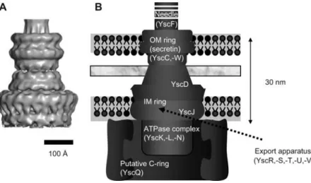
fla-Figure 1 Needle complex (NC) structure.
The NC is modeled by cryo-EM and single particle analysis (Hodgkinson et al., 2009) (left) and a cartoon of the basal body is shown for comparison with the flagellar basal body, identifying the main substructures and their constituents. The fact that several elements of the basal body are missing in the NC is illustrated.
gellar basal body. This structure, called the needle complex (NC) or injectisome basal body, consists of two pairs of rings that span the inner (IM) and outer (OM) bacterial mem-branes, joined together by a narrower cylinder and terminat-ed by a neterminat-edle, filament or pilus (Kubori et al., 1998; Blocker et al., 1999; Kimbrough and Miller, 2000; Daniell et al., 2001; Jin and He, 2001; Sekiya et al., 2001; Morita-Ishihara et al., 2006; Sani et al., 2007; Hodgkinson et al., 2009).
Single particles of NCs fromS. enterica SPI-1 (Marlovits
et al., 2004, 2006) and fromS. flexneri (Blocker et al., 2001;
Sani et al., 2007; Hodgkinson et al., 2009) were character-ized by cryo-electron microscopy (cryo-EM). These recons-titutions show that an inner rod with a central channel of approximately 2–3 nm is anchored at the basis of the cham-ber by a socket-like structure and traverses the chamcham-ber of the NC. This channel extends all the way to the tip of the needle. Some sample heterogeneity was reported among the
S. enterica SPI-1 NCs. The lower rings of the majority of
the complexes have a rotational symmetry of 20 or 21 but some are smaller (19-fold) or larger (22-fold). By contrast, the three-dimensional (3D) reconstruction of theS. flexneri
NC, reveals an homogeneous 12-fold symmetry (Hodgkin-son et al., 2009). The external diameter of the lower double ring in both structures is 21–22 nm (Marlovits et al., 2004; Hodgkinson et al., 2009). This value is significantly smaller than the diameter measured by transmission EM (TEM; approx. 40 nm; Kubori et al., 1998). It is also significantly smaller than the diameter of the C-ring of the flagellar basal body (40 nm; Macnab, 2003). This discrepancy indicates that the NC structures reconstructed have no C-ring, most prob-ably because it breaks off during the purification procedure. Actually, the extracted NC not only misses the C-ring but also the ATPase and the transmembrane (TM) proteins thought to form the export apparatus (Figure 1). Electron microscopy has thus provided excellent information about the global structure of the injectisome but the determination
of the precise localization of most structural components of the injectisome base and their secondary structure still requires a large effort.
The OM-rings
A triple ring spans the OM layer (Hodgkinson et al., 2009). It consists of a 12–14-mer of a protein belonging to the YscC family of secretins (Figure 1) (Koster et al., 1997; Kubori et al., 2000; Tamano et al., 2000; Blocker et al., 2001; Burghout et al., 2004b). Secretins are found not only in injectisomes but also in the type II secretion apparatus, in type IV pili (Collins et al., 2004; Chami et al., 2005) as well as in fila-mentous phages (Russel, 1994). They have a conserved C-terminal membrane spanning domain, predicted to be a b-barrel, and a variable N-terminal domain extending in the periplasm. The proper insertion of secretins in the OM requires the assistance of a lipoprotein (YscW family) anchored in the OM (Crago and Koronakis, 1998; Daefler and Russel, 1998; Burghout et al., 2004a). The crystal struc-ture of the N-terminal domain of EscC, the homolog of YscC in EPECs was recently solved (Spreter et al., 2009). The IM-ring and the connector
In the flagellum, the ring spanning the IM is called the MS-ring. It is made of FliF and spans the outer leaflet of the plasma membrane (Ueno et al., 1994). The homologs of FliF are lipoproteins YscJ (Yersinia)/PrgK (S. enterica)/MxiJ (S. flexneri)/EscJ (EPEC). The crystal structure of EscJ, the
EPEC ortholog of YscJ (Crepin et al., 2005; Yip et al., 2005) allowed building of a 24-subunit ring model (Yip et al., 2005). However, attempts to dock the EscJ structure into the recent cryo-EM map of theS. flexneri NC, suggest that MxiJ
might rather be a 12-mer (Hodgkinson et al., 2009). A pro-tein from the less-conserved YscD (Yersinia)/PrgH (S. ente-rica)/MxiC (S. flexneri) family is proposed to participate in
connect the IM- and OM-rings (Spreter et al., 2009). The structure of the periplasmic domain of PrgH reveals strong similarity to the EscJ/PrgK IM-ring component and to the periplasmic domain of the secretin (Spreter et al., 2009). This strong similarity in fold and architecture between the three proteins suggests the conservation of a fold that potentially provides a common ring-building motif for the assembly of the symmetrical ring structures that constitute the T3SS basal body (Spreter et al., 2009).
The C-ring
In the flagellum, the most internal part of the basal body is the 45–50 nm C-ring (cytosolic), made of 31–38 copies of Fli(MN3) (Driks and DeRosier, 1990; Khan et al., 1992; Kubori et al., 1997; Young et al., 2003; Thomas et al., 2006). A 3D reconstruction of the C-ring has been proposed (Tho-mas et al., 2006). It is an essential component of the switch complex reversing the rotation of the motor but it also acts as a non-essential affinity cup-like structure during flagellar T3S to enhance the specificity and efficiency of the secretion process (Macnab, 2003; Erhardt and Hughes, 2010). As men-tioned before, there is no C-ring visible in the cryo-EM reconstructions of theS. enterica SPI-1 and S. flexneri NCs
(Marlovits et al., 2004, 2006; Hodgkinson et al., 2009) but proteins of the YscQ family have a significant similarity to FliN and FliM and genetic data indicate that such proteins are essential components in all injectisome families. In Pseu-domonas syringae, the ortholog of YscQ even appears as two
products called HrcQAand HrcQB, which interact with each other. The overall fold of HrcQB is remarkably similar to that of FliN (Fadouloglou et al., 2004). In agreement with this, immunogold-labeling experiments have shown that the
S. flexneri ortholog of YscQ, localizes to a lower portion of
the injectisome via interaction with the orthologs of YscD and YscJ that form the IM-ring (Morita-Ishihara et al., 2006). Together these data indicate that there is a C-ring in the injec-tisome even though it could not be visualized by cryo-EM thus far. It would form a platform at the cytoplasm/inner membrane interface for the recruitment of the ATPase com-plex (see below) (Jackson and Plano, 2000) and substrate-chaperone complexes (Morita-Ishihara et al., 2006; Spaeth et al., 2009).
The export apparatus
Among the eight proteins that are highly conserved between flagella and injectisomes, five (YscR, S, T, U, V) are TM a-helical proteins (Plano and Straley, 1993; Allaoui et al., 1994; Fields et al., 1994). They are thought to form the trans-location channel(s) embedded in a patch of IM enclosed within the IM-ring but this view awaits biochemical confir-mation. Two of them (YscU, -V) have a large C-terminal cytosolic domain. In YscU, this domain is autocleavable (Zarivach et al., 2008; Wiesand et al., 2009) and involved in the selection of the export substrates (Sorg et al., 2007). The stoichiometry of all these components is not known.
One of the conserved proteins is an AAAq
ATPase (YscN family) whose integrity is essential to T3S (Woestyn et al.,
1994). InvC fromS. enterica SPI-1 has been shown to unfold
the exported proteins in an ATP-dependent manner and to detach some T3S substrates from their cytoplasmic chaper-ones before their export (Akeda and Galan, 2005). It is also probable that the ATPase energizes export but the proton motive force is also involved (Wilharm et al., 2004; Mina-mino and Namba, 2008; Paul et al., 2008). HrcN, the ATPase from P. syringae, forms hexamers and dodecamers that are
activated by oligomerization and peripherally associated with the cytoplasmic side of the inner membrane (Pozidis et al., 2003). According to cryo-EM, it has a 2.0–3.8 nm wide inner channel (Muller et al., 2006). This structure compares to that of the flagellar ATPase FliI. As for HrcN, its oligomerization and enzyme activity are coupled (Claret et al., 2003). Recent-ly, the structure of the catalytic domain from EscN (50% identical to YscN) from EPEC was solved by crystallography at 1.8 A˚ resolution. Along with in vitro and in vivo muta-tional analysis, these data show that, in spite of the expected similarity with the F1 ATPase, there are important structural differences that dictate their unique secretory role (Zarivach et al., 2007). YscN was shown to interact with YscQ, YscK and YscL (Jackson and Plano, 2000; Blaylock et al., 2006). Likewise, in S. flexneri, the ATPase Spa47 was shown to
interact with MxiK and MxiN, the probable orthologs of YscK and YscL (Jouihri et al., 2003). These observations fit with previous data showing that the flagellar ATPase FliI interacts with FliH (the so-called ATPase regulator), the ortholog of YscL and that the FliHI complex interacts with FliN from the C-ring (Minamino and MacNab, 2000; Gon-zalez-Pedrajo et al., 2002). Given that YscL interacts also with YscQ, the component of the putative C-ring, YscL could be the protein tethering the ATPase to the export chan-nel (Minamino and MacNab, 2000; Blaylock et al., 2006). The stoichiometry of YscK and YscL is still not known. Interestingly, FliH/YscL-like proteins represent fusions of domains from the b and d subunits of the second-stalk com-ponents of the FoF1ATPase (Pallen et al., 2006), pointing to some similarity between this nanomachine and the injectisome.
A rod?
In the flagellum, FlgBCF and -G form the so-called rod, a tube extending the hook inside the basal body. Some indirect biochemical and EM evidence suggests that, inS. enterica
SPI-1, PrgJ forms such an ‘inner rod’ structure (Kubori et al., 2000; Sukhan et al., 2003; Marlovits et al., 2004). There is noYersinia Ysc homolog of PrgJ but YscI has properties
similar to those of the needle subunit. Based on this, some authors propose that YscI forms a rod inside the basal body of theYersinia injectisome (Wood et al., 2008) but this awaits
some direct biochemical evidence. The needle
The needle is a straight hollow tube with an inner diameter of approximately 2.5 nm. It is constructed via the helical polymerization of approximately 150 subunits of the YscF family (Kubori et al., 2000; Hoiczyk and Blobel, 2001).
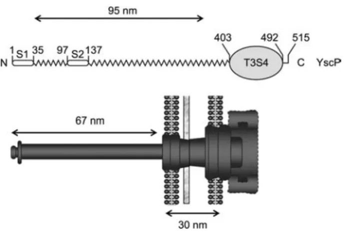
Figure 2 Illustration of the molecular ruler concept.
Above, a schematic representation of the YscP molecule with the two export signals (S1 and S2) at the N-terminus and the T3S4 domain at the C-terminus is shown. The ruler domain is represented by the zigzag line. Below is a schematic representation of the injec-tisome. The protein is aligned with the nanomachine in the way it is proposed to operate: the N-terminus towards the tip and the T3S4 domain in the cytosolic part of the basal body.
Although the needle subunits (approx. 9 kDa) are signifi-cantly smaller than flagellin (approx. 45 kDa), the helical parameters are similar (5.6 units per turn, helical pitch of 2.4 nm; Cordes et al., 2003). The crystal structure of MxiH, the S. flexneri needle subunit consists of two extended and
bent antiparallel a-helices connected by a short turn (Deane et al., 2006), a structure similar to that of the D0 portion of flagellin (Yonekura et al., 2003). The structure of MxiH, combined to the EM 3D reconstructions, allowed the build-ing of an atomic model (Deane et al., 2006). Scannbuild-ing-TEM (STEM) analyses of Y. enterocolitica needles showed that
they end with a distinct tip structure consisting of a homo-pentamer of the protein LcrV (Mueller et al., 2005; Deane et al., 2006; Broz et al., 2007) the crystal structure of which is known (Derewenda et al., 2004). An active injectisome in contact with a target cell terminates with a translocation pore that is inserted in the plasma membrane of the target cell (Hakansson et al., 1996; Blocker et al., 1999; Neyt and Cor-nelis, 1999). The assembly of this pore, upon cell contact, requires two hydrophobic proteins called YopB and YopD (Cornelis and Wolf-Watz, 1997). The needle-tip structure is thought to serve as a scaffold or assembly platform for the translocation pore (Goure et al., 2004).
Needle length control and substrate specificity switch
The final length of the needle varies between different bac-teria or even strains but the length of the needle is regulated as is the case for the flagellar hook (55 nm). In Y. entero-colitica strain E40, the needle length is 65"10 nm (Journet
et al., 2003) whereas in otherY. enterocolitica strains it
var-ies from 49"6 to 87"11 nm (Wagner et al., 2009). During morphogenesis, the needle components, as with the hook and the filament of the flagellum, are sequentially exported by the T3S apparatus itself (Sukhan et al., 2001), traveling through the growing structure and polymerizing at its distal end (Li et al., 2002; Macnab, 2003). There is no clear hier-archy in the synthesis of the injectisome components and substrates. Thus, the export apparatus is expected to switch its substrate specificity over time so that needle subunits (early substrates) are exported before the tip structure subunit LcrV (intermediate substrate) and the effectors (late sub-strates). This substrate specificity switch presumably leads to the arrest of needle growth (Ferris and Minamino, 2006). The switch to export late substrates is triggered by the protein FliK, in the flagellum system (Hirano et al., 1994; Minamino et al., 1999, 2004) and YscP in the injectisome (Journet et al., 2003). The 515-residue YscP (Figure 2) is itself an early substrate of the machine driven by two independent N-ter-minal export signals (Agrain et al., 2005b). The switch activ-ity is exerted by residues 405–500 (Agrain et al., 2005a), a domain called T3S4 for type 3 secretion substrate specificity switch. This domain is thought to interact with YscU (FlhB in the flagellum), a component of the basal body that is also involved in setting the hierarchy of export (Hirano et al., 1994; Sorg et al., 2007). The central domain of YscP is pre-dicted to be helical and there is a linear correlation between the number of residues in the protein and the needle length,
suggesting that YscP acts as a molecular ruler or a molecular timer. There is also an inverse correlation between the helical content of this central domain and the needle length, indi-cating that the functional ruler is partially helical (Wagner et al., 2009). The calculated length when the helical content is preserved correlates strikingly with the measured needle length, with a constant difference of approximately 29 nm, which corresponds to the size of the basal body (Figure 2). These data support the ruler model and show that the func-tional ruler has a helical structure (Wagner et al., 2009).
References
Agrain, C., Callebaut, I., Journet, L., Sorg, I., Paroz, C., Mota, L.J., et al. (2005a). Characterization of a type III secretion substrate specificity switch (T3S4) domain in YscP fromYersinia ente-rocolitica. Mol. Microbiol. 56, 54–67.
Agrain, C., Sorg, I., Paroz, C., and Cornelis, G.R. (2005b). Secretion of YscP fromYersinia enterocolitica is essential to control the
length of the injectisome needle but not to change the type III secretion substrate specificity. Mol. Microbiol.57, 1415–1427.
Akeda, Y. and Galan, J.E. (2005). Chaperone release and unfolding of substrates in type III secretion. Nature437, 911–915.
Alfano, J.R. and Collmer, A. (2004). Type III secretion system effec-tor proteins: double agents in bacterial disease and plant defense. Annu. Rev. Phytopathol.42, 385–414.
Allaoui, A., Woestyn, S., Sluiters, C., and Cornelis, G.R. (1994). YscU, aYersinia enterocolitica inner membrane protein involved
in Yop secretion. J. Bacteriol.176, 4534–4542.
Blaylock, B., Riordan, K.E., Missiakas, D.M., and Schneewind, O. (2006). Characterization of theYersinia enterocolitica type III
secretion ATPase YscN and its regulator, YscL. J. Bacteriol.188,
3525–3534.
Blocker, A., Gounon, P., Larquet, E., Niebuhr, K., Cabiaux, V., Par-sot, C., et al. (1999). The tripartite type III secreton ofShigella flexneri inserts IpaB and IpaC into host membranes. J. Cell Biol. 147, 683–693.
Blocker, A., Jouihri, N., Larquet, E., Gounon, P., Ebel, F., Parsot, C., et al. (2001). Structure and composition of theShigella flex-neri ‘needle complex’, a part of its type III secreton. Mol.
Micro-biol.39, 652–663.
Bogdanove, A.J., Beer, S.V., Bonas, U., Boucher, C.A., Collmer, A., Coplin, D.L., et al. (1996). Unified nomenclature for broadly conserved hrp genes of phytopathogenic bacteria. Mol. Micro-biol.20, 681–683.
Broz, P., Mueller, C.A., Muller, S.A., Philippsen, A., Sorg, I., Engel, A., et al. (2007). Function and molecular architecture of the Yer-sinia injectisome tip complex. Mol. Microbiol. 65, 1311–1320.
Burghout, P., Beckers, F., de Wit, E., van Boxtel, R., Cornelis, G.R., Tommassen, J., et al. (2004a). Role of the pilot protein YscW in the biogenesis of the YscC secretin inYersinia enterocolitica.
J. Bacteriol.186, 5366–5375.
Burghout, P., van Boxtel, R., Van Gelder, P., Ringler, P., Muller, S.A., Tommassen, J., et al. (2004b). Structure and electrophysio-logical properties of the YscC secretin from the type III secretion system of Yersinia enterocolitica. J. Bacteriol. 186, 4645–
4654.
Burr, S.E., Wahli, T., Segner, H., Pugovkin, D., and Frey, J. (2003). Association of type III secretion genes with virulence of Aero-monas salmonicida subsp. salmonicida. Dis. Aquat. Organ. 57,
167–171.
Chami, M., Guilvout, I., Gregorini, M., Remigy, H.W., Muller, S.A., Valerio, M., et al. (2005). Structural insights into the secretin PulD and its trypsin-resistant core. J. Biol. Chem.280, 37732–
37741.
Claret, L., Calder, S.R., Higgins, M., and Hughes, C. (2003). Olig-omerization and activation of the FliI ATPase central to bacterial flagellum assembly. Mol. Microbiol.48, 1349–1355.
Collins, R.F., Frye, S.A., Kitmitto, A., Ford, R.C., Tonjum, T., and Derrick, J.P. (2004). Structure of theNeisseria meningitidis outer
membrane PilQ secretin complex at 12 A˚ resolution. J. Biol. Chem.279, 39750–39756.
Cordes, F.S., Komoriya, K., Larquet, E., Yang, S., Egelman, E.H., Blocker, A., et al. (2003). Helical structure of the needle of the type III secretion system ofShigella flexneri. J. Biol. Chem. 278,
17103–17107.
Cornelis, G.R. and Van Gijsegem, F. (2000). Assembly and function of type III secretory systems. Annu. Rev. Microbiol.54, 735–
774.
Cornelis, G.R. and Wolf-Watz, H. (1997). TheYersinia Yop virulon:
a bacterial system for subverting eukaryotic cells. Mol. Micro-biol.23, 861–867.
Crago, A.M. and Koronakis, V. (1998).Salmonella InvG forms a
ring-like multimer that requires the InvH lipoprotein for outer membrane localization. Mol. Microbiol.30, 47–56.
Crepin, V.F., Prasannan, S., Shaw, R.K., Wilson, R.K., Creasey, E., Abe, C.M., et al. (2005). Structural and functional studies of the enteropathogenicEscherichia coli type III needle complex
pro-tein EscJ. Mol. Microbiol.55, 1658–1670.
Daefler, S. and Russel, M. (1998). The Salmonella typhimurium
InvH protein is an outer membrane lipoprotein required for the proper localization of InvG. Mol. Microbiol.28, 1367–1380.
Daniell, S.J., Takahashi, N., Wilson, R., Friedberg, D., Rosenshine, I., Booy, F.P., et al. (2001). The filamentous type III secretion translocon of enteropathogenicEscherichia coli. Cell Microbiol. 3, 865–871.
Deane, J.E., Roversi, P., Cordes, F.S., Johnson, S., Kenjale, R., Daniell, S., et al. (2006). Molecular model of a type III secretion system needle: implications for host-cell sensing. Proc. Natl. Acad. Sci. USA103, 12529–12533.
Derewenda, U., Mateja, A., Devedjiev, Y., Routzahn, K.M., Evdo-kimov, A.G., Derewenda, Z.S., et al. (2004). The structure of
Yersinia pestis V-antigen, an essential virulence factor and
medi-ator of immunity against plague. Structure (Camb.)12, 301–306.
Driks, A. and DeRosier, D.J. (1990). Additional structures associ-ated with bacterial flagellar basal body. J. Mol. Biol. 211,
669–672.
Erhardt, M. and Hughes, K.T. (2010). C-ring requirement in flagel-lar type III secretion is bypassed by FlhDC upregulation. Mol. Microbiol.75, 376–393.
Fadouloglou, V.E., Tampakaki, A.P., Glykos, N.M., Bastaki, M.N., Hadden, J.M., Phillips, S.E., et al. (2004). Structure of HrcQB-C, a conserved component of the bacterial type III secretion systems. Proc. Natl. Acad. Sci. USA101, 70–75.
Ferris, H.U. and Minamino, T. (2006). Flipping the switch: bringing order to flagellar assembly. Trends Microbiol.14, 519–526.
Fields, K.A., Plano, G.V., and Straley, S.C. (1994). A low-Ca2q
response (LCR) secretion (ysc) locus lies within the lcrB region of the LCR plasmid inYersinia pestis. J. Bacteriol. 176, 569–
579.
Galan, J.E. and Collmer, A. (1999). Type III secretion machines: bacterial devices for protein delivery into host cells. Science284,
1322–1328.
Gonzalez-Pedrajo, B., Fraser, G.M., Minamino, T., and Macnab, R.M. (2002). Molecular dissection ofSalmonella FliH, a
regu-lator of the ATPase FliI and the type III flagellar protein export pathway. Mol. Microbiol.45, 967–982.
Gophna, U., Ron, E.Z., and Graur, D. (2003). Bacterial type III secretion systems are ancient and evolved by multiple horizon-tal-transfer events. Gene312, 151–163.
Goure, J., Pastor, A., Faudry, E., Chabert, J., Dessen, A., and Attree, I. (2004). The V antigen ofPseudomonas aeruginosa is required
for assembly of the functional PopB/PopD translocation pore in host cell membranes. Infect. Immun.72, 4741–4750.
Grant, S.R., Fisher, E.J., Chang, J.H., Mole, B.M., and Dangl, J.L. (2006). Subterfuge and manipulation: type III effector proteins of phytopathogenic bacteria. Annu. Rev. Microbiol. 60,
425–449.
Hakansson, S., Galyov, E.E., Rosqvist, R., and Wolf-Watz, H. (1996). TheYersinia YpkA Ser/Thr kinase is translocated and
subsequently targeted to the inner surface of the HeLa cell plas-ma membrane. Mol. Microbiol.20, 593–603.
Hirano, T., Yamaguchi, S., Oosawa, K., and Aizawa, S. (1994). Roles of FliK and FlhB in determination of flagellar hook length inSalmonella typhimurium. J. Bacteriol. 176, 5439–5449.
Hodgkinson, J.L., Horsley, A., Stabat, D., Simon, M., Johnson, S., da Fonseca, P.C., et al. (2009). Three-dimensional reconstruction of the Shigella T3SS transmembrane regions reveals 12-fold
symmetry and novel features throughout. Nat. Struct. Mol. Biol.
16, 477–485.
Hoiczyk, E. and Blobel, G. (2001). Polymerization of a single pro-tein of the pathogenYersinia enterocolitica into needles
punc-tures eukaryotic cells. Proc. Natl. Acad. Sci. USA 98, 4669–
4674.
Jackson, M.W. and Plano, G.V. (2000). Interactions between type III secretion apparatus components fromYersinia pestis detected
using the yeast two-hybrid system. FEMS Microbiol. Lett.186,
85–90.
Jin, Q. and He, S.Y. (2001). Role of the Hrp pilus in type III protein secretion inPseudomonas syringae. Science 294, 2556–2558.
Jouihri, N., Sory, M.P., Page, A.L., Gounon, P., Parsot, C., and Allaoui, A. (2003). MxiK and MxiN interact with the Spa47 ATPase and are required for transit of the needle components MxiH and MxiI, but not of Ipa proteins, through the type III
secretion apparatus of Shigella flexneri. Mol. Microbiol. 49,
755–767.
Journet, L., Agrain, C., Broz, P., and Cornelis, G.R. (2003). The needle length of bacterial injectisomes is determined by a molec-ular ruler. Science302, 1757–1760.
Khan, I.H., Reese, T.S., and Khan, S. (1992). The cytoplasmic com-ponent of the bacterial flagellar motor. Proc. Natl. Acad. Sci. USA89, 5956–5960.
Kimbrough, T.G. and Miller, S.I. (2000). Contribution ofSalmonella typhimurium type III secretion components to needle complex
formation. Proc. Natl. Acad. Sci. USA97, 11008–11013.
Koster, M., Bitter, W., de Cock, H., Allaoui, A., Cornelis, G.R., and Tommassen, J. (1997). The outer membrane component, YscC, of the Yop secretion machinery ofYersinia enterocolitica forms
a ring-shaped multimeric complex. Mol. Microbiol. 26, 789–
797.
Kubori, T., Yamaguchi, S., and Aizawa, S. (1997). Assembly of the switch complex onto the MS ring complex ofSalmonella typhi-murium does not require any other flagellar proteins. J. Bacteriol. 179, 813–817.
Kubori, T., Matsushima, Y., Nakamura, D., Uralil, J., Lara-Tejero, M., Sukhan, A., et al. (1998). Supramolecular structure of the
Salmonella typhimurium type III protein secretion system.
Sci-ence280, 602–605.
Kubori, T., Sukhan, A., Aizawa, S.I., and Galan, J.E. (2000). Molec-ular characterization and assembly of the needle complex of the
Salmonella typhimurium type III protein secretion system. Proc.
Natl. Acad. Sci. USA97, 10225–10230.
Li, C.M., Brown, I., Mansfield, J., Stevens, C., Boureau, T., Romantschuk, M., et al. (2002). The Hrp pilus ofPseudomonas syringae elongates from its tip and acts as a conduit for
trans-location of the effector protein HrpZ. EMBO J.21, 1909–1915.
Macnab, R.M. (2003). How bacteria assemble flagella. Annu. Rev. Microbiol.57, 77–100.
Marlovits, T.C., Kubori, T., Sukhan, A., Thomas, D.R., Galan, J.E., and Unger, V.M. (2004). Structural insights into the assembly of the type III secretion needle complex. Science306, 1040–1042.
Marlovits, T.C., Kubori, T., Lara-Tejero, M., Thomas, D., Unger, V.M., and Galan, J.E. (2006). Assembly of the inner rod deter-mines needle length in the type III secretion injectisome. Nature
441, 637–640.
Minamino, T. and MacNab, R.M. (2000). Interactions among com-ponents of theSalmonella flagellar export apparatus and its
sub-strates. Mol. Microbiol.35, 1052–1064.
Minamino, T. and Namba, K. (2008). Distinct roles of the FliI ATP-ase and proton motive force in bacterial flagellar protein export. Nature451, 485–488.
Minamino, T., Gonzalez-Pedrajo, B., Yamaguchi, K., Aizawa, S.I., and Macnab, R.M. (1999). FliK, the protein responsible for fla-gellar hook length control in Salmonella, is exported during
hook assembly. Mol. Microbiol.34, 295–304.
Minamino, T., Saijo-Hamano, Y., Furukawa, Y., Gonzalez-Pedrajo, B., Macnab, R.M., and Namba, K. (2004). Domain organization and function ofSalmonella FliK, a flagellar hook-length control
protein. J. Mol. Biol.341, 491–502.
Morita-Ishihara, T., Ogawa, M., Sagara, H., Yoshida, M., Katayama, E., and Sasakawa, C. (2006).Shigella Spa33 is an essential
C-ring component of type III secretion machinery. J. Biol. Chem.
281, 599–607.
Mota, L.J. and Cornelis, G.R. (2005). The bacterial injection kit: type III secretion systems. Ann. Med.37, 234–249.
Mueller, C.A., Broz, P., Muller, S.A., Ringler, P., Erne-Brand, F., Sorg, I., et al. (2005). The V-antigen ofYersinia forms a distinct
structure at the tip of injectisome needles. Science310, 674–
676.
Muller, S.A., Pozidis, C., Stone, R., Meesters, C., Chami, M., Engel, A., et al. (2006). Double hexameric ring assembly of the type III protein translocase ATPase HrcN. Mol. Microbiol.61, 119–
125.
Neyt, C. and Cornelis, G.R. (1999). Insertion of a Yop translocation pore into the macrophage plasma membrane byYersinia ente-rocolitica: requirement for translocators YopB and YopD, but not
LcrG. Mol. Microbiol.33, 971–981.
Pallen, M.J., Beatson, S.A., and Bailey, C.M. (2005). Bioinforma-tics, genomics and evolution of non-flagellar type-III secretion systems: a Darwinian perspective. FEMS Microbiol. Rev.29,
201–229.
Pallen, M.J., Bailey, C.M., and Beatson, S.A. (2006). Evolutionary links between FliH/YscL-like proteins from bacterial type III secretion systems and second-stalk components of the FoF1 and vacuolar ATPases. Protein Sci.15, 935–941.
Paul, K., Erhardt, M., Hirano, T., Blair, D.F., and Hughes, K.T. (2008). Energy source of flagellar type III secretion. Nature451,
489–492.
Plano, G.V. and Straley, S.C. (1993). Multiple effects of lcrD muta-tions inYersinia pestis. J. Bacteriol. 175, 3536–3545.
Pozidis, C., Chalkiadaki, A., Gomez-Serrano, A., Stahlberg, H., Brown, I., Tampakaki, A.P., et al. (2003). Type III protein trans-locase: HrcN is a peripheral ATPase that is activated by oligo-merization. J. Biol. Chem.278, 25816–25824.
Roy-Burman, A., Savel, R.H., Racine, S., Swanson, B.L., Revadi-gar, N.S., Fujimoto, J., et al. (2001). Type III protein secretion is associated with death in lower respiratory and systemic Pseu-domonas aeruginosa infections. J. Infect. Dis. 183, 1767–1774.
Russel, M. (1994). Phage assembly: a paradigm for bacterial viru-lence factor export? Science265, 612–614.
Sani, M., Allaoui, A., Fusetti, F., Oostergetel, G.T., Keegstra, W., and Boekema, E.J. (2007). Structural organization of the needle complex of the type III secretion apparatus ofShigella flexneri.
Micron38, 291–301.
Sekiya, K., Ohishi, M., Ogino, T., Tamano, K., Sasakawa, C., and Abe, A. (2001). Supermolecular structure of the enteropathogen-icEscherichia coli type III secretion system and its direct
inter-action with the EspA-sheath-like structure. Proc. Natl. Acad. Sci. USA98, 11638–11643.
Sorg, I., Wagner, S., Amstutz, M., Muller, S.A., Broz, P., Lussi, Y., et al. (2007). YscU recognizes translocators as export substrates of theYersinia injectisome. EMBO J. 26, 3015–3024.
Spaeth, K.E., Chen, Y.S., and Valdivia, R.H. (2009). TheChlamydia
type III secretion system C-ring engages a chaperone-effector protein complex. PLoS Pathog.5, e1000579.
Spreter, T., Yip, C.K., Sanowar, S., Andre, I., Kimbrough, T.G., Vuckovic, M., et al. (2009). A conserved structural motif medi-ates formation of the periplasmic rings in the type III secretion system. Nat. Struct. Mol. Biol.16, 468–476.
Sukhan, A., Kubori, T., Wilson, J., and Galan, J.E. (2001). Genetic analysis of assembly of theSalmonella enterica serovar Typhi-murium type III secretion-associated needle complex. J.
Bacte-riol.183, 1159–1167.
Sukhan, A., Kubori, T., and Galan, J.E. (2003). Synthesis and local-ization of theSalmonella SPI-1 type III secretion needle
com-plex proteins PrgI and PrgJ. J. Bacteriol.185, 3480–3483.
Tamano, K., Aizawa, S., Katayama, E., Nonaka, T., Imajoh-Ohmi, S., Kuwae, A., et al. (2000). Supramolecular structure of the
Shigella type III secretion machinery: the needle part is
change-able in length and essential for delivery of effectors. EMBO J.
Thomas, D.R., Francis, N.R., Xu, C., and DeRosier, D.J. (2006). The three-dimensional structure of the flagellar rotor from a clockwise-locked mutant ofSalmonella enterica serovar Typhi-murium. J. Bacteriol. 188, 7039–7048.
Troisfontaines, P. and Cornelis, G.R. (2005). Type III secretion: more systems than you think. Physiology (Bethesda)20, 326–
339.
Ueno, T., Oosawa, K., and Aizawa, S. (1994). Domain structures of the MS ring component protein (FliF) of the flagellar basal body ofSalmonella typhimurium. J. Mol. Biol. 236, 546–555.
Van Gijsegem, F., Gough, C., Zischek, C., Niqueux, E., Arlat, M., Genin, S., et al. (1995). The hrp gene locus of Pseudomonas solanacearum, which controls the production of a type III
secre-tion system, encodes eight proteins related to components of the bacterial flagellar biogenesis complex. Mol. Microbiol. 15,
1095–1114.
Wagner, S., Sorg, I., Degiacomi, M., Journet, L., Dal Peraro, M., and Cornelis, G.R. (2009). The helical content of the YscP molecular ruler determines the length of the Yersinia
injecti-some. Mol. Microbiol.71, 692–701.
Wiesand, U., Sorg, I., Amstutz, M., Wagner, S., van den Heuvel, J., Luhrs, T., et al. (2009). Structure of the type III secretion rec-ognition protein YscU fromYersinia enterocolitica. J. Mol. Biol. 385, 854–866.
Wilharm, G., Lehmann, V., Krauss, K., Lehnert, B., Richter, S., Ruckdeschel, K., et al. (2004).Yersinia enterocolitica type III
secretion depends on the proton motive force but not on the flagellar motor components MotA and MotB. Infect. Immun.72,
4004–4009.
Woestyn, S., Allaoui, A., Wattiau, P., and Cornelis, G.R. (1994). YscN, the putative energizer of the Yersinia Yop secretion
machinery. J. Bacteriol.176, 1561–1569.
Wood, S.E., Jin, J., and Lloyd, S.A. (2008). YscP and YscU switch the substrate specificity of theYersinia type III secretion system
by regulating export of the inner rod protein YscI. J. Bacteriol.
190, 4252–4262.
Yip, C.K., Kimbrough, T.G., Felise, H.B., Vuckovic, M., Thomas, N.A., Pfuetzner, R.A., et al. (2005). Structural characterization of the molecular platform for type III secretion system assembly. Nature435, 702–707.
Yonekura, K., Maki-Yonekura, S., and Namba, K. (2003). Complete atomic model of the bacterial flagellar filament by electron cryo-microscopy. Nature424, 643–650.
Young, H.S., Dang, H., Lai, Y., DeRosier, D.J., and Khan, S. (2003). Variable symmetry inSalmonella typhimurium flagellar motors.
Biophys. J.84, 571–577.
Zarivach, R., Vuckovic, M., Deng, W., Finlay, B.B., and Strynadka, N.C. (2007). Structural analysis of a prototypical ATPase from the type III secretion system. Nat. Struct. Mol. Biol.14, 131–
137.
Zarivach, R., Deng, W., Vuckovic, M., Felise, H.B., Nguyen, H.V., Miller, S.I., et al. (2008). Structural analysis of the essential self-cleaving type III secretion proteins EscU and SpaS. Nature453,
124–127.