Ventricular myocardial architecture as visualised in postmortem swine
hearts using magnetic resonance diffusion tensor imaging
Peter Schmid
a,*, Thomas Jaermann
a, Peter Boesiger
a, Peter F. Niederer
a,
Paul P. Lunkenheimer
b, Colin W. Cryer
c, Robert H. Anderson
daInstitute for Biomedical Engineering, University of Zurich and Swiss Federal Institute of Technology (ETH), Gloriastrasse 35, CH-8092 Zurich, Switzerland b
Clinic and Policlinic for Thorax, Heart and Vessel Surgery, University of Munster, Munster, Germany
cInstitute for Numerical Mathematics, University of Munster, Munster, Germany dCardiac Unit, Institute of Child Health, University College London, London, UK
Received 26 August 2004; accepted 5 November 2004; Available online 18 January 2005
Abstract
Objective: The three-dimensional arrangement of the ventricular myocardial architecture remains controversial, in part because histological assessment is difficult to achieve, while anatomic dissections are, of necessity, destructive. In this study, we describe how the use of magnetic resonance diffusion tensor imaging has permitted us to reconstruct with precision the architecture of the ventricular myocardial fibres in the post-mortem swine heart. Methods and Results: We obtained diffusion-weighted spin-echo measurements of autopsied porcine hearts using a whole body MR system. We calculated the diffusion tensor and the corresponding eigenvectors on a voxel-by-voxel basis. This permitted us to colour code the fibres, and reconstruct them by connecting voxels in direction of the largest eigenvector. Such reconstructions show that, in the middle layer of the left ventricle, most of the fibres have a circular orientation, albit that a far from negligible component runs in a transverse direction. With increasing distance from the epicardium, the orientation of the fibres shows a continuous change in angulation with respect to an axis normal to the epicardium. Conclusion: Our data presented here supports the concept that the ventricular mass is arranged as a complex three-dimensional mesh of tangential and intruding fibres. The data offers no support for the concept of a ‘unique myocardial band’. The method has the potential to detecting deviations from this basic normal architecture, being capable of reconstructing the ventricular mass so as to assess the spatial coordinates of any single fibre strand. The technique, therefore, has major potential clinical applications in the setting of the failing or malformed heart, potentially being able to identify either systematic or regional disarray of the myocardial fibres.
q2005 Elsevier B.V. All rights reserved.
Keywords: Magnetic resonance imaging; Myocardium; Structure
1. Introduction
Despite many anatomic investigations, the architecture of the ventricular myocardial fibres remains controversial
[1–7]. This is because, using histological techniques, it is difficult to obtain a comprehensive three-dimensional visualisation of the entire ventricular mass, while anatomic dissections are of necessity destructive, and introduce potential artefacts. Because of these technical problems, it remains difficult to prove that, as is claimed by the majority, the ventricular mass is arranged on the basis of an interweaving network of myocardial fibres[1,2,4–6], since a small minority continue to argue[7]that the musculature is arranged in the form of a ‘unique myocardial band’[8].
In this study, we describe how the use of magnetic resonance (MR) diffusion tensor imaging (DTI) [9] permits
the architecture of the ventricular myocardial fibres to be assessed with precision in the intact autopsied heart, disproving unequivocally the notion that the myocardial mass is arranged in the form of the purported ‘unique band’
[7,8].
2. Materials and methods
We obtained eight porcine hearts from the slaughter-house immediately after death. So as to minimise artefacts during our studies, we removed the subepicardial fat, the coronary vessels, the atria, and the papillary muscles of the atrioventricular valves. We then housed the hearts in a plastic box filled with 2% agarose gel doped with 0.5% copper sulphate to enhance the signal contrast between the myocardium and the gel, taking care to avoid the inclusion of air bubbles within the ventricular contour while filling the ventricular cavities with gel.
www.elsevier.com/locate/ejcts
1010-7940/$ - see front matter q 2005 Elsevier B.V. All rights reserved. doi:10.1016/j.ejcts.2004.11.036
* Corresponding author. Tel.: C41 1 632 45 64; fax: C41 1 632 11 93. E-mail address: [email protected] (P. Schmid).
The hearts were then imaged using a 3 Tesla Philips Intera whole body MR system (Philips Medical Systems, Best, The Netherlands), attaching four rectangular surface coils (210! 110 mm) to the box. The data were obtained from diffusion weighted spin-echo measurements using two different protocols for diffusion tensor imaging, namely single shot echo planar imaging (sshEPI) for scanning the entire heart, and scans with enhanced in-plane resolution in the sub millimetre realm with very low geometric distortions for the purposes of validation. The parallel imaging technique sensitivity encoding (SENSE) [10] was used in order to improve the quality of the images[11].
Whole heart SENSE-sshEPI-scans (TE/TRZ73/5560 ms) were carried out with diffusion sensitized gradients (bZ800 s/mm2) along six directions. Additionally, a baseline image with minimal diffusion weighting (b!20 s/mm2) for each slice was acquired. After SENSE reconstruction, each slice matrix consisted of 128!128 voxels, with a nominal resolution of 1.0!1.0!4 mm3.
We used a plain diffusion weighted spin-echo scheme for the validation of the scans with enhanced resolution. The acquisition parameters were as follows: acquisition matrixZ 1442, TRZ1800 s, TEZ17 ms. Calculation of diffusion tensors was achieved using matrices of 256!256 voxels, with a nominal in-plane resolution of 0.43!0.43 mm2.
Subsequent to scanning, eddy current-induced image warping was reduced with a correlation-based 2D-affine registration algorithm. The independent elements of the diffusion tensor were obtained on a pixel-by-pixel basis by singular value decomposition. After diagonalisation, the principal eigenvalues and eigenvectors were determined, thus permitting the creation of colour-coded vector maps. Each individual vector is associated to the first principal eigenvector, and corresponds to the main diffusion orien-tation, thus reflecting the orientation of the myocardial fibres. The orientation of the vector is then colour-coded by a coordinate system assigning one of three colours to each of the three orthogonal axes (Figs. 1–5). Oblique orientations are coloured as superimpositions of the particular axes involved. The length of the vector of each pixel is arbitrary,
and is kept the same. InFig. 4, we have reconstructed the whole heart stack, consisting of 20 contiguous short-axis slices, to show the diffusion vectors of three parallel planes orthogonal to the acquired slices. The different lengths of the projected vectors reflect their orientations in three-dimensional space. For instance, a short red vector corresponds to a fibre orientated orthogonal to the picture plane. To delineate the global architecture (Fig. 5), we traced the arrangement of the fibre tracts using the FACT algorithm, as described elsewhere[12].
3. Results
So as to validate the technique, we first reconstructed the data obtained from an isolated papillary muscle (Fig. 1). The vectors (Fig. 1(b) and (d)) imaged were mainly aligned parallel to the long axis of the muscle, with some pathways converging at the head of the muscle, and others crossing at its base. Histology of the muscle (Fig. 1(c)) showed a comparable and well-defined longitudinal orientation of the myocardial fibres. It should be noted, however, that histologic sections are 10 mm thick, and typically demon-strate single fibres, whereas the resonance imaging averages over a thickness of 2–3 mm. The comparison, therefore, is
Fig. 1. The papillary muscle from a porcine heart (a) as shown using diffusion tensor imaging (b) and standard histology, with an inset at higher magnification, showing the pathways of diffusion (d) aligned mainly parallel to the long axis of the muscle (c) and the myocardial fibres. Note that even minor deviations from the main direction at the head and base of the muscle coincide in the two techniques.
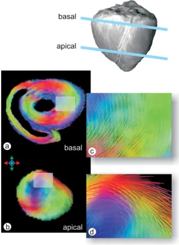
Fig. 2. Short axis (SA) slices taken at basal (a) and apical (b) levels of the ventricular mass. Insets from the basal and apical cross sections (c) and (d) exhibit the diffusion images with more detail. The green colour indicates a main diffusion in supero-inferior, red in right-left, and blue in through-plane directions. The predominant pathways of diffusion are circular.
limited to macroscopic features. In particular, the branching of individual fibres cannot be revealed by the diffusion tensor imaging.
Figs. 2–5show representative images that document the overall potential of the method, in particular its resolution and limitations. In Fig. 2, we have selected two cross-sections of the left ventricle (Fig. 2(a) and (b)), with
the thin-walled right ventricle also visible. Insets from the basal (Fig. 2(c)) and apical (Fig. 2(d)) cross-sections show the diffusion images in greater detail. The alignment of the fibres is predominantly circular course, particularly in the middle layer of the left ventricle. Some subepicardial pathways, however, are seen to spiral in an outward direction towards the epicardial surface.
Focussing on the middle layer of the left ventricle at higher magnification (Fig. 3) confirms that most of the fibres have a circular orientation. The subepicardial layers, in contrast, spiral in anticlockwise fashion from inside to outside, while the subendocardial layers take the form of individual pathways aligned in an almost radial direction.
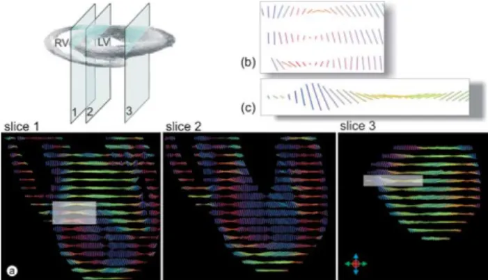
InFig. 4, we display three longitudinal sections through the left ventricle taken at various depths, starting from the mid-layers of the septum (slice 1), through the mid-portion (slice 2), and then through the inner layers of the wall near the obtuse margin (slice 3). In the second and third slices, the pathways show a continuous change in angulation with
Fig. 3. Short axis (SA) slice at basal level of the left ventricle. A magnified histological section is shown on the upper panel (a) and the corresponding measurement from diffusion tensor imaging on the lower panel (b). The colours indicate the main directions of diffusion as described inFig. 2.
Fig. 4. Long axis (LA) slices of left and right ventricle through the left ventricle at various depths (a) starting from the mid-layers of the septum (slice 1), continuing through the mid-portion (slice 2), and then through the inner layers of the wall near the obtuse margin (slice 3). In the second and third slices, with increasing distance from the epicardium, the orientation of the pathways of diffusion undergoes a continuous change in angulation with respect to an axis normal to the epicardium (b) and (c). In the third slice, the posterior free wall has been cut, exposing obliquely ascending inner pathways at its centre.
Fig. 5. All the data obtained from the diffusion tensor imaging measurements of one porcine heart has been reconstructed to show the three-dimensional fibre field. The base is seen from above (a) with the thick-walled left ventricle (LV) on top and the thin-walled right ventricle below. The reconstruction is also shown as seen from the front (b), with the right ventricle to the left hand. The overall arrangement is that of a complex three-dimensional mesh, with no evidence to support the notion that the fibres are arranged as a solitary band winding its way around both ventricular cavities.
growing distance from the epicardium when related to an axis normal to the epicardium (Fig. 4(b) and (c)). Thus, the myocardial fibres change their orientation, from possessing longitudinal alignments in the outermost and innermost layers, but adopting a more equatorial orientation in the middle layer. The pathways showing strictly equatorial alignment are located within the outer third of the ventricular wall. The third slice cuts the posterior free wall of the left ventricle, exposing the obliquely ascending pathways from the inner layer that occupy its centre. The alignment of fibres in the outer areas, however, shows a pronounced turn from the basic longitudinal arrangement as is seen in the second slice.
In Fig. 5, we show a three-dimensional reconstruction containing all the data from one porcine heart. Then overall arrangement is that of an interweaving mass of tangential and intruding fibres. No evidence is found to support the notion that the musculature is arranged in the form of a unique band taking its origin at the pulmonary trunk and coursing through the walls of both ventricles to insert at the aortic root.
4. Discussion
The technique we have described offers the possibility to go beyond anatomical imaging, and to probe the micro-structure of the myocardium at a sub-voxel level. The technique works by characterising the local mobility of water molecules in all three spatial dimensions. The process of diffusion reflects the microscopic structure of the surrounding tissue. By probing the diffusion in multiple directions, it is possible to determine a vector in direction of the largest eigenvalue of diffusion in each imaging voxel. The technique has already been validated for analysis of net-works of neural fibres in the brain[13]. In the heart, the cellular membrane of the myocytes, made up as with other cellular membranes of a lipoprotein bilayer, together with the supporting fibrous connective tissue and the contained vasculature, should similarly induce anisotropy in the diffusion of water molecules. It is this anisotropy that is recorded to characterise the orientation of the myocardial fibres. Magnetic resonance techniques have already been used to produce images of the fibre orientation of the human ventricular mass, albeit not with the precision achieved in our study[14–20].
When the architecture of the myocardium is studied by tomographic techniques, then any section taken, be it longitudinal, horizontal, radial, or tangential, can reveal only two of the dimensions of an arrangement that is known to vary throughout the overall ventricular mass. Because of this inherent geometrical fact, it follows that a comprehen-sive view of the myocardial architecture is impossible to obtain with precision using anatomical dissection. The imaging of the true spatial network, of course, could be provided by superimposition of a large number of serial sections obtained by standard histological procedures, but such exact reconstruction has thus far been difficult to achieve. Our current studies, adding to the findings from the previous investigations[14–20], show that this deficit can be
overcome by using magnetic resonance diffusion tensor imaging. The technique permits non-invasive analysis of the alignment of the myocardial fibres making up the entirety of the ventricular mass.
When considered in the setting of the overall ventricular mass, previous histological findings have shown that the architecture is dominated by the coupling and branching of the myocytes with their neighbours, not only in an end-to-end fashion, but also laterally [1,3,7,21,22]. Myocytes coupled axially in this fashion form fibre strands. The strands thus formed are alike in extent, shape, and ultrastructure. Thus, there is no-well defined beginning and ending of the contractile chains within the myocardium, unlike the situation seen in skeletal muscle. Axial coupling on the one hand, and spatial netting on the other hand, produce the histological picture of a meshwork. Previous histological studies [6] on the meshwork have demonstrated that the orientation of the fibre strands falls within a narrow cone around a mean tangential direction. The prevailing orien-tation is parallel to the epicardial and endocardial surfaces, with the angle of inclination changing from about 558 upward to about 558 downward. Not surprisingly, the data presented here supports the majority of previous observations on the architecture of the ventricular mass, albeit that those supporting the concept of the ‘unique myocardial band’ seem oblivious to the existence of these studies, making the extravagant claim that ‘previous accounts of the architec-ture of the ventricular mass are not deficient—they just do not exist as such’[8]. Our current reconstructions show, on the contrary, that not only did the previous accounts exist
[1,2,5], but they were remarkably accurate. It is the ‘unique myocardial band’[7]that does not exist, since the heart is not arranged in the form of a skeletal muscle.
Our new method, however, does far more than confirm the long-held concepts for the structure of the ventricular mass. It is likely to have the potential of detecting any marked inhomogeneities in the architecture of the ventri-cular musculature of the failing heart, features which have probably underestimated in the past because of a lack of methodology with which to detect them. Thus, our findings show that the described method is capable of reconstructing the ventricular mass so as to assess the spatial coordinates of any single fibre strand. This means that, for the first time, we will be able to assess the true orientation of the fibres, comparing them with mathematical approximations that describe ventricular mechanics and dynamics[23–25]. The technique is also likely to have clinical applications in the assessment of cardiac failure due to systematic and regional disarray of the myocardial fibres. As yet, nonetheless, our method, because of the duration of the measurements, has been confined to examinations of autopsied hearts. Its major value thus far, therefore, is to confirm that the ventricular mass is arranged in the form of a complex three-dimensional meshwork, and not as a ‘unique myocardial band’.
References
[1] Anderson RH, Becker AF. The orientation of fibres within the ventricular mass. In: Cardiac anatomy. London: Churchill Livingstone; 1980. p. 14–26.
[2] Greenbaum RA, Ho SY, Gibson DG, Becker AE, Anderson RH. Left ventricular fibre architecture in man. Br Heart J 1981;45:248–63. [3] LeGrice IJ, Smaill BH, Chai LZ, Edgar SG, Gavin JB, Hunter PJ. Laminar
structure of the heart: ventricular myocyte arrangement and connective tissue architecture in the dog. Am J Physiol 1995;269:H571–H82. [4] Lunkenheimer PP, Redmann K, Dietl KH, Cryer C, Richter KD,
Whimster WF, Niederer P. The heart’s fibre alignment assessed by comparing two digitizing systems. Methodological investigation into the inclination angle towards wall thickness. Technol Health Care 1997;5: 65–77.
[5] Sanchez-Quintana D, Garcia-Martinez V, Hurle JM. Myocardial fiber architecture in the human heart. Acta Anat 1990;138:352–8.
[6] Streeter DD. Gross morphology and fiber geometry of the heart. In: Handbook of physiology, section 2: the cardiovascular system, volume 1: the heart. Bethesda, MD: American Physiological Society; 1979 p. 61– 112.
[7] Buckberg GD. The Structure and function of the helical heart and its buttress wrapping. In: Seminars in thoracic and cardivascular surgery 2001. p. 320–332.
[8] Torrent-Guasp F, Kocica MJ, Corno AF, Carrera-Costa F. Systolic ventricular filling—reply to the letter to the editor. Eur J Cardiothorac Surg 2004;26:461–2.
[9] Basser PJ, Mattiello J, LeBihan D. MR diffusion tensor spectroscopy and imaging. Biophys J 1994;66:259–67.
[10] Pruessmann KP, Weiger M, Scheidegger MB, Boesiger P. SENSE: sensitivity encoding for fast MRI. Magn Reson Med 1999;42:952–62. [11] Jaermann T, Crelier G, Pruessmann KP, Golay X, Netsch T, van
Muiswinkel AM, Mori S, van Zijl PC, Valavanis A, Kollias S, Boesiger P. SENSE-DTI at 3 T. Magn Reson Med 2004;51:230–6.
[12] Mori S, Crain BJ, Chacko VP, van Zijl PC. Three-dimensional tracking of axonal projections in the brain by magnetic resonance imaging. Ann Neurol 1999;45:265–9.
[13] Jones DK, Simmons A, Williams SC, Horsfield MA. Non-invasive assess-ment of axonal fiber connectivity in the human brain via diffusion tensor MRI. Magn Reson Med 1999;42:37–41.
[14] Hsu EW, Muzikant AL, Matulevicius SA, Penland RC, Henriquez CS. Magnetic resonance myocardial fiber-orientation mapping with direct histological correlation. Am J Physiol 1998;274:H1627–H34.
[15] Forder JR, Bui JD, Buckley DL, Blackband SJ. MR imaging measurement of compartmental water diffusion in perfused heart slices. Am J Physiol Heart Circ Physiol 2001;281:H1280–H5.
[16] Hsu EW, Buckley DL, Bui JD, Blackband SJ, Forder JR. Two-component diffusion tensor MRI of isolated perfused hearts. Magn Reson Med 2001; 45:1039–45.
[17] Geerts L, Bovendeerd P, Nicolay K, Arts T. Characterization of the normal cardiac myofiber field in goat measured with MR-diffusion tensor imaging. Am J Physiol Heart Circ Physiol 2002;283:H139–H45. [18] Dou J, Tseng WY, Reese TG, Wedeen VJ. Combined diffusion and strain
MRI reveals structure and function of human myocardial laminar sheets in vivo. Magn Reson Med 2003;50:107–13.
[19] Forder JR, Pohost GM. Cardiovascular nuclear magnetic resonance: basic and clinical applications. J Clin Invest 2003;111:1630–9.
[20] Tseng WY, Reese TG, Weisskoff RM, Wedeen VJ. Cardiac diffusion tensor MRI in vivo without strain correction. Magn Reson Med 1999;42:393–403. [21] Grant RP. Notes on the muscular architecture of the left ventricle.
Circulation 1965;32:301–8.
[22] Cryer CW, Navidi-Kasmai H, Lunkenheimer PP, Redmann K. Computation of the alignment of myocardial contractile pathways using a magnetic tablet and an optical method. Technol Health Care 1997;5:79–94. [23] Bovendeerd PH, Huyghe JM, Arts T, van Campen DH, Reneman RS.
Influence of endocardial–epicardial crossover of muscle fibers on left ventricular wall mechanics. J Biomech 1994;27:941–51.
[24] Hunter PJ, McCulloch AD, Nielsen PMF, Smaill BH. A finite element model of passive ventricular mechanics. In: Spilker RL, Simon BR, editors. Computational methods in bioengineering. New York: ASME, BED; 1988. p. 387–97.
[25] Nielsen PM, Le Grice IJ, Smaill BH, Hunter PJ. Mathematical model of geometry and fibrous structure of the heart. Am J Physiol 1991;260: H1365–H78.
Appendix A. Editorial comment
New technology and old responsibilities
Gerald D. Buckberg*
David Geffen School of Medicine at UCLA, 10833 Le Conte Avenue, Rm 62-258 CHS, Los Angeles, CA 90095, USA Options of Bio-Engineering, California Institute of Technology, 1200 E. California Blvd., Pasadena, CA 91125, USA
The purpose of placing this report of diffusion tensor imaging into a surgical journal is not clear. This is a time-consuming study that is technically well done to permit data accumulation.The acquisitions for a complete DTI data set usually take about 9 h, yielding about 200,000 helix angle measurements from each heart. There is extensive back-ground for use of this MRI method in the literature, including histological evaluation by Scollan et al. and Chen et al.[1,2], and the central theme of each paper validates the suggestion of Streeter[3]about the oblique and helical configuration of the fiber orientation. Diffusion tensor imaging papers [4]
provide clear eigenvector pixel images of orientation angulation going from epicardium to endocardium of around C60 to K608, yet the authors do not include this helical pattern in their data base and refer to this blueprint in only one sentence in their discussion.
This paper amplifies the sustained effort from Lunkenheimer and Anderson to show that the ventricular
band concept of Torrent-Guasp is wrong. The diffusion tensor method in dead hearts simply supports the sugges-tions of Streeter about the helical formation of the ventricle. In fact, each prior MRI paper defines the angle of inclination in the dead heart as the helical angle, with clockwise and counterclockwise fibers traversing the ven-tricular wall, through 0, to define the C60 and K608 inclination angle, as well as transverse dimensions. Similar examples also appear in studies by Hsu, Forder, Geerts (cited in their references), Costa et al [5]using strain, and many others that look at the helical configuration of fibers with diffusion tensor MRI methods.
Unfortunately, this concept of helical inclination is completely missing from their results, with a description focus that is directed toward the circular middle portion. Architecture has a structure function counterpart, and the recognized oblique fiber orientation is consistent with the twisting action of torsion that is very evident by tagged MRI studies [6,7]. The authors focus upon the transverse orientation supports the constriction and dilation described by William Harvey[8], who also viewed the dead heart, and thus could not recognize the twisting elements of function that are so visible in the experimental laboratory and in the operating room in conical hearts with normal excitation contraction coupling. How does transverse architecture explain the twisting function, and should not the recognized but excluded oblique pattern also be cited to promote better understanding of living action?