M A J O R A R T I C L E
Prolonged Outbreak of
Mycobacterium chimaera
Infection After Open-Chest Heart Surgery
Hugo Sax,1,aGuido Bloemberg,2,aBarbara Hasse,1,aRami Sommerstein,1Philipp Kohler,1Yvonne Achermann,1 Matthias Rössle,3Volkmar Falk,4Stefan P. Kuster,1Erik C. Böttger,2,band Rainer Weber1,b1Division of Infectious Diseases and Hospital Epidemiology, University Hospital Zurich,2Institute of Medical Microbiology, National Centre for
Mycobacteria, University of Zurich,3Institute of Surgical Pathology, and4Division of Cardiac Surgery, University Hospital Zurich, Switzerland
Background. Invasive Mycobacterium chimaera infections were diagnosed in 2012 in 2 heart surgery patients on extracorporeal circulation. We launched an outbreak investigation to identify the source and extent of the potential outbreak and to implement preventive measures.
Methods. We collected water samples from operating theaters, intensive care units, and wards, including air samples from operating theaters.Mycobacterium chimaera strains were characterized by randomly amplified poly-morphic DNA polymerase chain reaction (RAPD-PCR). Case detection was performed based on archived histopa-thology samples andM. chimaera isolates since 2006, and the patient population at risk was prospectively surveyed. Results. We identified 6 male patients aged between 49 and 64 years with prosthetic valve endocarditis or vas-cular graft infection due toM. chimaera, which became clinically manifest with a latency of between 1.5 and 3.6 years after surgery.Mycobacterium chimaera was isolated from cardiac tissue specimens, blood cultures, or other biopsy specimens. We were able also to cultureM. chimaera from water circuits of heater-cooler units connected to the cardiopulmonary bypass, and air samples collected when the units were in use. RAPD-PCR demonstrated identical patterns amongM. chimaera strains from heater-cooler unit water circuits and air samples, and strains in 2 patient clusters.
Conclusions. The epidemiological and microbiological features of this prolonged outbreak provided evidence for the airborne transmission ofM. chimaera from contaminated heater-cooler unit water tanks to patients during open-heart surgery.
Keywords. outbreak; Mycobacterium chimaera; nontuberculous mycobacteria; open-chest heart surgery; infection control.
Several outbreaks of surgical site infections with nontu-berculous mycobacteria following heart surgery have been described [1–8]. However, the etiology concerned exclusively fast-growing nontuberculous mycobacteria, such asMycobacterium fortuitum, Mycobacterium che-lonae, and Mycobacterium wolinskyi. Source identifica-tion failed in most cases. One outbreak with 6 cases of
wound infections or endocarditis due to fast-growing mycobacteria was putatively linked to contaminated water used for the cooling of cardioplegia solution [4].Mycobacterium chelonae was involved in prosthetic valve endocarditis due to implant contamination dur-ing the manufacturdur-ing process [6]. An outbreak with M. fortuitum in 3 patients was due to a single con-taminated patch used for septum defect repair and di-vided among 8 children [7]. Recently, an outbreak of M. wolinskyi occurred at a single center where heater-cooler units were contaminated with other mycobacte-ria [8]. Mycobacterium chimaera was distinguished within theMycobacterium avium complex (MAC) as a novel species in 2004 and is considered to be a human pathogen, mainly in cystic fibrosis patients with pulmonary infections [9,10]. Accurate identifica-tion ofM. chimaera requires a molecular diagnostic
Received 7 December 2014; accepted 26 February 2015; electronically published 11 March 2015.
a
H. S., G. B., and B. H. contributed equally to this work.
b
E. C. B. and R. W. contributed equally to this work.
Correspondence: Hugo Sax, MD, HAL 14, Division of Infectious Diseases and Hospital Epidemiology, University Hospital Zurich, Raemistrasse 100, 8091 Zurich, Switzerland (hugo.sax@usz.ch).
Clinical Infectious Diseases® 2015;61(1):67–75
© The Author 2015. Published by Oxford University Press on behalf of the Infectious Diseases Society of America. All rights reserved. For Permissions, please e-mail: journals.permissions@oup.com.
DOI: 10.1093/cid/civ198
laboratory for 16S ribosomal RNA (rRNA) gene sequencing and homology analysis. Recently, we described extrapulmonary infections withM. chimaera involving 2 cases of prosthetic valve endocarditis and bloodstream infection after open-chest heart surgery concerning an annuloplasty ring and an artificial heart valve implant, respectively [11].
In October 2012, the infection control team at our institution was notified of 2 cases of invasive M. chimaera infection [11]. Due to identical randomly amplified polymorphic DNA poly-merase chain reaction (RAPD-PCR) patterns, a point source at the hospital seemed probable. We describe the in-depth out-break investigation to detect the source, including retrospective case detection, prospective surveillance, on-site observations, and targeted microbiological sampling of patients and the hos-pital environment.
METHODS
Setting
The University Hospital of Zurich is an 870-bed tertiary care cen-ter. The Zurich Heart Centre is an entity within the hospital where approximately 1400 patients undergo cardiovascular sur-gery annually, including coronary artery bypass graft procedures, valve replacement and repair, aortic surgery, placement of im-plantable cardiac devices, and heart transplantation. Of these, ap-proximately 600 procedures require extracorporeal circulation. Surgery is performed in 3 adjacent operating rooms and a sepa-rate hybrid suite. Postoperatively, patients are transferred to the nearby cardiovascular intensive care unit (ICU) and then to sur-gical wards located in the same building, constructed in 1953.
Retrospective and Prospective Case Detection
A case was defined as a patient with proven invasive M. chimaera infection following open-chest heart surgery performed at the hospital since August 2006. In addition to the American Tho-racic Society statement on diagnosis, treatment, and prevention of nontuberculous mycobacterial diseases that require positive blood or bone marrow cultures, our definition of invasive non-tuberculous mycobacteria disease included cultures or molecu-lar methods positive for M. chimaera in heart valves with histopathological signs of infection [12].
For the purpose of retrospective case detection, allM. chi-maera isolates recovered by culture since 2006 at the University of Zurich Institute of Medical Microbiology were reviewed. Available tissue samples from heart surgery patients with culture-negative endocarditis were reexamined for evidence of infection with nontuberculous mycobacteria. Prospectively, in addition to microbiological surveillance, we maintained a high level of clinical alert to detect new cases through the infec-tious disease consultation service by daily participation in clin-ical rounds at the ICU, active involvement in any infectious
disease issues following heart surgery, and daily rounds at the Institute of Medical Microbiology diagnostic laboratory.
Observations
We repeatedly observed activity in anesthesia induction rooms, cardiovascular operating rooms, ICUs, and wards. A valve re-placement intervention was videotaped in its entirety for offline analysis. Considering the characteristics ofM. chimaera, we fo-cused on procedures involving water both inside and outside the operating rooms.
Microbiology
Water and air samples were collected at different times and locations in the operating rooms, ICUs, and wards. Microbio-logical techniques used in this study for the detection and iden-tification of nontuberculous mycobacteria have been described previously [11]. Sodium hydroxide was used for decontamina-tion of specimens from sterile sites andN-acetyl-L -cysteine-sodium hydroxide was used for respiratory and environmental samples. Mycobacteria were cultured by standard methods using the mycobacteria growth indicator tube (MGIT) 960 system (Becton Dickinson Microbiology Systems, Sparks, Maryland) or Middlebrook 7H11 agar plates incubated at 37°C for 7 weeks or until positive.
One-liter water samples werefiltered and plated on Middle-brook 7H11 agar; samples of 50 mL were then centrifuged and cultured using the MGIT 960 system. Environmental swabs were rinsed with sterile water to recuperate bacterial cells. The resulting suspension was decontaminated before being used for culturing. Bioaerosol sampling with culture was performed using Middlebrook 7H11 agar plates inserted in an air sampler (MAS-100 NT; MBV, Staefa, Switzerland) run-ning for 2.5 minutes at a rate of 100 L/minute. The 16S rRNA gene sequencing was performed as described previously [13]. Sequences were analyzed using the SmartGene IDNS software and databases (SmartGene, Zug, Switzerland). RAPD-PCR with chromosomal DNA and primers IS986-FP and OPA18 was used for genotyping as described previously [11,14]. Antimi-crobial susceptibility testing of theM. chimaera patients’ isolates was performed in the MGIT 960 system equipped with the Tuberculosis Exist module [15] for rifampin, rifabutin, amikacin, ofloxacin, moxifloxacin, clarithromycin, and ethambutol.
Outbreak Management and Ethics Approval
We established an interdisciplinary panel of clinicians, micro-biologists, technicians, and hospital administrators at the hospital and the Institute of Medical Microbiology to optimize knowledge and risk management. Case patients were seen at the hospital as inpatients or outpatients. The independent ethics committee of the Canton of Zurich waived the necessity for formal submission due to the quality assurance nature of the investigation.
RESULTS
Retrospective histopathological review of the available tissue sam-ples of 10 patients from our hospital with culture-negative endo-carditis revealed no further case with evidence of infection with nontuberculous mycobacteria. Among the 28 patients with posi-tiveM. chimaera cultures isolated at the Institute of Medical Mi-crobiology since 2006, 6 were current outbreak cases, 6 had invasive pulmonary disease, and 16 were colonized according to established definitions for infections with nontuberculous myco-bacteria [12]. Eight randomly selectedM. chimaera patient isolates not included among our cases showed each individual nonmatch-ing RAPD-PCR patterns. A convenience sample of perioperative blood cultures of 32 patients collected during the outbreak period remained negative for nontuberculous mycobacteria.
Cases
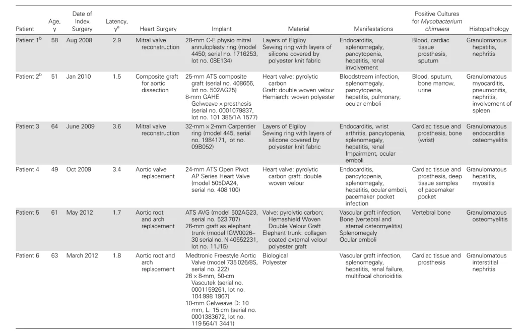
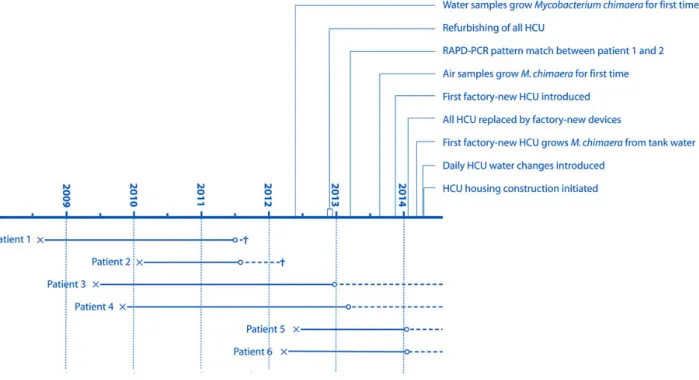
A total of 6 male patients withM. chimaera met our case defi-nition. Patient characteristics are shown in Table1. All had un-dergone open-chest heart surgery involving implants at the University Hospital of Zurich between 2008 and 2012. Latency between surgery and manifest infection ranged between 1.5 and 3.6 years (Figure1). In all cases except 1 patient with sustained M. chimaera bacteremia, the cardiac implant showed echocar-diographic signs of endocarditis. Accompanying symptoms, signs, and disorders were fatigue, fever, hepatitis, renal insuf fi-ciency, splenomegaly, and pancytopenia. Host defense was compromised in 2 patients. One patient had lowered CD4 cell counts for no obvious reason, and 1 had received steroid med-ication for a supposed granulomatous autoimmune disease. As prosthetic valves and aortic grafts differed in type, manufactur-er, and lot, a production-related contamination cause appeared highly unlikely (Table 1). No potential common exposure sources outside the hospital could be identified among patients. RAPD-PCR analysis of isolates of the 6 cases repeatedly re-vealed 2 clusters with similar patterns involving 2 patients and 3 patients each (Figure2). The isolate of 1 patient had a unique RAPD-PCR pattern.
Targeted antibiotic therapy consisted of a prolonged combi-nation of clarithromycin, rifabutin, and ethambutol, combined with either amikacin or moxifloxacin. Three patients experi-enced breakthrough infections with splenic embolus, pacemaker pocket infection, and progressive mitral valve endocarditis, re-spectively. Three patients underwent valve replacement surgery; 1 patient required repeated surgical debridement. Two other patients died, despite antibiotic treatment [11].
Observations
We identified 2 sources of stagnant nonsterile water in the op-erating room consisting of a heater-cooler unit connected to the extracorporeal circuit, and a separate device providing warm
water to patient warming blankets. Patients received water from a drinking water fountain in the ICU, and they were ex-posed to showers and tap water onfloor wards. Additional water used in the operating room was sterile.
During open-chest heart surgery, heater-cooler units are gen-erally used for the dual purpose of warming patients and cool-ing of the cardioplegia solution [16]. These are stand-alone units on wheels placed inside the operating room with reser-voirs of tap water as a thermic transfer medium. The heater-cooler model used in the investigated period consists of a water tank with different compartments containing heating and cooling aggregates, all housed in a stainless steel case. Water from a 6-liter tank is pumped through a silicone tube to the heart–lung machine where it runs through the water phase of a single-use heat exchanger against the blood phase, separated by highly heat-conductive material. A second circuit, designed to feed a warming blanket, was never used. A third cir-cuit delivers cold water from two 3-liter tanks to the single-use heat exchange unit for the cardioplegia solution. Temperature in the tanks may range between 2°C and 41°C during operation, returning to room temperature on standby. The tanks are not airtight due to overflow tubes, and multiple probes and stirring devices are inserted in their roof. Below the tank, a fan forces air through a heat exchanger for cooling. Heater-cooler units are usually located 1–2 m from the surgical sterile field.
Heater-cooler unit employment and water changes were not documented in the past. In 2012, the manufacturer (Sorin Group, Milan, Italy) issued a maintenance protocol. Recom-mendations were to change the water every second week using a bacteriafilter with 0.2-µm pores, initially adding 100 mL of 3% w/v hydrogen peroxide as disinfectant, then 50 mL every 5 days. Every 3 months, a 15-minute extra disinfection cycle had to be run by adding 200 mL of 5% w/v Clorox Regular Bleach. The manual states to use decalcified water exclusively. During the operation, water tanks must be refilled occasionally. Spilling is common. The 2 single-use heat exchangers are tested for airtightness during assembly of the heart–lung machine by applying 250 mm Hg of pressure during 30 seconds.
The second nonsterile water source in the operating room concerned a body temperature regulation system (Blanketrol III, Cincinnati Sub-Zero Medical, Cincinnati, Ohio), a stand-alone device featuring a water tank with tubes feeding warm water to a blanket placed beneath the patient during surgery.
Sterile conditioned water is used to rinse the surgical site. In addition, we identified no risk of contamination related to han-dling or storage of prostheses at the hospital.
Environmental Cultures
Mycobacterium chimaera was cultured from 5 heater-cooler units and characterized by RAPD-PCR. Similar RAPD-PCR patterns were observed forM. chimaera isolates from heater-cooler unit
Table 1. Characteristics of Cases WithMycobacterium chimaera Infection After Open-Chest Heart Surgery Patient Age, y Date of Index Surgery Latency,
ya Heart Surgery Implant Material Manifestations
Positive Cultures for Mycobacterium
chimaera Histopathology
Patient 1b 58 Aug 2008 2.9 Mitral valve
reconstruction
28-mm C-E physio mitral annuloplasty ring (model 4450; serial no. 1716253, lot no. 08E134)
Layers of Elgiloy
Sewing ring with layers of silicone covered by polyester knit fabric
Endocarditis, splenomegaly, pancytopenia, hepatitis, renal involvement Blood, cardiac tissue prosthesis, sputum Granulomatous hepatitis, nephritis
Patient 2b 51 Jan 2010 1.5 Composite graft
for aortic dissection
25-mm ATS composite graft (serial no. 408656, lot no. 502AG25) 8-mm GAHE
Gelweave × prosthesis (serial no. 0001079837, lot no. 101 385/1A 1577)
Heart valve: pyrolytic carbon
Graft: double woven velour Hemiarch: woven polyester
Bloodstream infection, splenomegaly, pancytopenia, hepatitis, pulmonary, ocular emboli Blood, sputum, bone marrow, urine Granulomatous myocarditis, pneumonitis, nephritis, involvement of spleen
Patient 3 64 June 2009 3.6 Mitral valve
reconstruction
32-mm × 2-mm Carpentier ring (model 445, serial no. 1984171, lot no. 09B052)
Layers of Elgiloy
Sewing ring with layers of silicone covered by polyester knit fabric
Endocarditis, wrist arthritis, pancytopenia, splenomegaly, hepatitis, renal Impairment, ocular emboli
Cardiac tissue and prosthesis, bone (wrist)
Granulomatous endocarditis osteomyelitis
Patient 4 49 Oct 2009 3.4 Aortic valve
replacement
24-mm ATS Open Pivot AP Series Heart Valve (model 505DA24, serial no. 408 100)
Heart valve: pyrolytic carbon graft: double woven velour
Endocarditis, pancytopenia, splenomegaly, hepatitis, ocular emboli, pacemaker pocket infection
Cardiac tissue and prosthesis, deep tissue samples of pacemaker pocket Granulomatous hepatitis, myositis
Patient 5 61 May 2012 1.7 Aortic root
and arch replacement
ATS AVG (model 502AG23, serial no. 523 707) 26-mm graft as elephant
trunk (model IGW0026– 30 serial no. N 40552231, lot no. 11J15)
Valve: pyrolytic carbon; Hemashield Woven Double Velour Graft Elephant trunk: collagen
coated external velour polyester graft
Vascular graft infection, Bone (vertebral and
sternal osteomyelitis) Splenomegaly Ocular emboli
Vertebral bone Granulomatous osteomyelitis
Patient 6 63 March 2012 1.8 Aortic root and
arch replacement
Medtronic Freestyle Aortic Valve (model 735 026/8S, serial no. 222)
26 × 8-mm, 50-cm Vascutek (serial no. 0001159261, lot no. 104 998 1967) 10-mm Gelweave D: 10 mm, L: 15 cm (serial no. 0001383672, lot no. 119 564/1 3441) Biological Polyester
Vascular graft infection, splenomegaly, hepatitis, renal failure, multifocal chorioiditis
Cardiac tissue and prosthesis
Granulomatous interstitial nephritis
a
Latency between open-chest heart surgery and diagnosis of M. chimaera infection.
70
•
CID 2015:61 (1 July)•
Sax et alno. 1 and an air sample associated with the same heater-cooler unit (Figure2). Different RAPD-PCR patterns were observed for M. chimaera isolates sampled from the different heater-cooler units (Figure 2), indicating a wide diversity ofM. chimaera strains in the different units. An exact match of RAPD-PCR
patterns betweenM. chimaera strains from the environment andM. chimaera strains isolated from infected patients could not be established (Figure2). Ten cultures from warming blan-ket devices remained negative (Supplementary Table). All drinking water fountains in the hospital ICUs tested positive
Figure 1. Evolution of the 6 cases ofMycobacterium chimaera infection and investigational activity. Abbreviations: x, open-chest heart surgery;○,M. chi-maera diagnosis; - - -, antibiotic and, in some cases surgical, treatment; +, fatality; HCU, heater-cooler unit; RAPD-PCR, randomly amplified polymorphic DNA polymerase chain reaction.
Figure 2. Mycobacterium chimaera strain typing using randomly amplified polymorphic DNA polymerase chain reaction (RAPD-PCR). A and B, RAPD-PCR patterns usingM. chimaera chromosomal DNA were generated with primer IS986-FP (A) and primer OPA18 (B). Lanes 1–6, M. chimaera clinical isolates from patients 1, 2, 4, 3, 5, and 6 as referred to in Table1. Lanes 7–15, Environmental M. chimaera culture isolates obtained from heater-cooler units and air sampling. Lane 7, Water tank of heater-cooler unit no. 1. Lane 8, Water from overflow tube heater-cooler unit no. 1. Lane 9, Air sampling operation room ventilation block while heater-cooler unit no. 1 was running. Lane 10, Air sampling <1 m before running heater-cooler unit no. 1. Lane 11, Water of patient circuit heater-cooler unit no. 3. Lane 12, Water from cardioplegia circuit from heater-cooler unit no. 3. Lane 13, Air sampling operation room >1 m before running heater-cooler unit no. 3. Lane 14, Water of patient circuit heater-cooler unit no. 6. Lane 15, Water of cardioplegia circuit heater-cooler unit no. 6.
forM. chimaera, whereas all samples taken from taps in the op-erating room, ICUs, and surgical wards remained negative (Figure 3; Supplementary Table). Water from showers on wards also tested negative.
Preventive Measures
In parallel with accumulating evidence for the transmission pathway, preventive actions and notification to the national reg-ulatory bodies and the manufacturer of the heater-cooler units were undertaken to minimize the infectious risk of patients un-dergoing open-chest heart surgery at the Zurich Heart Centre and elsewhere (Figure1). We use only factory-new, heater-cooler units with daily water changes over 0.2-µm bacteriafilters and
perform regular water and air surveillance cultures. Since March 2014, heater-cooler unit water and operating room air samples have remained negative forM. chimaera. However, in Septem-ber 2014, it grew again from 1 heater-cooler unit sample. The construction of custom-built containers with high-efficiency particulate airfilters to house heater-cooler units that cannot be placed outside the operating room is now under way.
DISCUSSION
We have linked a cluster of 6 cases of invasive infection with M. chimaera after cardiac surgery to a source of contaminated
Figure 3. Situational schema of the microbiologic environmental investigation results. Schematic positive (red plus signs) and negative (green negative signs)Mycobacterium chimaera results from environmental investigation. In the cardiovascular operating room, situations 1–3 show different configurations of a heater-cooler unit running (ON) or switched off (OFF) and categorical results from heater-cooler unit tank water, air close to heater-cooler units, and air in ventilation exhaust, testing positive or negative forM. chimaera.
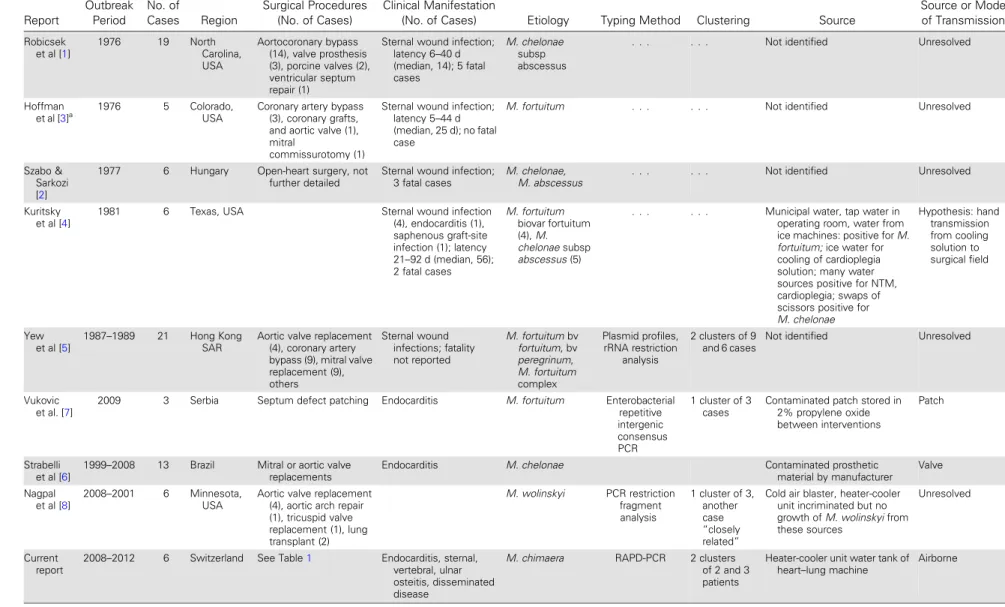
Table 2. Reported Outbreaks of Nontuberculous Mycobacteria in Open-Chest Heart Surgery, 1976–2014 Report Outbreak Period No. of Cases Region Surgical Procedures (No. of Cases) Clinical Manifestation
(No. of Cases) Etiology Typing Method Clustering Source
Source or Mode of Transmission Robicsek et al [1] 1976 19 North Carolina, USA Aortocoronary bypass (14), valve prosthesis (3), porcine valves (2), ventricular septum repair (1)
Sternal wound infection; latency 6–40 d (median, 14); 5 fatal cases M. chelonae subsp abscessus
. . . Not identified Unresolved
Hoffman
et al [3]a 1976 5 Colorado,
USA
Coronary artery bypass (3), coronary grafts, and aortic valve (1), mitral
commissurotomy (1)
Sternal wound infection; latency 5–44 d (median, 25 d); no fatal case
M. fortuitum . . . Not identified Unresolved
Szabo & Sarkozi [2]
1977 6 Hungary Open-heart surgery, not
further detailed
Sternal wound infection; 3 fatal cases
M. chelonae,
M. abscessus . . . Not identified Unresolved
Kuritsky et al [4]
1981 6 Texas, USA Sternal wound infection
(4), endocarditis (1), saphenous graft-site infection (1); latency 21–92 d (median, 56); 2 fatal cases M. fortuitum biovar fortuitum (4), M. chelonae subsp abscessus (5)
. . . Municipal water, tap water in
operating room, water from ice machines: positive for M. fortuitum; ice water for cooling of cardioplegia solution; many water sources positive for NTM, cardioplegia; swaps of scissors positive for M. chelonae Hypothesis: hand transmission from cooling solution to surgical field Yew et al [5] 1987–1989 21 Hong Kong SAR
Aortic valve replacement (4), coronary artery bypass (9), mitral valve replacement (9), others Sternal wound infections; fatality not reported M. fortuitum bv fortuitum, bv peregrinum, M. fortuitum complex Plasmid profiles, rRNA restriction analysis 2 clusters of 9 and 6 cases
Not identified Unresolved
Vukovic et al. [7]
2009 3 Serbia Septum defect patching Endocarditis M. fortuitum Enterobacterial
repetitive intergenic consensus PCR 1 cluster of 3 cases
Contaminated patch stored in 2% propylene oxide between interventions
Patch
Strabelli et al [6]
1999–2008 13 Brazil Mitral or aortic valve
replacements
Endocarditis M. chelonae Contaminated prosthetic
material by manufacturer Valve Nagpal et al [8] 2008–2001 6 Minnesota, USA
Aortic valve replacement (4), aortic arch repair (1), tricuspid valve replacement (1), lung transplant (2) M. wolinskyi PCR restriction fragment analysis 1 cluster of 3, another case “closely related”
Cold air blaster, heater-cooler unit incriminated but no growth of M. wolinskyi from these sources
Unresolved
Current report
2008–2012 6 Switzerland See Table1 Endocarditis, sternal,
vertebral, ulnar osteitis, disseminated disease
M. chimaera RAPD-PCR 2 clusters
of 2 and 3 patients
Heater-cooler unit water tank of
heart–lung machine Airborne
Abbreviations: NTM, nontuberculous mycobacteria; PCR, polymerase chain reaction; RAPD, randomly amplified polymorphic DNA; rRNA, ribosomal RNA; SAR, Special Administrative Region. a
This publication reports again the outbreak already described in reference [1].
M. chimaer a Outbr eak in Heart Surger y
•
CID 2015:61 (1 July)•
73water in heater-cooler units connected to the heart–lung ma-chine. Outbreaks with fast-growing nontuberculous mycobacte-ria in cardiac surgery have been reported previously, but remained mostly without source identification (Table2). In this study, a distinct source and an airborne transmission route have been established with high plausibility. Heater-cooler units are universally used in open-heart surgery. However, as the clinical manifestations associated with nontuberculous my-cobacteria are delayed and insidious, it is possible that similar problems may have remained undetected in other institutions. Thus, it is likely that more cases may become manifest in years to come, despite effective control measures, similar to the re-ported large outbreak of slow-growingMycobacterium xenopi following spinal surgery [17].
Mycobacterium chimaera has been isolated from water systems at the home of patients with MAC lung disease [18]. In general, nontuberculous mycobacteria, both of the fast- and slow-growing type, readily colonize the hospital environment [19–23]. They are linked mostly to humidity, but have been isolated also from air samples [3, 24–26] and environmental swabs in operating rooms [3]. Resistance of nontuberculous mycobacteria against frequently used disinfectants, such as chlorines and ozone, facil-itates their persistence in water systems [27], aided also by biofilm formation. In addition, MAC members have been shown to pref-erentially colonize warm water sources, which might explain at least in part their propensity to aerosolize [28,29].
We hypothesized that the current outbreak was due to the presence ofM. chimaera in the hospital water system, which sub-sequently contaminated heater-cooler unit water tanks. Due to advantageous conditions in these devices, mycobacteria probably multiplied and formed biofilms. During operation, mycobacteria became dispersed from the heater-cooler units into the air of the operating room, thereby causing infection. This transmission hy-pothesis is supported by severalfindings. Air sampling cultures became positive only when a heater-cooler unit was running, but not when it was turned off. Air samples taken early in the course of a surgical intervention consistently grew a lower num-ber of colony-forming units than those taken later (data not shown). Furthermore, some strains isolated from air and water samples showed matching RAPD-PCR patterns. Factory-new, heater-cooler units initially never grewM. chimaera, but one factory-new device became colonized after 3 months’ use, despite maintenance according to the manufacturer’s instructions.
The transmission hypothesis is challenged by our inability to demonstrate exactly matching RAPD-PCR patterns between en-vironmental and patient samples. However, this can easily be ex-plained by several reasons, the most important being the long time lag between patient exposure and sampling of heater-cooler units and the wide diversity of strains found in the heater-cooler unit water tanks. In addition, no records of heater-cooler unit use according to each individual surgical intervention were available.
We assume that transmission ofM. chimaera was via aerosoli-zation from the water tanks. The water inside the tank is kept in motion by stirring devices, thus producing bubbles known to aerosolize fast- and slow-growing nontuberculous mycobacteria [30]. Alternatively, droplets from tubes or connections could have reached the turbulent airflow produced by the fan of the heat exchanger in the lower part of the heater-cooler unit. Airborne translocation of viable nontuberculous mycobacteria over a distance of several meters has been documented in an outbreak ofMycobacterium abscessus/chelonae in metal-workers [31].
Water samples from operating room taps, including those used to refill the heater-cooler unit water tanks, and taps in the ICU and wards remained negative forM. chimaera. However, positiveM. chimaera cultures from water fountains connected to the hospital water system in several different locations in the ICUs suggest a pathogen of hospital origin, or even from the municipal water system.
Two alternative transmission routes are possible. First, pa-tient blood may have been contaminated by a leakage in the membrane in one of the heat exchange units, which would clas-sify the positive air samples as an epiphenomenon. Although such a transmission route has been demonstrated before [32], it is improbable here because perioperative blood cultures of a series of patients remained negative. Second, infection outside the operating room, such as by ingestion of contaminated water or aerosols in showers, is equally unlikely. Showers tested negative, and drinking water is an unlikely infectious route for invasive infections.
In conclusion, these results show that a slow-growing nontu-berculous mycobacterium,M. chimaera, was the cause of a pro-longed healthcare-associated outbreak of invasive infection in patients who underwent open-chest heart surgery. The out-break could be traced to heater-cooler unit reservoirs and an airborne transmission pathway with a high degree of certainty. Ourfindings suggest that heater-cooler units should be regard-ed as a potential source of bacterial infections in cardiac surgery as argued in the past [33]. In the framework of a Swiss govern-ment initiative, several other hospitals have found their heater-cooler units to be growingM. chimaera. In one case, this was linked also to positive air cultures. It remains to be investigated how widespread this risk is for patient safety and what consti-tutes the most effective measures for its prevention.
Supplementary Data
Supplementary materialsare available atClinical Infectious Diseases online (http://cid.oxfordjournals.org). Supplementary materials consist of data provided by the author that are published to benefit the reader. The posted materials are not copyedited. The contents of all supplementary data are the sole responsibility of the authors. Questions or messages regarding errors should be addressed to the author.
Notes
Acknowledgments. We would like to acknowledge the contribution of the many collaborators of University Hospital Zurich to this investigation. We are indebted to Rosemary Sudan for her editorial assistance.
Author contributions. H. S. supervised the outbreak investigation, and drafted andfinalized the manuscript. G. B. performed microbiological investigations. B. H. led case detection and clinical investigations in case patients. R. S., P. K., Y. A., and S. P. K. performed the outbreak investiga-tions. M. R. undertook histopathological investigainvestiga-tions. V. F. contributed to the surgical evaluation of case patients and surgical procedures. E. C. B. su-pervised the microbiological investigations. R. W. susu-pervised the outbreak and clinical investigations. All authors contributed to data interpretation and preparation of the manuscript.
Potential conflicts of interest. All authors: No potential conflicts of interest.
All authors have submitted the ICMJE Form for Disclosure of Potential Conflicts of Interest. Conflicts that the editors consider relevant to the con-tent of the manuscript have been disclosed.
References
1. Robicsek F, Daugherty HK, Cook JW, et al.Mycobacterium fortuitum epidemics after open-heart surgery. J Thorac Cardiovasc Surg1978; 75:91–6.
2. Szabo I, Sarkozi K.Mycobacterium chelonei endemy after heart surgery with fatal consequences. Am Rev Resp Dis1980; 121:607.
3. Hoffman PC, Fraser DW, Robicsek F, O’Bar PR, Mauney CU. Two out-breaks of sternal wound infection due to organisms of the Mycobacte-rium fortuitum complex. J Infect Dis 1981; 143:533–42.
4. Kuritsky JN, Bullen MG, Broome CV, Silcox VA, Good RC, Wallace RJ. Sternal wound infections and endocarditis due to organisms of the My-cobacterium fortuitum complex. Ann Intern Med 1983; 98:938–9. 5. Yew WW, Wong PC, Woo HS, Yip CW, Chan CY, Cheng FB.
Charac-terization of Mycobacterium fortuitum isolates from sternotomy wounds by antimicrobial susceptibilities, plasmid profiles, and ribosom-al ribonucleic acid gene restriction patterns. Diagn Microbiol Infect Dis 1993; 17:111–7.
6. Strabelli TMV, Siciliano RF, Castelli JB, et al.Mycobacterium chelonae valve endocarditis resulting from contaminated biological prostheses. J Infect2010; 60:467–73.
7. Vukovic D, Parezanovic V, Savic B, et al.Mycobacterium fortuitum en-docarditis associated with cardiac surgery, Serbia. Emerg Infect Dis 2013; 19:517–9.
8. Nagpal A, Wentink JE, Berbari EF, et al. A cluster ofMycobacterium wolinskyi surgical site infections at an academic medical center. Infect Control Hosp Epidemiol2014; 35:1169–75.
9. Tortoli E, Rindi L, Garcia MJ, et al. Proposal to elevate the genetic var-iant MAC-A, included in theMycobacterium avium complex, to species rank asMycobacterium chimaera sp. nov. Int J Syst Evol Microbiol 2004; 54:1277–85.
10. Cohen-Bacrie S, David M, Stremler N, Dubus J-C, Rolain J-M, Drancourt M.Mycobacterium chimaera pulmonary infection compli-cating cysticfibrosis: a case report. J Med Case Rep 2011; 5:473. 11. Achermann Y, Rossle M, Hoffmann M, et al. Prosthetic valve
endocar-ditis and bloodstream infection due toMycobacterium chimaera. J Clin Microbiol2013; 51:1769–73.
12. Griffith DE, Aksamit T, Brown-Elliott BA, et al. An official ATS/IDSA statement: diagnosis, treatment, and prevention of nontuberculous my-cobacterial diseases. Am J Respir Crit Care Med2007; 175:367–416. 13. Bosshard PP, Zbinden R, Böddinghaus B, Altwegg M, Böttger EC. 16S
rRNA gene sequencing versus the API 20 NE system and the VITEK 2 ID-GNB card for identification of nonfermenting gram-negative bacte-ria in the clinical laboratory. J Clin Microbiol2006; 44:1359–66.
14. Zhang Y, Rajagopalan M, Brown BA, Wallace RJJ. Randomly amplified polymorphic DNA PCR for comparison ofMycobacterium abscessus strains from nosocomial outbreaks. J Clin Microbiol1997; 35:3132–9. 15. Hombach M, Somoskovi A, Hömke R, Ritter C, Böttger EC. Drug
susceptibility distributions in slowly growing non-tuberculous myco-bacteria using MGIT 960 TB eXiST. Int J Med Microbiol2013; 303: 270–6.
16. High KM, Bashein G, Kurusz M. Principles of oxygenator function: gas exchange, heat transfer, and operation. In: Gravlee GP, Davis RF, Kurusz M, Utley JR, eds. Cardiopulmonary bypass principles and practice. Phil-adelphia, PA: Wolter Kluwer Health,2008:47–62.
17. Astagneau P, Desplaces N, Vincent V, et al.Mycobacterium xenopi spi-nal infections after discovertebral surgery: investigation and screening of a large outbreak. Lancet2001; 358:747–51.
18. Wallace RJJ, Iakhiaeva E, Williams MD, et al. Absence of Mycobacteri-um intracellulare and presence of MycobacteriMycobacteri-um chimaera in house-hold water and biofilm samples of patients in the United States with Mycobacterium avium complex respiratory disease. J Clin Microbiol 2013; 51:1747–52.
19. Chang C-T, Wang L-Y, Liao C-Y, Huang S-P. Identification of nontu-berculous mycobacteria existing in tap water by PCR-restriction frag-ment length polymorphism. Appl Environ Microbiol2002; 68:3159–61. 20. Shin JH, Lee EJ, Lee HR, et al. Prevalence of non-tuberculous
mycobac-teria in a hospital environment. J Hosp Infect2007; 65:143–8. 21. Sahly El HM, Septimus E, Soini H, et al.Mycobacterium simiae
pseudo-outbreak resulting from a contaminated hospital water supply in Hous-ton, Texas. Clin Infect Dis2002; 35:802–7.
22. Galassi L, Donato R, Tortoli E, Santianni D, Del R. Nontuberculous mycobacteria in hospital water systems: application of HPLC for iden-tification of environmental mycobacteria. J Water Health 2003; 1: 133–9.
23. Williams MM, Armbruster CR, Arduino MJ. Plumbing of hospital pre-mises is a reservoir for opportunistically pathogenic microorganisms: a review. Biofouling2013; 29:147–62.
24. Glazer CS, Martyny JW, Lee B, et al. Nontuberculous mycobacteria in aerosol droplets and bulk water samples from therapy pools and hot tubs. J Occup Environ Hyg2007; 4:831–40.
25. Falkinham JO 3rd. Mycobacterial aerosols and respiratory disease. Emerg Infect Dis2003; 9:763–7.
26. Falkinham JO 3rd, Iseman MD, de Haas P, van Soolingen D. Mycobac-terium avium in a shower linked to pulmonary disease. J Water Health 2008; 6:209–13.
27. Taylor RH, Falkinham JO 3rd, Norton CD, Le Chevallier MW. Chlo-rine, chloramine, chlorine dioxide, and ozone susceptibility of Mycobac-terium avium. Appl Environ Microbiol 2000; 66:1702–5.
28. Moulin du GC, Stottmeier KD, Pelletier PA, Tsang AY, Hedley-Whyte J. Concentration ofMycobacterium avium by hospital hot water systems. JAMA1988; 260:1599–601.
29. Parker BC, Ford MA, Gruft H, Falkinham JO. Epidemiology of infection by nontuberculous mycobacteria. IV. Preferential aerosolization of Mycobacterium intracellulare from natural waters. Am Rev Respir Dis 1983; 128:652–6.
30. Falkinham JO 3rd. Factors influencing the aerosolization of mycobacteria. In: Monahan EC, Van Patten MA, eds. The climate and health implica-tions of bubble-mediated sea-air exchange. Groton, CT:1989:17–25. 31. Centers for Disease Control and Prevention (CDC). Respiratory illness
in workers exposed to metalworkingfluid contaminated with nontuber-culous mycobacteria—Ohio, 2001. MMWR Morb Mortal Wkly Rep 2002; 51:349–52.
32. Geldof WC, Brom AG. Infections through blood from heart-lung machine. Thorax1972; 27:395–7.
33. Weltkemper HH, Spilker A, Knobl HJ, Körfer R. The heater-cooler unit— a conceivable source of infection. J Am Soc Extra-Corporeal Tech2002; 34:276–80.