Lung volume reduction surgery combined with cardiac interventions
q
Ralph A. Schmid
a, Uz Stammberger
a, Sven Hillinger
a, Paul R. Vogt
a, Franz W. Amman
b,
Erich W. Russi
c, Walter Weder
a,*
aDivision of Thoracic Surgery, Department of Surgery, Department of Internal Medicine, University Hospital, ZuÈrich, Switzerland bPulmonary Division, Department of Internal Medicine, University Hospital, ZuÈrich, Switzerland
cDivision of Cardiology, Department of Internal Medicine, University Hospital, ZuÈrich, Switzerland
Received 22 September 1998; received in revised form 16 February 1999; accepted 2 March 1999
Abstract
Objective: Postoperative course and functional outcome were evaluated in patients who underwent lung volume reduction surgery (LVRS) or in combination with valve replacement (VR), percutaneous transluminal coronary angioplasty (PTCA), placement of a stent, or coronary artery bypass grafting (CABG). Methods: Patients with severe bronchial obstruction and hyperin¯ation due to pulmonary emphysema were evaluated for lung volume reduction surgery. Cardiac disorders were screened by history and physical examination and assessed by coronary angiography. Nine patients were accepted for LVRS in combination with an intervention for coronary artery disease (CAD). In addition, three patients with valve disease and severe emphysema were accepted for valve replacement (two aortic-, one mitral valve) only in combination with LVRS. Functional results over the ®rst 6 months were analysed. Results: Pulmonary function testing
demonstrates a signi®cant improvement in postoperative FEV1in patients who underwent LVRS combined with an intervention for CAD.
This was re¯ected in reduction of overin¯ation (residual volume/total lung capacity (RV/TLC)), and improvement in the 12-min walking distance and dyspnea. Median hospital stay was 15 days (10±33). One patient in the CAD group died due to pulmonary edema on day 2 postoperatively. One of the three patients who underwent valve replacement and LVRS died on day 14 postoperatively following intestinal infarction. Both survivors improved in pulmonary function, dyspnea score and exercise capacity. Complications in all 12 patients included pneumothorax (n 2), hematothorax (n 1) and urosepsis (n 1). Conclusion: Functional improvement after LVRS in patients with CAD is equal to patients without CAD. Mortality in patients who underwent LVRS after PTCA or CABG was comparable to patients without CAD. LVRS enables valve replacement in selected patients with severe emphysema otherwise inoperable. q 1999 Elsevier Science 1reland Ltd. All rights reserved.
Keywords: Lung volume reduction surgery; Cardiac disease; Outcome
1. Introduction
Lung volume reduction surgery (LVRS) for patients with end-stage emphysema and severe hyperin¯ation results in decreased dyspnea and improved pulmonary function [1,2]. Most of the patients with end-stage emphysema have a history of smoking and are therefore at increased risk for coronary artery disease (CAD). Previously, we demon-strated that in 15% of patients qualifying for LVRS relevant CAD is present despite any clinical signs for CAD increas-ing the risk for perioperative complications [3]. In total 12 of 124 patients who underwent LVRS at our institution were treated for both, cardiac disease and emphysema.
In this study we retrospectively evaluated the periopera-tive complications and functional outcome of these 12 patients which were operated in combination with cardiac interventions (PTCA, coronary stenting, CABG, valve replacement (VR)).
2. Patients and methods
Until August 1998, 285 patients with severe emphysema were evaluated for LVRS at the University of Zurich. Poten-tial candidates were considered for this type of surgery according to selection and exclusion criteria previously published [2±4]. Brie¯y, the patient suffers from severe chronic obstructive pulmonary disease (COPD) with a forced expiratory volume in 1 second (FEV 1) ,35% predicted and static lung volumes demonstrate hyperin¯ation (RV . 200%, TLC . 130% predicted). Radiological signs
1010-7940/99/$ - see front matter q 1999 Elsevier Science 1reland Ltd. All rights reserved. PII: S1010-7940(99)00090-1
qPresented at the 12th Annual Meeting of the European Association for
Cardio-thoracic Surgery, Brussels, Belgium, September 20±23, 1998. * Corresponding author. Tel.: 1 41-1-255-8802; fax: 1 41-1-255-8805.
of emphysema are present on conventional chest radiograph and emphysema is con®rmed on a high resolution CT scan. Exclusion criteria are age over 75 years, PaCO2. 55 mmHg,
diffusing capacity for carbon oxide (CO) (singlebreath) ,20% predicted, bronchiectasis, acute bronchopulmonary infection, neoplastic disease with life expectancy ,2 years, psychiatric disturbance, previous Q-wave infarction and/or congestive heart failure, mean pulmonary artery pressure .35 mmHg. All patients were screened for cardiac disorders by history and physical examination. Routine right heart catheterization was not performed. As we demonstrated in a previous study, only patients with hypercarbia had elevated pulmonary artery pressures [5]. Nine patients who were evaluated for LVRS did not ful®ll the study criteria because of relevant, but asymptomatic CAD which was con®rmed by coronary angiography. Signi®cant CAD was de®ned as narrowing of one or more vessels by at least 70% or of the left main coronary artery by at least 50%. These nine patients underwent LVRS in combination with treatment for CAD.
In addition, three patients with valvular heart disease and severe emphysema, which were considered inoperable due to their extremely limited pulmonary function, underwent valve replacement in combination with LVRS.
2.1. Patients with CAD
Coronary angiography revealed relevant coronary artery disease in nine patients (one female) with a median age of 66 (56±74) years. Three patients had three-vessel disease, four two-vessel-, and two one-vessel disease. One patient with predominantly unilateral diffuse emphysema under-went unilateral lung volume reduction surgery on the right side following coronary artery bypass grafting (£5) in one session. LVRS was performed through the median sternot-omy when the patient was still on bypass using ELC45 staplers (Ethicon, Endo-Surgery, Switzerland) buttressed with bovine pericardium (Peri-Strips Drye, Biovascular INC, Saint Paul MN). In the second patient LVRS was performed bilaterally by video-endoscopic approach six months after CABG (£4). The other patients underwent PTCA (n 7) and/or placement of a stent (n 4) 4±6 weeks prior to bilateral thoracoscopic LVRS. The patients received Ticlidw(Ticlopidin) 2 £ 250 mg/day and Aspirin
100 mg/day for 4 weeks. The medication was stopped 1 week prior to LVRS.
2.2. Patients with valve disease
All three patients (67±70 years) who underwent LVRS in combination with replacement of the mitral or aortic valve were initially not accepted for a cardiac surgical interven-tion because of severe COPD with emphysema. The ratio-nale for the combined treatment was to improve pulmonary function postoperatively with the aim to faciliate weaning from the respirator [6]. Bilateral LVRS was performed in one patient immediately after aortic valve replacement
through the median sternotomy. In the other two patients LVRS was postponed because of intraoperative complica-tions during the cardiac intervention.
2.3. Surgical technique
Our standard procedure is lung volume reduction surgery (LVRS) performed bilaterally by video-assisted thoraco-scopy (VAT), as described previously (2]. Brie¯y, three 11.5 mm trocars are placed in the 7th or 8th ICS and a 5.5-mm trocar in the 4th ICS. A 10-mm, 258 angled thor-acoscope is used. The resection is aimed at the most destroyed tissues previously identi®ed by CT scans and perfusion scintigraphy.
In cases with upper lobe predominance or a diffuse type, 20±30% of the lung volume is resected from the apical upper lobe in the shape of an inverted `hockey stick'. In the other cases the resection is aimed at the most destroyed areas. Two chest tubes on each side are connected to a chest tube drainage system with Heimlich valves or suction of 10 to 20 cm H2O.
In the patients who underwent CABG or valve replace-ment combined with LVRS in one operation, LVRS was performed through the median sternotomy when the patient was still on bypass. The resection was performed using an Endolinear cutter (ELC45 Ethicon, Endo-Surgery, Switzer-land) buttressed with bovine pericardium (Peri-Strips Drye, Biovascular INC, Saint Paul MN).
2.4. Functional assessment
Lung volumes were measured in a standardized manner (Sensor Medics 66200 Autobox; Yorba Linda, CA) [7]. Results were expressed as the best values after inhalation of two puffs of salbutamol. Diffusing capacity for carbon monoxide was measured by the single breath technique (66200/Sensor Medics). Reference values were according to the European community for steel and coal [8].
Exercise capacity was assessed by the 12-min walking test. The patients walked along the same hospital hallway without oxygen supplementation encouraged by a techni-cian [9].
2.5. Statistical analysis
Data analysis was performed by analysis of variance (ANOVA) with planned comparison using a commercially available program (STATISTICA for Windows, Version 4.5).
Continuous data are given as mean ^ standard error of the mean (SEM). Demographic parameters are given as median and range. P-Values less than 0.05 were considered signi®cant.
3. Results
3.1. Lung volume reduction surgery and coronary artery disease
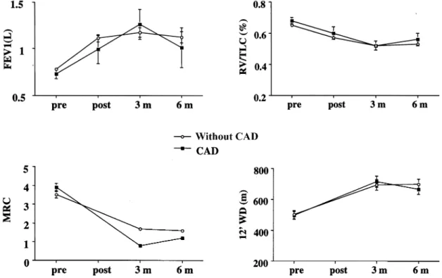
The results of pulmonary function testing of the nine CAD patients pre- and postoperatively, at 3 and 6 months after LVRS are summarized in Table 1. We ®nd an improve-ment in FEV1 over the ®rst six months following LVRS in patients who underwent an intervention for CAD in combi-nation with LVRS (Fig. 1). Furthermore, a reduction of pulmonary overin¯ation as assessed by the RV/TLC ratio is observed. Diffusing capacitiy for carbon monoxide remained unchanged. MRC was signi®cantly lower in both postoperative follow-up examinations. The outcome
of 12-min walking distance (WD) was slightly improved but not signi®cantly different from preoperative values. 3.2. Lung volume reduction surgery and valve replacement
All three patients who underwent LVRS in combination with replacement of the mitral- or aortic valve were treated outside the prospective LVRS protocol, and therefore did not meet all criteria to qualify for LVRS according to our study protocol.
3.2.1. Patient 1
In a 73-year-old female with known COPD and emphy-sema (FEV1 0.8 l, 42% predicted, TLC 5.45 l, 110% predicted, RV 3.5L, 165% predicted) a severe aortic valve
Table 1
Postoperative pulmonary function, exercise capacity (12-min WD (m)), and modi®ed medical council dyspnoe score (MRC) in patients with CAD and before valve replacement over the ®rst 6 months postoperatively
Pre (n 9) Post (n 6) 3 months (n 5) 6 months (n 6) FEV1(l) 0.73 ^ 0.05 0.99 ^ 0.15a 1.26 ^ 0.16b 1.01 ^ 0.21 FEV1(%) 25 ^ 1.8 37 ^ 3.9a 45 ^ 5.1c 38 ^ 5.8a RV/TLC (%) 0.68 ^ 0.02 0.60 ^ 0.04 0.52 ^ 0.03c 0.56 ^ 0.04b DLCO (%) 39 ^ 3.2 33 ^ 2.7 38 ^ 4.6 39 ^ 4.9 MRC 3.9 ^ 0.1 0.8 ^ 0.4c 1.2 ^ 0.48c 12-min WD (m) 498 ^ 89 715 ^ 74 664 ^ 46 aP , 0:05: bP , 0:01:
cP , 0:001 versus preop. values. There were no statistially signi®cant differences between groups at any point in time.
Fig. 1. Respiratory function (FEV1 (L), RV/TLC, DLCO (%)), modi®ed medical research council dyspnoe score (MRC), and exercise capacity (12-min WD (m)) preoperatively and over the ®rst 6 months postoperatively. In the patients with CAD: pre: n 9; post: n 6; at 3 months: n 5; and at 6 months: n 6, and patients without CAD (mean ^ SEM).
Table 2 Coro nary angiogra phy ®nd ings, hemodyn amic data, and sympt oms in candi dates for LVR S with sig ni®cant lesi ons. Patient /age/sex Corona ry angiogra phy ®nd ings a Hemo dynami cs Symp toms/ myocar dial infarc tion Procedure Co urse EF (%) b CO (l/mi n) c PAP S/D/M (mm Hg) d 1/62/M 80±90% LA D, 90% LC X 65 4.68 27/ 11/18 Asym ptomat ic, silent M1 CABG eg U neventf ul 2/67/M 70% LAD, 90% RCA, 70±90% LCX 68 8.91 ù Asym ptomat ic CABG U neventf ul 3/68/M 75±80% LA D, 60±90% RCA 87 4.5 ù h Asym ptomat ic PTCA f1 stent U neventf ul 4/71/M 75% LAD 62 4.72 30/ 15/21 Asym ptomat ic PTCA U neventf ul 5/75/M 65% LAD ,70% RCA, 85% LCX 61 10.4 51/ 11/29 Asym ptomat ic PTCA 1 stent U neventf ul 6/58/F 75% RCA 68 4.33 24/ 9/15 Asym ptomat ic PTCA U neventf ul 7/69/M 90% LAD, 100% RCA, 70±9 0% LCX 55 ù ù Hist ory of M1 Stent Pos toperative death 8/68/M 50±60% LA D, 70±90% RCA 71 4.74 35/ 17/23 Asym ptomat ic PTCA 1 stent U neventf ul 9/64/M 70±90% LA D, 70±90% RCA 72 4.95 23/ 13/17 Asym ptomat ic, silent M1 PTCA 1 stent U neventf ul aLAD ,lef tanteri or descendi ng (art ery); RCA, right coronar y arte ry; LC X, left circum ¯ex (art ery). bEF, ejection fraction (left ve ntricular). cCO, card iac out put. dPAP S/D/M ,pulmona ry artery pressure syst olic/d iastolic/mean . eCAB G, coronary arter y bypas s gra fting. gCABG and LVR S in one opera tion, all other pr ocedures befor e LVRS. hù, onl y co ronary angi ograph y w as perfom ed. fPTCA, percutaneous translum inal coronary angi oplasty .
stenosis (mean gradient 85 mmHg) was diagnosed. She suffered from severe dyspnea during normal daily activity and severe orthopnea at night.
Combined aortic valve replacement and bilateral lung volume reduction surgery was performed in the same session via median sternotomy. Postoperatively, severe pulmonary arterial hypertension developed and was treated with NO inhalation. The patient was weaned from the respirator and extubated on the 12th postoperative day. Antibiotic therapy was required for a unilateral pneumonia and the patient was leaving the hospital on day 28 post-operatively.
In the 3 months follow-up examination bronchial obstruc-tion had decreased as well as pulmonary hyperin¯aobstruc-tion (FEV1 0.88L, 44% predicted, TLC 4.72 l, 95% predicted, RV 2.7 l, 128% predicted).
3.2.2. Patient 2
A 67-year-old female with biventricular cardiac insuf®-ciency (NYHA 3-4), mitral valve stenosis and a mild aortic valve insuf®ciency was refused in 1994 for mitral valve replacement because of severe COPD (FEV1 0.84L, 36% predicted, RV/TLC ratio 0.75, diffusing capacity for carbon monoxide (DLCO) 39% predicted). The patient was a heavy smoker for 25 years and was on long-term oxygen therapy since the beginning of 1996. She suffered from severe dyspnoea (Medial research council (MRC) dyspnea score: 4), and was very limited in her exercise capacity (12-min walking distance 540 m).
A combined mitral valve replacement and lung volume
reduction surgery in the same session was planned. After valve replacement, however, a type A dissection occured. LVRS was postponed and performed 3 days later which faciliated successful weaning fom the respirator.
The late postoperative course was further complicated by urosepsis and the patient was discharged from the hospital on day 37 postoperatively.
Three months after the operation, dyspnea and exercise capacity were markedly improved (MRC: 1; 12-min walk-ing distance 675 m), and lung function showed less obstruc-tion (FEV1 1.47 l, 67% predicted) and overin¯aobstruc-tion (RV/ TLC 0.54). DLCO revealed a slight improvement to 49% predicted. Long-term oxygen therapy was no longer neces-sary. One year after the operation the patient was in good general condition with FEV1 of 53% predicted, RV/TLC ratio 0.50, and a 12-min walking distance of 808 m. 3.2.3. Patient 3
A 68-year-old previous heavy smoker suffered from COPD with alpha-1-antitrypsin de®ciency and severe bullous emphysema. Furthermore a combined aortic valve disorder with dominant stenosis (mean gradient 35 mmHg) was known. Since pulmonary emboli were suspected, the patient received coumarine since year one. FEV1 was 1.3 l (47% pred.).
After several hospitalisations for pulmonary decompen-sation a simultanous valve replacement and LVRS was planned. Due to accidental perforation of the left ventricle during the aortic valve replacement, bilateral LVRS was performed by video-assisted thoracoscopies (VATS) 10
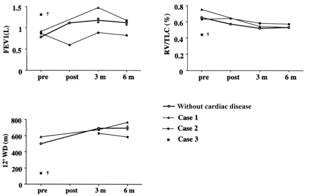
Fig. 2. Individual pulmonary functional parameters (FEV1 (L), RV/TLC) and exercise capacity (12-min WD (m)) of the three patients before and after valve replacement in comparison to patients without cardiac disease (mean ^ SEM).
days after the cardiac intervention. At this time point a pulmonary infection with a multiresistant Pseudomonas was diagnosed.
After a prolonged weaning period and slow improvement of the lung function the patient developed severe pneumo-nia. In the later postoperative course infectious parameters increased. On the 15th postoperative day the patient died from multi-organ failure following intestinal infarction. 3.2.4. Morbidity and mortality
One patient in the group with CAD died after develop-ment of pulmonary edema on day 2 postoperatively and one patient after valve replacement and LVRS died because of intestinal infarction on day 14 postoperatively (Table 2).
Postoperative complications after LVRS following a cardiac intervention include late pneumothorax (n 2), hematothorax (n 1) and urosepsis (n 1).
3.2.5. Drainage time and hospital stay
In the 12 patients with combined intervention median drainage time was 11.5 days (range 5±30 days).
The median hospital stay was 15 days (range 10±33 days). In contrast, the two surviving patients after valve replacement were hospitalised longer, for 28 and 37 days respectively.
4. Discussion
In this study we retrospectively evaluated the functional outcome of patients who underwent LVRS in combination with valve replacement, PTCA, placement of a coronary stent, or CABG. We found that LVRS can be performed safely in selected patients who were previously treated for CAD. The functional outcome in these patients is equal to LVRS patients without CAD over the ®rst 6 months (Fig. 1). In addition, we could demonstrate that LVRS enables valve replacement in selected patients with severe COPD and emphysema who were previously considered to be inoper-able.
At the University of Zurich the LVRS program has been started in early 1994. All patients were included in a prospective study. Nearly 300 patients have been evaluated and 124 underwent surgery for emphysema. LVRS at our institution is mainly performed bilateral in one session by a video-thoracoscopic approach.
Smoking is the main risk factor for emphysema as well as for coronary artery disease. Therefore, in these patients additional risk factors as CAD have to be excluded. Exercise testing, however, in COPD patients is often not possible due to severe pulmonary limitation.
In a previous prospective study we could demonstrate that clinically silent, but relevant CAD is a frequent ®nding in emphysema patients, otherwise qualifying for LVRS [3]. We found that in 15% of LVRS candidates at least one relevant coronary artery stenosis (.70% or a 50% stenosis
of the left main coronary artery) is present. After treatment of CAD with PTCA, stent, or CABG, LVRS can be performed safely with a low mortality and morbidity similar to the group of patients without CAD.
When LVRS was performed on a patient while on cardi-opulmonary bypass, buttressing with bovine pericardial strips was always used with the aim to prevent bleeding in the fragile lung tissue of the emphysematous lung. The improvement in pulmonary function and exercise perfor-mance was identical in the nine CAD patients as compared to patients without CAD. These data suggest that a combi-nation of both interventions is feasible. However, in our experience it seems to be favourable to select the patients carefully and to operate preferentially on patients which are more likely to improve functionally after LVRS (e.g. hetero-genous emphysema with good target areas) [10].
The rationale for LVRS in patients with valvular heart disease was different [6]. The combined procedure was only performed when the patient was severely limited preoperatively and unable to maintain even everyday aciv-ities after failure of all conservative treatment modalaciv-ities. Basically, the patients were considered inoperable for valve replacement due to their respiratory insuf®ciency resulting from severe COPD with emphysema. LVRS was performed with the aim to improve pulmonary func-tion to faciliate postoperative weaning from the respirator. The intraoperative course of two of the described patients during the cardiac intervention was complicated and LVRS was postponed and performed 3 and 10 days later under more stable conditions. Two patients after valve replace-ment and LVRS showed equal or even improved pulmon-ary function and exercise performance 3±6 months postoperatively (Fig. 2).
Since bilateral procedures offer more functional improve-ment [11], unilateral LVRS is performed only in patients with severe emphysematous destruction predominantly on one side. All patients with CAD or valve replacement were heavy smokers with the exception of one CAD patient with diffuse unilateral emphysema. A bilateral approach was favoured to achive maximal respiratory bene®t in these high risk patients. A unilateral approach was not due to intraoperative complications in our patients.
Postoperative pulmonary improvement in patients who underwent CABG or valve replacement are even more impressive since it has been demonstrated in previous studies that in patients with normal preoperative respiratory function who undergo cardiac surgery (CABG or valve replacement) FEV1 and FVC decrease postoperatively by at least 10% over several months [12].
Our experience with surgical treatment for empysema and cardiac valve disorder demonstrates that combined interventions can be performed successfully in selected patients. The morbidity and mortality are acceptable. However, this surgical concept can only be recommended for centers with a large experience in postoperative manage-ment of patients with severe emphysema.
References
[1] Cooper JD, Patterson GA, Sundaresan RS, Trulock EP, Yusen RD, Pohl MS, Lefrak SS. Results of 150 consecutive bilateral lung volume reduction procedures in patients with severe emphysema. J Thorac Cardiovasc Surg 1996;112:1319±1330.
[2] Binggisser R, Zollinger A, Hauser M, Bloch KE, Russi EW, Weder W. Bilateral lung volume reduction surgery for diffuse pulmonary emphysema by video-assisted thoracoscopy. J Thorac Cardiovasc Surg 1996;97:875±882.
[3] Turnheer R, Muntwhyler J, Stammberger U, Bloch KE, Zollinger A, Weder W, Russi EW. Coronary artery disease in patients undergoing lung volume reduction surgery for emphysema. Chest 1997;112:122± 128.
[4] Russi EW, Stammberger U, Weder W. Lung volume reduction surgery for emphysema. Eur Resp J 1997;19:208±218.
[5] Thurnheer R, Bingisser R, Stammberger U, et al. Effect of lung volume reduction surgery on pulmonary hemodynamics in severe pulmonary emphysema. Eur J Cardiothorac Surg 1998;13:253±258. [6] Schmid RA, Vogt P, Stocker R, Zalunardo M, Russi EW, Weder W.
Lung volume reduction surgery for a patient receiving mechanical ventilation after a complex cardiac operation. J Thorac Cardiovasc Surg 1997;115:236±237.
[7] Quanjer PH, Tammeling GJ, Cotes JE, Pedersen OF, Peslin R, Yernault JC. Lung volumes and forced ventilatory ¯ows. Report working party standardization of lung function tests, European community for steel and coal. Of®cial statement of the European respiratory society. Eur Respir J 1993;16(Suppl 6):5±40.
[8] Cotes JE, Chinn DJ, Quanjer PH, Roca J, Yernault JC. Report work-ing party. standartization of lung function tests. European community for steel and coal. Of®cial statement of the European respirastory society: lung volumes and forced respiratory ¯ows. Eur Respir J 1993;16(Suppl 3):41±52.
[9] McGavin CR, Gupta SP, McHardy GJ. Twelve-minute walking test for assessing disability in chronic bronchitis. Br Med J 1976;1:822± 823.
[10] Weder W, Thurnheer R, Stammberger U, Burge M, Russi EW, Bloch KE. Radiologic emphysema morphology is associated with outcome after surgical lung volume reduction. Ann Thorac Surg 1997;64(2):313±320.
[11] McKenna Jr RJ, Brenner M, Fischel RJ, Gelb AF. Should volume reduction for emphysema be unilateral or bilateral?. J Thorac Cardi-ovasc Surg 1996;112:1331±1339.
[12] Dubois P, Dubois G, Delwiche JP, Schoevaerdts JC, Kremer R. Lung function and cardiac surgery. Acta Cardiol 1991;4:439±451.
Appendix A. Conference discussion
Dr H. Toomes (Gerlingen, Germany): I want to ask about your strategy once more. Do you always plan to make the operations, lung and cardiac, simultaneously? Or why don't you do it simultaneously?
Dr Schmid: I think this is risk stratifying. In the valve patient, it was always planned to perform LVRS through the median sternotomy in the same session at the end of the procedure when the patient was still on bypass. We used buttressing of the staple line in all cases to prevent parenchymal haemorrhage, and we did not have any bleeding problems. On two occasions, during the cardiac intervention severe intraoperative complications occurred, which prolonged bypass time, and we had to delay the lung volume reduction procedure. One patient we tried to wean from the respirator, but it was impossible. Lung volume reduction surgery was performed and 10 days later we could extubate the patient. This case was published in the Journal of Thoracic and Cardiovascular Surgery in 1998.
Dr F. Venuta (Rome, Italy): I did not understand which procedures you did ®rst in the series of patients that you treated at the same time with cardiac procedure and lung volume reduction. I mean, did you do the lung volume reduction before putting the patient on bypass, or after?
Dr Schimd: After the cardiac operation, but still on bypass.
Dr Venuta: So you reversed heparin and then you did the lung volume reduction?
Dr Schmid: No, we performed LVRS. When the patient was, as I just mentioned, still on bypass.
Dr T. Dosios (Athens, Greece): I understood that all your patients had coronary arteriogram done before the operation. Is it correct?
Dr Schmid: In the very initial experience of lung volume reduction surgery, we performed it in all patients. Evaluating our data, we found that it is only indicated when you have clinical suspicion, or certain risk factors, except smoking of course. In general, we do now perform LVRS without coronary angiography.
Dr P. Baptista (Carnaxide, Portugal): From what I understood in the beginning, you did coronary angiograms in all patients proposed to lung reduction. And then you said you only did it when there was suspicion of cardiac pathology. Why not do just stress efforts in every patient, which is something we can do very easily?
Dr Schmid: The emphysema patients usually can not perform ergome-try because of their pulmonary limitation. Therefore, if there is any clinical suspicion for coronary heart disease we liberally perform angiography.