. . . .
. . . .
Injectable living marrow stromal cell-based
autologous tissue engineered heart valves: first
experiences with a one-step intervention in primates
Benedikt Weber
1,2,3†, Jacques Scherman
4,5†, Maximilian Y. Emmert
1,2,3†,
Juerg Gruenenfelder
2,3, Renier Verbeek
5, Mona Bracher
5, Melanie Black
5,
Jeroen Kortsmit
5, Thomas Franz
5, Roman Schoenauer
1,2, Laura Baumgartner
1,2,
Chad Brokopp
1,2, Irina Agarkova
1,2, Petra Wolint
1,2, Gregor Zund
1,2, Volkmar Falk
3,
Peter Zilla
4,5, and Simon P. Hoerstrup
1,2,3*
1
Swiss Center for Regenerative Medicine, Zurich, Switzerland;2
Department of Surgical Research, University Hospital of Zurich, Zurich, Switzerland;3
Clinic for Cardiovascular
Surgery, University Hospital of Zurich, Raemistrasse 100, 8091 CH-Zu¨rich, Switzerland;4
Chris Barnard Division of Cardiothoracic Surgery, Groote Schuur Hospital, University of
Cape Town, Cape Town, South Africa; and5
Cardiovascular Research Unit, Cape Heart Centre, University of Cape Town, Cape Town, South Africa Received 5 November 2010; revised 7 January 2011; accepted 7 February 2011; online publish-ahead-of-print 17 March 2011
This paper was guest edited by Prof. Stephan Janssens, University Hospital Gasthuisberg, Leuven, Belgium.
Aims A living heart valve with regeneration capacity based on autologous cells and minimally invasive implantation
technol-ogy would represent a substantial improvement upon contemporary heart valve prostheses. This study investigates the feasibility of injectable, marrow stromal cell-based, autologous, living tissue engineered heart valves (TEHV) generated and implanted in a one-step intervention in non-human primates.
Methods and results
Trileaflet heart valves were fabricated from non-woven biodegradable synthetic composite scaffolds and integrated into self-expanding nitinol stents. During the same intervention autologous bone marrow-derived mononuclear cells were harvested, seeded onto the scaffold matrix, and implanted transapically as pulmonary valve replacements into non-human primates (n ¼ 6). The transapical implantations were successful in all animals and the overall procedure time from cell harvest to TEHV implantation was 118 + 17 min. In vivo functionality assessed by echocardiography revealed preserved valvular structures and adequate functionality up to 4 weeks post implantation. Substantial cellular remodelling and in-growth into the scaffold materials resulted in layered, endothelialized tissues as visualized by histology and immunohistochemistry. Biomechanical analysis showed non-linear stress – strain curves of the leaflets, indicating replacement of the initial biodegradable matrix by living tissue.
Conclusion Here, we provide a novel concept demonstrating that heart valve tissue engineering based on a minimally invasive technique for both cell harvest and valve delivery as a one-step intervention is feasible in non-human primates. This innovative approach may overcome the limitations of contemporary surgical and interventional bioprosthetic heart valve prostheses.
-Keywords Minimally invasive † Heart valve † Tissue engineering † Bone marrow † Stem cells † Primate model
Introduction
Valvular heart disease represents a major cause of morbidity and
mortality worldwide.1 Besides conventional treatment modalities
based on surgical valve repair or replacement, the recent clinical implementation of minimally invasive implantation techniques is expected to have a major impact on the management of patients
with valvular heart disease.2Various percutaneous catheter-based
†These authors contributed equally to this paper.
*Corresponding author. Tel:+41 44 255 3644, Fax: +41 44 255 4369, Email:[email protected]
as well as transapical surgical implantation approaches have been developed and successfully used in both experimental and clinical settings, representing a promising alternative to conventional
heart valve surgery.3–5However, despite these auspicious
thera-peutic advances, currently available valvular substitutes for mini-mally invasive replacement procedures are bio-prosthetic and as such inherently prone to calcification and progressive dysfunctional degeneration suggesting their primary clinical application in elderly
patients.6
Living autologous heart valve substitutes with regeneration and growth potential could overcome the limitations of today’s valvular prostheses and enable the application of minimally invasive treat-ment modalities to a broader patient population, including young
patients.7 We recently demonstrated the principal feasibility of
merging the two innovative heart valve replacement technologies of heart valve tissue engineering (HVTE) and minimally invasive
delivery in an ovine model.8 A clinically relevant HVTE concept
ideally comprises minimally invasive techniques for both cell harvest and valve delivery. Therefore, several potential cell
sources and protocols have been assessed;9–12however, most of
them requiring an extensive and complex ex vivo cell and tissue
culture phase. Shin´oka et al.13–17described a method of creating
tissue-engineered vascular grafts (TEVG) by using bone marrow-derived mononuclear cells (BMCs) without cell expansion culture. Constructed from biodegradable polyester tubes seeded with autologous cells, these grafts demonstrated functionality and appeared to transform into living vascular grafts. Initial clinical pilot studies evaluating BMC-seeded vascular grafts as venous con-duits for congenital heart surgery revealed adequate safety profiles
and functionality up to 8 years.18–21
The present study investigates for the first time the implantation of autologous BMC-based tissue engineered heart valves (TEHV) by minimally invasive implantation technologies in a primate model; thereby approximating the human situation as much as possible in a pre-clinical large animal model. The presented auto-logous approach provides a minimally invasive, one-step heart valve replacement procedure from cell harvest to in vitro engineer-ing and transapical delivery of livengineer-ing TEHV.
Methods
Scaffold fabrication
Trileaflet heart valve scaffolds were fabricated from non-woven poly-glycolic acid meshes (PGA; Cellon, Luxembourg), coated with 1.75% poly-4-hydroxybutyrate (P4HB; TEPHA, Inc., USA; see Supplementary material online, Methods A). Thereafter, the scaffolds were integrated into radially self-expandable nitinol stents (length ¼ 30 mm; OD ¼ 20 mm; pfm AG, Germany) by attaching the scaffold matrix to the inner surface of the nitinol stent wires. After vacuum drying for 24 h, the scaffolds were sterilized overnight by using ethylene oxide (EtO) gas sterilization.
Isolation of primate bone marrow-derived
mononuclear cells
A volume of 66.5 + 14.4 mL of bone marrow was aspirated from the sternum of adult Chacma Baboons into a heparinized syringe (50 – 100 U/mL) using a 12-Gauge threpine needle. Bone marrow-derived
mononuclear cells were obtained by centrifuging the samples on a histopaque density gradient (Sigma Chemical Co., USA) for 30 min at 1500 r.p.m (500 g). The viability of the isolated buffy coat was deter-mined by flow cytometry and Trypan Blue staining after erythrocyte elimination and quantification with a haemocytometer (see Sup-plementary material online, Methods B). All animals received human care and the study was approved by the institutional review boards (Department of Surgery Research Committee: Approval Ref. 2009/ 096, Animal Research Ethics Committee; Approval Ref. 009/035, Faculty of Health Sciences, University of Cape Town) and in compli-ance with the Guide for the Care and Use of Laboratory Animals, published by the National Institutes of Health (NIH publication No. 85-23). The transport of explanted tissue for histological analysis was in accordance with the Convention on International Trade in Endan-gered Species (CITES) regulations for transport of protected species (CITES Export Permission No. 091992; Import Permission No. 3710/ 09). Unseeded controls were not included for ethical reasons given the results of previous experiments in the sheep model. When using the same tissue engineering technology, unseeded (only fibrin-coated) scaffolds implanted in the orthotopic pulmonary position of sheep demonstrated severe structural failure already after 4 weeks (see Supplementary material online, Figure S1).
Phenotyping of bone marrow-derived
mononuclear cells
Small samples of isolated BMCs of each animal were used for haema-toxylin –eosin (H&E) staining and immunocytochemical analysis. The cells were fixed with methanol and immunofluorescence staining was performed using for the following epitopes: CD45, CD34, CD146, CD90, CD166, CD44, and DAPI. Phalloidin Alexa 633 was used as control staining. Primary antibodies were detected with Cyanine-2 or Cyanine-3 goat anti-mouse antibodies (see Supplementary material online, Methods B and C). In order to evaluate the in vivo fate of isolated primate BMCs (n ¼ 2) seeded cells were tracked for 4 weeks in vivo using the CellTraceTM CFSE Cell Proliferation Kit (C34554, Invitrogen
Corp., USA; see Supplementary material online, Methods D).
Bone marrow-derived mononuclear cell
seeding and characterization
Seeding of the BMCs onto the stented heart valve scaffolds (8 + 3× 10⁶cells/cm2) was performed using fibrin (Sigma Chemical Co., St.
Louis, MO, USA) as a cell carrier.22 After seeding, the constructs were placed into vented 50 mL tubes for 10 min to allow the fibrin – thrombin solution to clot adequately. Thereafter, the seeded construct was loaded into the delivery device by decreasing the diameter from 20 to 8 mm and transferred to the theatre. Mesenchymal stem cells (MSCs) were obtained from bone marrow aspirate according to the method of Pittenger et al.23and were differentiated into pre-adipocytic and osteo-blastic phenotypes (see Supplementary material online, Methods J) to confirm their differentiation potential.
Tissue engineered heart valves implantation
and in vivo functionality
For evaluation of in vivo functionality, TEHV were minimally invasively delivered into the pulmonary valve position using a mini-sternotomy and antegrade transapical approach. The valves were crimped and loaded onto a custom-made inducing system (OD ¼ 8 mm) consisting of a rigid tube and pusher. The right ventricle was punctured using needle through purse-string sutures. Next, the inducer system was inserted and the TEHV was delivered into the pulmonary artery under guidance of fluoroscopy (BV, Pulsera, Philips, Medical Systems,
The Netherlands) and transoesophageal echocardiography (2D-transoesophageal echo cardiography (TEE), Philips, Medical Systems, The Netherlands). The radial expansion of the nitinol stent forced the native pulmonary valve leaflets against the pulmonary artery wall and kept them fixed. One construct was deployed in a supravalvular position not excluding the native valvular leaflets. The appropriate position and functionality of the implanted valve was con-firmed by angiography. In addition, the in vivo functionality was moni-tored using transoesophageal, transthoracic, and/or epicardial echocardiography during the procedure, immediately after implan-tation, weekly up to 4 weeks, and prior to sacrifice of the animal (see Supplementary material online, Methods E). The pulmonary artery pressure was measured in order to exclude stent-associated stenosis of the pulmonary outflow tract. Anticoagulation (aspirin and warfarin) was maintained for 4 weeks. The animals were sacrificed (potassium euthanization and exsanguination) after 12 h (n ¼ 1) and 4 weeks (n ¼ 5) and the TEHV were explanted.
Qualitative explant tissue analysis
Tissue composition of explanted heart valve constructs was analysed qualitatively using immuno-/histology and compared with native heart valve leaflets. The tissue sections were studied using Azan staining, Von Kossa staining, H&E staining, Masson – Trichrome staining, and Elastin-van-Gieson (eVG) staining. In addition, immunohistochemistry was performed using the Ventana Benchmark automated staining system (Ventana Medical Systems, Tuscon, AZ, USA) and antibodies for alpha smooth muscle actin (a-SMA), Desmin, CD 68, CD34, CD31, eNOS, HAM-56, and von Willebrand factor (see Supplemen-tary material online, Methods F). Representative tissue samples of TEHV were analysed using Scanning Electron Microscopy (see Sup-plementary material online, Methods G).
Quantitative explant tissue analysis
Explanted TEHV (4 week explants, orthotopical position) were lyophi-lized and analysed by biochemical assays for total DNA, hydroxyproline (HYP) as well as glycosaminogylcans (GAG) content (see Supplemen-tary material online, Methods H). For measuring the DNA amount, the Hoechst dye method24was used. The GAG content was deter-mined using a modified version of the protocol described by Farndale et al.25and a standard curve prepared from chondroitin sulphate from shark cartilage (Sigma Chemical Co., USA). Hydroxyproline was deter-mined with a modified version of the protocol provided by Huszar et al.26The mechanical properties of the leaflets and the valve conduits were assessed independently in radial direction by uniaxial tensile testing to determine ultimate tensile stress (UTS) as well as Young’s modulus (YM; see Supplementary material online, Methods I).
Statistical analysis
All quantitative data are presented as mean + standard deviation (SPSS 17.0, IBM, Somers, NY, USA).
Results
Viable bone marrow-derived
mononuclear cells harvested from the
sternum: a cocktail of stem cells and
leucocytes
The isolation of BMCs rendered 175 + 82× 106cells per animal
for scaffold seeding. Cellular viability was in excess of 95% as
verified by whole-blood flow cytometry (Figure 1A) and Trypan
Blue exclusion staining (Figure1B). Isolated primate mononuclear
cells (Figure1C) stained positively for common leucocytic antigens
including CD45, CD44, and CD15 (see Supplementary material online, Figure S2A – C ). According to the flow cytometric analysis the marrow mononuclear cells comprised distinct leucocytic cell populations including granulocytes (20.8%), monocytes (7.6%), and lymphocytes (67.6%). The amount of dead cells, indicated by a population of low forward scatters (FSC)-height and increased
side scatters (SSC)-height in the scatter plot was ,5% (Figure1A).
Primate bone marrow aspirate comprised CD34+
haematopoie-tic stem cells as well as MSCs staining positively for CD44, CD90, CD146, and CD166, but negatively for CD45 and CD34,
repre-senting a common staining pattern for MSCs (Figure 1G and H;
see Supplementary material online, Figure S2D). The differentiation potential of isolated primate MSCs was assessed exemplarily using pre-adipocytic and osteoblastic differentiation assays (see Sup-plementary material online, Figure 2E and F ). After isolation,
BMCs were labelled (Figure1D – F ) and successfully seeded onto
PGA/P4HB heart valve scaffolds (Figure 2A – D) and the TEHV
could be inserted into the sheath system for transapical delivery.
Minimally invasive delivery: the
transapical implantation of tissue
engineered heart valves
The transapical implantations were successful in all animals. Of all six animals five valves were deployed in the orthotopic valvular
position (Figure3A – C and E), whereas one valve was positioned
supravalvularly thereby not excluding the native valvular leaflets. In one of the orthotopically implanted animals perioperative cor-onary perfusion complications not related to the TEHV functional-ity resulted in termination 12 h after implantation. Positioning of the TEHV was verified intraoperatively using TEE and fluoroscopy
(Figure 4C and D; see Supplementary material online, Trailer S1).
Neither migration of the TEHV nor paravalvular leakage has been observed. Proper opening and closing behaviour was
demon-strated by TEE measurements (Figure 4E – H; See Supplementary
material online, Trailer S2 and S3). The mean crimping time of
the TEHV (Figure3D and A – C), from insertion into the application
system until surgical deployment, was 17 + 8 min (Figure3D and E).
The mean duration of the entire procedure, from bone marrow harvest until valve delivery, was 118 + 17 min.
In vivo performance of tissue engineered
heart valves
Valve functionality with leaflet mobility and sufficient opening and closing behaviour was observed after implantation. No paravalvular leakage was detected during the follow-up period. Weekly TTE/ TEE follow-up revealed a slight but not significant increase in
trans-valvular peak pressure (TVG; Table 1) starting around 25 +
7 mmHg (TEE) perioperatively, up to 30 + 7 mmHg after 3 weeks (TTE). At explantation (4 weeks), TVG gradient was clearly reduced to 16.1 + 1.9 mmHg (TEE). Moderate regurgita-tion was detected in three animals, which remained unchanged over the whole period of implantation time until harvest. Proper opening and closing behaviour could be demonstrated by
TTE/TEE measurements (Figure4D – G; see Supplementary material online, Trailer S2 and S3).
Explant macroscopy of tissue engineered
heart valves
Explantation of the early TEHV (12 h) revealed an intact leaflet structure dominated by a fibrin-coated PGA scaffold matrix with lack of evidence for thrombus formation. Harvest of the remaining TEHV 4 weeks after delivery showed constructs that were well integrated into the adjacent tissue-by-tissue covering the stent
margin and encircling stent strut endings (Figure4A). The valvular
leaflets of the four orthotopically positioned constructs presented
as pliable, well-defined cusps (Figure 4B). Although the leaflets
appeared to be shortened in radial diameter, sufficient co-aptation was demonstrated throughout the entire experimental period.
Interestingly, in the haemodynamically non-fully loaded TEHV deployed in the supravalvular position the leaflet structure was almost entirely missing. Instead two small parietal nodes could be identified, most likely representing remnants of the original
scaf-fold pockets (Figure4C).
Scanning electron microscopy
The surface of the early explants (12 h) was mainly characterized by previously seeded mononuclear cells embedded into a fibrin
matrix densely covering the entire scaffold (Figure5A and B).
Analy-sis of the 4-week explants revealed that three of the orthotopically implanted leaflets showed already a mature structure, character-ized by a well-defined endothelial lining on the conduit wall as
well as partially on the valvular leaflets (Figure 5E and F ). In
several spots these endothelial coverages were confluent and
Figure 1 Isolation of bone marrow-derived mononuclear cells. Flow cytometry scatter (A) plot of nucleated cells with microfluidic erythro-cyte lysis labelled by population with as forward scatters vs. side scatters. The fraction of dead cells, indicated by low forward scatters/high side scatters values, is ,5%. This is confirmed by Trypan Blue exclusion staining (TBS; B). Mononuclear cells (C; H&E staining) were labelled with CFSE (D) and seeded onto the polyglycolic acid matrix (E). After 4 weeks no fluorescent cells could be detected in the explants tissue (F ). A fraction of adherent bone marrow-derived mononuclear cells stained positively for CD44, CD90, CD146, CD166, and negatively for CD45 and CD34 indicating a mesenchymal stem cell character (G – L). Specific staining is shown in red, DAPI (nuclei in green), and Phalloidin (f-actin in blue) were used as control stainings.
not distinguishable from native primate (Figure 5C) or human
(Figure 5D) valvular endothelium. The other orthotopic explant
presented with a more premature surface remodelling, character-ized by areas with extensive fibrin formation as well as leucocyte and thrombocyte attachment. Interestingly, in this explant all stages of surface remodelling could be detected, from beginning
provisional thrombocyte-mediated surface coverage (Figure5G – I )
to areas of almost confluent endothelial covering. The non-orthotopically positioned construct also revealed a well estab-lished endothelial coverage with the remaining leaflet structure firmly integrated into the conduit wall.
Histology and immunohistochemistry
Haematoxylin – eosin staining (Figure 6A – D) of the explants
demonstrated layered tissue morphology, characterized by a central core of non-degraded PGA matrix surrounded by dense tissue formation on the luminal as well as on the vascular side. Masson’s Trichrome and eVG staining confirmed the presence of collagen predominantly in the outer layers of the conduit wall
and the leaflet (Figure6E – L). As confirmed by SEM, the surface
of the conduit was covered by a confluent endothelial layer in large areas of the constructs. Cells of the constructs’ surface
layer expressed CD31 (Figure 6M – P) and vWF, resembling the
staining pattern of native valve endothelial cells (Figure6M).
More-over, a-SMA expression could be detected in interstitial cells of
the newly formed surface layers of the conduit (Figure6S) and
par-tially of the leaflet (Figure6T). In contrast to previous studies,8the
leaflets’ hinge region was free of a-SMA and/or Desmin positivity (see Supplementary material online, Figure S3C and D). The early (12 h) as well as the late (4 weeks) explants stained positively for CD68 (see Supplementary material online, Figure S3G – I ), indi-cating a distinct monocytic infiltration and continuous remodelling. Cell tracking analysis using CFSE-dye and confocal microscopy
(Figure 1D and E) revealed no signal within the explanted tissue
(Figure 1F), indicating that most of the seeded BMCs were not
present after 4 weeks in vivo.
Biomechanical and extracellular matrix
analysis: the remodelling of viable tissue
Mechanical evaluation was performed separately for the valvular leaflets and the wall region. Analysis of the 4 week explants revealed that the tissue strength (UTS 0.19 + 0.12 MPa) as well as the stiffness (YM 1.0 + 0.54 MPa) of the conduit wall was higher than that of the valvular leaflet itself (UTS 0.08 + 0.05 MPa; YM 0.19 + 0.08 MPa). The stress – strain curves of both the heart valve leaflet and the conduit wall revealed non-linear behaviour until rupture. The YM obtained in the low strain (0.0 – 10.0%) as well as the high strain (.10.0%) showed to be significantly different (all P’s ,0.05), which is characteristic for properties of biological tissue (see Supplementary materialFigure 2 Bone marrow-derived tissue engineered heart valves. After isolation of bone marrow-derived mononuclear cells, stented polygly-colic acid scaffold matrices (A and B) were seeded with cells using fibrin as a cell carrier (C and D).
online, Figure S4), indicating the in vivo replacement of the
biode-gradable matrix by living tissue (Figure7A).
Quantitative extracellular matrix (ECM) analysis of the TEHV explants showed that the GAG as well as the DNA content of the constructs’ leaflets was higher than compared with native tissue
(GAG 140.28 + 36.17%; DNA 168.10 + 87.07%), indicating high cellular infiltration as well as ECM remodelling. Consistent with
pre-vious studies,8the collagen content in the leaflets and the conduit
wall was substantially lower in the newly formed tissues than
com-pared with native controls (HYP 59.24 + 16.87%; Figure7B).
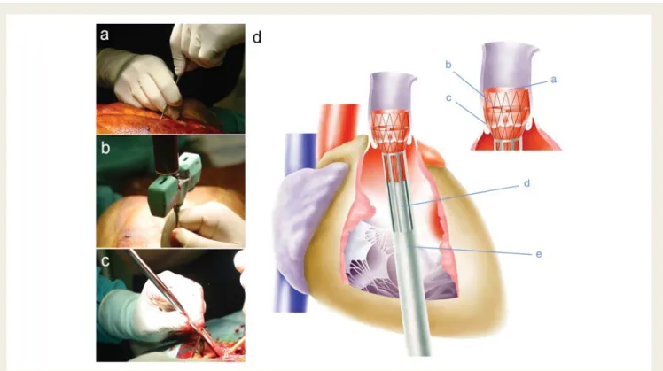
Figure 3 Minimally invasive tissue engineered heart valves implantation. After sternal bone marrow puncture (A) and aspiration of fluid (B), the tissue engineered heart valve was loaded into the delivery device (C ), inserted into the right ventricle (D) and deployed in the pulmonary position under sonographic and fluoroscopic guidance. The crimped tissue engineered heart valve (a), integrated into the nitinol stent (b), is carefully deployed into the pulmonary position (c) by slowly advancing the inner pusher (d) into the sheath system (e).
Figure 4 Explant analysis of tissue engineered heart valves. After 4 weeks in vivo the stented constructs were well integrated into the adjacent tissue (A). Orthotopical tissue engineered heart valves (B) presented with a cusp-like leaflet structure, with shorter leaflets than native controls. In a final transoesophageal echocardiography-assessment the leaflet co-aptation (C; asterisk indicates leaflets) as well as opening movements of all three leaflets could be visualized (D – G).
Discussion
Minimally invasive heart valve replacement techniques have recently emerged as a promising technology in interventional
car-diovascular therapeutics.2,6,27,28Despite the potential of this novel
technology, currently used replacement materials for transcatheter
or transapical valves are bioprosthetic and therefore highly prone
to calcification as well as structural degeneration.29,30Living TEHV,
based on rapidly biodegrading scaffolds and autologous cells could overcome these limitations with characteristics such as repair, remodelling, and growth. We recently demonstrated the principal feasibility of merging tissue engineering and minimally invasive
. . . .
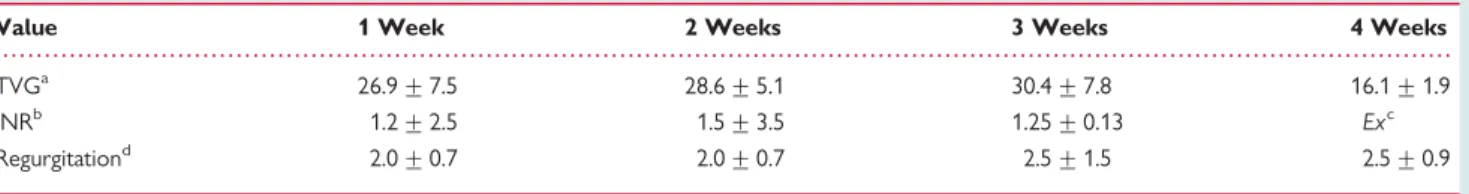
Table 1 Transvalvular peak pressure gradient (mmHg), international normalized ratio, and grade of regurgitation
during 4 weeks follow-up of orthotopically delivered tissue engineered heart valve (mean +++++ SD)
Value 1 Week 2 Weeks 3 Weeks 4 Weeks
TVGa 26.9 + 7.5 28.6 + 5.1 30.4 + 7.8 16.1 + 1.9
INRb 1.2 + 2.5 1.5 + 3.5 1.25 + 0.13 Exc
Regurgitationd 2.0 + 0.7 2.0 + 0.7 2.5 + 1.5 2.5 + 0.9
a
TVG, transvalvular peak pressure gradient;
b
INR, international normalized ratio;
c
Explantation;
d
Regurgitation—Grading: 0/1/2/3/4—None/trivial/mild/moderate/severe.
Figure 5 Scanning electron microscopy of the polyglycolic acid – poly-4-hydroxybutyrate scaffold (A and B), primate (C ), and human (D) control leaflets. In most areas the surface of the 4 week explants showed confluent (E and F ) or initial (G) endothelial coverage. In some areas the surface remodelling was still evident involving thrombocyte attachment (H ) and leucocyte attraction (I ).
heart valve replacement technologies.8Ideally, a clinically relevant HVTE concept must involve low-invasive techniques for both cell harvest and valve delivery. Consequently, several non-invasive
potential cell sources comprising stem cells have been assessed with regard to the clinical realization of the concept. However, most of these concepts require extensive, logistically complex,
Figure 6 Histology of the explanted living tissue engineered heart valves. Explanted tissues showed layered tissue architecture visualized by haematoxylin – eosin staining (A – D). In Masson’s Trichrome staining (E – H) and Elastin-van-Gieson staining (I – L) collagen fibres were predomi-nantly found in the outer tissue layer of the conduit and leaflets. Cells of the conduit (O) and leaflet (P) surface layer expressed CD31 (M – P). a-SMA expression (Q – T) could be detected in the outer layers of the conduit (S) and the leaflet (T ), but was missing in the proximal leaflet region. Arrows indicate reminiscent scaffold material.
Figure 7 Qualitative tissue analysis. Mechanical properties of explanted tissue engineered leaflets after 4 weeks in vivo are displayed as stress – strain curves [MPa/%] characteristic for biological tissue (A). Extracellular matrix analysis of the explants for glycosaminogylcans and DNA shows enhanced values compared with native tissues. Hydroxyproline normalized to tissue weight is decreased compared with native controls (B).
and time-consuming in vitro tissue engineering processes that limit their clinical feasibility.
The present study demonstrates for the first time the feasibility of merging heart valve tissue engineering based on a low-invasive stem cell source, with a minimally invasive, injectable implantation technology combined in a single intervention. Stem cell sources previously used for HVTE, including MSCs, foetal stem cells, and
blood-derived progenitors,8–12 necessitate cell harvest in a
par-ticular intervention, followed by protracted and complex in vitro cell expansion and implantation of fabricated TEHV in a second
procedure.8 To the contrary, autologous BMCs can be isolated
in sufficient numbers from sternum bone marrow aspirates, thereby enabling immediate re-implantation as autologous TEHV. Therefore, the presented one-step approach suggests a highly clini-cally relevant concept and may represent a significant step towards the routine utilization of TEHVs.
The principle of using BMCs for cardiovascular tissue
engineer-ing was described by Shin’oka et al.13and has been assessed in
pre-clinical14–16 as well as initial clinical investigations.18–21 It was
hypothesized that multipotent bone marrow-derived stem cells
proliferate, differentiate, and constitute new tissues.19In a recent
study, Roh et al.17elucidated the potential underlying molecular
mechanism of autologous BMC-induced tissue formation. In
con-trast to previous assumptions, no evidence for (trans-)
differentiation of bone marrow-derived stem cells into mature vas-cular cells has been found. Interestingly, seeded BMCs were not detectable in the tissue engineered constructs 1 week after implan-tation. In fact, it was shown that BMC-seeded biodegradable
scaf-folds transform into functional blood vessels via an
inflammation-mediated process of vascular remodelling. While the role of BMCs in this remodelling process remains largely con-jectural, a growing body of evidence suggests their role within inflammation-mediated processes.
This paracrine role of seeded BMCs as a constituent of an inflammatory vascular remodelling process is consistent with mul-tiple studies reporting that (trans-)differentiation of bone
marrow-derived stem cells in vivo is deemed rare.10,31–36The present study
concurs with this mechanism, as the initially labelled BMCs were not detectable within the explants 4 weeks after implantation. Also, a distinct remodelling process was evident, characterized by a monocytic infiltration of the leaflets as well as the conduit wall. Besides this critical role of seeded BMCs for in vivo
remodel-ling and tissue formation,17,19 the precise contribution of the
detectable multipotent BMC stem cell fractions is of great interest
to understand the underlying regenerative mechanism.37–40
Experimentally, it would have been elegant to include an unseeded control group in this study. However, previous exper-iments in the sheep model using the same tissue engineering tech-nology and unseeded (only fibrin-coated) scaffolds implanted in the orthotopic pulmonary position demonstrated severe structural failure after 4 weeks (see Supplementary material online, Figure S1). Given the fact that remodelling phenomena in the ovine model are expected to be substantially more intense than in the
human/primate cardiovascular system,41 the implantation of
unseeded controls into primates appeared ethically not justifiable. A very interesting observation of the current study was the influ-ence of haemodynamic loading on remodelling, tissue formation,
and maturation in vivo. This is a known phenomenon from embryo-logy and has been used for in vitro tissue engineering experiments utilizing biomimetic flow and pressure conditions to achieve
optimal tissue maturation in so-called bioreactors.42,43 In the
present investigation, the role of mechanical loading on functional remodelling was uniquely demonstrated by the supra-valvular posi-tioned TEHV not excluding the native valvular leaflets. In contrast to the orthotopic implants, the non-loaded supravalvular TEHV lacked any leaflet structure after 4 weeks, indicating missing tissue formation as well as BMC-mediated leaflet remodelling.
Macroscopically the orthotopic constructs presented with cusp-like leaflet structures and did not appear thickened compared with
previous reports based on the sheep model.8,44Although principal
valvular functionality was demonstrated, mild to moderate regurgi-tation was observed by echocardiography in three of five valves in the presence of leaflet co-aptation. This may be explained by the rather prototypic design of the TEHV in this proof of concept study. Furthermore, a minimal structural shortening of the TEHV cusps was detectable which will be addressed in future TEHV scaf-fold designs e.g. by oversizing of the co-aptation areas of the starter matrices. Further enhancement of cellular attraction and, thus, tissue formation particularly in the distal leaflet region may
provide solutions.45Finally, following this feasibility study
introdu-cing injectable TEHV generated and minimally invasively implanted in a single intervention, extended long-term studies are mandatory in order to further elucidate the fate of such living autologous cell-based engineered heart valves as well as the underlying tissue remodelling mechanisms observed in vivo. Particularly, the precise role of seeded stem cells with respect to their function in chemo-attraction and tissue formation has to be systematically assessed. However, addressing these questions was beyond the scope of this pilot-study and will be investigated in future experiments.
Conclusions
These first results of combining minimally invasive valve replace-ment procedures with heart valve tissue engineering in a single intervention in a preclinical primate model are promising and demonstrate the feasibility of using BMCs for the fabrication of TEHV. Moreover, utilizing the body’s natural abilities to regenerate TEHV in vivo, may greatly simplify, and improve the clinical feasi-bility of the autologous cell-based TEHV approach. Such auto-logous and living heart valves with repair and regeneration capacities may represent the next generation of transcatheter and transapical heart valves overcoming the time limitations of the currently used bioprosthetic valves suggesting their future clini-cal application also beyond elderly patients.
Supplementary material
Supplementary material is available at European Heart Journal online.
Acknowledgements
The authors thank Deon Bezuidenhout, Ronnett Seldon, Elizabeth VanderMerwe, Ursula Steckholzer, Pia Fuchs, Klaus Marquardt and
Silvia Behnke for their technical support, as well as all the members of the Animal Unit, Faculty of Health Sciences, University of Cape Town for their excellent contribution.
Funding
This work was supported by the Swiss South African Joint Research Programme of the State Secretariat for Education and Research, Swiss and South African Governments [EX25-2010], the Research Infrastructure Support Programme [UID 65720], the National Research Foundation and the Department of Science and Technology South Africa, the University Research Committee - University of Cape Town, the Claude Leon Foundation, the Swiss National Science Foun-dation [32-122273] as well as the 7th Framework Programme, Life Valve, European Commission [242008].
Conflict of interest: S.P.H. and G.Z. are scientific advisors of Xeltis AG, Switzerland.
References
1. Supino PG, Borer JS, Preibisz J. The epidemiology of valvular heart disease: a growing public health problem. Heart Fail Clin 2006;2:379 – 393.
2. Lurz P, Bonhoeffer P, Taylor AM. Percutaneous pulmonary valve implantation: an update. Expert Rev Cardiovasc Ther 2009;7:823 – 833.
3. Asoh K, Walsh M, Hickey E, Nagiub M, Chaturvedi R, Lee KJ, Benson LN. Percu-taneous pulmonary valve implantation within bioprosthetic valves. Eur Heart J 2010;31:1404 – 1409.
4. Momenah TS, El Oakley R, Al Najashi K, Khoshhal S, Al Qethamy H, Bonhoeffer P. Extended application of percutaneous pulmonary valve implan-tation. J Am Coll Cardiol 2009;53:1859 – 1863.
5. Walther T, Schuler G, Borger MA, Kempfert J, Seeburger J, Ru¨ckert Y, Ender J, Linke A, Scholz M, Falk V, Mohr FW. Transapical aortic valve implantation in 100 consecutive patients: comparison to propensity-matched conventional aortic valve replacement. Eur Heart J 2010;31:1398 – 1403.
6. Walther T, Simon P, Dewey T, Wimmer-Greinecker G, Falk V, Kasimir MT, Doss M, Borger MA, Schuler G, Glogar D, Fehske W, Wolner E, Mohr FW, Mack M. Transapical minimally invasive aortic valve implantation: multicenter experience. Circulation 2007;611:I240 – I245.
7. Fernandes SM, Khairy P, Sanders SP, Colan SD. Bicuspid aortic valve morphology and interventions in the young. J Am Coll Cardiol 2007;49: 2211 – 2214.
8. Schmidt D, Dijkman PE, Driessen-Mol A, Stenger R, Mariani C, Puolakka A, Rissanen M, Deichmann T, Odermatt B, Weber B, Emmert MY, Zund G, Baaijens FPT, Hoerstrup SP. Minimally invasive implantation of living tissue engin-eered heart valves—a comprehensive approach from autologous vascular cells to stem cells. J Am Coll Cardiol 2010;56:510 – 520.
9. Hoerstrup SP, Kadner A, Melnitchouk S, Trojan A, Eid K, Tracy J, Sodian R, Visjager JF, Kolb SA, Grunenfelder J, Zund G, Turina MI. Tissue engineering of functional trileaflet heart valves from human marrow stromal cells. Circulation 2002;106:I143 – I150.
10. Sutherland FW, Perry TE, Yu Y, Sherwood MC, Rabkin E, Masuda Y, Garcia GA, McLellan DL, Engelmayr GC Jr, Sacks MS, Schoen FJ, Mayer JE Jr. From stem
cells to viable autologous semilunar heart valve. Circulation 2005;111:
2783 – 2791.
11. Schmidt D, Mol A, Breymann C, Achermann J, Odermatt B, Go¨ssi M, Neuenschwander S, Preˆtre R, Genoni M, Zund G, Hoerstrup SP. Living autolo-gous heart valves engineered from human prenatally harvested progenitors. Circu-lation 2006;114:I125 – I131.
12. Schmidt D, Achermann J, Odermatt B, Breymann C, Mol A, Genoni M, Zund G, Hoerstrup SP. Prenatally fabricated autologous human living heart valves based on amniotic fluid derived progenitor cells as single cell source. Circulation 2007;116: I64 – I70.
13. Hibino N, Shin’oka T, Matsumura G, Ikada Y, Kurosawa H. The tissue-engineered vascular graft using bone marrow without culture. J Thorac Cardiovasc Surg 2005; 129:1064 – 1070.
14. Matsumura G, Ishihara Y, Miyagawa-Tomita S, Ikada Y, Matsuda S, Kurosawa H, Shin’oka T. Evaluation of tissue-engineered vascular autografts. Tissue Eng 2006; 12:3075 – 3083.
15. Brennan MP, Dardik A, Hibino N, Roh JD, Nelson GN, Papademitris X, Shinoka T, Breuer CK. Tissue-engineered vascular grafts demonstrate evidence of growth
and development when implanted in a juvenile animal model. Ann Surg 2008; 248:370 – 377.
16. Mirensky TL, Nelson GN, Brennan MP, Roh JD, Hibino N, Yi T, Shinoka T, Breuer CK. Tissue-engineered arterial grafts: long-term results after implantation in a small animal model. J Pediatr Surg 2009;44:1127 – 1132.
17. Roh JD, Sawh-Martinez R, Brennan MP, Jay SM, Devine L, Rao DA, Yi T, Mirensky TL, Nalbandian A, Udelsman B, Hibino N, Shinoka T, Saltzman WM, Snyder E, Kyriakides TR, Pober JS, Breuer CK. Tissue-engineered vascular grafts transform into mature blood vessels via an inflammation-mediated process of vascular remodeling. Proc Natl Acad Sci U S A 2010;107: 4669 – 4674.
18. Shin’oka T, Imai Y, Ikada Y. Transplantation of a tissue-engineered pulmonary artery. N Engl J Med 2001;344:532 – 533.
19. Matsumura G, Miyagawa-Tomita S, Shin’oka T, Ikada Y, Kurosawa H. First
evi-dence that bone marrow cells contribute to the construction of
tissue-engineered vascular autografts in vivo. Circulation 2003;108:1729 – 1734. 20. Isomatsu Y, Shin’oka T, Matsumura G, Hibino N, Konuma T, Nagatsu M,
Kurosawa H. Extracardiac total cavopulmonary connection using a
tissue-engineered graft. J Thorac Cardiovasc Surg 2003;126:1958 – 1962. 21. Shin’oka T, Matsumura G, Hibino N, Naito Y, Watanabe M, Konuma T,
Sakamoto T, Nagatsu M, Kurosawa H. Midterm clinical result of tissue-engineered vascular autografts seeded with autologous bone marrow cells. J Thorac Cardiovasc Surg 2005;129:1330 – 1338.
22. Mol A, van Lieshout MI, Dam-de Veen CG, Neuenschwander S, Hoerstrup SP, Baaijens FP, Bouten CV. Fibrin as a cell carrier in cardiovascular tissue engineering applications. Biomaterials 2005;26:3113 – 3121.
23. Pittenger MF, Mackay AM, Beck SC, Jaiswal RK, Douglas R, Mosca JD, Moorman MA, Simonetti DW, Craig S, Marshak DR. Multilineage potential of adult human mesenchymal stem cells. Science 1999;284:143 – 147.
24. Cesarone CF, Bolognesi C, Santi L. Improved microfluorometric DNA determi-nation in biological material using 33258 Hoechst. Anal Biochem 1979;100: 188 – 197.
25. Farndale RW, Buttle DJ, Barrett AJ. Improved quantitation and discrimination of sulphated glycosaminoglycans by use of dimethylmethylene blue. Biochem Biophys Acta 1986;883:173 – 177.
26. Huszar G, Maiocco J, Naftolin F. Monitoring of collagen and collagen fragments in chromatography of protein mixtures. Anal Biochem 1980;105:424 – 429. 27. Bonhoeffer P, Boudjemline Y, Qureshi SA, Le Bidois J, Iserin L, Acar P, Merckx J,
Kachaner J, Sidi D. Percutaneous insertion of the pulmonary valve. J Am Coll Cardiol 2002;39:1664 – 1669.
28. Cribier A, Eltchaninoff H, Tron C, Bauer F, Agatiello C, Sebagh L, Bash A, Nusimovici D, Litzler PY, Bessou JP, Leon MB. Early experience with percuta-neous transcatheter implantation of heart valve prosthesis for the treatment of end-stage inoperable patients with calcific aortic stenosis. J Am Coll Cardiol 2004;43:698 – 703.
29. Coats L, Bonhoeffer P. New percutaneous treatments for valve disease. Heart 2007;93:639 – 644.
30. Schoen FJ. Evolving concepts of cardiac valve dynamics: the continuum of devel-opment, functional structure, pathobiology, and tissue engineering. Circulation 2008;118:1864 – 1880.
31. Zentilin L, Tafuro S, Zacchigna S, Arsic N, Pattarini L, Sinigaglia M, Giacca M. Bone marrow mononuclear cells are recruited to the sites of VEGF-induced neovascu-larization but are not incorporated into the newly formed vessels. Blood 2006; 107:3546 – 3554.
32. Murry CE, Soonpaa MH, Reinecke H, Nakajima H, Nakajima HO, Rubart M, Pasumarthi KB, Virag JI, Bartelmez SH, Poppa V, Bradford G, Dowell JD, Williams DA, Field LJ. Haematopoietic stem cells do not transdifferentiate into cardiac myocytes in myocardial infarcts. Nature 2004;428:664 – 668.
33. Balsam LB, Wagers AJ, Christensen JL, Kofidis T, Weissman IL, Robbins RC. Hae-matopoietic stem cells adopt mature haeHae-matopoietic fates in ischaemic myocar-dium. Nature 2004;428:668 – 673.
34. Wagers AJ, Sherwood RI, Christensen JL, Weissman IL. Little evidence for devel-opmental plasticity of adult hematopoietic stem cells. Science 2002;297: 2256 – 2259.
35. Terada N, Hamazaki T, Oka M, Hoki M, Mastalerz DM, Nakano Y, Meyer EM, Morel L, Petersen BE, Scott EW. Bone marrow cells adopt the phenotype of other cells by spontaneous cell fusion. Nature 2002;416:542 – 545.
36. Nygren JM, Jovinge S, Breitbach M, Sa¨we´n P, Ro¨ll W, Hescheler J, Taneera J, Fleischmann BK, Jacobsen SE. Bone marrow-derived hematopoietic cells generate cardiomyocytes at a low frequency through cell fusion, but not transdifferentia-tion. Nat Med 2004;10:494 – 501.
37. Kinnaird T, Stabile E, Burnett MS, Shou M, Lee CW, Barr S, Fuchs S, Epstein SE. Local delivery of marrow-derived stromal cells augments collateral perfusion through paracrine mechanisms. Circulation 2004;109:1543 – 1549.
38. Gnecchi M, He H, Liang OD, Melo LG, Morello F, Mu H, Noiseux N, Zhang L, Pratt RE, Ingwall JS, Dzau VJ. Paracrine action accounts for marked protection of ischemic heart by Akt-modified mesenchymal stem cells. Nat Med 2005;11: 367 – 368.
39. O’Neill TJ IV, Wamhoff BR, Owens GK, Skalak TC. Mobilization of bone marrow-derived cells enhances the angiogenic response to hypoxia without transdifferen-tiation into endothelial cells. Circ Res 2005;97:1027 – 1035.
40. Uemura R, Xu M, Ahmad N, Ashraf M. Bone marrow stem cells prevent left ven-tricular remodeling of ischemic heart through paracrine signaling. Circ Res 2006; 98:1414 – 1421.
41. Schoen FJ, Levy RJ. Calcification of tissue heart valve substitutes: progress toward understanding and prevention. Ann Thorac Surg 2005;79:1072 – 1080.
42. Hoerstrup SP, Sodian R, Sperling JS, Vacanti JP, Mayer JE Jr. New pulsatile bio-reactor for in vitro formation of tissue engineered heart valves. Tissue Eng 2000;61:75 – 79.
43. Mol A, Driessen NJ, Rutten MC, Hoerstrup SP, Bouten CV, Baaijens FP. Tissue engineering of human heart valve leaflets: a novel bioreactor for a strain-based conditioning approach. Ann Biomed Eng 2005;33:1778 – 1788.
44. Flanagan TC, Sachweh JS, Frese J, Schno¨ring H, Gronloh N, Koch S, Tolba RH, Schmitz-Rode T, Jockenhoevel S. In vivo remodeling and structural characteriz-ation of fibrin-based tissue-engineered heart valves in the adult sheep model. Tissue Eng Part A 2009;15:2965 – 2976.
45. Lutolf MP, Gilbert PM, Blau HM. Designing materials to direct stem-cell fate. Nature 2009;462:433 – 441.
CARDIOVASCULAR FLASHLIGHT
. . . .
doi:10.1093/eurheartj/ehr240
Online publish-ahead-of-print 15 July 2011
Isolated arrhythmogenic left ventricular cardiomyopathy identified
by cardiac magnetic resonance imaging
Ingo Paetsch1*, Sebastian Reith1, Nikolaus Gassler2, and Cosima Jahnke1 1
Department of Cardiology, University Hospital RWTH Aachen, Pauwelsstrasse 30, 52074 Aachen, Germany and2Institute of Pathology and Electron Microscopy Facility,
RWTH Aachen University, Aachen, Germany
*Corresponding author. Tel:+49 241 80 89705, Fax: +49 241 80 82131, Email:[email protected]
A 78-year-old man experienced a first-time syncope with fall resulting in raccoon eyes and a single episode of non-sustained ventricular tachycardia (.10 beats) was docu-mented. Creatine kinase and troponin T were only weakly abnormal. Echocardiography and invasive coronary angiography were unremarkable.
Cardiac magnetic resonance (CMR) imaging revealed the presence of intramyocardial fatty spots and streaks of the left ventricular (LV) inferolateral wall and the inter-ventricular septum (arrowheads in Panel A, steady-state
free precession (SSFP) imaging; Panels B and C,
T2-weighted black blood imaging without and with fat sup-pression; white arrows in Panel D, delayed enhancement imaging with diffuse and streaky intramyocardial signal enhancement; see Supplementary material online, Movie 1). Right ventricular morphology and regional and global function were normal. Lipomatous metaplasia of chronic myocardial infarction was ruled out by missing regional subendocardial delayed enhancement. Hence, CMR find-ings were suggestive of isolated arrhythmogenic dysplasia of the LV.
Histopathological work-up including transmission
elec-tron microscopy of a deep LV biopsy demonstrated disrupted myocardial architecture with extensive fibrofatty replacement [Panels E and F, haematoxylin and eosin staining; Panels G and H, transmission electron microscopy, white arrows indicate cardiomyo-cytes with several cytoplasmic vacuoles separated by a collagen rich matrix (asterisk); arrowheads, nuclei of fibrocardiomyo-cytes; black-lined white arrows, lipid droplets; rectangle in G delineates the magnified image area in Panel H].
A ‘left-dominant’ subtype of arrhythmogenic right ventricular cardiomyopathy (ARVC) has been recognized with multicentre studies reporting LV involvement in ARVC to occur with disease progression in .75% of cases. However, primary LV involvement has been acknowledged as a phenotype expression of ARVC only recently.
Supplementary material: Supplementary material is available at European Heart Journal online.
![Figure 7 Qualitative tissue analysis. Mechanical properties of explanted tissue engineered leaflets after 4 weeks in vivo are displayed as stress – strain curves [MPa/%] characteristic for biological tissue (A)](https://thumb-eu.123doks.com/thumbv2/123doknet/14927072.664163/8.892.77.827.88.487/qualitative-mechanical-properties-explanted-engineered-displayed-characteristic-biological.webp)