Role of nutrient-sensing taste 1 receptor (T1R) family members
in gastrointestinal chemosensing
Soraya P. Shirazi-Beechey
1*, Kristian Daly
1, Miran Al-Rammahi
1, Andrew W. Moran
1and David Bravo
21
Epithelial Function and Development Group, Department of Functional and Comparative Genomics,
Institute of Integrative Biology, University of Liverpool, Liverpool L69 7ZB, UK
2
Pancosma SA, Geneva, Switzerland
(Submitted 26 February 2013 – Final revision received 8 April 2013 – Accepted 11 April 2013)
Abstract
Luminal nutrient sensing by G-protein-coupled receptors (GPCR) expressed on the apical domain of enteroendocrine cells activates intracellular pathways leading to secretion of gut hormones that control vital physiological processes such as digestion, absorption, food intake and glucose homeostasis. The taste 1 receptor (T1R) family of GPCR consists of three members: T1R1; T1R2; T1R3. Expression of T1R1, T1R2 and T1R3 at mRNA and protein levels has been demonstrated in the intestinal tissue of various species. It has been shown that T1R2– T1R3, in association with G-protein gustducin, is expressed in intestinal K and L endocrine cells, where it acts as the intestinal glucose (sweet) sensor. A number of studies have demonstrated that activation of T1R2 – T1R3 by natural sugars and artificial sweeteners leads to secretion of glucagon-like peptides 1&2 (GLP-1 and GLP-2) and glucose dependent insulinotropic peptide (GIP). GLP-1 and GIP enhance insulin secretion; GLP-2 increases intestinal growth and glucose absorption. T1R1 –T1R3 combination co-expressed on the apical domain of cholecystokinin (CCK) expressing cells is a luminal sensor for a number ofL-amino acids; with amino acid-activation of the receptor eliciting CCK secretion. This article focuses on the role of the gut-expressed T1R1, T1R2 and T1R3 in intestinal sweet andL-amino acid sensing. The impact of exploiting T1R2 – T1R3 as a nutritional target for enhancing intestinal glucose absorption and gut structural maturity in young animals is also highlighted.
Key words:T1R1-T1R3: T1R2-T1R3: Intestine: sensing: SGLT1: GLP-2: CCK
G-protein-coupled receptors (GPCR) represent the largest family of cell-surface mediators of signal transduction(1). GPCR have attracted significant attention in terms of continued identification and characterisation, with recognition that they are targets for novel drug discovery. With more recent evidence demonstrating that nutrient sensing in the gastrointestinal tract is accomplished by a number of GPCR(2), the role of these receptors as important nutritional targets is becoming evident. Nutrient-sensing GPCR for a variety of nutrients have been identified in the intestinal epithelium. They are expressed on the apical domain of enteroendocrine (sensor) cells of the gut and are directly activated by nutrients(3 – 9). Nutrient sensing initiates a cascade of events involving hormonal and neural pathways. This culminates in functional responses that ultimately regulate vital processes such as nutrient digestion and absorption, food intake, insulin secretion and metabolism. This brief article focuses on the role of the taste receptor 1 family of GPCR, T1R1, T1R2, and T1R3, in sweet andL-amino acid sensing, with particular focus on its role in glucose
absorption, glucose homeostasis and satiety. Moreover, the impact of exploiting the T1R2 – T1R3 heterodimer as a nutritional target for enhancing intestinal glucose (salt and water) absorption and gut structural maturity in young animals is highlighted.
Sweet and
L-amino acid sensing in the lingual epithelium
The T1R family present in the taste cells of the lingual epithelium consists of three members: T1R1; T1R2; T1R3(10,11). These receptors are distantly related to metabotropic glutamate receptors (mGluR), extracellular Ca2þ-sensing receptor (CaSR) and g-aminobutyric acid type B receptor(10). Based on electro-physiological studies, heterologous expression of taste receptor subunits and behavioural assays of knockout mice, the heterodimeric combination of T1R2 – T1R3 has been shown to function as a broad-specificity sweet sensor for natural sugars, sweet proteins and artificial sweeteners, whereas the combi-nation of T1R1 – T1R3 has been identified as a broad-spectrum
* Corresponding author: S. P. Shirazi-Beechey, email spsb@liverpool.ac.uk
Abbreviations: CaSR, Ca2þ-sensing receptor; CCK, cholecystokinin; GIP, glucose-dependent insulinotropic peptide; GLP, glucagon-like peptide; GLUT,
glutamate; GPCR, G-protein-coupled receptors; IMP, monophosphate esters of inosine; LEU, leucine; PHE, phenylalanine; SGLT1, Naþ/glucose cotransporter-1; T1R, taste receptor 1; TRP, tryptophan.
qThe Authors 2013
British
Journal
of
L-amino acid sensor, responsible for mediating the perception of the savoury ‘umami’ taste of monosodium glutamate(11,12). Both the T1R2 – T1R3 and T1R1 – T1R3 heterodimers are coupled to the heterotrimeric G-protein gustducin to transmit intracellular signals(13).
In rodents and many other mammalian species, the lingual epithelium T1R1 – T1R3 heterodimer responds to most of the twenty standard L-amino acids in the millimolar range(12). However, the T1R1 – T1R3 heterodimer is not activated by L-tryptophan (TRP)(12). The human T1R1 – T1R3 complex functions as a much more specific receptor, responding selectively to monosodium glutamate (GLUT) and aspartate (as well as to the GLUT analogue L-AP4)(10,11). A salient feature of amino acid taste in animals and umami taste in humans is the synergistic enhancement of potency when GLUT or other amino acids combine with the monophosphate esters of inosine or gua-nosine nucleotides (IMP and GMP)(14 – 16). Both GLUT and IMP/ GMP bind to adjacent domains on the N-terminal Venus flytrap module of T1R1(17), while potentiation of intracellular signal transmission by IMP is mediated through a-gustducin(18). Gurmarin, a thirty-five-residue polypeptide from the Indian-originated tree Gymnema sylvestre (Gurmar), can inhibit both sweet andL-amino acid sensing by binding to the Venus flytrap domain of T1R3, inhibiting its function(19 – 24).
Intestinal sweet sensing
Work carried out in many laboratories has demonstrated that T1R family members and gustducin are co-expressed in enter-oendocrine cells in a range of species(5,7,24 – 33), suggesting that taste-sensing mechanisms exist in the gastrointestinal tract.
It is well established that enteroendocrine L and K cells secrete glucagon-like peptides (GLP) (1 and 2) and glucose-dependent insulinotropic peptide (GIP), respectively, on encountering glucose in the intestinal lumen. GLP-1 and GIP, known as incretins, enhance insulin secretion, while GLP-2 increases intestinal growth and glucose absorption(34 – 36). The infusion of intestinal lumen with the D-isoforms of glucose, galactose and fructose and non-metabolisable analogues of glucose, 3-O-methyl-glucose and a-methyl-glucose, causes the secretion of GIP and GLP-1 in rats, pigs and humans(37 – 39). Furthermore, it has been shown that the T1R2 – T1R3 heterodimer together with the a-subunit of gustducin resides in K and L endocrine cells containing GIP, GLP-1 and GLP-2, respectively(7,8,30,33).
Functional evidence for the role of the T1R2 – T1R3 hetero-dimer in intestinal glucose (sweet) sensing, inducing GLP-1, GLP-2 and GIP release, has been provided using endocrine cell lines, native intestinal tissue explants and knockout mice deficient in a-gustducin or T1R3(7,24,32,33). The murine endocrine cell line GLUTag exhibits markedly increased GLP-1 secretion upon exposure to the artificial sweetener sucralose; this secretion is blocked by gurmarin, indicating that sucralose-induced GLP-1 release occurs through the acti-vation of the T1R2 – T1R3 heterodimer(7). Similar results were obtained for sucralose-induced GLP-1 release in the human L endocrine cell line NCI-H716, which was blocked either by RNA interference targeting of a-gustducin or by the human
sweet taste receptor antagonist lactisole(5). Furthermore, the plasma levels of GLP-1 and GIP following the introduction of glucose directly into the proximal intestine are reduced in a-gustducin or T1R3 knockout mice, compared with wild-type controls(40). Moreover, these knockout mice have abnormal insulin profile and prolonged postprandial blood glucose responses in response to luminal glucose(40). Further work car-ried out by Geraedts et al.(32)has shown that luminal glucose, fructose or sucralose evoke release of GLP-1 from mouse ileal explants embedded in an Ussing chamber, and that secretion of GLP-1 does not occur in tissue explants from T1R3 knockout mice(32). Moreover, exposure off mouse intestinal explants to either glucose or sucralose results in the secretion of GLP-1 and GLP-2, in a dose-dependent manner, and that this secretion is inhibited in the presence of gurmarin, a specific inhibitor of T1R3(33) (Fig. 1). Notably, the levels of GLP-1 and GLP-2 released by control and glucose-stimulated tissues were similar to those observed in in vivo studies in rats and human subjects given glucose orally or maintained as controls(41,42), supporting the suitability of intestinal tissue explants for such studies. In these assays, the endocrine cells reside in their native niche, and it appears that maintaining contacts with neighbouring cells is important for endocrine cells to retain their functional viability(43). Collectively, the data suggest that the sensing of sugars by the T1R2 – T1R3 heterodimer coupled to gustducin expressed in L and K endocrine cells leads to the release of GLP-1, GLP-2 and GIP.
However, there are reports indicating that sweeteners do not trigger the release of incretins. Parker et al.(44) have reported that primary cultures of adult mouse intestine do not secrete GIP in response to sucralose. This is not surprising, since they have indicated that these isolated cells do not express the T1R2 – T1R3 heterodimer(44). There are also reports that oral ingestion or intragastric infusion of artificial sweeteners does not increase the secretion of incretins in rats(45)or humans(46). By feeding rats a single concentration of sweeteners (50 mg or 1 g/kg body weight, depending on the sweetener), Fujita et al.(45)have concluded that sweeteners do not acutely induce the release of incretin hormones. Ma et al.(46) have also reported that 0·4 or 4 mM-sucralose given by intragastric infusion does not induce the secretion of incretins. Interestingly, lactisole, which inhibits T1R3 function, reduces the blood levels of GLP-1 in humans receiving an intragastric glucose load(47).
Many artificial sweeteners are partly absorbed in the stomach and subsequently secreted in the urine(48). Therefore, the lack of response observed by these workers may be due to the concentration of the sweeteners being below the threshold level required for activating the candidate receptor and/or the lack of availability of the sweetener at the distinct target region of the intestine. Further work is required to unravel these controversies.
The majority of membrane-bound proteins, including GPCR, are low-abundance proteins(49 – 53). In our experience more sensitive SYBR green assay rather than TaqMan-based assay and/or increased amounts of template complementary DNA (up to 250 ng/reaction) are effective in detecting the expression of T1R family members, having low abundance
British
Journal
of
mRNA. This is perhaps why one or two laboratories have failed to detect the expression of T1R1, T1R2, T1R3 and gustducin in purified primary enteroendocrine cells using quantitative PCR(44,54,55). Other factors, such as the prevailing cell isolation conditions or the small proportion of purified L or K cells expressing taste receptor subunits and gustducin, have also been proposed to be responsible for the lack of detection of taste receptor elements in purified L and K cells(56).
There are some reports proposing that T1R subunits are expressed in the colon; however, their precise functions require further investigations. Iwatsuki et al.(57)have demonstrated the expression of T1R2 – LacZ in mouse small and large intestinal tissues. Geraedts et al.(32) have reported glucose-stimulated GLP-1 secretion from Ussing chamber-embedded large intestinal explants of T1R3, but not T1R2, knockout mice. They have concluded that T1R3-dependent and independent pathways are involved in the regulation of GLP-1 secretion in the colon(32).
It should be borne in mind that L cells in the small and large intestines may have different phenotypes. Furthermore, in the lumen of the native colonic tissue, there is hardly any free glucose available. Glucose is rapidly metabolised to SCFA by colonic microbiota. SCFA induce the release of GLP-1 via colonic endocrine L-cell GPR43 (FFAR2)(58). There-fore, studies directed at the sensing of nutrients in the hindgut must always consider the digestive activity of the microbiota.
Mechanisms underlying intestinal sweet sensing and
glucose transport regulation: application to the
maintenance of gut health in weaning piglets
The major route for the absorption of dietary glucose (and galactose) from the lumen of the intestine into enterocytes is via the apical membrane Naþ/glucose cotransporter-1 (SGLT1)(59 – 62). The absorption of glucose by SGLT1 also activates salt (NaCl) and water absorption; this is used as the route for oral rehydration therapy(63). Thus, the regulation of SGLT1 is essential for the provision of glucose to the body and avoidance of intestinal malabsorption. A number of studies(60,61,64 – 67) have established that the expression of intestinal SGLT1 is enhanced in response to a range of monosac-charides, including non-metabolisable analogues of glucose. Furthermore, it has been shown that the pathway underlying monosaccharide-enhanced SGLT1 expression involves a luminal membrane GPCR glucose sensor(66).
Convincing evidence for the involvement of gut-expressed T1R2 – T1R3 heterodimer and gustducin in intestinal sweet transduction and SGLT1 regulation has been provided by studies using mice in which the genes for either a-gustducin or the sweet receptor subunit, T1R3, had been deleted. The elimination of sweet transduction in mice in vivo has been shown to prevent the dietary monosaccharide-induced up-regulation of SGLT1 expression that is observed in wild-type mice(7). Furthermore, it has been demonstrated that artificial
0 10 20 30 ** † ** † *** *** *** ‡ ‡‡‡ ** ‡ ‡‡ * * (a) Control Glucose GLP-1 (pmol/l) 0 2 5 10 20 50 0 10 20 30 40 (c) Sucralose (mmol/l) GLP-1 (pmol/l) Sucralose (mmol/l) 0 2 5 10 20 50 0 200 400 600 (d) GLP-2 (pmol/l) (b) Control Glucose 0 100 200 300 400 GLP-2 (pmol/l)
Fig. 1. Glucagon-like peptide (GLP)-1 and GLP-2 secretion, from mouse small intestine in response to glucose or sucralose. Mouse small-intestinal tissue explants were incubated for 1 h at 378C in incubation media supplemented with: 10 % (w/v) glucose or untreated (controls), in the absence ( ) or presence ( ) of 5 mg/ml gurmarin ((a) and (b)); the indicated concentrations of sucralose or untreated (control) in the absence ( ) or presence ( ) of 5 mg/ml gurmarin ((c) and (d)). Data are means, with standard errors represented by vertical bars. Mean value was significantly different from that of the untreated control in the absence of gurmarin: * P, 0·05, ** P, 0·01, *** P, 0·001. † Mean value was significantly different from that for glucose supplementation in the absence of gurmarin (P, 0·05). Mean value was significantly different from that for sucralose supplementation at the same concentration in the absence of gurmarin: ‡ P, 0·05, ‡‡ P, 0·01, ‡‡‡ P, 0·001. Reprinted with permission from Daly et al.(33).
British
Journal
of
sweeteners when included in the diet also enhance the expression of SGLT1(7). In cats (Felidae family) and chickens, naturally occurring ‘T1R2 knockout’ models, there is a good correlation between the absence of T1R2 expression and the inability to increase SGLT1 expression in response to increased dietary sugars(30,68,69). All together, the data support the notion that the T1R2 – T1R3 heterodimer, in association with gustducin, senses dietary sugars to regulate the expression of intestinal SGLT1(7).
To unravel the underlying mechanism by which sugar activation by the T1R2 – T1R3 heterodimer, expressed on the apical domain of endocrine cells, leads to the up-regulation of SGLT1 expression in neighbouring enterocytes, the underlying chemosensing mechanism has been investigated. It is well established that systemic infusion of GLP-2 enhances intestinal growth and SGLT1 expression(35,36,70 – 72). Moreover, it has been demonstrated that in vivo vascular infusion of GLP-2 increases, with a similar magnitude, the maximal rate of Naþ-dependent glucose transport, Naþ-dependent phlorizin binding and SGLT1 protein abundance in the intestinal brush border membrane. This GLP-2 effect was inhibited by brefeldin A(72), an inhibitor of protein translocation from the trans-Golgi apparatus to the plasma membrane(73 – 75), suggesting that GLP-2, increases the number if SGLT1 protein molecules in the brush border membrane(72).
As shown in Fig. 1, the exposure of mouse small intestinal explants to glucose or sucralose evokes the secretion of GLP-2, in a dose-dependent manner, which is inhibited in the presence of gurmarin, indicating that glucose/sucralose-induced GLP-2 release occurs via the activation of the T1R2 – T1R3 heterodimer. Since the GLP-2 receptor is expressed in enteric neurons(76), and not in absorptive enter-ocytes, a direct paracrine effect of GLP-2 on the neighbouring enterocytes is excluded. The knowledge that direct adminis-tration of GLP-2 to enteric neurons induces a neuronal response(76,77)and that electric stimulation of enteric neurons results in the up-regulation of SGLT1 expression, which is inhibited by nerve blocking agents (our own observation), implies that the binding of GLP-2 to its receptor in enteric neurons stimulates a reflex response that results in increased functional expression of SGLT1 in absorptive enterocytes.
Impact
With an intensive livestock production, a shorter suckling period increases productivity in terms of numbers of piglets born. However, early weaning has adverse effects on the intestinal function of piglets, leading to nutrient malab-sorption, diarrhoea, malnutrition and dehydration(78 – 80). A number of field trials (involving more than 4500 piglets) have shown that artificial sweeteners, included in piglet feed, are effective in preventing post-weaning intestinal disorders, enhancing the growth and well-being of early-weaned piglets(81). It is notable that despite the increased palatability of feed containing artificial sweeteners, no steady increase in feed intake has been observed. However, a consistent enhancement of feed conversion efficiency (i.e. kg body mass gained per kg feed intake) has been observed, and
the reason for this, until recently, was unknown. The understanding of the molecular basis by which artificial sweeteners enhance gut structural maturity and increase intes-tinal glucose (salt and water) absorption has led to an effective utilisation of sweeteners as dietary supplements, routinely included in the diet of early-weaned piglets to prevent post-weaning intestinal disorders.
Intestinal sensing of
L-amino acids
Protein hydrolysates, peptides and amino acids elicit the secretion of cholecystokinin (CCK) both in vivo and in vitro(82 – 91). CCK plays a variety of roles in digestive processes, such as slowing of gastric emptying, mediation of intestinal motility and stimulation of pancreatic and gall bladder secretions(92 – 95). It also inhibits food intake in a manner consistent with a role in satiety(96). Amino acids, in particular L-phenylalanine (PHE), at physiological concen-trations (10 – 50 mmol/l)(97,98) increase plasma CCK levels and reduce food intake in humans, monkeys, dogs and rodents(99 – 102). Leucine (LEU), a branched-chain amino acid, induces the release of CCK in cats(103).
T1R1 and T1R3 are expressed in mouse intestinal tissue(24,26,31) and in mouse enteroendocrine STC-1 cells(24). Immunohistochemistry, using triple immunolabelling, has demonstrated co-expression of T1R1, T1R3 and CCK in the same endocrine cell in the mouse proximal intestine(24). Furthermore, confocal microscopy has shown the expression of T1R1/T1R3 to be confined to the apical region, with CCK residing at the basal domain of the same endocrine cells. Immunohistochemical localisation, using double immuno-labelling, of mouse proximal intestinal serial sections has confirmed that T1R1 is not expressed by S, K or L endocrine cells and that T1R1 expression is confined to CCK-containing I cells(24). The endocrine cells containing CCK also possess T1R1, T1R3 and a-gustducin(24).
Functional evidence for the role of the T1R1 – T1R3 heterodimer in intestinal L-amino acid sensing and eliciting CCK release has been provided by using the STC-1 cell line and mouse proximal intestinal explants. The exposure of STC-1 cells to the individual L-amino acids PHE, TRP, LEU and GLUT provokes the secretion of CCK(24). In contrast, the D-isoforms of these amino acids have no effect, providing supportive evidence for the specific effect of L-isoforms on the induction of CCK secretion. Furthermore, the inhibition of T1R1 expression in STC-1 cells by RNA interference leads to a significant decrease in CCK secretion in response to PHE, LEU and GLUT, but not to TRP(24). TRP is a high potency activator of CaSR(104), but inactive for the T1R1 – T1R3 heterodimer(12). IMP, the specific potentiator of the T1R1 – T1R3 heterodimer, significantly enhances the release of CCK by STC-1 cells in response to PHE, LEU and GLUT, but not to TRP. Moreover, pre-incubation of STC-1 cells with gurmarin inhibits the secretion of CCK significantly in response to PHE, LEU and GLUT, but has no effect on TRP-induced CCK release(24), collectively indicating that the T1R1 – T1R3 heterodimer functions as a sensor for PHE-, LEU- and GLUT-induced CCK release in STC-1 cells.
British
Journal
of
Mouse proximal intestinal explants secrete CCK in response to PHE, LEU and GLUT and this secretion is enhanced by the addition of IMP. However, IMP has no effect on TRP-induced CCK secretion. Moreover, the release of CCK in response to PHE, LEU and GLUT, but not to TRP, is inhibited dramatically by pre-incubation of the tissue with gurmarin(24). Therefore, the functional properties and cellular location of gut-expressed T1R1 – T1R3 heterodimer support its role as a luminal sensor for L-amino acid-induced CCK secretion in mouse proximal intestine.
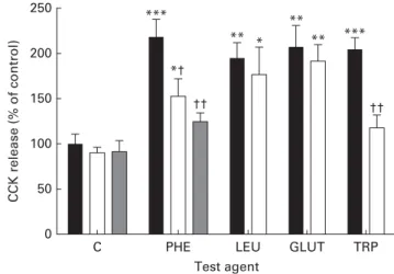
Using isolated and purified mouse mucosal enhanced green fluorescent protein-expressing CCK cells, Wang et al.(105)and Liou et al.(106) have shown that aromatic amino acids L-PHE andL-TRP stimulate the release of CCK through CaSR(105,106). We have shown that the addition of a CaSR antagonist, NPS2143, inhibits PHE-stimulated CCK release partially and TRP-induced CCK secretion totally in mouse proximal intestinal tissue explants, with no effect on LEU- or GLUT-induced CCK secretion (see Fig. 2). The partial and total inhibition of CaSR-mediated PHE- and TRP-induced CCK secretion is consistent with data presented by Wang et al.(105), using purified CCK – enhanced green fluorescent protein cells in the presence and absence of another CaSR antagonist, Calhex 231(105).
Therefore, it appears that both receptors T1R1 – T1R3 and CaSR are capable of sensing L-PHE. Interestingly, in support of this, the addition of NPS2143 together with gurmarin totally inhibits PHE-induced CCK release from mouse proximal intes-tinal tissue(24)(see Fig. 2). The experimental data suggest that CaSR acts as an intestinal amino acid receptor specifically
sensingL-aromatic amino acids, while the T1R1 – T1R3 hetero-dimer responds to a number of amino acids provoking CCK secretion.
Nutrient sensing GPCR are attractive and orally accessible targets for manipulations by functional foods and supple-ments. This has applications for maintaining health and preventing disease.
Acknowledgements
S. P. S.-B. acknowledges the University of Liverpool, the Wellcome Trust and Pancosma for providing financial support. The authors thank peer reviewers for providing constructive and critical comments. The authors’ contributions are as follows: K. D., M. A.-R. and A. W. M. carried out the experiments; D. B. provided scientific and practical advice; S. P. S.-B. wrote the article. S. P. S.-B., K. D., M. A.-R. and A. W. M. declare no conflicts of interest. D. B. is an employee of Pancosma.
References
1. Milligan G & McGrath JC (2009) GPCR theme editorial. Br J Pharmacol 158, 1 – 4.
2. Wellendorph P, Johansen LD & Bra¨uner-Osborne H (2010) The emerging role of promiscuous 7TM receptors as chemosensors for food intake. Vitam Horm 84, 151 – 184. 3. Dyer J, Daly K, Salmon KS, et al. (2007) Intestinal glucose
sensing and regulation of intestinal glucose absorption. Biochem Soc Trans 35, 1191 – 1194.
4. Hirasawa A, Tsumaya K, Awaji T, et al. (2005) Free fatty acids regulate gut incretin glucagon-like peptide-1 secretion through GPR120. Nat Med 11, 90 – 94.
5. Jang HJ, Kokrashvili Z, Theodorakis MJ, et al. (2007) Gut-expressed gustducin and taste receptors regulate secretion of glucagon-like peptide-1. Proc Natl Acad Sci U S A 104, 15069 – 15074.
6. Liou AP, Lu X, Sei Y, et al. (2011) The G-protein-coupled receptor GPR40 directly mediates long-chain fatty acid-induced secretion of cholecystokinin. Gastroenterology 140, 903 – 912.
7. Margolskee RF, Dyer J, Kokrashvili Z, et al. (2007) T1R3 and gustducin in gut sense sugars to regulate expression of Naþ-glucose cotransporter 1. Proc Natl Acad Sci U S A 104, 15 075 – 15 080.
8. Moran AW, Al-Rammahi MA, Arora DK, et al. (2010)
Expression of Naþ/glucose co-transporter 1 (SGLT1) is enhanced by supplementation of the diet of weaning pig-lets with artificial sweeteners. Br J Nutr 104, 637 – 646.
9. Hansen KB, Rosenkilde MM, Knop FK, et al. (2011)
2-Oleoyl glycerol is a GPR119 agonist and signals GLP-1
release in humans. J Clin Endocrinol Metab 96,
E1409 – E1417.
10. Chandrashekar J, Hoon MA, Ryba NJ, et al. (2006) The receptors and cells for mammalian taste. Nature 444, 288 – 294.
11. Li X, Staszewski L, Xu H, et al. (2002) Human receptors for sweet and umami taste. Proc Natl Acad Sci U S A 99, 4692 – 4696.
12. Nelson G, Chandrashekar J, Hoon MA, et al. (2002) An amino-acid taste receptor. Nature 416, 199 – 202.
13. McLaughlin SK, McKinnon PJ & Margolskee RF (1992) Gustducin is a taste cell-specific G protein closely related to the transducins. Nature 357, 563 – 569.
0 50 100 150 *** ** ** ** *** †† * *† †† 200 250 CCK release (% of control) Test agent
C PHE LEU GLUT TRP
Fig. 2. Effect of calcium-sensing receptor antagonist NPS2143 onL-amino acid-induced cholecystokinin (CCK) release by mouse proximal small intestine. Mouse proximal intestinal tissue explants were incubated for 1 h at 378C in Hank’s Balanced Salt Solution (HBSS) (containing 1·26 mM-Ca2þ) –20 mM -HEPES (pH 7·4) supplemented withL-amino acids or were untreated, in the absence ( ) or presence of 25 mM-NPS2143 ( ) or 25 mM-NPS2143 þ 30 mg/ml gurmarin ( ). CCK release is shown as a percentage of that in untreated control tissue. C, untreated; PHE, phenylalanine (20 mmol/l); LEU, leucine (20 mmol/l); GLUT, glutamate (20 mmol/l); TRP, tryptophan (20 mmol/l). Data are means, with standard errors represented by vertical bars. Mean value was significantly different from that of the corresponding control (C): * P, 0·05, ** P, 0·01, *** P, 0·001. Mean value was significantly different from that for the same test agent in the absence of NPS2143 and/or gurmarin: † P, 0·05, †† P, 0·01. Reprinted with permission from Daly et al.(24).
British
Journal
of
14. Yamaguchi S (1970) The synergistic taste effect of mono-sodium glutamate and dimono-sodium 50-inosinate. J Food Sci 32, 473 – 478.
15. Yasumatsu K, Ogiwara Y, Takai S, et al. (2012) Umami taste in mice uses multiple receptors and transduction pathways. J Physiol 590, 1155 – 1170.
16. Yoshii K, Yokouchi C & Kurihara K (1986) Synergistic effects of 50-nucleotides on rat taste responses to various
amino acids. Brain Res 367, 45 – 51.
17. Zhang F, Klebansky B, Fine RM, et al. (2008) Molecular mechanism for the umami taste synergism. Proc Natl Acad Sci U S A 105, 20930 – 20934.
18. He W, Yasumatsu K, Varadarajan V, et al. (2004) Umami
taste responses are mediated by a-transducin and
a-gustducin. J Neurosci 24, 7674 – 7680.
19. Imoto T, Miyasaka A, Ishima R, et al. (1991) A novel peptide
isolated from the leaves of Gymnema sylvestre.
I. Characterization and its suppressive effect on the neural responses to sweet stimuli in the rat. Comp Biochem Physiol A 100, 309 – 314.
20. Ninomiya Y & Imoto T (1995) Gurmarin inhibition of sweet taste responses in mice. Am J Physiol Regul Integr Comp Physiol 268, R1019 – R1025.
21. Ninomiya Y, Nakashima K, Fukuda A, et al. (2000)
Responses to umami substances in taste bud cells inner-vated by the chorda tympani and glossopharyngeal nerves. J Nutr 130, 950S – 953S.
22. Yamamoto T, Matsuo R, Fujimoto Y, et al. (1991) Electro-physiological and behavioral studies on the taste of umami substances in the rat. Physiol Behav 49, 919 – 925.
23. Yasumatsu K, Ohkuri T, Sanematsu K, et al. (2009)
Genetically-increased taste cell population with Ga
-gustducin-coupled sweet receptors is associated with increase of gurmarin-sensitive taste nerve fibers in mice. BMC Neurosci 10, 152.
24. Daly K, Al-Rammahi M, Moran A, et al. (2013) Sensing of amino acids by the gut-expressed taste receptor T1R1 – T1R3 stimulates CCK secretion. Am J Physiol Gastro-intest Liver Physiol 304, G271 – G282.
25. Wu SV, Rozengurt N, Yang M, et al. (2002) Expression of bitter taste receptors of the T2R family in the gastro-intestinal tract and enteroendocrine STC-1 cells. Proc Natl Acad Sci U S A 99, 2392 – 2397.
26. Dyer J, Salmon KS, Zibrik L, et al. (2005) Expression of sweet taste receptors of the T1R family in the intestinal tract and enteroendocrine cells. Biochem Soc Trans 33, 302 – 305.
27. Rozengurt N, Wu SV, Chen MC, et al. (2006) Colocalization of the alpha-subunit of gustducin with PYY and GLP-1 in L cells of human colon. Am J Physiol Gastrointest Liver Physiol 291, G792 – G802.
28. Sutherland K, Young RL, Cooper NJ, et al. (2007) Phenotypic characterization of taste cells of the mouse small intestine. Am J Physiol Gastrointest Liver Physiol 292, G1420 – G1428. 29. Young RL, Sutherland K, Pezos N, et al. (2009) Expression of
taste molecules in the upper gastrointestinal tract in humans with and without type 2 diabetes. Gut 58, 337 – 346. 30. Batchelor DJ, Al-Rammahi M, Moran AW, et al. (2010)
Sodium/glucose cotransporter-1, sweet receptor and disac-charidase expression in the intestine of the domestic dog and cat: species of different dietary habit. Am J Physiol Regul Integr Comp Physiol 300, R67 – R75.
31. Wang JH, Inoue T, Higashiyama M, et al. (2011) Umami receptor activation increases duodenal bicarbonate secretion via glucagon-like peptide-2 release in rats. J Pharmacol Exp Ther 339, 464 – 473.
32. Geraedts MC, Takahashi T, Vigues S, et al. (2012) Trans-formation of postingestive glucose responses after deletion of sweet taste receptor subunits or gastric bypass surgery. Am J Physiol Endocrinol Metab 303, E464 – E474.
33. Daly K, Al-Rammahi M, Arora DK, et al. (2012) Expression of sweet receptor components in equine small intestine: relevance to intestinal glucose transport. Am J Physiol Regul Integr Comp Physiol 303, R199 – R208.
34. Rehfeld JF (2004) A centenary of gastrointestinal endocrin-ology. Horm Metab Res 36, 735 – 741.
35. Cottrell JJ, Stoll B, Buddington RK, et al. (2006) Glucagon-like peptide-2 protects against TPN-induced intestinal hexose malabsorption in enterally refed piglets. Am J Physiol Gastrointest Liver Physiol 290, G293 – G300. 36. Sangild PT, Tappenden KA, Malo C, et al. (2006)
Glucagon-like peptide 2 stimulates intestinal nutrient absorption in parenterally fed newborn pigs. J Pediatr Gastroenterol Nutr 43, 160 – 167.
37. Dumoulin V, Moro F, Barcelo A, et al. (1998) Peptide YY, glucagon-like peptide-1, and neurotensin responses to luminal factors in the isolated vascularly perfused rat ileum. Endocrinol 139, 3780 – 3786.
38. Orskov C, Holst JJ, Khuhtsen K, et al. (1986) Glucagon-like peptides GLP-1 and GLP-2, predicted products of the glucagon gene, are secreted separately from pig small intes-tine but not pancreas. Endocrinol 119, 1467 – 1475. 39. Layer P, Holst JJ, Grandt D, et al. (1995) Ileal release of
glucagon-like peptide-1 (GLP-1). Association with
inhibition of gastric acid secretion in humans. Digest Dis Sci 40, 1074 – 1082.
40. Kokrashvili Z, Mosinger B & Margolskee RF (2009) Taste signaling elements expressed in gut enteroendocrine cells regulate nutrient-responsive secretion of gut hormones. Am J Clin Nutr 90, 822S – 825S.
41. Brubaker PL, Crivici A, Izzo A, et al. (1997) Circulating and tissue forms of the intestinal growth factor, glucagon-like peptide-2. Endocrinology 138, 4837 – 4843.
42. Smushkin G, Sathananthan A, Man CD, et al. (2012) Defects in GLP-1 response to an oral challenge do not play a significant role in the pathogenesis of prediabetes. J Clin Endocrinol Metab 97, 589 – 598.
43. Rogers GJ, Tolhurst G, Ramzan A, et al. (2011) Electrical activity-triggered glucagon-like peptide-1 secretion from primary murine L-cells. J Physiol 589, 1081 – 1093. 44. Parker HE, Habib AM, Rogers GJ, et al. (2009)
Nutrient-dependent secretion of glucose-Nutrient-dependent insulinotropic polypeptide from primary murine K cells. Diabetologia 52, 289 – 298.
45. Fujita Y, Wideman RD, Speck M, et al. (2009) Incretin release from gut is acutely enhanced by sugar but not by sweeteners in vivo. Am J Physiol Endocrinol Metab 296, E473 – E479.
46. Ma J, Bellon M, Wishart JM, et al. (2009) Effect of the artificial sweetener, sucralose, on gastric emptying and incretin hormone release in healthy subjects. Am J Physiol Gastrointest Liver Physiol 296, G735 – G739.
47. Steinert RE, Gerspach AC, Gutmann H, et al. (2011) The functional involvement of gut-expressed sweet taste receptors in glucose-stimulated secretion of glucagon-like peptide-1 (GLP-1) and peptide YY (PYY). Clin Nutr 30, 524 – 532.
48. Renwick AG (1986) The metabolism of intense sweeteners. Xenobiotica 16, 1057 – 1071.
49. Akermoun M, Koglin M, Zvalova-Iooss D, et al. (2005) Characterization of 16 human G protein-coupled receptors
British
Journal
of
expressed in baculovirus-infected insect cells. Protein Expr Purif 44, 65 – 74.
50. Rasmussen SG, Choi HJ, Rosenbaum DM, et al. (2007) Crystal structure of the human beta2 adrenergic G-protein-coupled receptor. Nature 450, 383 – 387.
51. Lai X (2013) A reproducible method to enrich membrane proteins with high-purity and high-yield for an LC – MS/
MS approach in quantitative membrane proteomics.
Electrophoresis 34, 809 – 817.
52. Naganathan S, Ye S, Sakmar TP, et al. (2013) Site-specific
epitope tagging of G protein-coupled receptors by
bioorthogonal modification of a genetically encoded unna-tural amino acid. Biochemistry 52, 1028 – 1036.
53. Choksawangkarn W, Kim SK, Cannon JR, et al. (2013) Enrichment of plasma membrane proteins using nanoparti-cle pellinanoparti-cles: comparison between silica and higher density nanoparticles. J Proteome Res 12, 1134 – 1141.
54. Reimann F, Habib AM, Tolhurst G, et al. (2008) Glucose sen-sing in L cells: a primary cell study. Cell Metab 8, 532 – 539. 55. Oya M, Kitaguchi T, Pais R, et al. (2013) The G
protein-coupled receptor family C group 6 subtype A (GPRC6A) receptor is involved in amino acid-induced glucagon-like peptide-1 secretion from GLUTag cells. J Biol Chem 288, 4513 – 4521.
56. Young RL (2011) Sensing via intestinal sweet taste path-ways. Front Neurosci 5, 23.
57. Iwatsuki K, Nomura M, Shibata A, et al. (2010) Generation
and characterization of T1R2-LacZ knock-in mouse.
Biochem Biophys Res Commun 402, 495 – 499.
58. Tolhurst G, Heffron H, Lam YS, et al. (2012) Short-chain fatty acids stimulate glucagon-like peptide-1 secretion via the G-protein-coupled receptor FFAR2. Diabetes 61, 364 – 371.
59. Dyer J, Al-Rammahi M, Waterfall L, et al. (2009) Adaptive response of equine intestinal Naþ/glucose co-transporter (SGLT1) to an increase in dietary soluble carbohydrate. Pflugers Arch 458, 419 – 430.
60. Moran AW, Al-Rammahi MA, Arora DK, et al. (2010)
Expression of Naþ/glucose co-transporter 1 (SGLT1) in the intestine of piglets weaned to different concentrations of dietary carbohydrate. Br J Nutr 104, 647 – 655.
61. Gorboulev V, Schu¨rmann A, Vallon V, et al. (2012) Na(þ )-D-glucose cotransporter SGLT1 is pivotal for intestinal glu-cose absorption and gluglu-cose-dependent incretin secretion. Diabetes 61, 187 – 196.
62. Gruzdkov AA, Gromova LV, Grefner NM, et al. (2012) Kinetics and mechanisms of glucose absorption in the rat small intestine under physiological conditions. J Biophys Chem 3, 191 – 200.
63. Hirschhorn N & Greenough WB 3rd. (1991) Progress in oral rehydration therapy. Sci Am 264, 50 – 56.
64. Ferraris RP & Diamond JM (1989) Specific regulation of intestinal nutrient transporters by their dietary substrates. Annu Rev Physiol 51, 125 – 141.
65. Shirazi-Beechey SP, Hirayama BA, Wang Y, et al. (1991) Ontogenic development of lamb intestinal sodium-glucose co-transporter is regulated by diet. J Physiol 437, 699 – 708. 66. Dyer J, Vayro S, King TP, et al. (2003) Glucose sensing in the intestinal epithelium. Eur J Biochem 270, 3377 – 3388. 67. Stearns AT, Balakrishnan A, Rhoads DB, et al. (2010) Rapid
upregulation of sodium-glucose transporter SGLT1 in response to intestinal sweet taste stimulation. Ann Surg 251, 865 – 871.
68. Buddington RK, Chen JW & Diamond JM (1991) Dietary regulation of intestinal brush-border sugar and amino acid transport in carnivores. Am J Physiol 261, R793 – R801.
69. Barfull A, Garriga C, Mitjans M, et al. (2002) Ontogenetic expression and regulation of Na(þ )-D-glucose cotranspor-ter in jejunum of domestic chicken. Am J Physiol Gastroint-est Liver Physiol 282, G559 – G564.
70. Tsai CH, Hill M, Asa SL, et al. (1997) Intestinal growth-promoting properties of glucagon-like peptide-2 in mice. Am J Physiol 273, E77 – E84.
71. Ramsanahie A, Duxbury MS, Grikscheit TC, et al. (2003) Effect of GLP-2 on mucosal morphology and SGLT1 expression in tissue-engineered neointestine. Am J Physiol Gastrointest Liver Physiol 285, G1345 – G1352.
72. Cheeseman CI (1997) Upregulation of SGLT-1 transport activity in rat jejunum induced by GLP-2 infusion in vivo. Am J Physiol 273, R1965 – R1971.
73. Hunziker W, Whitney JA & Mellman I (1991) Selective inhibition of transcytosis by brefeldin A in MDCK cells. Cell 67, 617 – 627.
74. Lippincott-Schwartz J, Yuan L, Tipper C, et al. (1991) Brefeldin A’s effects on endosomes, lysosomes, and the TGN suggest a general mechanism for regulating organelle structure and membrane traffic. Cell 67, 601 – 616. 75. Helms J & Rothman JE (1992) Inhibition by brefeldin A of a
Golgi membrane enzyme that catalyses exchange of gua-nine nucleotide bound to ARF. Nature 360, 352 – 354. 76. Bjerknes M & Cheng H (2001) Modulation of specific
intestinal epithelial progenitors by enteric neurons. Proc Natl Acad Sci U S A 98, 12497 – 12502.
77. Mills JC & Gordon JI (2001) The intestinal stem cell niche: there grows the neighborhood. Proc Natl Acad Sci U S A 98, 12334 – 12336.
78. Everts H, van Beers-Schreurs HM & Vellenga L (1999) Nutrition of young piglets in relation to weaning problems. Tijdschr Diergeneeskd 124, 44 – 47.
79. Nabuurs MJ (1998) Weaning piglets as a model for studying pathophysiology of diarrhea. Vet Q 20, S42 – S45.
80. Nabuurs MJ, Hoogendoorn A & van Zijderveld-van Bemmel A (1996) Effect of supplementary feeding during the sucking period on net absorption from the small intestine of weaned pigs. Res Vet Sci 61, 72 – 77.
81. Sterk A, Schlegel P, Mul AJ, et al. (2008) Effects of sweeteners on individual feed intake characteristics and performance in group-housed weanling pigs. J Anim Sci 86, 2990 – 2997. 82. Konturek SJ, Radecki T, Thor P, et al. (1973) Release of
cholecystokinin by amino acids. Proc Soc Exp Biol Med 143, 305 – 309.
83. Chang CH, Chey WY, Sun Q, et al. (1994) Characterization of the release of cholecystokinin from a murine neuro-endocrine tumor cell line, STC-1. Biochim Biophys Acta 1221, 339 – 347.
84. Diepvens K, Ha¨berer D & Westerterp-Plantenga M (2008) Different proteins and biopeptides differently affect satiety
and anorexigenic/orexigenic hormones in healthy
humans. Int J Obes (Lond) 32, 510 – 518.
85. Foltz M, Ansems P, Schwarz J, et al. (2008) Protein hydroly-sates induce CCK release from enteroendocrine cells and act as partial agonists of the CCK1 receptor. J Agric Food Chem 56, 837 – 843.
86. Hira T, Maekawa T, Asano K, et al. (2009) Cholecystokinin secretion induced by beta-conglycinin peptone depends on Galphaq-mediated pathways in enteroendocrine cells. Eur J Nutr 48, 124 – 127.
87. Liou AP, Chavez DI, Espero E, et al. (2011) Protein hydrolysate-induced cholecystokinin secretion from enteroendocrine cells is indirectly mediated by the intestinal oligopeptide transporter PepT1. Am J Physiol Gastrointest Liver Physiol 300, G895 – G902.
British
Journal
of
88. Nakajima S, Hira T, Eto Y, et al. (2010) Soybean beta 51-63 peptide stimulates cholecystokinin secretion via a calcium-sensing receptor in enteroendocrine STC-1 cells. Regul Pept 159, 148 – 155.
89. Ne´moz-Gaillard E, Bernard C, Abello J, et al. (1998) Regulation of cholecystokinin secretion by peptones and peptidomimetic antibiotics in STC-1 cells. Endocrinology 139, 932 – 938.
90. Nishi T, Hara H, Hira T, et al. (2001) Dietary protein peptic hydrolysates stimulate cholecystokinin release via direct sensing by rat intestinal mucosal cells. Exp Biol Med (Maywood) 226, 1031 – 1036.
91. Sufian MK, Hira T, Asano K, et al. (2007) Peptides derived from dolicholin, a phaseolin-like protein in country beans (Dolichos lablab), potently stimulate cholecystokinin secretion from enteroendocrine STC-1 cells. J Agric Food Chem 55, 8980 – 8986.
92. Dockray GJ (2012) Cholecystokinin. Curr Opin Endocrinol Diabetes Obes 19, 8 – 12.
93. Guan D & Green GM (1996) Significance of peptic digestion in rat pancreatic secretory response to dietary protein. Am J Physiol Gastrointest Liver Physiol 271, G42 – G47.
94. Ivy AC & Oldberg E (1928) A hormone mechanism for gallbladder contraction and evacuation. Am J Physiol 86, 599 – 613.
95. White WO, Schwartz GJ & Moran TH (2000) Role of endogenous CCK in the inhibition of gastric emptying by peptone and intralipid in rats. Regul Pept 88, 47 – 53. 96. Moran TH (2009) Gut peptides in the control of food
intake. Int J Obes (Lond) 33, S7 – S10.
97. Hira T, Nakajima S, Eto Y, et al. (2008) Calcium-sensing receptor mediates phenylalanine-induced cholecystokinin
secretion in enteroendocrine STC-1 cells. FEBS J 275, 4620 – 4626.
98. Liddle RA (1994) Cholecystokinin. In Gut Peptides: Biochemistry and Physiology, [JH Walsh and GJ Dockray, editors]. New York: Raven.
99. Anika SM, Houpt TR & Houpt KA (1977) Satiety elicited by cholecystokinin in intact and vagotomized rats. Physiol Behav 19, 761 – 766.
100. Koop I & Buchan AM (1992) Cholecystokinin release from isolated canine epithelial cells in short-term culture. Gastroenterology 102, 28 – 34.
101. Meyer JH, Kelly GA, Spingola LJ, et al. (1976) Canine gut receptors mediating pancreatic responses to luminal L-amino acids. Am J Physiol 231, 669 – 677.
102. Owyang C, May D & Louie DS (1986) Trypsin suppression of pancreatic enzyme secretion. Differential effect on cholecystokinin release and the enteropancreatic reflex. Gastroenterology 91, 637 – 643.
103. Backus RC, Howard KA & Rogers QR (1997) The potency of dietary amino acids in elevating plasma cholecystokinin
immunoreactivity in cats is related to amino acid
hydrophobicity. Regul Pept 72, 31 – 40.
104. Conigrave AD, Quinn SJ & Brown EM (2000)L-Amino acid sensing by the extracellular Ca2þ-sensing receptor. Proc Natl Acad Sci U S A 97, 4814 – 4819.
105. Wang Y, Chandra R, Samsa LA, et al. (2011) Amino acids stimulate cholecystokinin release through the Ca2þ-sensing
receptor. Am J Physiol Gastrointest Liver Physiol 300, G528 – G537.
106. Liou AP, Sei Y, Zhao X, et al. (2011) The extracellular calcium-sensing receptor is required for cholecystokinin secretion in response toL-phenylalanine in acutely isolated intestinal I cells. Am J Physiol Gastrointest Liver Physiol 300, G538 – G546.