Review
Connexins in leukocytes: shuttling messages?
Cindy W. Wong, Thomas Christen, Brenda R. Kwak*
Division of Cardiology, Department of Medicine, University Hospital of Geneva, Foundation for Medical Research, 64 Avenue de la Roseraie, CH-1211 Geneva 4, Switzerland
Received 13 October 2003; received in revised form 27 November 2003; accepted 11 December 2003 Time for primary review 16 days
Abstract
Gap junctions, formed by the connexin (Cx) protein family, are intercellular channels that permit the cytoplasmic exchange of ions and small metabolites between neighboring cells, a process called gap junction intercellular communication (GJIC). These channels possess unique properties, including distinctive permeabilities for various signaling molecules, which depend on the connexin member(s) that form them. Importantly, GJIC must be properly controlled as its misregulation might contribute to diseases. Morphological and functional studies have revealed ‘gap junction-like’ structures and cell-to-cell communication involving cells of the immune system. The connexins involved in such contacts have been partially identified in recent years. This review focuses on the potential physiological roles of gap junctions in the development and recruitment of leukocytes as well as in the regulation of the immune response. Furthermore, the importance of GJIC in immuno-inflammatory pathologies is illustrated in atherosclerosis.
D 2004 European Society of Cardiology. Published by Elsevier B.V. All rights reserved.
Keywords: Cell communication; Connexins; Gap junctions; Leukocytes; Atherosclerosis
1. Introduction
Inflammation has evolved as a mechanism to defend the body against invading microorganisms and to respond to injuries. In both physiological and inflammatory leukocyte migration, immune cells have to breach the vascular barrier, a process referred to as diapedesis, extravasation or endo-thelial transmigration. The endothelium changes when there is an injury in the underlying tissue, such that a normally non-permissive surface becomes permissive for the adhe-sion of circulating leukocytes thus supporting the recruit-ment of inflammatory cells from the blood stream. A multi-step adhesion cascade has been proposed for leukocyte recruitment consisting of four steps[1,2]. In the first step, leukocytes tether then roll on the endothelial cells (ECs). This is followed by a triggering step, via chemokines and their receptors, that leads to the rapid activation of leukocyte integrins, and then a third step at which point the leukocyte
adheres tightly onto the endothelial surface. Finally, diape-desis occurs and the leukocyte crawls across ECs. Recent reviews concerning endothelial junctions and leukocyte transmigration have highlighted how molecules of the adherens and tight junctions regulate leukocyte extravasa-tion[3,4]. Although gap junctions were previously thought not to play a role in leukocyte transmigration, recent data challenges this view.
Gap junctions, formed by the connexin (Cx) protein family, connect adjacent cells together thus providing a direct means of intercellular communication [5]. Six nexins assemble in the plasma membrane to make a con-nexon or hemichannel. Two hemichannels from neighboring cells join to form an intercellular channel that clusters with other intercellular channels to build a gap junction. There are presently over 20 connexins, each of which can create functional channels with certain isoforms [6,7]. Gap junc-tion channels formed by different connexins have unique properties including distinctive permeabilities for various dyes and signaling molecules[7,8]. Increasing data suggest that connexons expressed on the plasma membrane of a variety of cells might be more than just precursors for the formation of gap junction channels [9,10]. In fact, the
0008-6363/$ - see front matterD 2004 European Society of Cardiology. Published by Elsevier B.V. All rights reserved. doi:10.1016/j.cardiores.2003.12.015
* Corresponding author. Tel.: 7233; fax: +41-22-382-7245.
E-mail address: [email protected] (B.R. Kwak).
controlled opening and closing of hemichannels formed by various connexins have been observed. Furthermore, some hemichannels have been shown to be permeable to a number of substances, including important signaling mole-cules [10 – 13]. Much evidence, obtained from various cell systems, has shown the regulation of connexin expression and gap junctional intercellular communication (GJIC) by pro-inflammatory mediators, thus uncovering the impor-tance of gap junctions in the modulation of the inflamma-tory response[14 – 18]. In this part of the spotlight issue, we focus on gap junctions in leukocytes and the immune response, specifically on their importance in the process of leukocyte maturation and recruitment. In particular, the development of atherosclerosis is presented to illustrate the role played by GJIC in immuno-inflammatory diseases.
2. Evidence for gap junction communication in leukocytes
Leukocytes are principal players in the immune system that undergo a developing process in order to deal with infections and injuries inflicted to the body. To fight off unwanted aggressors, leukocytes must leave their site of production, circulate in the blood, and get recruited to the initial spot of injury. This process requires coordinated communications between leukocytes and other cells. The role of GJIC during these different steps is slowly emerging. 2.1. Leukocyte formation
All blood cells originate from a common hematopoietic stem cell. In the adult, the stem cells are found mainly in the bone marrow and thymus, where they can either divide to produce more pluripotent stem cells or differentiate to various committed progenitor cells, each able to generate only one or a few types of blood cells. This process is strictly regulated by the specialized hematopoietic microen-vironment, which includes stromal cells. Based on the gap junction-like structures observed in the bone marrow, it was hypothesized that gap junctions help to coordinate function-al networks of stromfunction-al cells that support blood cell forma-tion [19 – 22] and subsequent studies have supported this idea as described below.
In vitro studies of cultured bone marrow or thymic stromal cells revealed Cx43 as the principal gap junction protein expressed by these primary cell types [23 – 25]. In addition, Cx31 and Cx45 have been detected in some stromal cell lines [26]. Moreover, functional studies have demonstrated the transfer of dye or electrical current be-tween stromal cells[23,25]. The consistent observations of high levels of Cx43 expression in neonatal bone marrow and low expression of this protein in adult bone marrow has lead to the hypothesis that GJIC is required during periods of active hematopoiesis, as observed in the growing neonate
[24,27]. Consistent with this idea is the finding that
drug-induced elimination of committed hematopoietic cells led to a dramatic increase in Cx43 expression in the bone marrow that returned to normal when hematopoietic progenitors had replenished the hematopoietic compartments [24]. Con-versely, differentiation of stromal cells into adipocytes, a cell type found in the yellow non-hematopoitic bone mar-row, was associated with reduced Cx43 expression levels and decreased GJIC[28]. Evidence that the expression level of Cx43 is critical for normal hematopoiesis in vivo was obtained recently using Cx43-deficient mice [29]. Indeed, lack of Cx43 expression during embryogenesis compro-mised the terminal development of primary B and T lymphocytes. In addition, the authors observed similar defects in heterozygous Cx43 (Cx43+/ ) embryos that express reduced levels of Cx43. However, the hematopoietic system was returned to normal at 4 weeks of age in the Cx43+/ mice. Interestingly, the regeneration of lymphoid and myeloid cells in Cx43+/ mice was severely impaired after drug-induced elimination of hematopoietic cells, thus providing further support to the hypothesis that GJIC is mostly required during periods of active hematopoiesis.
Gap junctions between stromal and hematopoietic cells have also been observed in the bone marrow in situ and long-term cultures of bone marrow[21]. Subsequent dye transfer studies demonstrated functional coupling between these cell types. It is thus tempting to hypothesize that GJIC between stromal cells and hematopoietic cells may allow for direct transfer of stromal cell-derived signals into developing he-matopoietic cells, thus regulating blood cell formation. How-ever, the actual existence of such a pathway remains contro-versial. It has been reported that chemical blockade of GJIC between stromal and hematopoietic cells decreased blood cell formation, an effect that was more pronounced in primitive than in committed progenitors [30]. In contrast, other researchers have been unable to observe dye transfer between stromal and hematopoietic cells or to detect connexin expres-sion in hematopoietic progenitors by reverse transcription-polymerase chain reaction (RT-PCR) [23,26,29]. Taken to-gether, it appears that gap junctions may play a role in hematopoiesis, particularly during active periods when blood cell formation is initiating or regenerating. However, the question as to whether GJIC between stromal cells, between hematopoietic cells or between both cell types is most critical for active hematopoiesis remains to be answered.
2.2. Connexin expression in leukocytes
The presence of gap junctions between leukocytes (homocellular contacts) as well as between leukocytes and other cells (heterocellular contacts) have been extensively reported (Table 1). Furthermore, increasing information is becoming available on the connexin isoforms expressed in the different leukocytes as well as on the regulation/induc-tion of GJIC by pro-inflammatory mediators in these cells
(Table 2). Nevertheless, most of these reports result from in vitro studies and remain to be proven in vivo.
2.2.1. Neutrophils
Gap junction-like structures were initially observed by electron microscopy (EM) between hamster neutrophils as well as between neutrophils and ECs[31]and then detected between trout neutrophils and macrophages[32]. After initial
failure to detect connexin mRNA or protein in peripheral blood cells, early studies indicated that connexin expression in neutrophils is inducible. The presence of Cx43 was detected by immunofluorescent staining (IF), only after stimulation with lipopolysaccharide (LPS), in hamster and
Table 2
Connexin expression in leukocytes
Leukocytes Connexins Species Conditions Evidences References
Neutrophils Cx43 Human LPS or TNF-a IF, WB [33]
Human Untreated IF, WB [34]
Hamster LPS IF, WB, EM [31]
Cx40 Human LPS or TNF-a IF, WB [33]
Human Untreated IF, WB [34]
Cx37 Human Untreated IF, WB [34]
Monocytes/ Cx43 Human Atherosclerotic macrophage foam cells NB [40]
macrophages Human LPS and INFg or TNF-a and INFg WB, NB [39]
Mouse J774 macrophage cell line WB, NB [42]
Mouse LPS and INF-g IF, WB, EM [41]
Hamster LPS IF, WB, EM [31]
Murine Brain stab wounds or primary cultures of microglias
WB, EM [38]
Cx37 Human and mouse Early atheromas IF [43]
Lymphocytes Cx43 Human Peripheral blood or tonsil-derived cell WB, RT-PCR, FACS [54]
Mouse Lymph node-derived cells IF [81,82]
Cx40 Human Tonsil-derived-cells WB, RT-PCR, FACS [54]
Cx37 Human Peripheral blood IF [58]
Mouse Lymph node derived cells IF [81,82]; Discrepancy
with[54]
IF, immunofluorescence; WB, Western blot; NB, Northern blot; RT-PCR, reverse transcriptase-polymerase chain reaction; FACS, fluorescence-activated cell sorter; EM, electron microscopy; LPS, lipopolysaccharide; TNF-a, tumor necrosis factor-a; INF-g, interferon-g.
Table 1
Gap junction formation by leukocytes
Leukocytes Interactors Species Conditions Evidences References
Neutrophils Neutrophils Human LPS or TNF-a, unidentified EC derived factor(s)
Dye coupling [33]
Neutrophils Neutrophils Hamster Ischemia reperfusion injury EM [31]
Neutrophils EC line Human Untreated Dye coupling [34]
Neutrophils Epithelial cell line Human Untreated Dye coupling [34]
Neutrophils ECs Hamster Ischemia reperfusion injury EM [31]
Monocytes Monocytes Human LPS or TNF-a and INF-g Dye coupling [39]
Monocytes Monocytes Hamster Ischemia reperfusion injury EM [31]
Monocytes Neutrophils Hamster Ischemia reperfusion injury EM [31]
Macrophages Macrophages Murine Arranged in chains Electrical coupling [35]
Macrophages Macrophages Murine Peritonitis EM [37]
Macrophages Macrophages Canine GM-CFC EM [36]
Macrophages Neutrophils Trout Bacterial agents EM [32]
Macrophages Epithelial cell line Murine Peritonitis Dye coupling, EM [37]
Microglial cells Microglial cells Murine LPS and INF-g or TNF-a and INF-g or unstimulated
Dye coupling [38]
Lymphocytes Lymphocytes Human PHA Electrical coupling [46]
Lymphocytes Lymphocytes Human PHA Electrical coupling, EM [48]
Lymphocytes Lymphocytes Bovine PHA Electrical coupling [44,45]
Lymphocytes Lymphocytes Rabbit PHA EM [49]
Lymphocytes EC Murine Lymphnode in situ EM [50]
Lymphocytes Epidermal Langerhans cells Murine Antigen activated EM [51,52]
Lymphocytes Dermal Langerhans cells Human Dermatitis in situ EM [60]
Lymphocytes ECs Human HTLV-1 + Dye coupling [80]
Lymphocytes ECs Human Dye coupling [53]
EC, endothelial cell; LPS, lipopolysaccharide; TNF-a, tumor necrosis factor-a; INF-g, interferon-g; GM-CFC, granulocyte-macrophage colony-forming cell; PHA, phytohemagglutinin; HTLV-1+, human T-cell leukemia virus type 1+; IF, immunofluorescence; WB, Western blot; NB, Northern blot; RT-PCR, reverse transcriptase-polymerase chain reaction; FACS, fluorescence-activated cell sorter; EM, electron microscopy.
human neutrophils[31,33]. Subsequent studies revealed the expression of both Cx40 and Cx43, but not Cx32, in human neutrophils after stimulation with LPS or TNF-a[33]. Al-though these activated human neutrophils were able to form homocellular gap junctions in vitro, they were not dye-coupled unless treated with EC derived factor(s)[33]. More recently, it was demonstrated that unstimulated human neu-trophils express Cx37, Cx40 and Cx43[34]. Protein expres-sion at the cell surface was confirmed by Western blots that revealed the presence of all three connexins in the membrane fractions. Cx37 was mainly localized in the pseudopodia of neutrophils whereas Cx40 and Cx43 showed a more granular organization. Importantly, expression of these connexins permitted GJIC between neutrophils and ECs in a rapid bidirectional and adhesion-dependent manner, which was decreased after treatment with TNF-a[34]. Taken together, there is various support for homocellular and heterocellular gap junctions in neutrophils. However, additional evidence at the level of messenger RNA is needed to verify the connexin isoform expressed under the different conditions.
2.2.2. Monocytes/macrophages
The establishment of intercellular communication be-tween macrophages, based on electrical coupling of adher-ent murine macrophages, was first reported by Levy et al.
[35]. Subsequently, gap junctions were morphologically detected between progeny of canine macrophages by freeze fracture EM[36]. Gap junction structures have since been described by EM between murine macrophages and an intestinal epithelial cell line [37]; between hamster mono-cytes as well as monomono-cytes and neutrophils [31]; and between rainbow trout macrophages and neutrophils [32]. Further support for GJIC between monocytes/macrophages and other cells has come from dye transfer assays. Dye coupling was observed between murine peritoneal macro-phages as well as between murine macromacro-phages and intes-tinal epithelial cells [37]. At brain stab wounds and in primary culture of murine microglias, a low dye coupling was observed. This coupling was dramatically increased with the treatment of IFN-g and LPS or IFN-g and TNF-a as well as inhibited by a gap junction blocker [38]. In addition, freshly isolated human monocytes treated with LPS or TNF-a and IFN-g were dye-coupled[39]. However, these studies are in conflict with other reports that demon-strate the lack of GJIC between monocytes/macrophages and other cells. For example, the transfer of dye was not observed in untreated human or mouse monocytes/macro-phages [40,41], between human monocytes/macrophages and ECs or between human monocytes/macrophages and SMCs [39,40]. To date, the expression of two connexin isoforms in monocytes/macrophages has been reported. The presence of Cx43 was found in the mouse macrophage cell line J774 [42], activated peritoneal macrophages from hamsters and mice [31,41]; brain stab wound and primary cultures of murine microglia [38]; and human monocytes/ macrophages stimulated with TNF-a and INF-g or LPS and
INF-g [39]. Moreover, Cx43 mRNA was detected in mac-rophage foam cells of human atherosclerotic carotid arteries
[40]. Interestingly, we observed Cx37 but not Cx43 in macrophages of early atheromas [43]. The induced expres-sion of other connexins in monocytes/macrophages has been examined and neither Cx32 nor Cx40 were detected after treatment with LPS or TNF-a and IFN-g [39,40]. Until these discrepancies become resolved, it remains unclear whether monocytes/macrophages communicate via GJIC.
2.2.3. Lymphocytes
The initial observations that lymphocytes can establish intercellular communication were reported in the early 1970s. Two separate groups detected electrical coupling between lymphocytes isolated from bovine lymph nodes or human peripheral blood after stimulation with phytohae-magglutinin (PHA) [44 – 47]. Subsequently, the ultrastruc-tural detection of gap junctions between PHA stimulated lymphocytes was observed [48,49]. This was followed by ultrastructural reports of heterocellular gap junctions be-tween lymphocytes and ECs or Langerhans cells [50 – 52]. Moreover, bidirectional transfer of cytoplasmic fluorescent dyes between lymphocytes and the endothelium has been reported [53]. Connexin distribution and GJIC has been studied in human and mouse lymphocyte subpopulations. Human peripheral blood-derived T, B and natural killer (NK) lymphocytes express solely Cx43, whereas tonsil-derived T and B lymphocytes express both Cx40 and Cx43 [54]. It is worth mentioning that Cx26, Cx32, Cx37 and Cx45 were not detected in these cells by RT-PCR[54]. However, the expression of Cx37 protein in human periph-eral blood lymphocytes was previously reported by another group [55]. Both human and mouse lymphocytes display functional GJIC, as assayed by dye transfer, which can be reduced by pharmacological agents or synthetic peptides known to block gap junctional communication. Interesting-ly, lymphocytes increase connexin expression or translocate connexins towards cell – cell interfaces upon activation with either PHA or concanavalin A[56 – 58].
3. A role for gap junction communication in the immune response
Cell-to-cell interactions are of major importance for expanding the competency of cells in the immune system to control infections and maintain tolerance. Activation of the adaptive immune response includes the interaction between T cell antigen receptors and major histocompati-bility complex (MHC) molecule – peptide complexes. This nanometer scale gap between the T lymphocyte and the antigen-presenting cell (APC) is referred to as the immuno-logical synapse [59]. Specificity of these recognitions is critical, as reactions to microbial peptides are required for clearance of many infections and responses to self-derived peptides on APCs can give rise to autoimmunity. Despite
the early observation of gap junctions in lymphocytes and the extensive characterization of connexin in primary and secondary lymphoid organs, knowledge on the potential role of gap junctions in the immune system is limited. However, as specific connexin blocking peptides have become more accessible, research on this topic pushes forward quickly.
Gap junctions, composed of at least Cx43, between antigen-presenting Langerhans cells and T lymphocytes were observed both in vitro and in vivo[51,52,55,60]. Saez et al.[58]demonstrated first that synthetic peptides homol-ogous to the extracellular loop of Cx43 drastically reduced proliferation of mitogen-activated T cells, indicating that GJIC may play a role in the adaptive immune response. Subsequently, Oviedo-Orta et al.[57]elegantly showed that disruption of GJIC influenced fundamental aspects of lym-phocyte function, including immunoglobulin (Ig) secretion and cytokine production. Indeed, inhibition of GJIC by synthetic peptides homologous to the first and second extracellular loop of Cx43 markedly reduced secretion of IgM, IgG and IgA in mixed cultures of activated purified human B and T lymphocytes. In addition, they observed in these cultures complex temporal inhibitory effects on cyto-kine synthesis, especially on interleukin-10. Taken together, these results open up towards the novel hypothesis that connexins, and likely GJIC itself, may be an important component of the molecular mechanism underlying lym-phocyte activation and function in the immune response. Clearly, further studies are needed to identify which mole-cules are passing through gap junction channels between APCs and T cells as well as T and B lymphocytes. Considering the narrow space of the immunological syn-apse, involvement of gap junction hemichannels as passage-way for metabolites to the extracellular space and leading to paracrine cell – cell signaling should also be considered[61].
4. Gap junction communication in leukocyte recruitment Three connexins, namely Cx37, Cx40 and Cx43, have been detected in the vascular endothelium in situ. The precise distribution of these connexins within the vessel wall is known to be species and vessel specific[62]. Cultured human umbilical vein endothelial cells (HUVECs) also express the three vascular connexins and they mainly locate at cell – cell contacts [63]. Interestingly, TNF-a altered the connexin expression pattern in HUVECs and reduced GJIC between these ECs[63]. This reduction in GJIC within the endothe-lium might serve two important purposes. First, it might protect the endothelium by restricting the spread of injurious signals via EC gap junctions, thus limiting the area of inflammation. Second, as more connexons from the ECs become available for docking, they might form heterocellular gap junctions with leukocytes to control leukocyte migration across the endothelium. As discussed below, there are recent indications of gap junctions between ECs and leukocytes and that GJIC might play a role in leukocyte extravasation.
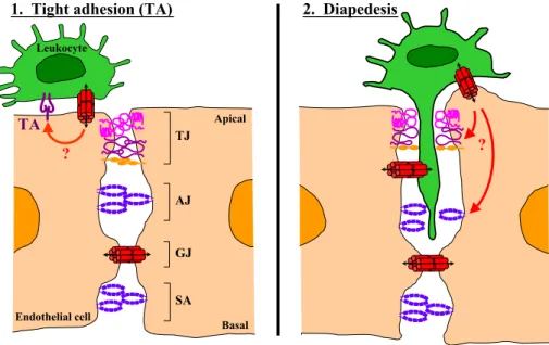
Cell communication via gap junctions during transmigra-tion was first described by Oviedi-Orta et al. [64]. In this study, they demonstrated by using dye transfer experiments that lymphocytes and ECs generate functional heterocellular gap junction channels during extravasation in vitro. Interest-ingly, blocking GJIC with pharmacological agents or con-nexin mimetic peptides caused only a modest reduction in transmigration of lymphocytes across an EC monolayer[61]. Neutrophils and HUVECs also form functional gap junction channels in vitro, as demonstrated by dye transfer experi-ments [34]. Moreover, this bidirectional coupling was re-duced when HUVECs were stimulated with TNF-a but not when stimulated with IFN-g or thrombin. Therefore, cou-pling between neutrophils and HUVECs is selectively mod-ulated during an inflammatory reaction, suggesting that this process might be of physiological relevance. Importantly, neutrophil transmigration was enhanced when GJIC was inhibited, suggesting a negative regulatory role for this coupling during the transmigration process. It was also shown in this study that strongly adherent neutrophils were more coupled than weakly adherent ones and that the adhesive properties between connexons played no role in this strength-ened cell adhesion process. This prompts a novel hypothesis that the tight adhesion, mediated by integrins and their ligands, between leukocytes and ECs might be modulated by signaling through gap junctions(Fig. 1). Finally, human monocytes were shown to form gap junctions with ECs in a blood brain barrier (BBB) model during the process of transmigration [39]. In addition, blockade of GJIC reduced the number of monocytes that transmigrated, suggesting that cell-to-cell signaling through gap junction channels might even affect the efficiency of the transmigration process across a tight endothelium. Transendothelial migration (TEM) of the different leukocytes appears to be differentially regulated by GJIC, such that inhibition of GJIC increased TEM of neu-trophils but decreased TEM of monocytes and had modest effects on lymphocyte TEM. Of major concern in the afore-mentioned studies is the specificity of the GJIC blocking reagents, pharmaceutical agents are plainly unspecific and the specificity of the mimetic peptides remains to be proven. Clearly, more work is required before definitive proof dem-onstrates that gap junctions do play a role in leukocyte TEM. Interestingly, regulation of leukocyte recruitment via GJIC might occur at different points of the multi-step adhesion cascade namely tight adhesion and diapedesis(Fig. 1). Based on current data, we hypothesize that there may be a cross talk between gap junctions (formed between leukocytes and ECs) and the integrin – IgSF CAM adhesion complex (also formed between leukocytes and ECs). This form of communication might then modulate the tight adhesion between leukocytes and ECs, controlling whether a leukocyte returns to the blood flow (the case for weak adhesion) or continues to transmi-grate into extravascular tissues (the case for firm adhesion). Likewise, we suggest that there may be cross talks between gap junctions (formed between leukocytes and ECs) with the EC tight and adherens junctions. The signals transmitted
would instruct the EC junctions to ‘‘open up’’ for leukocytes to pass through. As this line of research continues, one challenge will be to identify the signals that are being exchanged through gap junctions between leukocytes and ECs during different physiological states. This will certainly further our understanding of leukocyte migration during immune surveillance and inflammatory reactions that can cause diseases when improperly controlled.
5. Multiple roles for gap junction communication in atherosclerosis
Atherosclerosis is a progressive disease characterized in part by the accumulation of lipids, leukocytes, and smooth muscle cells (SMCs) in the intima of medium and large arteries[65]. This disease is presently the leading cause of illness and death in developed countries. The current view believes that inflammation is a major contributor to athero-genesis[66]. Moreover, evidence is growing that dysfunc-tional GJIC plays a role in the development of atherosclero-sis. Initially, Polacek et al.[40]reported the strong expression of Cx43 mRNA by macrophage foam cells in human athero-sclerotic carotid arteries. They extended this finding in a rabbit model of atherosclerosis, demonstrating that the ex-pression of Cx43 is upregulated in macrophage foam cells and downregulated in medial SMCs[67]. In another study,
Cx43 expression in intimal SMCs was shown to increase at early stages of human coronary atherosclerosis and to de-crease at later stages of the disease[68]. A genetic polymor-phism in the human Cx37 gene was reported as a potential prognostic marker for atherosclerotic plaque development
[69]. Furthermore, this Cx37 gene polymorphism was shown to possibly play a role in the manifestation of coronary atherosclerosis in Taiwan and Japan[70,71]. More recently, we demonstrated that expression of the three vascular con-nexins is altered in mouse and human atherosclerotic plaques
[43]and that the reduction of Cx43 expression inhibits the formation of atherosclerotic lesions in low-density lipopro-tein receptor-deficient (LDLR / ) mice[72,73]. These stud-ies have provided valuable clues as to how gap junction communication might play a role in the initiation as well as the progression of atherosclerotic plaque development.
5.1. The ‘‘initiation’’ and progression of an atherosclerotic plaque
The many risk factors that are implicated in atherogen-esis are linked by their common ability to promote inflam-matory reactions and injury to the endothelium. As a response to injury, the endothelium becomes dysfunctional leading to its increased expression of various cell adhesion molecules and secretion of chemoattractants to recruit specific leukocytes[74]. Leukocyte recruitment in the early
Fig. 1. The role of gap junctions in leukocyte recruitment to tissues. Sequential steps in leukocyte emigration, including tethering/rolling, activation, tight adhesion, and diapedesis (via inter-endothelial junctions TJ, AJ, GJ and SA), are controlled by specific cell adhesion molecules (CAMs). (1) Tight adhesion (TA), mediated by leukocyte integrins and endothelial IgSF CAMs (molecules depicted in dark purple), is triggered by signals such as chemokines. An alternative means to regulate this step might occur via the signaling molecules that pass through GJs formed between the leukocyte and EC (red arrow). This cross talk with the IgSF CAM – integrin complex may modulate the tightness of adhesion, consequently determining whether the leukocyte returns to the blood stream or continues to transmigrate. (2) Diapedesis, the passage of leukocytes across inter-endothelial junctions, appears to be controlled by both TJ and AJ; and recent reports suggest that GJ also participates in this process. The blockage of GJIC increases TEM of neutrophils but reduces TEM of monocytes/ macrophages and has a minor effect on lymphocyte TEM. In this model, it is hypothesized that GJIC between the leukocyte and EC cross talks with the EC TJ and AJ, signaling them to ‘‘open up’’ for the leukocytes to pass through (red arrows). IgSF, immunoglobulin supergene family; ECs, endothelial cells; TJ, tight junction; AJ, adherens junction; GJ, gap junctional channel; GJIC, gap junctional intercellular communication; SA, syndesmos/complexus adherens; TEM, transendothelial migration.
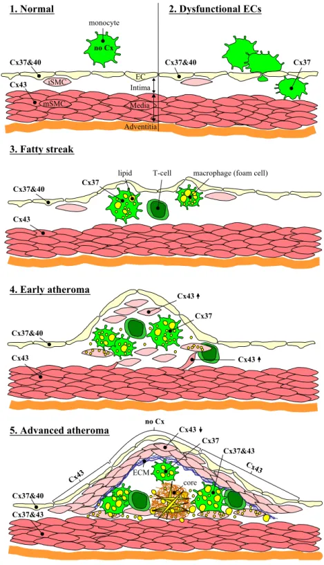
Fig. 2. Altered connexin expression during atherosclerotic plaque development. (1) Normal artery. Cx37 and Cx40 are expressed in ECs; Cx43 is expressed in mSMCs; connexins are not detected in circulating monocytes. (2) Dysfunctional ECs. As a response to injury, ECs become dysfunctional and recruit leukocytes, mainly monocytes/macrophages but also T lymphocytes, into the intima. Note the induced expression of Cx37 in intimal monocytes/macrophages. (3) Fatty streak. As leukocytes accumulate in the intima, monocytes mature into macrophages that take up lipid into their cytoplasm and become macrophage foam cells. Note the continued expression of Cx37 in the intima macrophages. (4) Early atheroma. Some mSMCs migrate into the intima, where the release of pro-inflammatory molecules by themselves and leukocytes induce iSMC proliferation. Lipids start to accumate in the extracellular space and in iSMCs. Note the increased expression of Cx43 in iSMCs compared to mSMCs, however the expression of connexins in ECs and mSMCs has not changed at this stage. (5) Advanced atheroma. A fibrous cap is formed by iSMCs and ECM that covers the lesion area. The central core of this lesion contains necrotic debris, extracellular lipids including cholesterol crystals. Note the disappearance of Cx37 and Cx40 in the diseased ECs, and the induced expression of Cx43 in ECs covering the shoulder regions of the lesion. In addition to Cx37, Cx43 is also detected in macrophage foam cells located in the shoulder regions. Another notable change at this stage is the reduced Cx43 expression in the iSMCs and the induced Cx37 expression in mSMCs. EC, endothelial cell; iSMC, intimal smooth muscle cell; mSMC, medial smooth muscle cell; Cx, connexin; ECM, extracellular matrix; core, lipid and/or necrotic core.
phases of atherosclerosis involves mainly monocytes [75]. However, T lymphocytes are also implicated in the early development of the disease [76]. After adhering to the dysfunctional endothelium, the monocyte transmigrates be-tween intact ECs to penetrate into the arterial intima. In the intima, monocytes proliferate and mature under the influ-ence of cytokines, chemokines and growth factors secreted by themselves and other atheroma-associated cells. Further-more, the induced expression of scavenger receptors permit macrophages to accumulate lipids within their cytoplasm and eventually progress to the arterial foam cells, a hallmark of the arterial lesion. These foam cells along with the T cells constitute the fatty streak known as the earliest form of atherosclerotic plaques.
Evidently, diapedesis of leukocytes is a prerequisite for the formation of atherosclerotic plaques. Thus, accelerating or decelerating monocyte/macrophage diapedesis might speed up or slow down atherosclerosis. In fact, transmigra-tion of monocytes/macrophages but not T lymphocytes is significantly reduced by inhibiting GJIC[39,64]. Moreover, reducing the expression of Cx43 in LDLR / mice de-creased the number of macrophages and T cells in the atheroma as well as the progression of atherosclerosis[72]. Taken together, it appears that GJIC is somehow modified in atherosclerosis leading to the enhanced leukocyte recruit-ment. A possible mechanism for this transformation may be via altered connexin expression resulting in the improper exchange of signaling molecules that cause miscommunica-tion. In fact, such alterations in connexin expression in leukocytes and the endothelium are known to occur during the development of atherosclerosis[43] (Fig. 2). Knowing that the properties of individual gap junction channels are distinct, it seems likely that the Cx37/Cx40 to Cx43 switch in the endothelium and the Cx37 to Cx37/Cx43 switch in macrophage foam cells will drastically change the messages exchanged between these cells. It is worth mentioning that since monocytes/macrophages can form gap junctions with adjacent monocytes/macrophages, and perhaps even with neighboring T lymphocytes and SMCs, miscommunication among these cells might play an additional role in plaque formation. For instance, lipid uptake by mature macrophages might rely on GJIC.
5.2. The stability and rupture of an atherosclerotic plaque The continued inflammatory response and accumulation of lipids work together with other events to promote atherosclerotic plaque growth and eventually rupture [66]. During the growing phase, medial SMCs migrate to the top of the intima where they multiply and produce components of the extracellular matrix (ECM). The SMCs and matrix molecules coalesce to form a strong fibrous cap that covers the original atherosclerotic site. Although this adds to the size of the plaque, it also seals the plaque off safely from the blood and reduces the chance of rupture. As this cap matures, some of the cells underneath die and lipids are
released. Therefore, this region is referred to as the lipid or necrotic core of the atherosclerotic lesion. Eventually, the fibrous cap of a plaque might break open, triggering a blood clot to develop over the rupture. Plaques that are most likely to break possess a thinned cap, a large lipid pool and many macrophages. This plaque phenotype is partially dependent on the activities of macrophages. Macrophage foam cells secrete pro-inflammatory cytokines that amplify the local inflammatory response in the lesion as well as reactive oxygen species that further induce macrophage proliferation and lipid uptake. In addition, the activated macrophages produce matrix metalloproteinases (MMPs) that can degrade the ECM thus weakening the plaque’s fibrous cap.
In addition to the initiation phase, GJIC might play a role in the progression of atherosclerotic plaques. For example, reducing Cx43 expression in LDLR / mice led to the development of atherosclerotic lesions that exhibited thicker fibrous caps with more collagen and SMCs, a phenotype associated with plaque stability [72]. Thus, it seems beneficial to reduce Cx43 mediated GJIC in atherosclerosis. Clearly, it will be interesting to see how changes in expression of other connexins might affect this disease. More recently, Eugenin et al. [39]showed that the increased GJIC in monocytes enhanced the release of MMP-2,3 but not MMP-9 by these cells. This amplified release of MMPs in atherosclerotic lesions could be dele-terious since it might promote plaque rupture and induce thrombosis. Although it remains to be proven, we envision that hemichannels on the macrophages in the lesions may also play a role in plaque development. For instance, the hemichannels might become misregulated such that they convert from their normally closed state to an open state leading to intracellular leakage and macrophage death. Taken together, altered GJIC may affect several processes required to promote atherosclerosis.
6. Perspectives and future directions
Are connexins in leukocytes forming gap junction (hemi-) channels to ‘‘shuttle messages’’? There is substantial evi-dence in support of leukocyte homocellular and heterocellu-lar gap junction assembly that allows for intercelluheterocellu-lar communication. On the contrary, it remains only speculative that hemichannels serve as bidirectional gateways between the intra- and extracellular space possibly leading to paracrine cell – cell signalling under particular circumstances. In vitro studies show that altered GJIC affects the migration and development of leukocytes, thus influencing the recruitment of leukocyte subtypes to sites of inflammation as well as the activation state of the immune system. Importantly, these observations are corroborated by recent in vivo studies on atherosclerosis. The dysregulation of GJIC is also implicated in other inflammatory diseases and reactions such as acute pancreatitis[18], cystic fibrosis[77], ischemia – reperfusion injury in liver[78]and heart[79], as well as wound repair in
skin[17]. We are far from identifying all the signals that go through gap junction (hemi-) channels and we know even less about when and how those molecules might cross talk with other molecules in any given situation. Perhaps, the answers to some of our key questions might not be so far away as we bridge ‘the gap’ between inter-disciplinary sciences.
Acknowledgements
The authors are especially grateful to Dr. Marc Chanson for helpful discussions and critical comments on the manuscript. This work is supported by grants from the Swiss National Science Foundation (#3234-066311.01, #PPOO-68883 and #3100-067777.02) and the Fondation Novartis.
References
[1] Butcher EC, Picker LJ. Lymphocyte homing and homeostasis. Sci-ence 1996;272:60 – 6.
[2] Springer TA. Traffic signals for lymphocyte recirculation and leuko-cyte emigration: the multistep paradigm. Cell 1994;76:301 – 14. [3] Aurrand-Lions M, Johnson-Leger C, Imhof BA. Role of
interendo-thelial adhesion molecules in the control of vascular functions. Vasc Pharmacol 2002;39:239 – 46.
[4] Luscinskas FW, Ma S, Nusrat A, Parkos CA, Shaw SK. The role of endothelial cell lateral junctions during leukocyte trafficking. Immu-nol Rev 2002;186:57 – 67.
[5] Evans WH, Martin PE. Gap junctions: structure and function (Re-view). Mol Membr Biol 2002;19:121 – 36.
[6] Willecke K, Eiberger J, Degen J, et al. Structural and functional diversity of connexin genes in the mouse and human genome. Biol Chem 2002;383:725 – 37.
[7] Harris AL. Emerging issues of connexin channels: biophysics fills the gap. Q Rev Biophys 2001;34:325 – 472.
[8] White TW. Nonredundant gap junction functions. News Physiol Sci 2003;18:95 – 9.
[9] Goodenough DA, Paul DL. Beyond the gap: functions of unpaired connexon channels. Nat Rev, Mol Cell Biol 2003;4:285 – 94. [10] Saez JC, Berthoud VM, Branes MC, Martinez AD, Beyer EC. Plasma
membrane channels formed by connexins: their regulation and func-tions. Physiol Rev 2003;83:1359 – 400.
[11] Saez JC, Contreras JE, Bukauskas FF, Retamal MA, Bennett MV. Gap junction hemichannels in astrocytes of the CNS. Acta Physiol Scand 2003;179:9 – 22.
[12] John S, Cesario D, Weiss JN. Gap junctional hemichannels in the heart. Acta Physiol Scand 2003;179:23 – 31.
[13] Vergara L, Bao X, Bello-Reuss E, Reuss L. Do connexin 43 gap-junctional hemichannels activate and cause cell damage during ATP depletion of renal-tubule cells? Acta Physiol Scand 2003;179:33 – 8. [14] Hu J, Cotgreave IA. Differential regulation of gap junctions by
proin-flammatory mediators in vitro. J Clin Invest 1997;99:2312 – 6. [15] Huang S, Dudez T, Scerri I, et al. Defective activation of c-Src in
cystic fibrosis airway epithelial cells results in loss of tumor necrosis factor-alpha-induced gap junction regulation. J Biol Chem 2003; 278:8326 – 32.
[16] De Maio A, Vega VL, Contreras JE. Gap junctions, homeostasis, and injury. J Cell Physiol 2002;191:269 – 82.
[17] Qiu C, Coutinho P, Frank S, et al. Targeting connexin43 expres-sion accelerates the rate of wound repair. Curr Biol 2003;13: 1697 – 703.
[18] Frossard JL, Rubbia-Brandt L, Wallig MA, et al. Severe acute pan-creatitis and reduced acinar cell apoptosis in the exocrine pancreas of mice deficient for the Cx32 gene. Gastroenterology 2003;124: 481 – 93.
[19] Watanabe Y. Fine structure of bone marrow stroma. Nippon Ketsueki Gakkai Zasshi 1985;48:1688 – 700.
[20] Campbell FR. Gap junctions between cells of bone marrow: an ultra-structural study using tannic acid. Anat Rec 1980;196:101 – 7. [21] Campbell FR. Ultrastructural studies of intercellular contacts
(junc-tions) in bone marrow. A review. Scand Electron Microsc 1986;196: 621 – 9.
[22] Rosendaal M, Gregan A, Green CR. Direct cell – cell communication in the blood-forming system. Tissue Cell 1991;23:457 – 70. [23] Dorshkind K, Green L, Godwin A, Fletcher WH. Connexin-43-type
gap junctions mediate communication between bone marrow stromal cells. Blood 1993;82:38 – 45.
[24] Rosendaal M, Green CR, Rahman A, Morgan D. Up-regulation of the connexin43+ gap junction network in haemopoietic tissue before the growth of stem cells. J Cell Sci 1994;107(Pt. 1):29 – 37.
[25] Alves LA, Campos de Carvalho AC, Lima Cirne EO, et al. Functional gap junctions in thymic epithelial cells are formed by connexin 43. Eur J Immunol 1995;25:431 – 7.
[26] Cancelas JA, Koevoet WL, de Koning AE, et al. Connexin-43 gap junctions are involved in multiconnexin-expressing stromal support of hemopoietic progenitors and stem cells. Blood 2000; 96:498 – 505.
[27] Krenacs T, Rosendaal M. Connexin43 gap junctions in normal, regen-erating, and cultured mouse bone marrow and in human leukemias: their possible involvement in blood formation. Am J Pathol 1998;152: 993 – 1004.
[28] Umezawa A, Hata J. Expression of gap-junctional protein (connexin 43 or alpha 1 gap junction) is down-regulated at the transcriptional level during adipocyte differentiation of H-1/A marrow stromal cells. Cell Struct Funct 1992;17:177 – 84.
[29] Montecino-Rodriguez E, Leathers H, Dorshkind K. Expression of connexin 43 (Cx43) is critical for normal hematopoiesis. Blood 2000;96:917 – 24.
[30] Rosendaal M, Mayen A, de Koning A, et al. Does transmembrane communication through gap junctions enable stem cells to overcome stromal inhibition? Leukemia 1997;11:1281 – 9.
[31] Jara PI, Boric MP, Saez JC. Leukocytes express connexin 43 after activation with lipopolysaccharide and appear to form gap junctions with endothelial cells after ischemia – reperfusion. Proc Natl Acad Sci U S A 1995;92:7011 – 5.
[32] Afonso A, Lousada S, Silva J, Ellis AE, Silva MT. Neutrophil and macrophage responses to inflammation in the peritoneal cavity of rainbow trout Oncorhynchus mykiss. A light and electron microscop-ic cytochemmicroscop-ical study. Dis Aquat Org 1998;34:27 – 37.
[33] Branes MC, Contreras JE, Saez JC. Activation of human polymor-phonuclear cells induces formation of functional gap junctions and expression of connexins. Med Sci Monit 2002;8:BR313 – 23. [34] Zahler S, Hoffmann A, Gloe T, Pohl U. Gap-junctional coupling
between neutrophils and endothelial cells: a novel modulator of trans-endothelial migration. J Leukoc Biol 2003;73:118 – 26.
[35] Levy JA, Weiss RM, Dirksen ER, Rosen MR. Possible communica-tion between murine macrophages oriented in linear chains in tissue culture. Exp Cell Res 1976;103:375 – 85.
[36] Porvaznik M, MacVittie TJ. Detection of gap junctions between the progeny of a canine macrophage colony-forming cell in vitro. J Cell Biol 1979;82:555 – 64.
[37] Martin CA, el-Sabban ME, Zhao L, Burakoff R, Homaidan FR. Ad-hesion and cytosolic dye transfer between macrophages and intestinal epithelial cells. Cell Adhes Commun 1998;5:83 – 95.
[38] Eugenin EA, Eckardt D, Theis M, et al. Microglia at brain stab wounds express connexin 43 and in vitro form functional gap junc-tions after treatment with interferon-gamma and tumor necrosis fac-tor-alpha. Proc Natl Acad Sci U S A 2001;98:4190 – 5.
[39] Eugenin EA, Branes MC, Berman JW, Saez JC. TNF-alpha plus IFN-gamma induce connexin43 expression and formation of gap junctions between human monocytes/macrophages that enhance physiological responses. J Immunol 2003;170:1320 – 8.
[40] Polacek D, Lal R, Volin MV, Davies PF. Gap junctional communica-tion between vascular cells. Induccommunica-tion of connexin43 messenger RNA in macrophage foam cells of atherosclerotic lesions. Am J Pathol 1993;142:593 – 606.
[41] Alves LA, Coutinho-Silva R, Persechini PM, et al. Are there func-tional gap junctions or juncfunc-tional hemichannels in macrophages? Blood 1996;88:328 – 34.
[42] Beyer EC, Steinberg TH. Evidence that the gap junction protein con-nexin-43 is the ATP-induced pore of mouse macrophages. J Biol Chem 1991;266:7971 – 4.
[43] Kwak BR, Mulhaupt F, Veillard N, Gros DB, Mach F. Altered pattern of vascular connexin expression in atherosclerotic plaques. Arterios-cler Thromb Vasc Biol 2002;22:225 – 30.
[44] Hulser DF, Peters JH. Intercellular communication in phytohemag-glutinin-induced lymphocyte agglutinates. Eur J Immunol 1971;1: 494 – 5.
[45] Hulser DF, Peters JH. Contact cooperation in stimulated lymphocytes: II. Electrophysiological investigations on intercellular communica-tion. Exp Cell Res 1972;74:319 – 26.
[46] Oliveira-Castro GM, Barcinski MA, Cukierman S. Intercellular com-munication in stimulated human lymphocytes. J Immunol 1973;111: 1616 – 9.
[47] Oliveira-Castro GM, Barcinski MA. Calcium-induced uncoupling in communicating human lymphocytes. Biochim Biophys Acta 1974; 352:338 – 43.
[48] Gaziri IF, Oliveira-Castro GM, Machado RD, Barcinski MA. Struc-ture and permeability of junctions in phytohemagglutinin stimulated human lymphocytes. Experientia 1975;31:172 – 4.
[49] Kapsenberg ML, Leene W. Formation of B type gap junctions be-tween PHA-stimulated rabbit lymphocytes. Exp Cell Res 1979;120: 211 – 22.
[50] Campbell FR. Intercellular contacts of lymphocytes during migration across high-endothelial venules of lymph nodes. An electron micro-scopic study. Anat Rec 1983;207:643 – 52.
[51] Concha M, Figueroa CD, Caorsi I. Ultrastructural characteristics of the contact zones between Langerhans cells and lymphocytes. J Pathol 1988;156:29 – 36.
[52] Concha M, Vidal A, Garces G, Figueroa CD, Caorsi I. Physical interaction between Langerhans cells and T-lymphocytes during an-tigen presentation in vitro. J Invest Dermatol 1993;100:429 – 34. [53] Guinan EC, Smith BR, Davies PF, Pober JS. Cytoplasmic transfer
between endothelium and lymphocytes: quantitation by flow cytom-etry. Am J Pathol 1988;132:406 – 9.
[54] Oviedo-Orta E, Hoy T, Evans WH. Intercellular communication in the immune system: differential expression of connexin40 and 43, and perturbation of gap junction channel functions in peripheral blood and tonsil human lymphocyte subpopulations. Immunology 2000;99: 578 – 90.
[55] Sa´ez JC, Araya R, Bran˜es MC, et al. Gap junctions in inflammatory responses: connexins, regulation and possible functional roles. In: Peracchia C, editor. Current Topics in Membranes. Gap Junctions— Molecular Basis of Cell Communication in Health and Diseases, vol. 49. San Diego: Academic Press; 2000. p. 555 – 79.
[56] Krenacs T, van Dartel M, Lindhout E, Rosendaal M. Direct cell/cell communication in the lymphoid germinal center: connexin43 gap junctions functionally couple follicular dendritic cells to each other and to B lymphocytes. Eur J Immunol 1997;27:1489 – 97.
[57] Oviedo-Orta E, Gasque P, Evans WH. Immunoglobulin and cyto-kine expression in mixed lymphocyte cultures is reduced by disrup-tion of gap juncdisrup-tion intercellular communicadisrup-tion. FASEB J 2001;15: 768 – 74.
[58] Sa´ez JC, Sepu´lveda MA, Araya R, Sa´ez CG, Palisson F. Concanav-alin A-activated lymphocytes form gap junctions that increase their
rate of DNA replication. In: Werner R, editor. Gap Junctions. Amster-dam: IOS Press; 1998. p. 372 – 81.
[59] Bromley SK, Burack WR, Johnson KG, et al. The immunological synapse. Annu Rev Immunol 2001;19:375 – 96.
[60] Brand CU, Hunziker T, Schaffner T, et al. Activated immunocom-petent cells in human skin lymph derived from irritant contact der-matitis: an immunomorphological study. Br J Dermatol 1995;132: 39 – 45.
[61] Oviedo-Orta E, Evans WH. Gap junctions and connexins: potential contributors to the immunological synapse. J Leukoc Biol 2002;72: 636 – 42.
[62] van Kempen MJ, Jongsma HJ. Distribution of connexin37, con-nexin40 and connexin43 in the aorta and coronary artery of several mammals. Histochem Cell Biol 1999;112:479 – 86.
[63] van Rijen HV, van Kempen MJ, Postma S, Jongsma HJ. Tumour necrosis factor alpha alters the expression of connexin43, connexin40, and connexin37 in human umbilical vein endothelial cells. Cytokine 1998;10:258 – 64.
[64] Oviedo-Orta E, Errington RJ, Evans WH. Gap junction intercellular communication during lymphocyte transendothelial migration. Cell Biol Int 2002;26:253 – 63.
[65] Glass CK, Witztum JL. Atherosclerosis. the road ahead. Cell 2001; 104:503 – 16.
[66] Libby P. Inflammation in atherosclerosis. Nature 2002;420: 868 – 74.
[67] Polacek D, Bech F, McKinsey JF, Davies PF. Connexin43 gene expression in the rabbit arterial wall: effects of hypercholesterol-emia, balloon injury and their combination. J Vasc Res 1997;34: 19 – 30.
[68] Blackburn JP, Peters NS, Yeh HI, et al. Upregulation of connexin43 gap junctions during early stages of human coronary atherosclerosis. Arterioscler Thromb Vasc Biol 1995;15:1219 – 28.
[69] Boerma M, Forsberg L, Van Zeijl L, et al. A genetic polymorphism in connexin 37 as a prognostic marker for atherosclerotic plaque devel-opment. J Intern Med 1999;246:211 – 8.
[70] Yeh HI, Chou Y, Liu HF, Chang SC, Tsai CH. Connexin37 gene polymorphism and coronary artery disease in Taiwan. Int J Cardiol 2001;81:251 – 5.
[71] Yamada Y, Izawa H, Ichihara S, et al. Prediction of the risk of myo-cardial infarction from polymorphisms in candidate genes. Eng Med J 2002;347:1916 – 23.
[72] Kwak BR, Veillard N, Pelli G, et al. Reduced connexin43 expression inhibits atherosclerotic lesion formation in low-density lipoprotein receptor-deficient mice. Circulation 2003;107:1033 – 9.
[73] Wong CW, Burger F, Pelli G, Mach F, Kwak BR. Dual benefit of reduced Cx43 on atherosclerosis in LDL receptor-deficient mice. Cell Commun Adhes 2003;10:395 – 400.
[74] Ross R. Atherosclerosis—an inflammatory disease. N Engl J Med 1999;340:115 – 26.
[75] Doukas J, Pober JS. Lymphocyte-mediated activation of cultured en-dothelial cells (EC). CD4+ T cells inhibit EC class II MHC expression despite secreting IFN-gamma and increasing EC class I MHC and intercellular adhesion molecule-1 expression. J Immunol 1990;145: 1088 – 98.
[76] Hansson GK, Holm J, Jonasson L. Detection of activated T lympho-cytes in the human atherosclerotic plaque. Am J Pathol 1989;135: 169 – 75.
[77] Chanson M, Scerri I, Suter S. Defective regulation of gap junctional coupling in cystic fibrosis pancreatic duct cells. J Clin Invest 1999; 103:1677 – 84.
[78] Gingalewski C, De Maio A. Differential decrease in connexin 32 expression in ischemic and nonischemic regions of rat liver during ischemia/reperfusion. J Cell Physiol 1997;171:20 – 7.
[79] Fernandez-Cobo M, Gingalewski C, Drujan D, De Maio A. Down-regulation of connexin 43 gene expression in rat heart during in-flammation. The role of tumour necrosis factor. Cytokine 1999;11: 216 – 24.
[80] El-Sabban ME, Merhi RA, Haidar HA, et al. Human T-cell lympho-tropic virus type 1-transformed cells induce angiogenesis and estab-lish functional gap junctions with endothelial cells. Blood 2002;99: 3383 – 9.
[81] Saez JC, Martinez AD, Branes MC, Gonzalez HE. Regulation of gap
junctions by protein phosphorylation. Braz J Med Biol Res 1998;31: 593 – 600.
[82] Saez JC, Branes MC, Corvalan LA, et al. Gap junctions in cells of the immune system: structure, regulation and possible functional roles. Braz J Med Biol Res 2000;33:447 – 55.