Primary diffuse large B-cell lymphoma of the breast:
prognostic factors and outcomes of a study by the
International Extranodal Lymphoma Study Group
G. Ryan
1*, G. Martinelli
2, M. Kuper-Hommel
3, R. Tsang
4, G. Pruneri
2, K. Yuen
1, D. Roos
5,
A. Lennard
6, L. Devizzi
7, S. Crabb
8, D. Hossfeld
9, G. Pratt
10, M. Dell’Olio
11, S. P. Choo
12,
R. G. Bociek
13, J. Radford
14, S. Lade
1, A. M. Gianni
5, E. Zucca
15, F. Cavalli
15& J. F. Seymour
1 1Peter MacCallum Cancer Centre and the University of Melbourne, Melbourne, Australia;2
Istituto Europeo di Oncologia, Milan, Italy;3
Bernhoven Hospital, Oss, The Netherlands;4
Princess Margaret Hospital, Toronto, Canada;5
Royal Adelaide Hospital, Adelaide, Australia;6
Royal Victoria Infirmary, Newcastle upon Tyne, UK;7
Istituto Nazionale Tumori, Milan, Italy;8
Southampton General Hospital, Southampton, UK;9
Universitatsklinikum Eppendorf, Hamburg, Germany;10
Royal Brisbane Hospital, Brisbane, Australia;11
Ospedale Casa Sollievo della Soffarenza, Casa Sollievo della Sofferenza, Istituto di Ricovero e Cura a Carattere Scientifico (IRCCS), San Giovanni Rotondo, Italy;12
National Cancer Centre, Singapore;13
University of Nebraska Medical Center, Omaha, NE, USA;14
Christie Hospital, Manchester, UK;15
Istituto Oncologico della Svizzera Italiana, Bellinzona, Switzerland
Received 13 June 2007; revised 23 August 2007; accepted 3 September 2007
Background:Primary diffuse large B-cell lymphoma (DLBCL) of breast is rare. We aimed to define clinical features, prognostic factors, patterns of failure, and treatment outcomes.
Patients and methods:A retrospective international study of 204 eligible patients presenting to the International Extranodal Lymphoma Study Group-affiliated institutions from 1980 to 2003.
Results:Median age was 64 years, with 95% of patients presenting with unilateral disease. Median overall survival (OS) was 8.0 years, and median progression-free survival 5.5 years. In multifactor analysis, favourable International Prognostic Index score, anthracycline-containing chemotherapy, and radiotherapy (RT) were significantly associated with longer OS (each P£ 0.03). There was no benefit from mastectomy, as opposed to biopsy or lumpectomy only. At a median follow-up time of 5.5 years, 37% of patients had progressed—16% in the same or contralateral breast, 5% in the central nervous system, and 14% in other extranodal sites.
Conclusions:The combination of limited surgery, anthracycline-containing chemotherapy, and involved-field RT produced the best outcome in the pre-rituximab era. A prospective trial on the basis of these results should be pursued to confirm these observations and to determine whether the impact of rituximab on the patterns of relapse and outcome parallels that of DLBCL presenting at other sites.
Key words:anthracycline-based chemotherapy, breast, large B-cell lymphoma, radiotherapy
introduction
Extranodal presentations of non-Hodgkin’s lymphoma (NHL) are increasingly frequent, with an estimated 40%–50% of patients with stage I/II NHL having extranodal disease. However, breast is an uncommon primary site, comprising only 2% of localized extranodal NHL presentations. Only a few hundred cases have been reported, most in small retrospective series [1–14], with only one prospective study identified [15].
This paucity of case material has clouded the prognosis and patterns of failure for patients with primary breast NHL, with wide variations in outcomes, and prognostic factors, reported, e.g. 5-year survival figures ranging from 26% to 66% in larger
series [2, 3, 9, 10, 12]. Certain common themes have emerged—diffuse large B-cell histology predominates, prognosis poorer than anticipated by stage, significant risk of contralateral breast involvement, and tendency to central nervous system (CNS) relapse. But are these themes real or the result of reporting bias and statistical variability?
In an attempt to more precisely define the specific features and outcomes of primary breast NHL, the International Extranodal Lymphoma Study Group (IELSG) has conducted a large retrospective study, aiming to incorporate the results into the design of a subsequent prospective study.
design and methods
Data on all cases of primary breast NHL presenting to participating institutions from January 1980 to December 2003 were collected retrospectively. Study eligibility required confirmed histological diagnosis
original
article
*Correspondence to: Dr G. Ryan, Peter MacCallum Cancer Centre, Locked Bag 1, A’Beckett Street, Melbourne, Victoria 8006, Australia. Tel:+61-3-9656-1111; Fax:+61-3-9656-1424; E-mail: [email protected]
and disease localized to one or both breasts 6 regional nodes. Patients presenting with either systemic disease with breast involvement or recurrent lymphoma in the breast following prior treatment were excluded. Various histological classifications were in use throughout the study period, and patients classified according to Kiel, Working Formulation, Revised European-American Lymphoma, or World Health Organization (WHO) were all eligible. Wherever possible, central pathology review was undertaken, and all cases were reclassified according to the WHO classification. This report is confined to the 204 cases with WHO (re)classification as diffuse large B-cell lymphoma (DLBCL) and adequate data.
Study-specific case record forms (CRFs) were provided. Collected data included patient and tumour characteristics, diagnostic test results, treatment parameters, and clinical outcomes. As this was a retrospective study, staging procedures were not standardized, and not all variables were available for each patient. Patients were staged according to the Ann Arbor classification [16], and the International Prognostic Index (IPI) score was determined according to published criteria [17]. The Ann Arbor staging of extranodal NHL involving bilateral paired organs remains contentious, but for this study, patients with bilateral disease were considered stage IV. CRFs were entered into one of two databases, which were merged for analysis.
Response was assessed after completion of planned initial therapy according to WHO response criteria [18]. All outcomes were calculated from treatment start date to date of the stated events—overall survival (OS) to death from any cause, progression-free survival (PFS) to disease progression, relapse or death from any cause, and cause-specific survival (CSS) to death from disease or treatment-related causes.
The median follow-up time, computed by the reverse Kaplan–Meier method [19], was 5.5 years. Log-rank and Cox regression methods were used to analyse time-to-event data. The exact log-rank test was used to carry out an unadjusted comparison between two groups; this test is on the basis of the exact distribution of the sum of independent hypergeometric random variables. For comparisons between more than two groups, the Mantel–Cox log-rank test was used, and a test for trend was carried out when the subgroups consisted of ordered categories; these tests are based on the approximation to the chi-square distribution. Competing risks analysis using the method of Kalbfleisch and Prentice [20] was used to estimate cumulative incidence of first progression at each predominant site (breast, regional nodes, CNS, and other sites). Correlations among potential prognostic factors were examined using the Fisher’s exact test for 2 · 2 tables; otherwise, a test for trend was used when any subgroups consisted of ordered categories. Two-sided tests were used throughout. P values of <0.05 were considered statistically significant. Given the exploratory and hypothesis-generating nature of the study, no formal adjustment for multiple comparisons was made. Analyses were carried out using S-PLUS [21] and SPSS [22] software.
results
patient characteristics
Table 1 documents baseline characteristics of the patient group. Ten patients were reported as having mixed histology, with a mucosal-associated lymphoid tissue component plus predominant DLBCL. The outcome for these patients was similar to the remainder of the cohort, and they are included in all further analyses.
The median age was 64 years (range 15–89 years), with all but five patients (2%) female. Performance status was 0 or 1 in 89% of patients. The median tumour size was 4.0 cm (range 1–20
Table 1. Patient characteristics at presentation
No. %
Diagnosis (WHO classification)
DLBCL 194 95
DLBCL + MALT 10 5
Age at treatment start date (years)
<60 81 40
60–69 53 26
‡70 70 34
ECOG performance status at presentation
0 129 63 1 53 26 2 10 5 3 2 1 Unknown 10 5 B symptoms Absent 195 96 Present 9 4
Primary site of lymphoma
Right breast 104 51
Left breast 87 43
Both breasts 11 5
Single breast, side unknown 2 1 Size of primary tumour (cm)a(% of 190)
<4.0 72 38
4.0–6.9 68 36
‡7.0 50 26
Nodal sites involvement at diagnosis
None 145 71
Axillary 51 25
Supraclavicular 6 axillary 7 3
Other 1 <1
Patient pregnant at diagnosis
No 204 100
Patient lactating at diagnosis
No 198 97
Yes 1 <1
Not applicable 5 2
Ann Arbor stage
IE unilateral 137 67 IIE unilateral 56 27 IVE bilateral 11 5 Lactate dehydrogenase Elevated 39 19 Normal 114 56 Unknown 51 25 IPIb 0 54 26 1 75 37 2 30 15 3 7 3 Unknown 38 19
aFor bilateral cases, this is the larger value of the left and right breast diameters.
bIPI was derived from the individual contributing data items if all available; otherwise, the score recorded on the case record form was used. WHO, World Health Organization; DLBCL, diffuse large B-cell lymphoma; MALT, mucosal-associated lymphoid tissue; ECOG, Eastern Cooperative Oncology Group; IPI, International Prognostic Index.
cm), with regional nodal involvement in 29%. Bilateral breast involvement was present in 5% of patients at presentation.
Fine-needle aspiration cytology was carried out before formal biopsy in 80 patients (39%), and was diagnostic of DLBCL in 52 patients (65%). Histology was reviewed centrally in 60 patients (29%), with a concordance rate of 95% with the local diagnosis. Patients with reviewed histology other than DLBCL were excluded from further analyses.
treatment and outcome
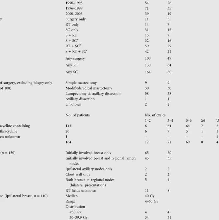
First-line therapy is documented in Table 2.
Treatment was at the discretion of the individual physician, with both single-modality and combination therapies used. Systemic chemotherapy (6 surgery) was given to 80% of patients, with 87% of regimens containing an anthracycline. Of those who received chemotherapy, 62% also received RT. Only eight patients received intrathecal chemotherapy as CNS prophylaxis.
A complete response to first-line therapy was achieved in 89% of patients, partial response in 4%, and progressive disease in 4% (n = 8, including three with bilateral presentation). Three percent of patients were unassessable, mainly due to early death from intercurrent disease.
Before proceeding with further outcome analyses, survival curves of patients who had central pathology review were compared with those whose pathology had not been reviewed. No significant difference in OS (P = 0.90) or PFS (P = 1.0) for the two groups was demonstrated. An apparent minor divergence in CSS after 5 years in favour of patients who had not had central pathology review almost certainly related to the small number of events rather than to a real difference. It was therefore considered appropriate to combine the two groups for subsequent analyses.
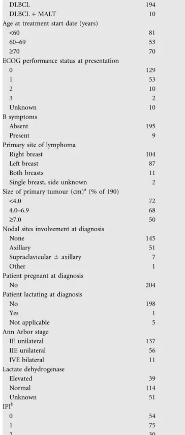
Median OS was 8.0 years [95% confidence interval (CI) 6.5–10.9 years], with 5- and 10-year OS rates 63% (95% CI 55% to 70%) and 47% (95% CI 38% to 56%), respectively (Figure 1). The median PFS was 5.5 years (95% CI 3.7–8.0 years), with median CSS not reached at the time of analysis. At a median follow-up time of 5.5 years, there were 76 progression events (37% of patients) and 81 deaths, with 54 deaths (26% of all patients) attributable to progressive lymphoma and/or treatment-related toxicity. Only one possible case of treatment-related second malignancy was identified, a patient who developed squamous cell carcinoma of the cervical oesophagus, within the RT field. Although the rate of progression and lymphoma-related death was highest in the first 3 years, there was a continuing pattern of progression and death up to 14 years from initial treatment. All ipsilateral progression events occurred within 2.6 years from commencement of treatment, whereas contralateral breast progressions occurred up to 13.3 years. Median OS following progression after first-line therapy was 1.0 years (95% CI 0.7–2.1 years), with 20% of patients with relapsed disease estimated to be alive at 5 years and 11% at 10 years after progression.
In the 11 patients with synchronous bilateral presentation, there were seven progressions during or following first-line therapy, with six deaths, all due to progressive disease, and all within 3 years of initial treatment (3-year PFS 36% and OS
46%). Only one patient developed first progression in CNS. The median PFS for this group was 1.3 years and the median OS 2.4 years, with wide CIs reflecting the small number of patients. In comparison to unilateral presentation, the hazard ratio (HR) for progression was 1.6 (P = 0.22) and for death was 1.9 (P = 0.15).
The outcome for males did not appear to differ from the remainder of the cohort, with 5-year OS of 60%, although the CIs were wide, given the small number of patients.
prognostic factor analysis and patterns of relapse
Unifactor and multifactor analyses of potential prognostic factors were carried out for all events (Table 3). A number of confounding associations were identified, e.g. use of
anthracycline negatively related to age and positively to nodal involvement, RT negatively related to age and IPI, thus only the more informative multifactor analyses are presented. The prognostic factors that retained statistical significance for OS were IPI (P < 0.001), anthracycline-containing chemotherapy (P = 0.02), and RT (P = 0.03). For PFS, significant prognostic factors were IPI (P = 0.01) and anthracycline-containing chemotherapy (P = 0.001). For CSS, IPI was once again significant (P = 0.002). There was a negative association of all outcome measures with the extent of surgery, with patients who underwent radical mastectomy having a statistically
significantly poorer CSS (P = 0.03). Although a greater number of cycles of any systemic chemotherapy (>3 cycles) was associated with an improved CSS (P = 0.04), for those patients receiving anthracycline-containing chemotherapy, this significance was lost, i.e. the importance of receiving anthracycline appeared to diminish the significance of the number of cycles of chemotherapy, as was also the case for OS and PFS. Other factors not found to have prognostic significance in multifactor analysis included presence of B symptoms, tumour diameter, nodal involvement, and treatment era. Bilaterality was not statistically significant on unifactor analysis, and was therefore included in multifactor analyses only as a component of IPI.
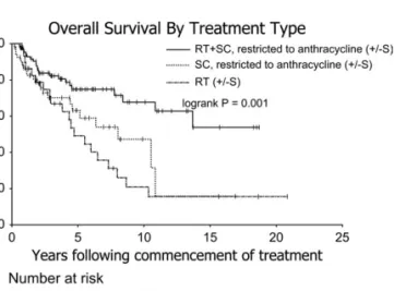
Figure 2 demonstrates the impact of anthracycline-containing chemotherapy on OS. Patients who received anthracycline had an OS of 73% at 5 years and 58% at 10 years. Survival curves for all patients who received anthracycline-containing chemotherapy and/or RT are shown in Figure 3. The combination of chemotherapy and RT was associated with the best outcomes (P = 0.001).
Seventy-six patients (37%) developed progression following first-line therapy, with 18 patients having ‡2 sites of
involvement at first progression. Breast was a site of first progression in 32 patients, regional nodes in 13, CNS in 11, other extranodal sites in 28, other nodal sites in 11, and bone marrow in two (sites unknown in five). Table 4 documents the cumulative incidence of progression at each predominant site.
In the 191 cases with unilateral presentation and known laterality, breast was the first site of progression in 28. Nine of these progressions were ipsilateral, with 19 in the contralateral breast. RT substantially reduced the risk of ipsilateral
number of ipsilateral breast progressions in the group who received RT, no dose relationship was explored. There was a trend in reduction of risk of locoregional progression with increasing extent of RT fields (0.021). However, due to the small number of nodal recurrences, no conclusion could be drawn regarding the benefit of stage I patients undergoing prophylactic nodal RT.
Anthracycline-containing chemotherapy appeared to reduce the risk of contralateral breast progression. The HR for anthracycline-containing chemotherapy compared with non-anthracycline-containing chemotherapy was 0.2 (P = 0.028). There was no evidence that RT to the involved breast reduced the risk of contralateral progression. OS following first progression in breast was similar whether the progression was
Table 2. First-line therapy
No. %
Year of commencement of first-line therapy 1980–1989 40 20
1990–1995 54 26
1996–1999 71 35
2000–2003 39 19
Treatment Surgery only 11 5
RT only 14 7 SC only 31 15 S + RT 15 7 S + SCa 32 16 RT + SCb 59 29 S + RT + SCc 42 21 Any surgery 100 49 Any RT 130 64 Any SC 164 80 Surgery
Type of surgery, excluding biopsy only (% of 100)
Simple mastectomy 9 9
Modified/radical mastectomy 30 30 Lumpectomy 6 axillary dissection 58 58
Axillary dissection 1 1
Unknown 2 2
SC
No. of patients No. of cycles
1–2 3–4 5–6 ‡6 Unknown Anthracycline containing 143 6 64 64 7 2 No anthracycline 20 6 7 5 1 1 Regimen unknown 1 – – – – 1 Total 164 12 71 69 8 4 RT
Fields (n = 130) Initially involved breast only 65 50 Initially involved breast and regional lymph
nodes
45 35
Ipsilateral axillary nodes only 2 2
Chest wall only 2 2
Both breasts 6 regional nodes (bilateral presentation)
5 4
RT fields unknown 11 8
RT dose (ipsilateral breast, n = 110) Median 40 Gy
Range 4–60 Gy Distribution <30 Gy 4 4 30–39.9 Gy 34 31 40–49.9 Gy 61 55 ‡50 Gy 11 10
aIntrathecal chemotherapy, one patient. bIntrathecal chemotherapy, five patients. cIntrathecal chemotherapy, two patients.
ipsilateral or contralateral, with 22% and 30%, respectively, alive at 5 years after progression (P = 0.62).
Breast was the first site of progression in four of the patients with bilateral breast involvement at presentation. All were dead within 12.5 months of progression.
CNS was the first site of progression in 11 patients (5%). There were no CNS progressions in the eight patients who received prophylactic intrathecal chemotherapy (P = 1.0). Given the small number of events, no prognostic factors for CNS progression could be identified.
discussion
The literature of primary breast NHL comprises many small retrospective series and just one prospective study, with inconsistent conclusions drawn, from breast being a site of presentation with a very poor prognosis, through to a prognosis no different from a similarly staged nodal presentation of DLBCL. This large multicentre study has the potential flaws of all retrospective studies, but nevertheless provides strong evidence that primary DLBCL of the breast is a distinct entity, with characteristic patterns of relapse differing from those of nodal DLBCL.
The Eastern Cooperative Oncology Group (ECOG) study 1484 [23] and the Southwest Oncology Group (SWOG) study 6736 [24, 25] established expected outcomes for patients with early stage DLBCL treated with anthracycline-containing chemotherapy (CHOP: cyclophosphamide, adriamycin, vincristine, prednisolone) 6 RT. The studies differed in eligibility criteria and treatment assignment, but included both nodal and extranodal stage I–II DLBCL. The 10-year survival outcomes for our entire cohort of patients with primary breast DLBCL are considerably inferior to those reported in the ECOG and SWOG studies. However, some of our patient group had undoubtedly been treated suboptimally. When the comparison is restricted to those of our patients who received anthracycline-containing systemic chemotherapy, the
inferiority of outcome is reduced; and in those patients treated with anthracycline-containing chemotherapy and RT, similar outcomes are achieved. The small group of patients with synchronous bilateral disease appears to be an exception, with substantial though non-significant increases in risk of early progression and poorer survival irrespective of treatment programme. The decision to regard such presentations as stage IV appears vindicated by the study data.
Sites of progression were predominantly extranodal in our patients, with the contralateral-paired organ at high risk, a phenomenon similarly described in primary testicular DLBCL [26, 27]. Contralateral progression was more common in this series than ipsilateral, the rate of ipsilateral progression having been substantially reduced by the use of RT in most patients. The role of prophylactic nodal irradiation remains unproven. One unexpected finding was that the risk of CNS relapse was relatively low, occurring in only 5% of patients. This is at variance with the reports of other smaller studies, and is considerably lower than the risk seen in primary testicular DLBCL [27]. It may be that primary breast DLBCL does not have the same tropism for CNS as does testicular DLBCL, and that this difference explains the generally superior survival for patients with primary breast DLBCL compared with that of patients with primary testicular DLBCL. However, it is possible that limiting the eligibility to patients with localized disease has led to underestimation of the rate of CNS involvement. We must regard this result with caution.
The discriminating ability of the IPI in patients with primary disease in breast has been validated in our study population. The IPI was a significant prognostic factor for OS, PFS, and CSS. The inclusion of an anthracycline in the chemotherapy regimen was, as expected, another important predictor of outcome, being significant for both OS and PFS. One factor found not to have prognostic significance was the extent of surgery—OS and PFS were not improved with more extensive surgery, and radical mastectomy was actually associated with poorer CSS. This finding has been described in other series, and although it may be a statistical quirk, it is possible that extensive surgery delayed the commencement of chemotherapy, with detrimental outcome.
The delivery of RT was a statistically significant predictor of improved OS in this study, and was associated with non-significant improvements in PFS, CSS, and risk of ipsilateral locoregional progression. Comparisons of survival outcomes by treatment type showed a statistically significant benefit resulting from the addition of RT to systemic chemotherapy, including those patients receiving anthracycline-containing regimens, and this benefit persisted with long-term follow-up. RT resulting in an improvement in PFS would not be an unexpected finding in patients with DLBCL, but the significant sustained survival benefit seen in the study population is at variance with many reported series of similarly staged predominantly nodal DLBCL. Two large prospective studies undertaken by the Groupe d’Etude des Lymphomes de l’Adulte (GELA) [28, 29] concluded that the addition of RT to anthracycline-containing chemotherapy did not result in a survival benefit, and was possibly detrimental to survival, presumably as a result of late radiation toxicity. However, radiation to the breast in the modern era can be delivered with
Figure 1. Cause-specific survival (CSS), overall survival (OS), and progression-free survival (PFS).
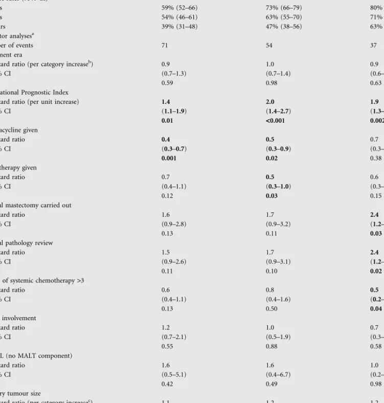
Table 3. Summary table of time-to-event analyses
Analysis PFS OS CSS
Event-free rates (95% CI)
3 years 59% (52–66) 73% (66–79) 80% (73–85) 5 years 54% (46–61) 63% (55–70) 71% (63–78) 10 years 39% (31–48) 47% (38–56) 63% (53–72) Multifactor analysesa Number of events 71 54 37 Treatment era
Hazard ratio (per category increaseb) 0.9 1.0 0.9
95% CI (0.7–1.3) (0.7–1.4) (0.6–1.4)
P 0.59 0.98 0.63
International Prognostic Index
Hazard ratio (per unit increase) 1.4 2.0 1.9
95% CI (1.1–1.9) (1.4–2.7) (1.3–2.8) P 0.01 <0.001 0.002 Anthracycline given Hazard ratio 0.4 0.5 0.7 95% CI (0.3–0.7) (0.3–0.9) (0.3–1.6) P 0.001 0.02 0.38 Radiotherapy given Hazard ratio 0.7 0.5 0.6 95% CI (0.4–1.1) (0.3–1.0) (0.3–1.2) P 0.12 0.03 0.15
Radical mastectomy carried out
Hazard ratio 1.6 1.7 2.4
95% CI (0.9–2.8) (0.9–3.2) (1.2–4.8)
P 0.13 0.11 0.03
Central pathology review
Hazard ratio 1.5 1.7 2.4
95% CI (0.9–2.6) (0.9–3.1) (1.2–4.8)
P 0.11 0.10 0.02
Cycles of systemic chemotherapy >3
Hazard ratio 0.6 0.8 0.5 95% CI (0.4–1.1) (0.4–1.6) (0.2–0.9) P 0.13 0.50 0.04 Nodal involvement Hazard ratio 1.2 1.0 0.7 95% CI (0.7–2.1) (0.5–1.9) (0.3–1.9) P 0.55 0.88 0.58
DLBCL (no MALT component)
Hazard ratio 1.6 1.6 1.0
95% CI (0.5–5.1) (0.4–6.7) (0.2–4.5)
P 0.42 0.49 0.98
Primary tumour size
Hazard ratio (per category increasec) 1.1 1.2 1.2
95% CI (0.8–1.5) (0.8–1.7) (0.8–1.9)
P 0.61 0.33 0.41
aResults from multifactor analyses were on the basis of 154 cases with no missing data on any of the factors examined. For the factors that were statistically significant following multifactor analysis, the adjusted hazard ratio and P value are presented, that is, adjusting for the significance of each other. For the factors that were not statistically significant following multifactor analysis, the hazard ratio and P value after adjusting for the significant factors are presented. bTreatment era categories: 1980–1989, 1990–1995, and 1996–2003.
cPrimary tumour size categories: <4, 4–6.9, and ‡7.0 cm.
PFS, progression-free survival; OS, overall survival; CSS, cause-specific survival; CI, confidence interval; DLBCL, diffuse large B-cell lymphoma; MALT, mucosal-associated lymphoid tissue.
modest acute and late toxicity, and in such a situation, the potential for a positive benefit is substantially increased. Thus, the concept of RT improving survival in primary DLBCL of the breast is not necessarily inconsistent with the GELA and other studies, and is deserving of further study.
The use of immunohistochemistry to assist in the classification and prognostication of lymphoma is now standard. Molecular analysis techniques using DNA microarrays are also becoming more common, facilitating identification of different molecular ‘signatures’ in lymphomas with identical morphology. Recent studies looking specifically at immunohistochemistry of primary breast DLBCL [30–32] have identified a number of different cytogenetic patterns with apparent prognostic implications. Immunohistochemical and molecular analyses of the cases submitted for central review are ongoing, and will provide further insights into the nature of primary breast DLBCL. These analyses will be the subject of a separate report.
In any retrospective study, accuracy and completeness of data may not be ideal, and controlling for all factors influencing outcome may have been imperfect in some analyses. Although the study results are consistent with our original hypothesis that primary breast DLBCL is intrinsically different from DLBCL presenting at other sites, the conclusions must be regarded as suggestive only, rather than definitive. On the basis of our study results, we conclude that the outcomes for patients with early stage primary breast DLBCL treated with optimal therapy are similar to those of nodal presentations of DLBCL. Recommended standard therapy for inclusion in future studies is limited surgery/biopsy, followed by ‡3 cycles of
anthracycline-containing chemotherapy and RT to the ipsilateral breast 6 regional nodes. CNS prophylaxis does not appear to be indicated routinely. Patients presenting with bilateral disease appear to be a poor prognosis group, and intensification of chemotherapy should be considered.
One final consideration, with a series acquired over a 20-year period, is how subsequent developments in treatment should be incorporated into management recommendations.
Anthracycline-containing chemotherapy remains standard, but the addition of rituximab to chemotherapy has been a major recent advance, with demonstrated improvement in outcomes of patients with DLBCL in a variety of sites [33–35]. Primary breast lymphoma, however, has been poorly represented in the patient group studied, and it should not be automatically assumed that the same improvements will be seen in this rare presentation of DLBCL. Thus, it is important that future prospective protocols for primary DLBCL of the breast incorporate rituximab and perhaps other targeted therapies arising from further research. How these might impact on other aspects of the treatment protocol will only become apparent through controlled prospective studies.
Figure 2. Overall survival by anthracycline status.
Figure 3. Overall survival by treatment type (chemotherapy restricted to anthracycline). RT, radiotherapy; SC, systemic chemotherapy; S, surgery.
Table 4. Cumulative incidence of each predominant site of progression
Progression site No. of casesa Estimated cumulative incidence (6 standard error) 3 years 5 years 10 years Breast (6 regional nodes, 6 CNS, 6 other sites) 32 14% 6 3% 16% 6 3% 17% 6 3% Regional nodes (6 CNS, 6 other sites but not breast) 5 2% 6 1% 3% 6 1% 3% 6 1% CNS (6 other sites but not breast or regional nodes) 9 3% 6 1% 5% 6 2% 6% 6 2% Sites other than breast, regional nodes or CNSb 25 11% 6 2% 11% 6 2% 16% 6 3% aProgression sites unknown in five patients.
bOther sites include the following: nodal, four; extranodal, 16 (including one case of spleen); nodal + extranodal, four; marrow, one. CNS, central nervous system.
funding
Oncosuisse (ICP OCS-01356-03-2003).
acknowledgements
The following members of the IELSG (listed in alphabetical order) contributed patients to the study: O. Bairey, Rabin Medical Centre, Beilinson Campus, Petah Tiqva, Israel; RGB, University of Nebraska Medical Centre, Omaha, NE, USA; L. Cavanna, Ospedale di Piacenza, Piacenza, Italy; SPC, National Cancer Centre, Singapore; D. Christie, East Coast Cancer Centre, Tugun, Australia; S. Cortelazzo, Ospedali Reuniti, Bergamo, Italy; SC, Southampton General Hospital, Southampton, UK; MDO, Casa Sollievo della Sofferenza IRCCS, San Giovanni Rotondo, Italy; A. De Renzo, Universita di Federico II, Napoli, Italy; LD, Istituto Nazionale Tumori, Milan, Italy; F. Di Raimondo, Ospedale Ferrarotto, Catania, Italy; M. Federico, Centro Oncologico Modenese, Modena, Italy; AMG, Istituto Nazionale Tumori, Milan, Italy; M. Goldaniga, Ospedale Maggiore IRCCS, Milano, Italy; M. H. Gomez, Instituto Nacional de Enfermedades Neoplasicas, Peru; A. Grigg, Royal Melbourne Hospital, Melbourne, Australia; MK-H, Bernhoven Hospital, Oss, The Netherlands; DH, Universitatsklinikum Eppendorf, Hamburg, Germany; F. Laveder, Ospedale S. Martino, Belluno, Italy; AL, Royal Victoria Infirmary, Newcastle upon Tyne, UK;
D. Lonergan, Prince of Wales Hospital, Sydney, Australia; GM, Istituto Europeo di Oncologia, Milan, Italy; G. Pinotti, Ospedale di Circolo, Varese, Italy; I. Poddubnaia, N. N. Blokhin Russian Cancer Research Centre, Moscow, Russia; GP, Royal Brisbane Hospital, Brisbane, Australia; J. M. Ribera, Hospital Universitari Germans Trias i Pujol, Badalona, Spain; J. Radford, Christie Hospital, Manchester, UK; DR, Royal Adelaide Hospital, Adelaide, Australia; GR, Peter MacCallum Cancer Centre, Melbourne, Australia; JFS, Peter MacCallum Cancer Centre, Melbourne, Australia; P. Solal-Ce´ligny, Centre J. Bernard, Le Mans, France; C. Stelitano, Azienda Ospedaliera ‘Bianchi-Melacrino-Morelli’, Reggio Calabria, Italy; RT, Princess Margaret Hospital, Toronto, Canada; M. L. Vigliotti, Centro di Riferimento Oncologico della Basilicata, Ospedale Oncologico Regionale, Rionero in Vulture, Potenza, Italy; U. Vitolo, Ospedale Molinette, Torino, Italy; R. Zanotti, University of Verona, Verona, Italy; P. L. Zinzani, S.Orsola-Malpighi, Bologna, Italy; and EZ, Instituto Oncologico della Svizzera Italiana, Bellinzona, Switzerland. We also acknowledge the work of the IELSG secretariat and the study data managers in Melbourne and Milan in ensuring the quality and integrity of the data.
references
1. DeBlasio D, McCormick B, Straus D et al. Definitive irradiation for localized non-Hodgkin’s lymphoma of breast. Int J Radiat Oncol Biol Phys 1989; 17: 843–846. 2. Giardini R, Piccolo C, Rilke F. Primary non-Hodgkin’s lymphomas of the female
breast. Cancer 1992; 69: 725–735.
3. Abbondanzo SL, Seidman JD, Lefkowitz M et al. Primary diffuse large B-cell lymphoma of the breast. A clinicopathologic study of 31 cases. Pathol Res Pract 1996; 192: 37–43.
4. Au WY, Chan AC, Chow LW, Liang R. Lymphoma of the breast in Hong Kong Chinese. Hematol Oncol 1997; 15: 33–38.
5. Ha CS, Dubey P, Goyal LK et al. Localized primary non-Hodgkin’s lymphoma of the breast. Am J Clin Oncol 1998; 21: 376–380.
6. Lyons JA, Myles J, Pohlman B et al. Treatment and prognosis of primary breast lymphoma: a review of 13 cases. Am J Clin Oncol 2000; 23: 334–336.
7. Wong WW, Schild SE, Halyard MY, Schomberg PJ. Primary non-Hodgkin lymphoma of the breast: the Mayo clinic experience. J Surg Oncol 2002; 80: 19–25.
8. Domchek SM, Hecht JL, Fleming MD et al. Lymphomas of the breast: primary and secondary involvement. Cancer 2002; 94: 6–13.
9. Kuper-Hommel MJ, Snijder S, Janssen-Heijnen ML et al. Treatment and survival of 38 female breast lymphomas: a population-based study with clinical and pathological reviews. Ann Hematol 2003; 82: 397–404.
10. Gholam D, Bibeau F, El Weshi A et al. Primary breast lymphoma. Leuk Lymphoma 2003; 44: 1173–1178.
11. Vignot S, Ledoussal V, Nodiot P et al. Non-Hodgkin’s lymphoma of the breast: a report of 19 cases and a review of the literature. Clin Lymphoma 2005; 6: 37–42.
12. Liu MT, Hsieh CY, Wang AY et al. Primary breast lymphoma: a pooled analysis of prognostic factors and survival in 93 cases. Ann Saudi Med 2005; 25: 288–293.
13. Choo SP, Lim ST, Wong EH, Tao M. Breast lymphoma: favorable prognosis after treatment with standard combination chemotherapy. Onkologie 2006; 29: 14–18.
14. Ryan GF, Roos DR, Seymour JF. Primary non-Hodgkin’s lymphoma of the breast: retrospective analysis of prognosis and patterns of failure in two Australian centers. Clin Lymphoma Myeloma 2006; 6: 337–341.
15. Aviles A, Delgado S, Nambo MJ et al. Primary breast lymphoma: results of a controlled clinical trial. Oncology 2005; 69: 256–260.
16. Carbone PP, Kaplan HS, Musshoff K et al. Report of the Committee on Hodgkin’s disease staging classification. Cancer Res 1971; 31: 1860–1861.
17. The International Non-Hodgkin’s Lymphoma Prognostic Factors Project. A predictive model for aggressive non-Hodgkin’s lymphoma. N Engl J Med 1993; 329: 987–994.
18. Miller AB, Hoogstraten B, Staquet M, Winkler A. Reporting results of cancer treatment. Cancer 1981; 47: 207–214.
19. Schemper M, Smith TL. A note on quantifying follow-up in studies of failure time. Control Clin Trials 1996; 17: 343–346.
20. Kalbfleisch JD, Prentice RL. The Statistical Analysis of Failure Time Data. New York: John Wiley and Sons 1980.
21. S-PLUS 2000 Release 3. Seattle, WA: MathSoft Inc, 1999. 22. SPSS for Windows Release 11.0.1. Chicago, IL: SPSS Inc, 2001. 23. Horning SJ, Weller E, Kim K et al. Chemotherapy with or without
radiotherapy in limited-stage diffuse aggressive non-Hodgkin’s lymphoma: Eastern Cooperative Oncology Group Study 1484. J Clin Oncol 2004; 22: 3032–3038.
24. Miller T, Dahlberg S, Cassady J et al. Chemotherapy alone compared with chemotherapy plus radiotherapy for localized intermediate- and high-grade non-Hodgkin’s lymphoma. N Engl J Med 1998; 339: 21–26.
25. Miller TP, LeBlanc M, Spier C et al. CHOP alone compared to CHOP plus radiotherapy for early stage aggressive non-Hodgkin’s lymphomas: update of the Southwest Oncology Group (SWOG) randomized trial. Blood 2001; 98: 724A (Abstr 3024).
26. Seymour JF, Solomon B, Wolf MM et al. Primary large-cell non-Hodgkin’s lymphoma of the testis. A retrospective analysis of patterns of failure and prognostic factors. Clin Lymphoma 2001; 2: 109–115.
27. Zucca E, Conconi A, Mughal T et al. Patterns of outcome and prognostic factors in primary large-cell lymphoma of the testis in a survey by the International Extranodal Lymphoma Study Group. J Clin Oncol 2003; 21: 20–27. 28. Reyes F, Lepage E, Ganem G et al. ACVBP versus CHOP plus radiotherapy for
localized aggressive lymphoma. N Engl J Med 2005; 352: 1197–1205. 29. Bonnet C, Fillet G, Mounier N et al. CHOP alone compared with CHOP plus
Groupe d’Etude des Lymphomes de l’Adulte. J Clin Oncol 2007; 25: 787–792.
30. Fruchart C, Denoux Y, Chasle J et al. High grade primary breast lymphoma: is it a different clinical entity? Breast Cancer Res Treat 2005; 93: 191–198. 31. Yoshida S, Nakamura N, Sasaki Y et al. Primary breast diffuse large B-cell
lymphoma shows a non-germinal B-cell phenotype. Mod Pathol 2005; 18: 398–405.
32. Lopez-Guillermo A, Colomo L, Jimenez M et al. Diffuse large B-Cell lymphoma: clinicobiological characterization and outcome according to the nodal or extranodal primary origin. J Clin Oncol 2005; 23: 2797–2804.
33. Coiffier B, Lepage E, Briere J et al. CHOP chemotherapy plus rituximab compared with CHOP alone in elderly patients with diffuse large-B-cell lymphoma. N Engl J Med 2002; 346: 235–242.
34. Pfreundschuh M, Trumper L, Osterborg A et al. CHOP-like chemotherapy plus rituximab versus CHOP-like chemotherapy alone in young patients with good-prognosis diffuse large B-cell lymphoma: a randomised controlled trial by the MabThera International Trial (MInT) Group. Lancet Oncol 2006; 7: 379–391. 35. Miller T, Unger J, Spier C et al. Effect of adding rituximab to three cycles of
CHOP plus involved field radiotherapy for limited stage aggressive diffuse large B-cell lymphoma (SWOG-0014). Blood 2004; 104: 48a–49a (Abstr 158).