DNA Damage and Its Links to Neurodegeneration
The MIT Faculty has made this article openly available.
Please share
how this access benefits you. Your story matters.
Citation
Madabhushi, Ram et al. “DNA Damage and Its Links to
Neurodegeneration.” Neuron 83, 2 (July 2014): 266–282 © 2014
Elsevier Inc
As Published
http://dx.doi.org/10.1016/j.neuron.2014.06.034
Publisher
Elsevier/Cell Press
Version
Author's final manuscript
Citable link
http://hdl.handle.net/1721.1/112701
Terms of Use
Creative Commons Attribution-NonCommercial-NoDerivs License
DNA damage and its links to neurodegeneration
Ram Madabhushi1,2, Ling Pan1,2, and Li-Huei Tsai1,2,*
1Picower Institute for Learning and Memory, Massachusetts Institute of Technology, Cambridge,
MA 02139 USA
2Department of Brain and Cognitive Sciences, Massachusetts Institute of Technology, Cambridge,
MA 02139 USA
Abstract
The integrity of our genetic material is under constant attack from numerous endogenous and exogenous agents. The consequences of a defective DNA damage response are well studied in proliferating cells, especially with regards to the development of cancer, yet its precise roles in the nervous system are relatively poorly understood. Here we attempt to provide a comprehensive overview of the consequences of genomic instability in the nervous system. We highlight the neuropathology of congenital syndromes that result from mutations in DNA repair factors and underscore the importance of the DNA damage response in neural development. In addition, we describe the findings of recent studies, which reveal that a robust DNA damage response is also intimately connected to aging and the manifestation of age-related neurodegenerative disorders such as Alzheimer’s disease and amyotrophic lateral sclerosis.
Introduction
Upon analyzing the data collected in the 2000 census, health officials arrived at the remarkable prediction that by the year 2050, approximately 800,000 Americans would live to see their hundredth birthday (Park, 2010). Even with the benefits of modern medical technology, it is miraculous that our bodies can sustain themselves for a century. Each cell in the human body incurs thousands of lesions per day to its constituent lipids, proteins, and nucleic acids from sources that range from the products of cellular metabolism to the myriad environmental chemicals, pollutants, and high-frequency electromagnetic radiation. While some cells only need endure this onslaught for a short time and are replaced continuously (for instance, epithelial cells lining the intestine have an average lifespan of 5 days), others such as neurons are retained for life and therefore require the means to cope with a lifetime of damage.
*To whom correspondence should be addressed: 77 Massachusetts Avenue, Building 46, Room 4235A, Cambridge, MA 02139, USA,
Publisher's Disclaimer: This is a PDF file of an unedited manuscript that has been accepted for publication. As a service to our
HHS Public Access
Author manuscript
Neuron
. Author manuscript; available in PMC 2017 August 21.Published in final edited form as:
Neuron. 2014 July 16; 83(2): 266–282. doi:10.1016/j.neuron.2014.06.034.
A
uthor Man
uscr
ipt
A
uthor Man
uscr
ipt
A
uthor Man
uscr
ipt
A
uthor Man
uscr
ipt
All biological macromolecules are susceptible to corruption; however, damage to a cell’s genomic DNA is particularly harmful because DNA is the blueprint for protein production and unlike other molecules, it cannot simply be replaced by re-synthesis. DNA damage induces mutations and chromosomal aberrations that can lead either to cellular dysfunction or to the formation of cancer, and encounters with certain DNA lesions can derail
transcription and replication, and thereby trigger cell death, senescence, and aging (Hoeijmakers, 2009). Accordingly, cells devote enormous resources for the purpose of genome maintenance and have evolved elaborate systems to repair damaged DNA. In this review, we focus on the consequences of DNA damage in the nervous system, taking into account the insights obtained from neurological disorders that manifest from a defective DNA damage response. A majority of these disorders are congenital; however several recent studies suggest that defective DNA repair also underlies brain aging and age-associated neurodegeneration and we also discuss the implications of these studies.
The cellular DNA damage response
On any given day, a listing of endogenous DNA damage experienced by a typical mammalian cell would read something as follows – 200 cytosine deaminations, 3000 guanine methylations, 10000 spontaneous depurinations, 10000–100000 oxidative lesions, 10000 single strand breaks, and 10–50 double strand breaks (Ames et al., 1993; Haber, 1999; Lindahl, 1993; Nakamura et al., 1998; Vilenchik and Knudson, 2003). To avert the potentially catastrophic consequences of these lesions, cells activate a highly evolved DNA damage response (DDR) that not only detects and repairs damaged DNA, but also
coordinates repair with other cellular processes, such as chromatin remodeling, transcription, cell cycle progression (in dividing cells), and apoptosis (Jackson and Bartek, 2009). A truly remarkable feature of the DDR is that each class of lesion elicits its own distinct damage detection and repair mechanism. For instance, thymidine dimers generated upon exposure to UV light are repaired using nucleotide excision repair, whereas a separate base excision repair pathway is utilized to repair oxidative lesions such as 8-oxo-dG. However, the same lesion can also be repaired using diverse mechanisms depending upon cell cycle stage, developmental status, and tissue type. As an example, whereas a majority of DNA double strand breaks are repaired through nonhomologous end joining (NHEJ), a specialized pathway called homologous recombination (HR) is employed to repair double strand breaks that are produced in the S and G2 phases of the cell cycle.
Although relatively stable compared to other macromolecules, DNA bases frequently undergo modification by alkylation, oxidation, and deamination. In fact, reactive oxygen species (ROS) alone generate more than 100 different oxidative base modifications and these alterations have the potential to be highly mutagenic (Iyama and Wilson, 2013). The brain is thought to metabolize as much as a fifth of consumed oxygen. Accordingly, a number of studies have shown that ROS are a major source of DNA damage in the brain. The base excision repair (BER) machinery has evolved to specifically solve these problems. The main strategy in BER consists of converting the large array of modified base substrates into a few intermediates that can then be processed by the core BER components of APE1, polβ, and XRCC1/LIG3. This step is mediated primarily by enzymes called DNA glycosylases
A
uthor Man
uscr
ipt
A
uthor Man
uscr
ipt
A
uthor Man
uscr
ipt
A
uthor Man
uscr
ipt
(humans contain at least 15) that specialize in detecting distinct modified bases and excising them through cleavage of the N-glycosidic bond (Lindahl, 1974).
Like BER, the nucleotide excision repair (NER) pathway also resolves modified bases and follows the general program of damage detection, excision, gap-filling DNA synthesis and ligation, the distinguishing feature of NER being that it senses structural distortions in the double helix rather than specific base modifications. This confers NER with the versatility to operate ona range of highly diverse substrates, such as the cyclobutane pyrimidine dimers (CPDs) and 6,4-photoproducts (6-4PPs) generated by UV radiation, DNA adducts that arise from intercalation of chemicals such as benzopyrene (a component of cigarette smoke), psoralen, and cisplatin, and cyclopurines formed by attacks of the hydroxyl radical on 2 ′-deoxyadenosine and 2′-deoxyguanosine. These “bulky” lesions typically obstruct the progression of transcription and replication machineries, and can thereby induce cellular dysfunction and apoptosis (de Laat et al., 1999). More than 30 different proteins work collaboratively in NER, which is broadly categorized into two classes – global genome NER (GG-NER), which resolves lesions throughout the genome, and transcription-coupled NER (TC-NER), which specializes in the removal of damage on the transcribed strand of DNA within active genes.
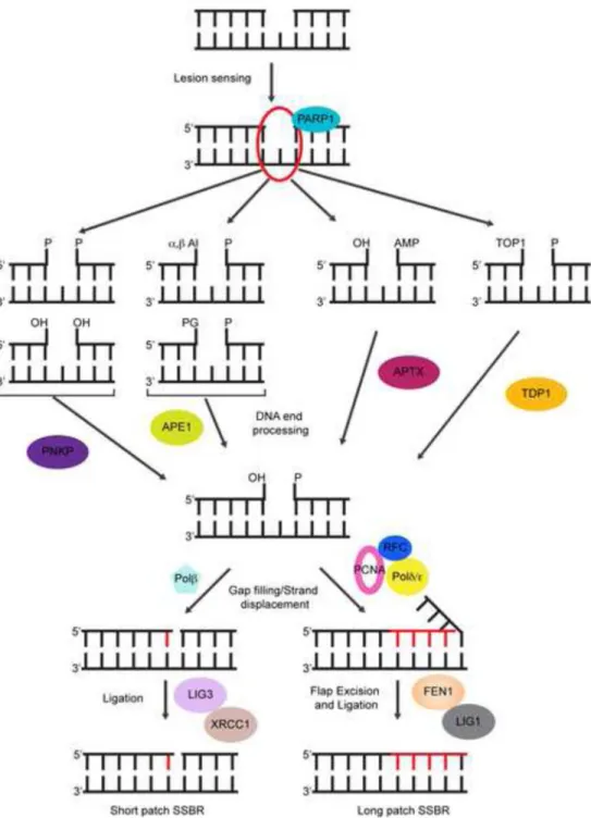
In addition to generating numerous base modifications, ROS-mediated attack on the DNA backbone and sugar fragmentation can also lead to the formation of DNA single strand breaks (SSBs). More indirectly, SSBs may also arise as intermediates of the BER pathway or as byproducts of abortive DNA topoisomerase I (TOP1) reactions (Caldecott, 2008).
The primary challenge in the repair of SSBs is to generate DNA ends that are compatible for ligation, which is to say, a 3′ hydroxyl and a 5′phosphate, and depending on the lesion this step may require diverse end-processing activities (Figure 1). Like in BER, any gap-filling synthesis is mediated by polβ and nicks are sealed by either XRCC1/LIG3 or FEN1/LIG1 (Figure 1). Under certain conditions, such as the generation of SSBs in close proximity to each other or encounters between an existing SSB and either the transcription or replication machineries (leading to their collapse) can cause SSBs breaks to be converted to DNA double strand breaks (DSBs). In addition to these, ionizing radiation and chemotherapeutic drugs are prominent environmental DSB-inducing agents. When compared to other lesions, DNA DSBs are rare events; however, DSBs are also extremely deleterious because they can cause large chromosome rearrangements that can either lead to cell death, or promote tumorigenesis (Jackson, 2002). Furthermore, even a few DSBs are sufficient to trigger apoptosis in proliferating cells (although whether neurons have a higher tolerance for DNA DSB accumulation has not been thoroughly investigated) (Rich et al., 2000). DSBs are repaired using one of two main pathways – nonhomologous end joining (NHEJ), which involves direct ligation of the broken DNA ends and is error prone, or homologous recombination (HR), which is selectively utilized in the S and G2 phases of the cell cycle and employs homologous sequences in the sister chromatid as a template to ensure error-free repair (Lombard et al., 2005). Because neurons are postmitotic cells, NHEJ is the primary pathway of DSB repair in neurons, although HR is likely important for DNA repairin neural progenitors and non-neuronal cells in the brain. In NHEJ, DSBs are recognized by the KU70/KU80 heterodimer, which binds the DNA ends and then recruits and activates the
A
uthor Man
uscr
ipt
A
uthor Man
uscr
ipt
A
uthor Man
uscr
ipt
A
uthor Man
uscr
ipt
catalytic subunit of the dependent protein kinase, PKcs. Activation of DNA-PKcs allows end-processing activities such as the ARTEMIS, APLF and PNKP to access the broken DNA ends and prepare them for ligation. The XRCC4/LIG4 complex is then
recruited, which acts in concert with proteins such as XLF to promote religation (Figure 2). However, it is important to note that while the importance of NHEJ in postmitotic neurons in vitro and in newly differentiated neurons in vivo has been established in various studies, its precise roles in neurons in the mature brain has not been characterized extensively. Studies that involve conditional ablation of NHEJ factors in the mature nervous system will provide key insights into these issues. Together, the collaborative efforts of these diverse DNA repair pathways ensures that cells remain functional despite the numerous lesions they accumulate daily.
Chromatin modifications in the DNA damage response
Both DNA damage and the concomitant response occur in the context of chromatin. Traditionally, this has been interpreted to mean that chromatin organization imposes a barrier that must be overcome to allow DNA repair activities to access damaged sites, following which the original chromatin configuration is restored. However, in contrast to this somewhat passive view of chromatin organization, more recent models emphasize that chromatin changes in the DDR play active roles in stabilization of the repair machinery, in the propagation of the DDR, and in the regulation of transcription in the vicinity of damaged sites (Smerdon, 1991; Soria et al., 2012). Furthermore, the discovery of roles for histone deacetylases and other chromatin compacting activities at the earliest stages following the induction of DNA damage suggest that chromatin changes at damaged sites are more dynamic than previously conceived.
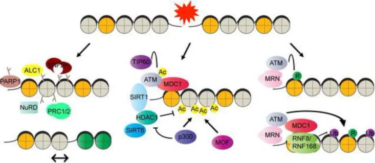
Generally, four main activities are thought to be important for chromatin changes in response to DNA damage – post-translational modifications of histone tails and chromatin modifying enzymes, ATP-dependent chromatin remodelers, histone variants, and histone chaperones. Although categorized in this manner, it is important to note that there is extensive crosstalk between these mechanisms during damage detection, repair and the restoration of chromatin organization following repair. ATP-dependent chromatin remodelers in DNA repair are summarized in Table 1 and the reader is referred to several excellent reviews on the topics of histone variants and chaperones in the DDR (Avvakumov et al., 2011; Soria et al., 2012). Here we limit our discussion to a broad overview of post-translational chromatin modifications in the DDR (Figure 3).
The modification of histone tails serves as an important way through which DNA
accessibility, chromatin dynamics, and the binding of non-histone proteins are regulated in the DDR. The N-terminal extensions of histones are the sites of most modifications, such as poly-ADP-ribosylation, phosphorylation, acetylation, methylation, ubiquitination, and sumoylation. One of the earliest detectable modifications following the induction of DNA strand breaks is poly-ADP-ribosylation (PARylation) that is mediated by poly-(ADP-ribose) polymerases (PARPs). PARP1 is a ubiquitous nuclear protein containing an N-terminal DNA binding domain composed primarily of two zinc finger motifs, a central BRCT motif containing auto-modification domain that mediates interaction with other DNA repair
A
uthor Man
uscr
ipt
A
uthor Man
uscr
ipt
A
uthor Man
uscr
ipt
A
uthor Man
uscr
ipt
proteins, and a C-terminal catalytic domain that binds NAD+ and transfers ADP-ribose from NAD+ to acceptor sites on proteins (Krishnakumar and Kraus, 2010). PARP1 senses DNA strand breaks and upon activation, catalyzes the assembly of poly-(ADP-ribose) (PAR) chains onto histones and other protein substrates including itself. Several lysine residues on histones, such as K13 of H2A, K30 of H2B, K27 and K37 of H3, and K16 of H4 have been identified as ADP-ribose acceptor sites on histones (Messner et al., 2010), although glutamate and aspartate residues have also been identified as acceptor sites on other targets. The PAR chains cause the nucleation of various chromatin modifiers. For instance, the recruitment of PAR-interacting factors such as ALC1 and the ATP-dependent chromatin remodeler, SNF2H, has also been shown to promote nucleosome sliding and greater
accessibility to DNA ends, whereas the recruitment of NuRD, polycomb, and macroH2A (an H2A variant) is thought to mediate chromatin looping and compaction (Ahel et al., 2009; Chou et al., 2010; Lukas et al., 2011). While chromatin modulation through PARylation mediates several important functions in the DDR, it is becoming increasingly clear that the regulation of PAR levels is also a critical determinant of survival. PAR levels are tightly regulated through the activities of PAR glycohyrolase (PARG), which cleaves PAR chains, and terminal ADP-ribose glycohydrolase (TARG), which removes the proximal ADP-ribose directly linked to the target protein. Defects in PARG and TARG1 have been shown to cause neurodegeneration (Tallis et al., 2014). In addition, prolonged PARP activity can cause the depletion of NAD+ and trigger an energy crisis within cells, leading to a form of apoptosis referred to as parthenos.
An extremely well-studied histone modification in the DDR is the phosphorylation of the histone H2A variant, H2AX, at Ser139 (γH2AX) (Burma et al., 2001). Three PI-3 kinases, ATM, ATR and DNA-PK, are known to phosphorylate H2AX. γH2AX appears immediately following formation of DNA DSBs, and can spread to upto a megabase in the vicinity of the DSB. The phosphorylation of H2AX at Ser139 is accompanied simultaneously by
dephosphorylation at Tyr142 and these events allow recognition of γH2AX by a protein called MDC1 (mediator of DNA damage checkpoint protein 1). The recruitment of MDC1 coordinates virtually every aspect of the DSB signaling response (Lukas et al., 2011). MDC1 allows for propagation of γH2AX adjacent to DSB sites, interacts with proteins such as ATM, TOPBP1 and CHK2 and activate checkpoint responses in proliferating cells, and also coordinates other post-translational modifications such as histone ubiquitination and acetylation. For instance, ATM-mediated phosphorylation of MDC1 stimulates the recruitment of the H2A ubiquitin ligases RNF8 and RNF168 (Lukas et al., 2011). Ubiquitination of H2A by RNF8 and RNF168 facilitates the binding sites of downstream factors, such as BRCA1(breast cancer 1), 53BP1 (p53-binding protein 1) and the E3 ubiquitin-protein ligase, RAD18, all of which are essential for DSB repair.
In addition to these modifications, the significance of histone acetylation in the DDR has become a major focus of recent studies. Increased histone acetylation following UV-damage is one of the first identified chromatin modifications associated with DNA damage. In particular, acetylation of histone H3 Lys56 (H3K56) is believed to promote nucleosome assembly in DNA repair and DNA synthesis (Das et al., 2009). Similarly, acetylation of H4K16 is known to unfold compact chromatin fibers (Shogren-Knaak and Peterson, 2006). Consistently, histone acetyltransferases (HATs) that mediate these modifications, such as
A
uthor Man
uscr
ipt
A
uthor Man
uscr
ipt
A
uthor Man
uscr
ipt
A
uthor Man
uscr
ipt
MOF, a specific H4K16 HAT, as well as Tip60 and CBP/P300, less specific HATs that acetylate both histone and non-histone proteins, play pivotal roles in DNA repair (Das et al., 2009; Ikura et al., 2000; Li et al., 2010).
Meanwhile, based on the notion that DNA repair requires chromatin relaxation, histone deacetylation and histone deacetylases (HDACs) were long believed to only participate at the later stages of DNA repair, primarily to restore chromatin structure. However, recent studies in several systems, including postmitotic neurons, have unveiled the roles of HDACs and chromatin compaction in the early phase of DNA repair (Dobbin et al., 2013; Miller et al., 2010; O’Hagan et al., 2008). Intriguingly, although acetylation of H3K56 and H4K16 are important for DNA repair, deacetylation of these residues has been observed to occur immediately after the formation of DSBs, whereas the re-acetylation is usually observed hours after DNA damage (Miller et al., 2010). In addition, H4K16 deacetylation facilitates 53BP1 foci formation and NHEJ repair (Hsiao and Mizzen, 2013). Given the positive role of acH4K16 in transcriptional activation (Taylor et al., 2013), it is also possible that the transient removal of this modification serves to inhibit local transcription. Incidentally, ATM activity was also shown to cause transcriptional silencing and prevent RNA polymerase II elongation-dependent chromatin decondensation in cis of DNA DSBs (Shanbhag et al., 2010). Histone deacetylation and chromatin compaction may also prevent the excessive processing of DNA ends and the uncontrolled expanding of repair factors into adjacent chromatin. SIRT6, HDAC1 and HDAC2 have all been implicated as H3K56 deacetylases, whereas HDAC1 and HDAC2 have been reported to deacetylate H4K16 (Miller et al., 2010; Toiber et al., 2013). SIRT6 increases the binding of SNF2H to nucleosomes and deacetylates H3K56 simultaneously. Decreased SNF2H-chromatin association, increased H3K56
acetylation and accumulated DNA damage are detected in the brains of Sirt6 KO mice, suggesting a physiological role of SIRT6 in maintaining genomic integrity in the central nervous system (Schwer et al., 2010; Toiber et al., 2013). In addition to its deacetylase activity, SIRT6 also has mono-ADP ribosyltransferase activity and is shown to promote BER repair through activating PARP1 (Mao et al., 2011; Mostoslavsky et al., 2006). Furthermore, the deacetylation of histones at DSBs is also associated with transient H3K9 methylation (Price and D’Andrea, 2013). Together, these results suggest that a compact chromatin state might prevail immediately after the formation of DNA damage. Such chromatin compaction could be important for the synapsis of DNA ends and to inhibit transcription in regions flanking damaged DNA. This transient compaction is then followed by an “open” chromatin state that allows repair proteins to be loaded to DNA damaged sites (Figure 4).
DNA repair during neural development
Human neural development commences in the third week of gestation with the specification of neural progenitors during gastrulation. From the end of gastrulation until about embryonic day 42 (E42), neural progenitors undergo symmetric divisions that enormously expand the size of the progenitor pool. Thereafter, neural progenitors switch to an “asymmetric” mode of division wherein each round yields one progenitor cell and one “post-mitotic” neuron. Newborn neurons then migrate from the proliferative zones to their final destinations in various regions of the CNS, and upon reaching their targets, undergo further differentiation and ultimately become integrated into functional networks.
A
uthor Man
uscr
ipt
A
uthor Man
uscr
ipt
A
uthor Man
uscr
ipt
A
uthor Man
uscr
ipt
DNA repair is extremely important in the early developmental stages because unrepaired lesions and mutations at this stage can have a huge effect on the formation of a functional nervous system (McKinnon, 2013). In fact, mouse models that involve germline deletions of various DNA repair factors clearly illustrate this point. For instance, as mentioned above, polβ is the polymerase that primarily mediates repair associated DNA synthesis in BER and SSBR. Targeted deletion of polβ causes neonatal lethality with widespread apoptosis of newly formed neurons in the developing CNS and PNS (Sugo et al., 2000). Similarly, deletion of either Xrcc2 or Lig4, which are essential for DSB repair through HR and NHEJ, respectively, results in embryonic lethality that is also associated with extensive apoptosis in the nervous system (Orii et al., 2006). Interestingly, Xrcc2−/− embryos display massive apoptosis in the brain by E10.5, a stage that corresponds to the period of neural progenitor proliferation, whereas no apoptotic cells are detectable in Lig4−/− brains until E12.5, a time period when neural progenitors are differentiating into neurons (Orii et al., 2006). These observations also reveal that cells rely on different repair pathways depending on their status in the developmental program. The reliance on HR-mediated DSB repair during progenitor proliferation has the added advantage that its utilization likely preserves genetic information. In contrast, HR is unlikely to operate in neurons that have exited the cell cycle and DSB repair through NHEJ becomes crucial under these conditions.
Defects in NER and neurodegeneration: the cancer connection
In addition to mouse models, the numerous congenital diseases that manifest from mutations in DNA repair factors also underscore the importance of maintaining genomic stability in the nervous system (Table 2). For instance, mutations in NER components result in syndromes such as xeroderma pigmentosum (XP), cockayne syndrome (CS), and trichothiodystrophy (TTD), all of which have neurological components. While all three disorders are
characterized by photosensitivity, patients with XP also show an elevated predisposition to various cancers, including skin, lung, and mucous membrane cancers, brain tumors, leukemia, and gastric carcinomas (Kraemer et al., 1987). In fact, such observations were the first to establish that the development of cancer is intimately related to the fidelity of DNA repair (Cleaver, 1968). However, about a quarter of XP patients also display a spectrum of neurological abnormalities that include microcephaly, mental retardation, deafness, cerebellar ataxia, and peripheral neuropathy, and these clinical presentations suggest that DNA repair defects are also linked to neurodegeneration (Iyama and Wilson, 2013; Mimaki et al., 1986). Interestingly, the fraction of XP patients that develop neurological phenotypes correspond to those with mutations in genes such as XPA, XPB, XPD, XPF, and XPG that would cripple both GG-NER and TC-NER (Iyama and Wilson, 2013). However, patients with mutations in XPC, and hence with defects in GG-NER alone, show no neurological impairments (Anttinen et al., 2008). Furthermore, patients with CS and TTD, who also have mutations in genes that specifically impair TC-NER, but not GG-NER, also show
neurological symptoms but no cancer predisposition (Iyama and Wilson, 2013). Thus, it appears that the nervous system is especially susceptible to perturbations in TC-NER.
A
uthor Man
uscr
ipt
A
uthor Man
uscr
ipt
A
uthor Man
uscr
ipt
A
uthor Man
uscr
ipt
Neurological consequences of unrepaired DNA strand breaks
In addition to defects in NER, neurological abnormalities have also been observed in individuals harboring hypomorphic mutations in certain SSBR and DSBR factors. Whereas these observations have been used as indicators of the importance of specific repair pathways in the nervous system, the non-overlapping phenotypes of mutations in genes within the same repair pathway have also raised new questions about whether the lack of DNA repair is in fact the underlying cause of neuropathology in these diseases. Despite this concern, however, it is impossible to ignore that mutations in diverse genes, whose products have well-characterized roles in the cellular DDR, have severe neuropathological effects. Perhaps the most detailed insights into the role of DNA damage in the nervous system have come from studies of ataxia telangiectasia (A-T) and related disorders. A-T is caused by mutations in ATM, a large serine/threonine kinase that is rapidly recruited to DNA DSBs and coordinates virtually all aspects of the cellular DSB response, including DNA repair, checkpoint activation and apoptosis (Shiloh and Ziv, 2013). Although A-T is a multisystem disease in which patients display radiosensitivity, immunodeficiency, and a predisposition to malignancy, its hallmark features are neurological. These include defects in movement and coordination (ataxia) that develop early in childhood and confine patients to a wheelchair by their teenage years, marked cerebellar atrophy, lack of natural eye movements (occulomotor apraxia), and slurred speech (dysarthria) (Biton et al., 2008). Like with XP, the coincidence of neurodegeneration and cancer predisposition in individuals with mutations in a DDR factor suggests that the neurological defects in A-T might also arise from defects in the DDR.
A related disorder related to A-T known simply as A-T like disease (ATLD) is caused by mutations in MRE11 (Stewart et al., 1999). ATLD is an extremely rare disease, with only nine families and a total of 20 affected patients identified worldwide (Palmeri et al., 2013). Like individuals with A-T, ATLD patients also display ataxia, dysarthria, and occulomotor apraxia, although these features appear later in ATLD compared to A-T (Taylor et al., 2004). In the DSB response, the MRN complex (comprising MRE11, RAD50, and NBS1) initially recognizes and binds the broken DNA ends and then rapidly recruits and activates ATM. The similarities between A-T and ATLD only reinforce the notion that a defective DDR
underlies the neuropathology in these diseases. However, the situation becomes complicated when one takes into account that mutations in another member of the MRN complex, NBS1, causes a disease called Nijmegen breakage syndrome (NBS), in which the primary
neuropathological feature is microcephaly and not the progressive cerebellar degeneration that characterizes A-T and ATLD (Digweed and Sperling, 2004). Furthermore, in contrast to the situation in the nervous system, NBS shares many of the other features of A-T, including radiosensitivity, immunodeficiency and cancer predisposition.
Although the reason for the differences between NBS, ATLD, and A-T are not fully
understood, an analysis of neural tissue from mouse models carrying human ATLD and NBS mutations is at least partially illuminating (Shull et al., 2009). When these mice were subjected to genotoxic stress induced by ionizing radiation, widespread apoptosis was observed in nervous systems of Nbs1 mutant mice, but not in the Mre11 mutants. Similar to
A
uthor Man
uscr
ipt
A
uthor Man
uscr
ipt
A
uthor Man
uscr
ipt
A
uthor Man
uscr
ipt
Mre11 mutants, Atm−/− mice were also resistant to DNA damage-induced apoptosis after ionizing radiation (Shull et al., 2009). From these studies, it appears that while both MRE11 and NBS1 are important for ATM activity, mutations in these components have different effects on ATM-mediated apoptosis following DNA damage induction. Thus, while human NBS mutations elevate the level of DNA damage, NBS neurons also seem to possess sufficient ATM activity to trigger neural apoptosis, which results in microcephaly. On the other hand, human ATLD and A-T mutations essentially preclude apoptotic ATM activity and thereby allow damaged neurons to survive. These dysfunctional neurons likely perish in the long run, which results in neurodegeneration. Such an explanation is also supported by an examination of mice lacking Ligase IV, which is a core component of the NHEJ
machinery and is essential for the repair of DNA DSBs. In humans, hypomorphic mutations in LIG4 result in LIG4 syndrome that is characterized by microcephaly and this feature is recapitulated in mice harboring a conditional deletion of Lig4 in the nervous system (O’Driscoll et al., 2001; Shull et al., 2009). Interestingly, the microcephaly in Ligase IV-deficient mice can be rescued either by introducing hypomorphic Mre11 mutations or by deleting Atm, but not through mutations in Nbs1 (Shull et al., 2009). Taken together, the overlapping features of A-T and related disorders and the functional relationship between ATM and MRN in the DDR strongly support the model that a defective DDR contributes significantly to neurodegeneration.
Notwithstanding the evidence presented above, it still remains to be shown precisely how defects in the DDR and DNA repair cause neurodegeneration. In the case of A-T and related disorders, the ideal scenario would consist of a mouse model(s) that faithfully recapitulates the phenotypes of the corresponding disease in humans and in which at least an accrual of DNA damage precedes neurodegeneration. However, whereas ATM-knockout mice exhibit many of the characteristics of A-T, they show almost none of the neurological phenotypes (Katyal and McKinnon, 2008). Similarly, mice carrying hypomorphic Mre11 and Nbs1 alleles are also devoid of neuropathology (Katyal and McKinnon, 2008). On the one hand, the relatively short life expectancy of mice might preclude the appearance of effects that manifest over two decades in humans. On the other hand, perhaps different thresholds exist for DNA damage-induced apoptosis between the two species. In any case, the lack of neuropathology in these mouse models (which recapitulate many of the non-neurological aspects of the respective human diseases) has been perplexing. If the issue is that mice have a higher threshold for DNA damage-induced apoptosis, then perhaps introducing mutations that more severely compromise the DDR could breach this threshold. Interestingly, a mouse model in which Nbs1 is conditionally deleted in the CNS seems to do exactly that (Frappart et al., 2005). While a null mutation in Nbs1 is embryonic lethal, its selective ablation in the CNS permits survival. However, the animals display both the microcephaly that is
characteristic of human NBS patients, as well as the severe cerebellar atrophy and ataxia that is seen in A-T (Frappart et al., 2005). A reason for this striking phenotype could be that MRN is essential for the activation of not only ATM, but also another related kinase called ATR that senses single-stranded DNA generated by stalled or collapsed replication forks, and coordinates the activation of cell cycle checkpoints. Thus, multiple DDR pathways and the survival of both proliferating progenitors and postmitotic neurons are likely
A
uthor Man
uscr
ipt
A
uthor Man
uscr
ipt
A
uthor Man
uscr
ipt
A
uthor Man
uscr
ipt
compromised by the loss of Nbs1 in the CNS. These effects provide an insight into what might be required to model human neurodegenerative diseases in the mouse.
Every single strand matters
Several lines of evidence suggest that SSBs might be at least as (if not more) crucial in the nervous system. SSBs arise three times more frequently than DSBs and unrepaired SSBs also elicit a strong apoptotic response (Rulten and Caldecott, 2013). In addition, while SSBs pose a problem for both proliferating and postmitotic cells, proliferating cells have more options to repair SSBs than postmitotic cells. For instance, DNA replication can convert SSBs into DSBs and these can be accurately repaired through HR in the S/G2 phases of the cell cycle, whereas such mechanisms are likely absent in cells like neurons (Rulten and Caldecott, 2013). The discovery of two disorders called ataxia with occulomotor apraxia-1 (AOA1) and spinocerebellar ataxia with axonal neuropathy (SCAN1) further highlights these points. The interesting feature is that the pathology in these disorders is almost exclusively restricted to the nervous system. The disease AOA1 is one of the most common forms of spinocerebellar ataxia and shares many phenotypic similarities with A-T, including age of onset, ataxia, occulomotor apraxia, and cerebellar atrophy caused by a severe loss of Purkinje cells (Date et al., 2001; Moreira et al., 2001). In addition, AOA1 patients also show cognitive impairments, hypoalbuminaemia, and hypercholesterolaemia. However, AOA1 patients do not display the radiosensitivity or a predisposition to cancer that is seen in A-T patients. AOA1 is caused by mutations in the gene aprataxin (APTX), which as mentioned above, is involved in processing DNA ends generated as a result of abortive ligation
reactions in the SSBR pathway (Date et al., 2001; Moreira et al., 2001). APTX encodes for a 342 amino acid polypeptide (although a splice variant of 356 amino acids is also thought to exist) that consists of three distinct domains – an N-terminal forkhead-associated (FHA) domain, a catalytic histidine triad (HIT) domain, and a C-terminal zinc finger (ZnF) motif (Rass et al., 2007). Through its FHA domain, APTX interacts with phosphorylated XRCC1 and XRCC4, whereas multiple domains of the protein bind PARP1 and p53 (Clements et al., 2004; Gueven et al., 2004). These interactions suggest that APTX might be important for both the repair of both SSBs and DSBs, although its specific role in DSBR remains unknown. The HIT-ZnF domain is responsible for the DNA deadenylase activity of APTX through which it resolves 5′-AMP termini and makes them compatible for religation (Ahel et al., 2006; Rass et al., 2008). A majority of the mutations in AOA1 map to the HIT domain of APTX (Rass et al., 2007). A number of studies using AOA1 cell lines have reported increased sensitivity to various DNA damaging agents and neurons lacking APTX show a specific defect in short-patch SSBR and accumulate adenylated DNA nicks (Gueven et al., 2004; Reynolds et al., 2009). However, like with other disease genes, Aptx−/− mice do not recapitulate the phenotypes of AOA1 patients, making it difficult to study the
neurodegenerative aspects of this disease in mice (Rulten and Caldecott, 2013).
Compared to AOA1, SCAN1 is an extremely rare disease and only nine patients from a single Saudi Arabian family have been discovered until now (Takashima et al., 2002). Like AOA1, patients with SCAN1 exhibit cerebellar atrophy and show no cancer predisposition; however, SCAN1 has a later onset than AOA1 (average age of onset is about 15 years) and SCAN1 patients show no cognitive impairment or occulomotor apraxia and present milder
A
uthor Man
uscr
ipt
A
uthor Man
uscr
ipt
A
uthor Man
uscr
ipt
A
uthor Man
uscr
ipt
hypercholesterolaemia and hypoalbuminaemia (Takashima et al., 2002). The underlying mutation in SCAN1 has been mapped to a gene that encodes for tyrosyl-DNA
phosphodiesterase 1 (TDP1) (Takashima et al., 2002). As its name suggests, TDP1 possesses the ability to hydrolyze a phosphotyrosyl linkage at the 3′ ends of DNA SSBs and DSBs (Yang et al., 1996). This sort of linkage usually arises from abortive TOP1 activity on the DNA (Pourquier et al., 1997). TOP1 is a topoisomerase that catalyzes the relaxation of DNA supercoils that form ahead of an advancing RNA or DNA polymerase. Normally, in this reaction, TOP1 generates an enzyme-bridged transient single strand break in which the 3′ end of the DNA becomes covalently attached to the active site tyrosine in TOP1. The break then causes the DNA to unwind and become relaxed, following which the enzyme religates the two ends. However, certain conditions, such as a collision between a replication fork or RNA polymerase with a TOP1-DNA complex or the exposure of cells to certain
topoisomerase poisons (such as camptothecin) can result in the formation of abortive TOP1-DNA complexes and resolving these intermediates requires TDP1.
Accordingly, SCAN1 cell lines accumulate more DNA SSBs in the presence of
camptothecin compared to control lines and are also defective in the repair of camptothecin-generated SSBs (El-Khamisy et al., 2005). In addition, SCAN1 cell lines also show defects in the repair of SSBs generated by treatment with hydrogen peroxide and ionizing radiation (El-Khamisy et al., 2005; Katyal et al., 2007). It is unclear whether these treatments also result in the accumulation of TOP1-DNA intermediates, although there is some evidence to suggest that TDP1 might also process other 3′ and 5′ termini that arise at SSBs and DSBs. Interestingly, and unlike a number of other mouse models, Tdp1−/− mice do show a progressive reduction in cerebellar size, which is consistent with the cerebellar atrophy in SCAN1 patients, although the mice do not develop ataxia (Katyal et al., 2007). Given that SCAN1 has a later onset, it is again likely that the short life expectancy in mice precludes them from developing other aspects of the disease. Nevertheless, the results from studies on AOA1 and SCAN1 suggest that the processing of SSBs and SSBR intermediates, especially abortive TOP1-DNA complexes, is extremely relevant in neurodegeneration.
DNA damage in the aging brain
No one is immune to aging, the progressive deterioration of bodily functions with time. Wrote Ralph Waldo Emerson, “…old age seems the only disease, all others run into this one”. In addition to being inevitable, the phenomenon of aging is also mysterious. For instance, in a classic paper titled Pleiotropy, Natural Selection and the Evolution of Senescence, George C. Williams (1957) observed, “It is remarkable that after a seemingly miraculous feat of morphogenesis a complex metazoan should be unable to perform the much simpler task of merely maintaining what is already formed” (Kirkwood, 2005;
Williams, 1957). Yet, maintenance is no simple task in a cellular environment that constantly threatens the stability of its constituents, especially its DNA.
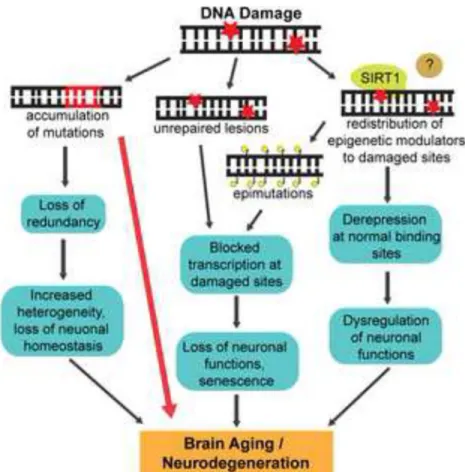
The consequences of genomic instability manifest in at least three important ways with age. The first is an accumulation of unrepaired DNA damage, which can arise from a decrease in DNA repair activities with age. For instance, a decline in the efficiency of BER and NHEJ due to a reduction in the activity of DNA glycosylases and DNA-PK, respectively, has been
A
uthor Man
uscr
ipt
A
uthor Man
uscr
ipt
A
uthor Man
uscr
ipt
A
uthor Man
uscr
ipt
reported in the literature. An age-dependent attenuation in DNA repair capacity has also been reported in the rodent and human brain. In an insightful study, microarray analysis of post-mortem human brain samples as a function of age revealed that genes encoding for critical neuronal functions, including synaptic plasticity, learning, and memory are downregulated after age 40 and concomitantly, the expression of stress response genes is upregulated (Lu et al., 2004). Importantly, this dramatic change in gene expression profiles is accompanied by an accumulation of oxidative lesions in the promoter regions of the downregulated genes (Lu et al., 2004), suggesting that an accrual of oxidative lesions could underlie the decline in cognitive abilities with age.
Another way in which DNA damage participates in aging is through the erroneous repair of DNA lesions that results in mutations (Vijg and Suh, 2013). In contrast to unrepaired lesions, which are reversible, mutations are irreversible and can therefore be highly problematic. For instance, the use of a transgenic mouse model that harbors a chromosomally integrated reporter that can be sequenced to assay for mutations as a function of age revealed that mutations in the liver almost quadrupled with age (Dolle et al., 1997). Interestingly, no such differences were found in the brain under these conditions. However, only a few chromosomal loci were sampled in this study and it is therefore formally possible that mutations accumulate at certain “hotspots” in the aging human brain. Mutations could also contribute to age-related neurodegeneration in a more indirect way. In an interesting study, single cell genomic analysis of post-mortem neurons from the human frontal cortex revealed that between 13 and 41% of neurons have copy number variations (CNVs) of at least one megabase (McConnell et al., 2013). The specific consequences of somatic mosaicism in the human brain is presently unknown; however, it would be interesting to determine whether CNVs in the brain increase as a function of age or in certain age-related neurodegenerative disorders. A more direct effect of mutations on neurodegeneration is clearly evident when these mutations compromise the activities of DNA repair/DDR factors (see below). In addition to direct alterations to the composition and structure of DNA, the formation of DNA damage also elicits substantial changes to
chromatin organization. While a number of these changes serve necessary functions in DDR signaling, there is also evidence to suggest that chromatin conformation might not be restored to its pre-damaged state following DNA repair (Oberdoerffer and Sinclair, 2007; Tamburini and Tyler, 2005). Thus, DNA damage could progressively alter chromatin conformation, and thereby, gene expression patterns, with age. In fact, a number of studies have reported age-associated changes in the epigenome (Krishnan et al., 2011; Peleg et al., 2010; Vijg and Suh, 2013), although precisely what fraction of these changes is a result of DNA damage remains unclear.
The studies described thus far highlight the local consequences of DNA damage, through lesions, mutations and epigenomic changes at the sites of damage. However, other studies, especially those conducted by Sinclair and colleagues, suggest that DNA damage can also trigger global changes in chromatin architecture (Oberdoerffer et al., 2008; Oberdoerffer and Sinclair, 2007). They observed that the exposure of cells to DNA damaging agents,
including hydrogen peroxide, and the generation of site-specific DNA DSBs leads to a redistribution of SIRT1 from various loci, including repetitive DNA elements, to the sites of DNA damage (Oberdoerffer et al., 2008). The localization of SIRT1 is essential for DNA
A
uthor Man
uscr
ipt
A
uthor Man
uscr
ipt
A
uthor Man
uscr
ipt
A
uthor Man
uscr
ipt
repair and therefore beneficial in the short-term; however, chronic genotoxic stress during and a persistent redistribution of SIRT1 causes large-scale transcriptional deregulation of genes normally targeted by SIRT1. Interestingly, the gene expression changes that result from SIRT1 redistribution parallel those in the aging mouse brain (Oberdoerffer et al., 2008). These results raise the possibility that pharmacological SIRT1 activation can impact aging in at least two ways: by stimulating the repair of damaged DNA and by promoting the transcriptional regulation of repetitive elements and other loci normally targeted by SIRT1. In addition to these mechanisms, telomere dysfunction is thought to be a major underlying factor in aging (Sahin and DePinho, 2012), although its specific roles in the aging human brain requires further characterization.
DNA damage in age-associated neurodegenerative disorders
In addition to normal aging, defective DNA repair has also been linked with age-associated neurodegenerative disorders such as Alzheimer’s disease (AD), Parkinson’s disease (PD), and amyotrophic lateral sclerosis (ALS). For instance, elevated levels of DNA strand breaks, a reduction in the levels of DSB repair proteins such as DNA-PKcs and MRN complex proteins, and decreased BER activity have been described in AD patients compared to age-matched controls (Adamec et al., 1999; Jacobsen et al., 2004; Mullaart et al., 1990; Shackelford, 2006). Similarly, elevated levels of oxidative lesions and SSBs have been reported in the neurons of ALS patients and damage to mitochondrial DNA has been documented in PD (Bender et al., 2006; Kraytsberg et al., 2006; Martin, 2001). While these studies certainly raise the possibility that defects in the DDR underlie brain aging and the development of age-related neurodegenerative disorders, it should be noted that these studies are largely correlative in nature and that our understanding of the specific contribution of DNA damage to the etiology of these disorders is still only rudimentary. To say that DNA damage has a causal effect in the neuropathology of AD, PD, or ALS requires specifying what lesions, if any, have a higher propensity to accumulate in the diseased neurons, identifying the molecular mechanisms that preclude the repair of these lesions, developing animal models in which lesion accumulation mimics at least some aspects of the
pathophysiology of human neurodegenerative disorders and ideally, that promoting DNA repair can alleviate these effects. While an understanding of each of these questions is currently limited, recent studies are fast changing the status quo.
A major issue concerns understanding which lesions are central to the progression of a given neurodegenerative disease. Generally, oxidative DNA lesions have received much attention because the brain has a relatively high metabolic rate, generates more ROS, and is thought to have a decreased ratio of antioxidant to pro-oxidant enzymes, all of which translates into a stateof elevated oxidative stress (Canugovi et al., 2013). As mentioned above, the BER pathway is primarily involved in the repair of oxidative lesions and consists of enzymes called DNA glycosylases that specialize in lesion recognition and initial processing. It has been reported that expression and activity of various BER factors changes both with age and in disorders such as AD. For instance, the expression of both UDG1 and βOGG1
glycosylases, as well as polβ, were found reduced in AD brains compared to age-matched controls (Canugovi et al., 2013). However, knockout mouse models of various DNA glycosylases show none of the drastic phenotypes of human neurodegenerative disorders. In
A
uthor Man
uscr
ipt
A
uthor Man
uscr
ipt
A
uthor Man
uscr
ipt
A
uthor Man
uscr
ipt
addition, transgenic mouse models of AD do not exhibit BER deficits. On the one hand, the lack of an overt phenotype in mice lacking DNA glycosylases is reminiscent of mouse models of the various neurodevelopmental disorders described above. On the other hand, whereas mutations in DDR factors actually underlie the neurodevelopmental disorders, mutations in BER factors have so far not been observed in neurodegenerative disorders. Thus, the specific contribution to age-related neurodegeneration of at least the subset of oxidative lesions that are repaired through BER remains unclear. In contrast to mouse models of BER mutants, conditional mouse models in which Ercc1 was specifically deleted in excitatory neurons of the forebrain showed reduced synaptic plasticity in the
hippocampus, as well as memory impairments that are characteristic features of age-related neurodegenerative disorders (Borgesius et al., 2011). Because Ercc1 is a component of the NER pathway, these results raise the possibility that NER deficits could underlie age-related neurodegeneration. However, Ercc1 also participates in the repair of DNA DSBs and crosslinks, and like with BER factors, NER mutations have so far not been identified in age-related neurodegenerative disorders.
In addition to oxidative lesions, the notion that DNA strand breaks might contribute significantly to the pathology of age-related neurodegenerative diseases has recently gained traction. First, DNA strand breaks are elevated in disorders such as AD and ALS (Adamec et al., 1999; Martin, 2001; Mullaart et al., 1990). In addition, elevated levels of DNA DSBs have now been reportedin several mouse models of neurodegeneration (Dobbin et al., 2013; Kim et al., 2008; Suberbielle et al., 2013). Among these, studies conducted on the p25/Cdk5 mouse model have been particularly illuminating. Cyclin-dependent kinase 5 (Cdk5) is a brain-specific serine/threonine kinase that requires its cyclin-like partner, p35, for catalytic activity (Lew et al., 1994; Tsai et al., 1994). Studies conducted over a dozen years have informed that in the AD brain as well as under other neurotoxic conditions, p35 undergoes proteolytic cleavage to generate p25 and that the association of p25 with Cdk5 changes the substrate specificity and subcellular localization of Cdk5 (Su and Tsai, 2011). These observations prompted the generation of the inducible p25/CDK5 mouse model (CK-p25 mice) (Cruz et al., 2003). Upon induction, CK-p25 mice express p25 in a forebrain-specific manner and systematically recapitulate various AD-like pathologies including the
accumulation of amyloid-β peptides, neurofibrillary tau tangles, astrogliosis, reduced synaptic density and neuronal loss in the forebrain (Cruz et al., 2006; Cruz et al., 2003). Remarkably, an analysis of pre-symptomatic CK-p25 mice revealed that an accrual of DNA DSBs in the forebrain precedes the appearance of all other pathological hallmarks and suggests that DSBs could be the initiating lesion of neurotoxicity in these mice (Kim et al., 2008). The unexpected discovery of DSBs in a mouse model of neurodegeneration triggered further investigations into the mechanisms underlying their accumulation in CK-p25 mice. These studies attributed the elevated DSBs to an inhibition of the class I histone deacetylase, HDAC1, in CK-p25 mice and found that overexpression of HDAC1 suppressed both the increased susceptibility to DSBs and the neuronal loss caused by p25 overexpression (Kim et al., 2008). However, a separate study noted that overexpression of the NAD+-dependent deacetylase, SIRT1, can also prevent neuronal loss in CK-p25 mice (Kim et al., 2007). Meanwhile, several groups working in dividing cells reported that SIRT1 is essential for the recruitment of NBS1 and RAD51 to the sites of DNA DSBs and that HDAC1 plays an
A
uthor Man
uscr
ipt
A
uthor Man
uscr
ipt
A
uthor Man
uscr
ipt
A
uthor Man
uscr
ipt
important role in DSB repair through the NHEJ pathway (Oberdoerffer et al., 2008; Yuan et al., 2007). These observations suggested that the neuroprotective functions of SIRT1 and HDAC1 in CK-p25 mice might arise from their roles in the neuronal DSB response. A direct exploration of the functions of SIRT1 and HDAC1 in the DSB response of postmitotic neurons has provided further insights into this question (Dobbin et al., 2013). Neurons lacking either SIRT1 or HDAC1 are more susceptible to DSB-inducing agents and are deficient in DSB repair. Both SIRT1 and HDAC1 also localize rapidly to the sites of DNA DSBs and SIRT1 specifically exhibits a co-dependent relationship with ATM for its recruitment to DSBs and also stimulates the autophosphorylation and activity of ATM (Dobbin et al., 2013). Interestingly however, SIRT1 also shares an enzyme-substrate relationship with HDAC1 in which SIRT1 deacetylates and stimulates the catalytic activity of HDAC1 and helps recruit HDAC1 to the sites of DNA damage. The effects of SIRT1 on HDAC1 activity can also be achieved by treatment with pharmacological activators of SIRT1 both in vitro and in vivo (Dobbin et al., 2013). Finally, pharmacological activation of SIRT1 is able to stimulate HDAC1 deacetylation, reduce DSB formation and improve neuronal survival in CK-p25 mice. Taken together, the studies involving CK-p25 mice not only implicate DSBs as a lesion that could underlie neurodegeneration, but also provide new clues into activities that could guard against genomic instability and preserve neuronal viability.
Although extremely cytotoxic, DSB formation is also incredibly rare even in proliferating cells, where DNA replication is an important source of these lesions. The notion of DSBs being important for age-related neurodegeneration therefore requires identifying the
processes that lead to their formation in the first place. Interestingly, a recent report indicates that physiological neural activity, including performing new learning tasks, itself can introduce DSBs within neurons (Suberbielle et al., 2013). Moreover, using an AD mouse model, the authors show that amyloid β generation exacerbates the accumulation of these DSBs (Suberbielle et al., 2013). At present, it is still unclear whether these DSBs serve a physiological purpose or whether they are merely a consequence of the changes that occur during neuronal activation, and further studies in this line should illuminate the precise risk posed by DSB formation induced during neural activity. Nonetheless, these results provide new evidence to suggest that DSBs are in fact produced in neurons under physiological conditions and that their repair could govern neuronal survival in neurodegenerative diseases.
A potential strategy to determine the role of DDR defects in age-related neurodegeneration consists of understanding whether mutations that cause the familial forms of these disorders also perturb the DDR. While such connections remain largely obscure, recent studies involving the RNA/DNA binding protein, FUS, could represent a breakthrough in this direction. In 2009, two studies identified more than a dozen mutations in FUS that are linked with familial ALS (fALS) and found that these mutations cause FUS to be deposited in the cytoplasm (Kwiatkowski et al., 2009; Vance et al., 2009). Based on its similarity to another RNA binding protein called TDP-43 that was also implicated in familial ALS, a majority of studies on FUS have since centered on its role in RNA processing (Lagier-Tourenne and Cleveland, 2009). Interestingly however, FUS (which stands for Fused in Sarcoma) was also shown to be important for genomic stability more than a decade ago. For instance, Fus−/−
A
uthor Man
uscr
ipt
A
uthor Man
uscr
ipt
A
uthor Man
uscr
ipt
A
uthor Man
uscr
ipt
mice suffer from high levels of genomic instability, defective B-lymphocyte development, male sterility, and undergo perinatal death, and FUS was shown to participate in D-loop formation, which is an intermediate step in DNA repair through HR (Baechtold et al., 1999; Hicks et al., 2000; Kuroda et al., 2000). Recently, several independent studies have
demonstrated the rapid recruitment of FUS to laser induced DNA damage sites, which is crucial for efficient DSB repair (Mastrocola et al., 2013; Rulten et al., 2013; Wang et al., 2013). FUS recruitment to DSBs depends on the enzymatic activity of PARP-1, but not on DNA-PK or ATM, and in FUS knockdown neurons, the response to treatment with DSB inducing agents is dampened. Moreover, stable tethering FUS to chromosomes in the absence of a DSB is sufficient to elicit the DDR (Wang et al., 2013). Thus, FUS appears to be an early component that participates in the initial steps of DDR signaling. Furthermore, FUS directly interacts with HDAC1, and the interacting domains map to the G-rich and C-terminal domains within FUS, where the majority of the fALS mutations are also
concentrated. fALS FUS mutants display an impaired interaction with HDAC1 and lead to deficient DNA repair (Wang et al., 2013). Importantly, when motor cortex samples from ALS patients harboring c-terminal FUS mutations were analyzed, it was found that the amount of DNA damage is significantly enriched compared to normal brain tissues (Wang et al., 2013). Together, these studies suggest that the dysfunction of FUS in DSB signaling and repair could contribute to the disease progression of FUS linked fALS.
In conclusion, it is becoming increasingly clear that the DNA damage response is important during both neural development and in the mature nervous system. Mutations in core DNA repair factors are either incompatible with life, or even when tolerated, manifest in severe neurodevelopmental disorders. On the other hand, determining the specific contribution of DNA damage to brain aging and neurodegeneration remains a complex problem. The vulnerability of postmitotic neurons to certain types of DNA damage (such as oxidative lesions or certain DNA strand break lesions) coupled with a gradual decline in the activities of corresponding repair mechanisms could lead to their accumulation with age and
contribute to brain aging and neurodegeneration. In addition, mutations in certain DDR factors (such as FUS) could exacerbate these effects and predispose individuals to neurodegeneration. In the future, identification of the specific lesions that accumulate in human age-related neurodegenerative diseases and the generation of new conditional mouse models are likely to provide key insights into which activities should be targeted in
therapeutic strategies to combat these disorders.
List of Abbreviations
DDR DNA damage response NHEJ nonhomologous end joining HR homologous recombination ROS reactive oxygen species BER base excision repair NER nucleotide excision repair
A
uthor Man
uscr
ipt
A
uthor Man
uscr
ipt
A
uthor Man
uscr
ipt
A
uthor Man
uscr
ipt
GG-NER global genome NER TC-NER transcription coupled NER SSBs single strand breaks DSBs double strand breaks PAR poly-(ADP-ribose) HAT histone acetyltransferase HDAC histone deacetylase XP xeroderma pigmentosum CS Cockayne syndrome TTD trichothiodystrophy A-T ataxia telangiectasia ATLD A-T like disease
NBS Nijmegen breakage syndrome AOA1 ataxia with occulomotor apraxia-1
SCAN1 spinocerebellar ataxia with axonal neuropathy CNV copy number variation
AD Alzheimer’s Disease PD Parkinson’s Disease
ALS Amyotrophic lateral sclerosis fALS familial Amyotrophic lateral sclerosis
References
Adamec E, Vonsattel JP, Nixon RA. DNA strand breaks in Alzheimer’s disease. Brain Res. 1999; 849:67–77. [PubMed: 10592288]
Ahel D, Horejsi Z, Wiechens N, Polo SE, Garcia-Wilson E, Ahel I, Flynn H, Skehel M, West SC, Jackson SP, et al. Poly(ADP-ribose)-dependent regulation of DNA repair by the chromatin remodeling enzyme ALC1. Science. 2009; 325:1240–1243. [PubMed: 19661379]
Ahel I, Rass U, El-Khamisy SF, Katyal S, Clements PM, McKinnon PJ, Caldecott KW, West SC. The neurodegenerative disease protein aprataxin resolves abortive DNA ligation intermediates. Nature. 2006; 443:713–716. [PubMed: 16964241]
Ames BN, Shigenaga MK, Hagen TM. Oxidants, antioxidants, and the degenerative diseases of aging. Proc Natl Acad Sci U S A. 1993; 90:7915–7922. [PubMed: 8367443]
Anttinen A, Koulu L, Nikoskelainen E, Portin R, Kurki T, Erkinjuntti M, Jaspers NG, Raams A, Green MH, Lehmann AR, et al. Neurological symptoms and natural course of xeroderma pigmentosum. Brain. 2008; 131:1979–1989. [PubMed: 18567921]
A
uthor Man
uscr
ipt
A
uthor Man
uscr
ipt
A
uthor Man
uscr
ipt
A
uthor Man
uscr
ipt
Avvakumov N, Nourani A, Cote J. Histone chaperones: modulators of chromatin marks. Mol Cell. 2011; 41:502–514. [PubMed: 21362547]
Baechtold H, Kuroda M, Sok J, Ron D, Lopez BS, Akhmedov AT. Human 75-kDa DNA-pairing protein is identical to the pro-oncoprotein TLS/FUS and is able to promote D-loop formation. J Biol Chem. 1999; 274:34337–34342. [PubMed: 10567410]
Bender A, Krishnan KJ, Morris CM, Taylor GA, Reeve AK, Perry RH, Jaros E, Hersheson JS, Betts J, Klopstock T, et al. High levels of mitochondrial DNA deletions in substantia nigra neurons in aging and Parkinson disease. Nat Genet. 2006; 38:515–517. [PubMed: 16604074]
Biton S, Barzilai A, Shiloh Y. The neurological phenotype of ataxia-telangiectasia: solving a persistent puzzle. DNA Repair (Amst). 2008; 7:1028–1038. [PubMed: 18456574]
Borgesius NZ, de Waard MC, van der Pluijm I, Omrani A, Zondag GC, van der Horst GT, Melton DW, Hoeijmakers JH, Jaarsma D, Elgersma Y. Accelerated age-related cognitive decline and
neurodegeneration, caused by deficient DNA repair. J Neurosci. 2011; 31:12543–12553. [PubMed: 21880916]
Burma S, Chen BP, Murphy M, Kurimasa A, Chen DJ. ATM phosphorylates histone H2AX in response to DNA double-strand breaks. J Biol Chem. 2001; 276:42462–42467. [PubMed: 11571274]
Caldecott KW. Single-strand break repair and genetic disease. Nat Rev Genet. 2008; 9:619–631. [PubMed: 18626472]
Canugovi C, Misiak M, Ferrarelli LK, Croteau DL, Bohr VA. The role of DNA repair in brain related disease pathology. DNA Repair (Amst). 2013; 12:578–587. [PubMed: 23721970]
Chou DM, Adamson B, Dephoure NE, Tan X, Nottke AC, Hurov KE, Gygi SP, Colaiacovo MP, Elledge SJ. A chromatin localization screen reveals poly (ADP ribose)-regulated recruitment of the repressive polycomb and NuRD complexes to sites of DNA damage. Proc Natl Acad Sci U S A. 2010; 107:18475–18480. [PubMed: 20937877]
Cleaver JE. Defective repair replication of DNA in xeroderma pigmentosum. Nature. 1968; 218:652– 656. [PubMed: 5655953]
Clements PM, Breslin C, Deeks ED, Byrd PJ, Ju L, Bieganowski P, Brenner C, Moreira MC, Taylor AM, Caldecott KW. The ataxia-oculomotor apraxia 1 gene product has a role distinct from ATM and interacts with the DNA strand break repair proteins XRCC1 and XRCC4. DNA Repair (Amst). 2004; 3:1493–1502. [PubMed: 15380105]
Cruz JC, Kim D, Moy LY, Dobbin MM, Sun X, Bronson RT, Tsai LH. p25/cyclin-dependent kinase 5 induces production and intraneuronal accumulation of amyloid beta in vivo. J Neurosci. 2006; 26:10536–10541. [PubMed: 17035538]
Cruz JC, Tseng HC, Goldman JA, Shih H, Tsai LH. Aberrant Cdk5 activation by p25 triggers pathological events leading to neurodegeneration and neurofibrillary tangles. Neuron. 2003; 40:471–483. [PubMed: 14642273]
Das C, Lucia MS, Hansen KC, Tyler JK. CBP/p300-mediated acetylation of histone H3 on lysine 56. Nature. 2009; 459:113–117. [PubMed: 19270680]
Date H, Onodera O, Tanaka H, Iwabuchi K, Uekawa K, Igarashi S, Koike R, Hiroi T, Yuasa T, Awaya Y, et al. Early-onset ataxia with ocular motor apraxia and hypoalbuminemia is caused by mutations in a new HIT superfamily gene. Nat Genet. 2001; 29:184–188. [PubMed: 11586299]
de Laat WL, Jaspers NG, Hoeijmakers JH. Molecular mechanism of nucleotide excision repair. Genes Dev. 1999; 13:768–785. [PubMed: 10197977]
Digweed M, Sperling K. Nijmegen breakage syndrome: clinical manifestation of defective response to DNA double-strand breaks. DNA Repair (Amst). 2004; 3:1207–1217. [PubMed: 15279809] Dobbin MM, Madabhushi R, Pan L, Chen Y, Kim D, Gao J, Ahanonu B, Pao PC, Qiu Y, Zhao Y, Tsai
LH. SIRT1 collaborates with ATM and HDAC1 to maintain genomic stability in neurons. Nat Neurosci. 2013; 16:1008–1015. [PubMed: 23852118]
Dolle ME, Giese H, Hopkins CL, Martus HJ, Hausdorff JM, Vijg J. Rapid accumulation of genome rearrangements in liver but not in brain of old mice. Nat Genet. 1997; 17:431–434. [PubMed: 9398844]
A
uthor Man
uscr
ipt
A
uthor Man
uscr
ipt
A
uthor Man
uscr
ipt
A
uthor Man
uscr
ipt
El-Khamisy SF, Saifi GM, Weinfeld M, Johansson F, Helleday T, Lupski JR, Caldecott KW. Defective DNA single-strand break repair in spinocerebellar ataxia with axonal neuropathy-1. Nature. 2005; 434:108–113. [PubMed: 15744309]
Frappart PO, Tong WM, Demuth I, Radovanovic I, Herceg Z, Aguzzi A, Digweed M, Wang ZQ. An essential function for NBS1 in the prevention of ataxia and cerebellar defects. Nat Med. 2005; 11:538–544. [PubMed: 15821748]
Gueven N, Becherel OJ, Kijas AW, Chen P, Howe O, Rudolph JH, Gatti R, Date H, Onodera O, Taucher-Scholz G, Lavin MF. Aprataxin, a novel protein that protects against genotoxic stress. Hum Mol Genet. 2004; 13:1081–1093. [PubMed: 15044383]
Haber JE. DNA recombination: the replication connection. Trends Biochem Sci. 1999; 24:271–275. [PubMed: 10390616]
Hicks GG, Singh N, Nashabi A, Mai S, Bozek G, Klewes L, Arapovic D, White EK, Koury MJ, Oltz EM, et al. Fus deficiency in mice results in defective B-lymphocyte development and activation, high levels of chromosomal instability and perinatal death. Nat Genet. 2000; 24:175–179. [PubMed: 10655065]
Hoeijmakers JH. DNA damage, aging, and cancer. N Engl J Med. 2009; 361:1475–1485. [PubMed: 19812404]
Hsiao KY, Mizzen CA. Histone H4 deacetylation facilitates 53BP1 DNA damage signaling and double-strand break repair. J Mol Cell Biol. 2013; 5:157–165. [PubMed: 23329852]
Ikura T, Ogryzko VV, Grigoriev M, Groisman R, Wang J, Horikoshi M, Scully R, Qin J, Nakatani Y. Involvement of the TIP60 histone acetylase complex in DNA repair and apoptosis. Cell. 2000; 102:463–473. [PubMed: 10966108]
Iyama T, Wilson DM 3rd. DNA repair mechanisms in dividing and non-dividing cells. DNA Repair (Amst). 2013; 12:620–636. [PubMed: 23684800]
Jackson SP. Sensing and repairing DNA double-strand breaks. Carcinogenesis. 2002; 23:687–696. [PubMed: 12016139]
Jackson SP, Bartek J. The DNA-damage response in human biology and disease. Nature. 2009; 461:1071–1078. [PubMed: 19847258]
Jacobsen E, Beach T, Shen Y, Li R, Chang Y. Deficiency of the Mre11 DNA repair complex in Alzheimer’s disease brains. Brain Res Mol Brain Res. 2004; 128:1–7. [PubMed: 15337312] Katyal S, el-Khamisy SF, Russell HR, Li Y, Ju L, Caldecott KW, McKinnon PJ. TDP1 facilitates
chromosomal single-strand break repair in neurons and is neuroprotective in vivo. The EMBO journal. 2007; 26:4720–4731. [PubMed: 17914460]
Katyal S, McKinnon PJ. DNA strand breaks, neurodegeneration and aging in the brain. Mech Ageing Dev. 2008; 129:483–491. [PubMed: 18455751]
Kim D, Frank CL, Dobbin MM, Tsunemoto RK, Tu W, Peng PL, Guan JS, Lee BH, Moy LY, Giusti P, et al. Deregulation of HDAC1 by p25/Cdk5 in neurotoxicity. Neuron. 2008; 60:803–817.
[PubMed: 19081376]
Kim D, Nguyen MD, Dobbin MM, Fischer A, Sananbenesi F, Rodgers JT, Delalle I, Baur JA, Sui G, Armour SM, et al. SIRT1 deacetylase protects against neurodegeneration in models for
Alzheimer’s disease and amyotrophic lateral sclerosis. The EMBO journal. 2007; 26:3169–3179. [PubMed: 17581637]
Kirkwood TB. Understanding the odd science of aging. Cell. 2005; 120:437–447. [PubMed: 15734677]
Kraemer KH, Lee MM, Scotto J. Xeroderma pigmentosum. Cutaneous, ocular, and neurologic abnormalities in 830 published cases. Arch Dermatol. 1987; 123:241–250. [PubMed: 3545087] Kraytsberg Y, Kudryavtseva E, McKee AC, Geula C, Kowall NW, Khrapko K. Mitochondrial DNA deletions are abundant and cause functional impairment in aged human substantia nigra neurons. Nat Genet. 2006; 38:518–520. [PubMed: 16604072]
Krishnakumar R, Kraus WL. The PARP side of the nucleus: molecular actions, physiological outcomes, and clinical targets. Mol Cell. 2010; 39:8–24. [PubMed: 20603072]
Krishnan V, Chow MZ, Wang Z, Zhang L, Liu B, Liu X, Zhou Z. Histone H4 lysine 16
hypoacetylation is associated with defective DNA repair and premature senescence in Zmpste24-deficient mice. Proc Natl Acad Sci U S A. 2011; 108:12325–12330. [PubMed: 21746928]