75
Herz 33 · 2008 · Nr. 1 © Urban & Vogel© Urban & Vogel 2008
Herz
Image of the Month
Herz 2008;33:75
DOI 10.1007/ s00059-008-3089-1
Coronary Angiography with Low-Dose
Computed Tomography at 1.4 mSv
Lars Husmann
1, Ines Valenta
1, Philipp A. Kaufmann
1, 21 Cardiovascular
Center, Univer-sity Hospital Zurich, Switzerland,
2 Zurich Center for
Integrative Human Physiology, University of Zurich, Switzerland
A 53-year-old woman with hypertension, dyslipid-emia, nicotine abuse, and a positive family history of coronary artery disease (CAD) was referred for cardiac work-up. The patient’s history revealed no evidence for CAD, but cardiac stress testing on a treadmill ergometer disclosed ST depression over > 3 min at a heart rate of 150 bpm. The patient was subsequently referred to computed tomography coronary angiography (CTCA). A low-dose CTCA protocol was performed on a LightSpeed VCT XT scanner (GE Healthcare), using prospective elec-trocardiogram (ECG) gating [1]. The smallest X-ray window was chosen (only 75% of the RR cycle); tube current (450 mAs) and tube voltage (100 kV) were adapted to the body mass index
(22.2 kg/m2), resulting in an effective dose of 1.4
mSv, as calculated from the product of the dose-length product and a conversion coefficient for the chest (k = 0.017 mSv/mGy × cm) [2]. A sin-gle dose of 2.5 mg sublingual isosorbide dinitrate was administered prior to CTCA scanning; no
β-blocking medication was given. During the scan,
the patient had a mean heart rate of 48 bpm, vary-ing between 46 and 49 bpm.
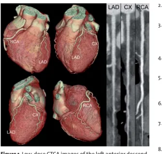
Image quality of CTCA was excellent in all coro-nary segments (Figure 1). There were no signs of ath-erosclerosis and CAD was excluded.
With prospective ECG gating, radiation in CTCA is only administered at predefined time points of the cardiac cycle, rather than throughout the entire cardiac cycle as in the helical mode used so far. Al-though CTCA has been demonstrated to reliably de-tect CAD [3–5], CT as a major source of radiation dose has remained an issue of concern [6]. Previous CTCA studies have reported estimated radiation doses of up to 21.4 mSv without the use of the ECG-pulsing technique [7] and down to 9.4 mSv with the use of ECG pulsing [8]. Our technique described here now allows a tremendous reduction of radiation dose down to 1.4 mSv. This case indicates that low-dose CTCA with prospective ECG gating is fea-sible with excellent image quality and low radiation exposure in dedicated patient populations.
References
1. Hsieh J, Londt J, Vass M, et al. Step-and-shoot data acquisi-tion and reconstrucacquisi-tion for cardiac x-ray computed tomog-raphy. Med Phys 2006;33:4236–48.
2. Einstein AJ, Moser KW, Thompson RC, et al. Radiation dose to patients from cardiac diagnostic imaging. Circulation 2007;116:1290–305.
3. Leber AW, Knez A, Ziegler F von, et al. Quantification of ob-structive and nonobob-structive coronary lesions by 64-slice computed tomography: a comparative study with quanti-tative coronary angiography and intravascular ultrasound. J Am Coll Cardiol 2005;46:147–54.
4. Leschka S, Alkadhi H, Plass A, et al. Accuracy of MSCT coro-nary angiography with 64-slice technology: first experi-ence. Eur Heart J 2005;26:1482–7.
5. Raff GL, Gallagher MJ, O’Neill WW, et al. Diagnostic accu-racy of noninvasive coronary angiography using 64-slice spiral computed tomography. J Am Coll Cardiol 2005;46: 552–7.
6. Brenner D, Hall E. Computed tomography – an increasing source of radiation exposure. N Engl J Med 2007;357: 2277–84.
7. Mollet NR, Cademartiri F, van Mieghem CA, et al. High-res-olution spiral computed tomography coronary angiogra-phy in patients referred for diagnostic conventional coro-nary angiography. Circulation 2005;112:2318–23.
8. Hausleiter J, Meyer T, Hadamitzky M, et al. Radiation dose estimates from cardiac multislice computed tomography in daily practice: impact of different scanning protocols on effective dose estimates. Circulation 2006;113:1305–10.
Figure 1. Low-dose CTCA images of the left anterior
descend-ing artery (LAD), the circumflex artery (CX), and the right coronary artery (RCA) rule out the presence of CAD.
Address for Correspondence Professor Philipp A. Kaufmann, MD Cardiovascular Center University Hospital Zurich NUK C 42 Rämistraße 100 8091 Zürich Switzerland Phone (+41/44) 255-4196, Fax -4414 e-mail: [email protected]