Self-expandable valved stent of large size:
off-bypass implantation in pulmonary position
Jun Qing Zhou
a, Antonio F. Corno
b,*, Christophe H. Huber
b,
Piergiorgio Tozzi
b, Ludwig K. von Segesser
baCardiothoracic Department, The No. 1 People’s Hospital of Shaoxing, Zhejiang, China b
Department of Cardiovascular Surgery, Centre Hospitalier Universitaire Vaudois (CHUV), 46 Rue du Bugnon, CH-1011 Lausanne, Switzerland Received 27 January 2003; received in revised form 12 March 2003; accepted 17 March 2003
Abstract
Objective: To evaluate the feasibility of the off-bypass implantation of a self-expandable valved stent of large size in pulmonary position. Materials and methods: A glutaraldehyde preserved valved bovine jugular xenograft with internal diameter ¼ 22 mm, mounted in two rings of nitinol ‘Z’ stent, expandable from 7 to 24 mm of internal diameter, was acutely evaluated in 6 adult pigs, mean body weight 55.6 kg (range 47 – 67 kg). Through a stent-graft delivery system (24 French) the self expandable valved stent was implanted off-bypass in pulmonary valve position by trans-ventricular approach through median sternotomy. Results: The mean diameter of the main pulmonary artery measured was 21.7 ^ 1.6 mm. The mean length of the self expandable valved stent was 23.1 ^ 0.7 mm, the mean internal diameter 21.6 ^ 0.7 mm and the mean external diameter 26.3 ^ 0.7 mm. The mean peak pressure gradient recorded across the valve was 6.33 ^ 2.8 mmHg (range 4.5 – 9.6 mmHg) at Doppler echocardiography, and 4.5 ^ 3.1 mmHg (range 0 – 7 mmHg) at invasive measurement, with a pulmonary blood flow of 3.03 ^ 0.05 l/min. Intra-vascular ultrasound showed complete opening and closure of the valve (mean area reduction from 315.08 ^ 54.13 to 0 mm2). Conclusions: (a) Off-bypass implantation of self-expandable valved stent is feasible in pulmonary position; (b) off-bypass surgical approach allows for valved stent implantation of adult size with adequate hemodynamic functioning; and (c) intra-vascular ultrasound makes implantation and evaluation easy and reproducible.
q2003 Elsevier Science B.V. All rights reserved.
Keywords: Biological valved conduits; Pulmonary valve implantation; Pulmonary valve regurgitation; Tetralogy of Fallot
1. Introduction
After previous experimental studies with percutaneous valve replacement in pulmonary [1] and aortic [2,3]
position, the percutaneous insertion of a biological valve in pulmonary position has been introduced in the clinical practice[4,5].
Insertion of a valve in pulmonary position is indicated mainly in two situations: (a) pulmonary valve regurgitation, generally after surgical repair for tetralogy of Fallot[6 – 14]; and (b) dysfunction of the biological valved conduit pre-viously implanted to establish the continuity between the right ventricle and the pulmonary artery during repair of complex congenital heart defects[15,16].
So far the conventional treatment for the above situations
consisted in pulmonary valve replacement [6 – 14] and replacement of the biological valved conduit[15 – 17], even if with uncertainty about the adequate timing for pulmonary valve insertion in order to prevent or reduce the incidence of sudden death, arrhythmias and right ventricular dysfunction
[5 – 14].
Several alternative strategies, particularly in the presence of a dysfunction of a biological valved conduit, have been considered within the last years, including other types of biological valved conduits [18 – 20] or endovascular stent implantation to dilate and delay the surgical replacement of the obstructed conduit[21 – 23].
The main limit in the use of endovascular stents to dilate an obstructed biological conduit implanted between the right ventricle and the pulmonary artery is that, even if the obstruction is relieved or substantially reduced without requiring for a re-operation on cardiopulmonary bypass, the patient remains with a pulmonary valve regurgitation,
www.elsevier.com/locate/ejcts
1010-7940/03/$ - see front matter q 2003 Elsevier Science B.V. All rights reserved. doi:10.1016/S1010-7940(03)00178-7
* Corresponding author. Tel.: þ 41-21-314-2280; fax: þ 41-21-314-2278. E-mail address: [email protected] (A.F. Corno).
frequently worse than before. The subsequent right ven-tricular volume overload can cause irreversible myocardial damages, with the known incidence of sudden death, arrhythmias and right ventricular dysfunction[5 – 14,24].
The advantages of the percutaneous insertion of the pulmonary valve over the conventional surgical techniques are quite evident in terms of avoiding an operation on cardiopulmonary bypass, and in the same time in the ability of implanting a functioning valve, therefore reducing or abolishing both the pressure and the volume overload on the right ventricle [1,4,5]. The major limit of this recently reported technique is the mismatch between size of the venous access and size of the introducer (at least 18 Fr), restricting the use of this strategy to older children and the size of the valve to an 18 mm internal diameter biological valve[5].
Because of the above problems, we devised an experi-mental study to evaluate an alternative strategy aiming at the off-bypass implantation of a self-expandable valved stent of large size in pulmonary position from right ventricular approach.
2. Materials and methods
A glutaraldehyde preserved valved bovine jugular xeno-graft with internal diameter ¼ 22 mm was mounted in two rings of non-thermosensitive nitinol ‘Z’ stents, expandable from 7 to 24 mm of internal diameter. In vitro static per-formance and dynamic test evaluation of this valved stent have been already reported[25]. The self expanding valved stent was prepared with a Teflon sheath stent-graft delivery system with overall diameter 8.0 mm ¼ 24 F.
Acute in vivo evaluation was performed in six adult pigs, mean body weight 55.6 kg (range 47 – 67 kg). After general anaesthesia, tracheal intubation and mechanical ventilation, with continuous monitoring of electrocardiogram, arterial and central venous pressure and oxygen saturation, the chest was opened through a conventional median sternotomy. Heparin was administered i.v. (1 mg/kg). After a short incision (4 mm) on the anterior aspect of the right ventricle, controlled by a purse string on 4-0 polypropylene suture, through the sheath stent-graft delivery system the valved stent was implanted off-bypass in pulmonary valve position by trans-ventricular approach (Fig. 1). The correct positioning of the valved stent was evaluated and confirmed before definitive deployment by intravascular ultrasound technique.
Valve function was assessed with colour Doppler echocardiography, flow and pressure drop measurements with Swan-Ganz Oximetry (Baxter-Edwards, CA, USA) catheter, as well as intravascular ultrasound (Boston Scientific Corporation, CA, USA) with a 6F, 12.5 MHz transducer and Acuson Corporation, CA, USA) with 10F, 7.5 MHz transducer in real time.
A high fidelity tip mounted Millar pressure transducer
system was used to invasively measure the pressure proximal and distal to the valve.
At the end of the study the animals were electively sacrificed to check the adequate position of the valved stent, as well as its deployment and anchorage and the presence of any deformation of the valve.
All animals received human care in compliance with the ‘Principles of Laboratory Animals’ formulated by the National Society of Medical Research and the ‘Guide for the Care and Use of Laboratory Animals’ prepared by the Institute of Laboratory Animal Resources and published by the National Institutes of Health (NIH publication 85-23, revised 1985). The protocol was approved by the insti-tutional Committee on Animal Research.
Statistical analysis: the Student’s t-test was utilized, and all data were expressed as mean ^ standard deviation.
3. Results
The mean diameter of the main pulmonary artery measured with intravascular ultrasound was 21.7 ^ 1.6 mm. The mean length of the valved stent was 23.1 ^ 0.7 mm, the mean internal diameter 21.6 ^ 0.7 mm and the mean external diameter 26.3 ^ 0.7 mm.
The mean peak pressure gradient recorded across the valve was 6.33 ^ 2.8 mmHg (range 4.5 – 9.6 mmHg) at Doppler echocardiography, and 4.5 ^ 3.1 mm Hg (range 0 – 7 mmHg) at invasive measurement, with a mean pulmonary blood flow of 3.03 ^ 0.05 l/min.
Intravascular ultrasound showed complete opening and closure of the valve (mean area reduction from
Fig. 1. After a short incision (4 mm) on the anterior aspect of the right ventricle, controlled by a purse string on 4-0 polypropylene suture, through the sheath stent-graft delivery system the valved stent has been implanted off-bypass in pulmonary valve position by trans-ventricular approach.
315.08 ^ 54.13 to 0 mm2) (Fig. 2). In all animals Doppler echocardiography confirmed the absence of any valve regurgitation as well as of paravalvular leak (Fig. 3).
No significant changes were recorded in electrocardio-gram, arterial and central venous pressure and oxygen satu-ration after self expandable valved stent implantation.
Post-mortem examination confirmed the adequate posi-tion of the valved stent in pulmonary posiposi-tion (Fig. 4), as well as ruled out any valve deformation or thrombus (Fig. 5).
4. Discussion
The implant of a valve in pulmonary position has the ideal purpose of abolishing the pulmonary valve regurgita-tion to prevent or reduce the incidence of sudden death, arrhythmias and right ventricular dysfunction[5 – 14]with the lowest possible surgical risk.
Because of the difficult balance between costs (risks) and benefits (competent pulmonary valve), timing and type of management so far have not reach general agreement[24].
The percutaneous insertion of a pulmonary valve, recently introduced in the clinical practice [4,5], presents the advantages of avoiding an operation on cardio-pulmonary bypass and in the same time the possibility of implanting a functioning valve, therefore reducing or abolishing both the pressure and the volume overload on the right ventricle[1,4,5]. The major current limit of this technique is the mismatch between size of the venous access and size of the introducer (at least 18 Fr), restricting the use to older children (. 25 kg of body weight) and the size of the valve to an 18 mm internal diameter biological valve [5].
Our experimental study proposes an alternative strategy allowing the off-bypass implantation of a self-expandable valved stent of large size (internal diameter ¼ 22 mm) in pulmonary position from right ventricular approach without cardiopulmonary bypass.
The implant of a functioning pulmonary valve can therefore been accomplished, overcoming the limits of the currently available techniques, with the only additional risks of a limited chest opening. In our initial experimental study
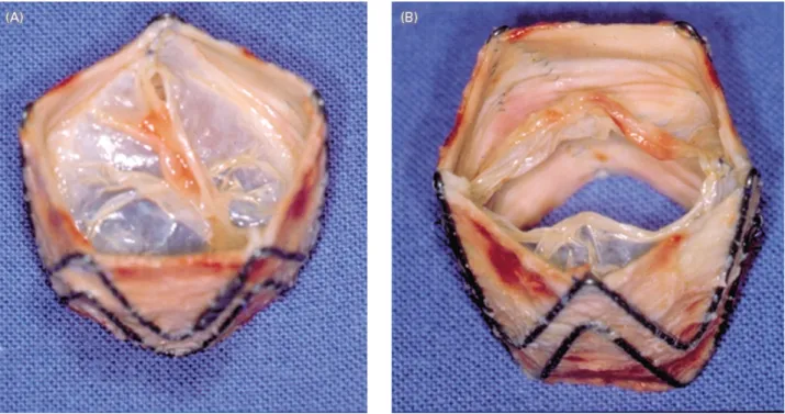
Fig. 2. Intravascular ultrasound showing complete opening (A); and closure (B) of the valve.
we used a median sternotomy because we wanted to evaluated the feasibility of the new technique. In fact, based on this first experience, the implant of the valved stent in pulmonary position can be accomplished through a limited left anterior thoracotomy, particularly advantageous in clinical practice in the presence of a previous median sternotomy.
With regard to the size of the valved stent, the mean
external diameter of 26.3 ^ 0.7 mm should allow adequate implant even in patients with dilated right ventricular outflow tract because of severe pulmonary valve regurgita-tion, like the situation encountered years after repair of tetralogy of Fallot with transannular patch.
With regard to the larger size of available biological valve, the internal diameter of 22 mm, because of the favourable effective orifice area, allows an adequate
Fig. 4. Post-mortem examination confirming the adequate position of the valved stent in pulmonary position (A); and showing the fingerprint of the Z stent on the pulmonary artery wall (B).
hemodynamics without pressure gradient in patients up to 91 kg of body weight, as we already reported[26].
Of course our positive preliminary experimental data will need to be validated by chronic studies.
4.1. Conclusions
(a) The off-bypass implantation of self-expandable valved stent is feasible in pulmonary position; (b) the off-bypass surgical approach allows for valved stent implan-tation of adult size with adequate hemodynamic function-ing; and (c) intravascular ultrasound makes implantation and evaluation easy and reproducible.
Acknowledgements
We deeply acknowledge the technical assistance received for and during the performance of the animal experiments from Monique Augstburger, Marko Burki, Gilles Godar, Iker Mallabiabarena, Antonio Mucciolo and Giuseppe Mucciolo.
References
[1] Bonhoeffer P, Boudjemline Y, Saliba Z, Hausse AO, Aggoun Y, Bonnet D, Sidi D, Kachaner J. Transcatheter implantation of a bovine valve in pulmonary position. A lamb study. Circulation 2000;102: 813 – 6.
[2] Boudjemline Y, Bonhoeffer P. Steps toward percutaneous aortic valve replacement. Circulation 2002;105:775– 8.
[3] Lutter G, Kuklinski D, Berg G, von Samson P, Martin J, Handke M, Uhrmeister P, Beyersdorf F. Percutaneous aortic valve replacement: an experimental study. I. Studies on implantation. J Thorac Cardiovasc Surg 2002;123:768 – 76.
[4] Bonhoeffer P, Boudjemline Y, Saliba Z, Merckx J, Aggoun Y, Bonnet D, Acar P, Le Bidois J, Sidi D, Kachaner J. Percutaneous replacement of pulmonary valve in a right-ventricle to pulmonary-artery prosthetic conduit with valve dysfunction. Lancet 2000;356:1403 – 5. [5] Bonhoeffer P, Boudjemline Y, Qureshi SA, Le Bidois J, Iserin L, Acar
P, Merckx J, Kachaner J, Sidi D. Percutaneous insertion of the pulmonary valve. J Am Coll Cardiol 2002;39:1664 – 9.
[6] Rocchini AP, Rosenthal A, Freed M. Chronic congestive heart failure after repair of tetralogy of Fallot. Circulation 1977;56:305 – 10. [7] Bove EL, Kavey REW, Byrum CJ, Sondheimer HM, Blackman MS,
Thomas FD. Improved right ventricular function following late pulmonary valve replacement for residual pulmonary insufficiency or stenosis. J Thorac Cardiovasc Surg 1985;90:50 – 5.
[8] Warner KB, Anderson JE, Fulton DR, Payne DD, Geggel RL, Marx GR. Restoration of the pulmonary valve reduces right ventricular volume overload after previous repair of tetralogy of Fallot. Circulation 1993;88(Suppl. II):189 – 97.
[9] Dietl CA, Cazzaniga ME, Dubner SJ, Perez-Balino NA, Torres AR, Favaloro RG. Life threatening arrhythmias and RV dysfunction after surgical repair of tetralogy of Fallot. Circulation 1994;90:7– 12. [10] Yemets IM, Williams WG, Webb GD, Harrison DA, McLaughlin PR,
Trusler GA, Coles JG, Rebeyka IM, Freedom RM. Pulmonary valve replacement late after repair of tetralogy of Fallot. Ann Thorac Surg 1997;64:526– 30.
[11] Therrien J, Siu SC, McLaughlin PR, Liu PP, Williams WG, Webb GD. Pulmonary valve replacement in adults after repair of tetralogy of Fallot: are we operating too late? J Am Coll Cardiol 2000;36:1670 – 5. [12] Discigil B, Dearani JA, Puga FJ, Schaff HV, Hagler DJ, Warnes CA, Danielson GK. Late pulmonary valve replacement after repair of tetralogy of Fallot. J Thorac Cardiovasc Surg 2001;121:344 – 51. [13] de Ruijter FTH, Weenink I, Hitchcock FJ, Meijboom EJ, Bennink
GBWE. Right ventricular dysfunction and pulmonary valve replace-ment after correction of tetralogy of Fallot. Ann Thorac Surg 2002;73: 1794 – 800.
[14] Kanter KR, Budde JM, Parks J, Tam VKH, Sharma S, Williams WH, Fyfe DA. One hundred pulmonary valve replacements in children after relief of right ventricular outflow tract obstruction. Ann Thorac Surg 2002;73:1801 – 7.
[15] Corno AF, Giamberti A, Giannico S, Marino B, Picardo S, Ballerini L, Marcelletti C. Long-term results after extracardiac valved conduits implanted for complex congenital heart disease. J Card Surg 1988;3: 495 – 500.
[16] Stark J, Bull C, Stajevic M, Jothi M, Elliott M, de Leval MR. Fate of subpulmonary homograft conduits: determinants of late homograft failure. J Thorac Cardiovasc Surg 1998;115:506– 16.
[17] Homann M, Haehnel JC, Mendler N, Paek SU, Holper K, Meisner H, Lange R. Reconstruction of the RVOT with valved biological conduits: 25 years experience with allografts and xenografts. Eur J Cardiothorac Surg 2000;17:624– 30.
[18] Corno AF, Hurni M, Griffin H, Galal OM, Payot M, Sekarski N, Tozzi P, von Segesser LK. Bovine jugular vein as right ventricle-to-pulmonary artery valved conduit. J Heart Valve Dis 2002;11:242– 7. [19] Stock UA, Nagashima M, Khalil PN, Nollert GD, Herden T, Sperling JS, Moran A, Lien J, Martin DP, Schoen FJ, Vacanti JP, Mayer JE. Tissue-engineered valved conduits the pulmonary circulation. J Thorac Cardiovasc Surg 2000;119:732– 40.
[20] Hoerstrup SP, Kadner A, Breymann C, Maurus CF, Guenter CI, Sodian R, Visjager JF, Zund G, Turina MI. Living, autologous pulmonary artery conduits tissue engineered from human umbilical cord cells. Ann Thorac Surg 2002;74:46– 52.
[21] Powell AJ, Lock JE, Keane JF, Perry SB. Prolongation of RV-PA conduit life span by percutaneous stent implantation. Intermediate-term results. Circulation 1995;92:282– 8.
[22] Saliba Z, Bonhoeffer P, Aggoun Y, Iserin L, Butera G, Bonnet D, Sidi D, Kachaner J. Treatment of obstruction of prosthetic conduits by percutaneous implantation of stents. Arch Mal Coeur Vaiss 1999;92: 591 – 6.
[23] Pedra CA, Justino H, Nykanen DG, VanArsdell G, Coles JG, Williams WG, Freedom RM, Benson LN. Percutaneous stent implantation to stenotic bioprosthetic valves in the pulmonary position. J Thorac Cardiovasc Surg 2002;124:82– 7.
[24] Hanley FL. Management of the congenitally abnormal right ventricular outflow tract. What is the right approach? J Thorac Cardiovasc Surg 2000;119:1 – 3.
[25] Corno AF, Zhou J, Tozzi P, von Segesser LK. Off-bypass implantation of a self-expandable valved stent between inferior vena cava and right atrium. Interactive Cardiovasc Thorac J 2003 (accepted for publication)
[26] Corno AF, Hurni M, Griffin H, Jeanrenaud X, von Segesser LK. Glutaraldehyde-fixed bovine jugular vein as a substitute for the pulmonary valve in the Ross operation. J Thorac Cardiovasc Surg 2001;122:493– 4.