Chronicle of a death foretold: Plasmodium liver stage parasites
decide on the fate of the host cell
Stefanie Graewe1, Rebecca R. Stanway2, Annika Rennenberg3 & Volker T. Heussler2
1Bernhard Nocht Institute for Tropical Medicine, Hamburg, Germany;2Institute of Cell Biology, University of Bern, Bern, Switzerland; and3Astra
GmbH, Hamburg, Germany
Correspondence: Volker T. Heussler, Institute of Cell Biology, University of Bern, Baltzerstrasse 4, 3012 Bern, Switzerland. Tel.: +41 31 631 4650; fax: +41 31 631 4615; e-mail: heussler@izb.unibe.ch Received 27 February 2011; accepted 22 June 2011. Final version published online 9 August 2011.
DOI: 10.1111/j.1574-6976.2011.00297.x Editor: Gerhard Braus
Keywords
malaria; Plasmodium liver stage; sporozoite; parasite–host interaction; exo-erythrocytic development; merosome.
Abstract
Protozoan parasites of the genus Plasmodium are the causative agents of malaria. Despite more than 100 years of research, the complex life cycle of the parasite still bears many surprises and it is safe to say that understanding the biology of the pathogen will keep scientists busy for many years to come. Malaria research has mainly concentrated on the pathological blood stage of Plasmodium parasites, leaving us with many questions concerning parasite development within the mosquito and during the exo-erythrocytic stage in the vertebrate host. After the discovery of the Plasmodium liver stage in the middle of the last century, it remained understudied for many years but the realization that it represents a promising target for vaccination approaches has brought it back into focus. The last decade saw many new and exciting discoveries concerning the exo-erythrocytic stage and in this review we will discuss the highlights of the latest developments in the field.
Malaria: infection of mice and men
Malaria remains one of the most devastating infectious diseases worldwide, infecting hundreds of millions of people every year. The disease is caused by protozoan parasites of the genus Plasmodium, which alternate between a mosquito vector and a vertebrate host. Most of the more than 200 known Plasmodium species infect rep-tiles and birds and only a relatively small number infect mammals, with five species being considered human pathogens (White, 2008). Some Plasmodium species that infect rodents have become invaluable tools to study gen-eral aspects of the biology of the mammalian Plasmodium species as their life cycles are very similar. This is particu-larly true of the sporozoite and liver stages of the parasite, where species infecting rodents have widely been used. Studying the biology of such stages for human Plasmo-dium species is difficult because it requires a safety level 3 facility for the maintenance of infected Anopheles mosqui-toes, whereas mosquitoes infected with rodent Plasmodia can be kept in insectaries with lower safety levels. The complete liver stage development of human Plasmodium species can only be studied in vitro in primary human
hepatocytes (Mazier et al., 1985) and in vivo in immuno-compromised chimpanzees (Daubersies et al., 2000; Per-laza et al., 2003). Together, these factors explain why most of our recent knowledge about the Plasmodium exo-erythrocytic stage is based on studies using rodent models. Although the focus of this review is the exo-erythrocytic form of the parasite, for a better understand-ing a brief and simplified description of the entire life cycle is provided in the following section.
The ins and outs of the Plasmodium life cycle
Once injected into the mammalian host by female Anoph-eles mosquitoes, the Plasmodium parasite must pass through a series of developmental stages to ultimately produce forms that can again infect mosquitoes. For a long time, it was postulated that mosquito-derived spor-ozoites directly infect red blood cells (RBCs) and replicate asexually. However, in the late 1940s, it was shown that sporozoites of mammal-infecting Plasmodium species ini-tially invade hepatocytes, where they replicate asexually to form thousands of merozoites (Fonseca et al., 1946;
MICR
Bastianelli, 1948; Shortt & Garnham, 1948). We now know that these merozoites are packaged into vesicles (merosomes) for safe transport into the bloodstream, where they can infect RBCs (Sturm et al., 2006). After several rounds of asexual reproduction in erythrocytes, which is probably necessary to generate a critical mass of infected cells for transmission, some parasites differentiate into sexual forms (gametocytes), which are infectious to mosquitoes. During a blood meal, a mosquito ingests sev-eral microliters of blood, containing as many as sevsev-eral thousand gametocytes together with millions of asexual forms that, unlike gametocytes, are simply digested in the midgut of the insect. Gametocytes, in response to changes in the environment from the warm-blooded mammalian host to the midgut of the mosquito, develop into gametes. Motile male gametes (microgametes) are liber-ated and fuse with female gametes (macrogametes), form-ing zygotes that advance to ookinetes. These motile forms penetrate the midgut of the insect and encapsulate under the basal lamina to form oocysts. Through extensive asex-ual replication thousands of sporozoites are formed, which are liberated into the hemolymph of the insect to be passively distributed throughout the entire insect body cavity. Eventually, they reach the salivary glands and actively penetrate them. After further maturation, they migrate to the ducts of the gland and can be transmitted when the mosquito takes a blood meal. The number of sporozoites inoculated by the mosquito during each bite is relatively low (50–100) (Rosenberg et al., 1990; Fris-chknecht et al., 2004; Medica & Sinnis, 2005) and thus the infection of hepatocytes was thought to be very effi-cient. Surprisingly, it was recently reported that only a third of the transmitted sporozoites penetrate a blood vessel and potentially reach the liver (Amino et al., 2006). This might be one of the reasons why even in hyper-endemic regions not every bite of an infectious mosquito results in manifestation of the disease.
This review concentrates on the biology of the sporozo-ite forms injected by the mosquito and their subsequent development during the liver stage. For the blood and insect stages, many excellent reviews have been published and readers interested in this stage are referred to them. Here, three main topics will be discussed: How do spor-ozoites leave the place of injection and reach the liver to infect hepatocytes and what happens to those that do not make it? How can sporozoites develop into thousands of merozoites in a very short time (2 days for rodent‐infec-tious species, 4–5 days for human‐infecrodent‐infec-tious species)? How do merozoites leave the liver tissue and gain access to the bloodstream where they infect RBCs? It should be kept in mind that the vast majority of work related to these questions has been performed using rodent malaria models but considering the similarities in mammalian‐
infecting Plasmodium life cycles, it is highly likely that the human‐infectious Plasmodium species behave similarly.
The strange journeys of Plasmodium sporozoites in the mammalian host
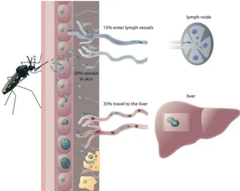
When Plasmodium parasites are transmitted by Anopheles mosquitoes into their mammalian host, they are con-fronted with extreme environmental changes as they move from the salivary gland of a cold-blooded insect host to the skin tissue of a warm-blooded mammalian host. Once injected into the skin, the motile sporozoites transmigrate several cells before eventually crossing endo-thelial cells to reach a blood vessel (Frischknecht et al., 2004; Vanderberg & Frevert, 2004; Amino et al., 2006). The phenomenon of transmigration is discussed in detail below. Surprisingly, only a portion of the injected spor-ozoites (c. 35%) enters a blood vessel and is carried by the bloodstream to the next destination, the liver (Fig. 1). A considerable number (c. 15%) ends up not in blood but in lymph vessels, which are a dead end for the parasite. An even bigger portion of sporozoites (c. 50%) does not leave the skin tissue at all. Interestingly, it has been shown that the parasites that do not manage to leave the skin or end up in the draining lymph node induce a strong cell-mediated immune response. There is evidence that this immune response may be the basis of protection against subsequent challenges (Sinnis & Zavala, 2008).
One-way road or the highway? Parasite development in the skin
So far it has been assumed that in vivo, sporozoites need to invade hepatocytes to complete exo-erythrocytic development, but recent studies suggest that there is an alternative infection route. In an experimental setup, rodent‐infectious Plasmodium berghei sporozoites entered cells in the skin and completed development into merozoites (Gueirard et al., 2010). Whether this can also take place in natural infections or for other Plasmodium species remains to be shown. Considering that sporozoites of avian‐infecting Plasmodium species invade and com-plete their development in a variety of cells including macrophages and endothelial cells (Frevert et al., 2008), infection of cell types other than hepatocytes might repre-sent an evolutionary conserved mechanism. The capability of infecting different cell types raises the question of how sporozoites recognize their host cells. There appear to be considerable differences among Plasmodium species in the receptors they require for infection. It has been shown that for successful invasion by Plasmodium falciparum and Plasmodium yoelii but not P. berghei, expression of
CD81 on the host cell surface is required (Silvie et al., 2006, 2007). It is therefore not surprising that in vitro, unlike P. falciparum and P. yoelii, P. berghei parasites can infect a wider variety of cells and it is possible that this is also the basis for their ability to infect skin cells in vivo. In this case, it is questionable whether the human patho-gen P. falciparum and other species that obviously need a well-defined set of cell surface markers for recognition of their host cell can infect cells other than hepatocytes.
Parasite development in hepatocytes: first contact
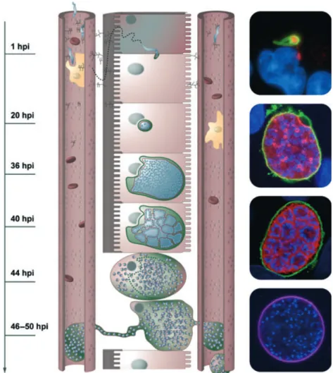
Even if Plasmodium sporozoites can infect a wider range of cells than originally thought, it is generally agreed that the main cell type in which they complete development are hepatocytes. To access them, the motile parasites need to cross the endothelium a second time after entering the bloodstream in the skin. After being passively transported by the bloodstream through the body, they eventually reach the liver but how does the parasite know where to leave the blood vessel? In the liver sinusoids, the blood flow is very slow and sporozoites are able to adhere to the endothelium. There they bind highly sulfated heparansulfate proteogly-cans (HSPGs) (Coppi et al., 2007), which are presented by hepatocytes through fenestrae, small channels in endothe-lial cells (Fig. 2). HSPGs are presented by many cell types but the sulfation level differs and is particularly high in the liver tissue. The contact of sporozoites with HSPGs starts a signaling cascade in the parasite, involving
calcium-dependent protein kinase 6 and other kinases, which finally results in the switch to an invasion mode (Coppi et al., 2007).
For many years, it was thought that the circumsporozo-ite surface protein (CSP) serves as the receptor on the migrating parasite that targets it to the HSPGs in the liver sinusoids (Menard, 2000; Sinnis & Nardin, 2002). However, a recent study provides clear evidence that full-length CSP does not interact specifically with HSPGs but is processed to expose the C-terminal cell-adhesive thrombo-spondin repeat (TSR) domain once the sporozoite recog-nizes HSPGs by other means (Coppi et al., 2011). Exposure of the TSR then allows the sporozoite to attach to the endo-thelium. Thus CSP is not responsible for hepatocyte detec-tion, but rather for an unspecific adherence to cells once processed to expose the TSR domain. This also explains very nicely how the parasite can rapidly switch to the inva-sion mode. Importantly, transgenic sporozoites expressing only the cell-adhesive TSR domain of CSP are constitu-tively in the adherence and invasion mode. They do not leave the site of injection but enter skin cells and develop into infectious merozoites, confirming a recent report suggesting that sporozoites can enter and fully develop in skin cells (Gueirard et al., 2010).
However, sensing the correct environment in liver sinusoids and switching to the invasion mode is not suffi-cient: the parasite is still on the wrong side of the endo-thelium and has to cross this barrier to reach its final destination, the hepatocytes. It is therefore likely that the invasion mode is not immediately triggered but rather is Fig. 1. Fate of Plasmodium sporozoites
injected into the skin by female Anopheles mosquitoes: In the skin, sporozoites become motile and either enter blood vessels to be passively transported to their final destination, the liver, or enter lymph vessels to end up in the draining lymph node where they are eliminated. The vast majority of injected sporozoites, however, remain in the skin and are removed by dendritic cells (yellow) or enter skin cells and develop into mature exo-erythrocytic forms.
a progressive event. It has been suggested that sporozoites glide along the endothelium until they reach one of the numerous Kupffer cells (resident macrophages in the liver) and then transmigrate them to reach the other side of the endothelium (Meis et al., 1983; Frevert et al., 2006). Indeed, it has convincingly been shown that sporozoites can transmigrate Kupffer cells and macro-phages in vitro (Pradel & Frevert, 2001). On the other hand, Meis et al. observed digested sporozoites in Kupffer cells suggesting that these cells actively phagocytose and destroy parasites (Meis et al., 1985a). Kupffer cells are not necessarily an integral part of the endothelium but
rather sit on top of endothelial cells, meaning that cross-ing these cells is of no obvious advantage to the parasite. Perhaps the ability to leave Kupffer cells is used instead as an immune escape strategy by the parasite to avoid destruction by phagocytosis, whereas the endothelium is crossed via a different route. Considering that sporozoites are able to transmigrate endothelial cells in the skin, it would not be surprising if they could do the same thing in the liver. It has been suggested that sporozoites can migrate through cells in at least two different ways (Mota et al., 2001; Pradel & Frevert, 2001). One has been described in the literature as an aggressive wounding of Fig. 2. Sporozoite entering the liver: in the liver sinusoids, the blood flow is slow and the sporozoites can attach to the endothelium by interacting with HSPGs presented by hepatocytes through small channels in endothelial cells. Upon crossing the endothelium, sporozoites transmigrate through several hepatocytes before settling in one and residing inside a parasitophorous vacuole. There it develops to a multinucleated schizont and finally, by membrane invagination, forms thousands of merozoites that are liberated from the PV into the host cell cytoplasm. PVM disruption induces host cell death and the formation of vesicles that are continuously filled with merozoites and reach into the blood vessel. Finally these vesicles (merosomes) bud off and are carried away by the bloodstream to reach the lungs, where they release merozoites to infect RBCs. The pictures on the right show representative immunofluorescence assays of exo-erythrocytic parasites. From the top: sporozoite just after invasion (green) expressing PbICP (red); schizont (PVM in green and parasite membrane in red); cytomere and merozoites inside a detached cell (staining as before); merosome (merosome membrane stained in purple); all nuclei are stained with DAPI (blue).
cells at the point of entrance and exit, where the parasite punches holes in the membrane (Mota et al., 2001). This means that some intracellular material will be released from the wounded cell, potentially attracting phagocytes to the site of transmigration. The second, alternative method of sporozoite transmigration involves an invagi-nation of the host cell plasma membrane at the point of entry (Pradel & Frevert, 2001) and likely a membrane fusion event at the exit site of the transmigrated cell. So far this mode of transmigration has only been found in Kupffer cells but it may well be that it is used for passage through endothelial cells as well. A similar mode of trans-cellular migration through cells is well known for immune cells in order to rapidly cross endothelia (Muller, 2010). The transmigrating immune cell induces channel formation in endothelial cells, a process involving membrane fusion events, but in doing so does not injure the transmigrated cell. Whether sporozoites in vivo trans-migrate the endothelium by cell wounding or by the less aggressive membrane invagination and fusion method remains to be shown, but the latter could be advanta-geous to the parasite because it is an immunologically quiet event. Another strong argument for silent transmi-gration via a membrane-surrounded channel is the in vivo observation that the sporozoite squeezes through a small constriction in the membrane of the transmigrated cell (Amino et al., 2008). This definitely fits better with the membrane invagination and membrane fusion model than with the wounding model, especially as the wound-ing model would suggest two parasite constriction events, one upon entry and one upon exit, whereas in vivo only one is seen.
Theoretically, there are alternative explanations for the parasite passing through a constriction (Frevert et al., 2006). In liver sinusoids, endothelial cells are known to have so-called fenestrae (De Leeuw et al., 1990). These are small channels in the cell, connecting the blood vessel on one side of the endothelium and the Space of Disse on the other. The size of these channels (0.1lm) is only a tenth of the diameter of a sporozoite (1 lm) but membranes are flexible, as are sporozoites, and it has been suggested that this route is used to directly access hepatocytes (Shin et al., 1982). Transmigration of sporo-zoites through a tight constriction might therefore reflect the passage through fenestrae in endothelial cells. Advanced intravital imaging techniques will hopefully help us fully understand this and other aspects of sporo-zoite transmigration in the near future.
The molecular events underlying transmigration are not yet understood. Several proteins have been identified as being specifically expressed in transmigrating parasites and have indeed found to be essential for transmigration (Ejigiri & Sinnis, 2009). A knockout of the corresponding
genes results in parasites that cannot transmigrate but are not impaired in invasion. However, the exact functions of these proteins remain to be determined.
Not falling for the first one: hepatocyte transmigration and invasion
Following migration through the endothelium, sporozo-ites need to cross the Space of Disse between endothelial cells and hepatocytes before they can eventually invade their final host cell. They do not, however, infect the first hepatocyte they encounter but transmigrate a number of cells before eventually invading one (Mota et al., 2001) (Supporting Information, Movies S1 and S2). Again, it is not clear which mode of transmigration the parasite uses, but it has been suggested that transmigration of sporozo-ites through hepatocytes by cell wounding causes the release of hepatocyte growth factor (HGF) by the wounded cells (Carrolo et al., 2003; Leiriao et al., 2005). HGF, in turn, could be beneficial for the invading parasite in that it promotes the survival of hepatocytes, which constitutively express the HGF receptor cMET. However, as transgenic parasite lines that are not able to transmigrate cells can still enter hepatocytes and develop successfully, HGF signaling does not appear to be essen-tial (Ishino et al., 2004). In addition, as explained above, it still remains to be demonstrated which mode of trans-migration sporozoites use to cross hepatocytes. Perhaps there is no cell wounding at all and the parasite again uses the silent mode to transmigrate, avoiding unwanted immune responses at the site of infection. Although other theories exist as to why transmigration by wounding might be important for the parasite maturation (Mota et al., 2002; Leiriao et al., 2005), it might also be that it has no specific function. The switch from the transmigra-tion mode to the invasion mode might instead be a progressive event. Thus sporozoites continue to transmi-grate cells until the complete switch to invasion mode has taken place. How the parasite decides to invade is still a mystery. For recognition and invasion, one would expect the sporozoite to require access to the host cell surface to allow receptor-ligand interaction to occur. While migrat-ing from cell to cell in a tightly packed space, it is diffi-cult to conceive how the parasite would perform these necessary interactions and therefore sense the host cell that it will ultimately invade rather than transmigrate. If, however, transmigration occurs in the silent mode also in hepatocytes, there is a very simple explanation: The para-site always induces invagination of the host cell mem-brane and leaves the cell by memmem-brane fusion. Once the parasite stops migrating, it is already within a vacuole inside a hepatocyte and just needs to avoid membrane fusion to stay in its final host cell.
Ejigiri et al., have recently suggested how sporozoites can successfully infect different cell types (Ejigiri & Sinnis, 2009). Their hypothesis is that the parasite injects its own ligands into the host cell membrane, which then bind to receptors on the sporozoite surface. This theory is supported by the fact that despite many years of research, thus far only two host cell-derived factors (CD81, SRBI) have been identified (Silvie et al., 2003; Rodrigues et al., 2008; Yalaoui et al., 2008), which have not been proven to bind directly to sporozoite molecules. It is possible that they facilitate parasite invasion in a different way, i.e. by allowing parasite ligands to be incorporated into the host cell mem-brane. Although this provides an elegant explanation for how the sporozoite can infect cells other than hepatocytes, it does not explain how the parasite incorporates the ligand through the membrane of the transmigrated cells into the membrane of the neighboring hepatocyte.
Having travelled all the way from the skin to the liver, migrated through several cells, squeezed through narrow gaps, after many signaling events and recognition of being at the correct location, the sporozoite is finally ready to invade. Although it is not clear on which basis the parasite chooses its final host cell, it is well established that during sporozoite invasion the host cell membrane invaginates to form a parasitophorous vacuole (PV) around the parasite (Movie S3). In analogy to the inva-sion of RBCs by merozoites, it can be assumed that the majority of host cell proteins are proteolytically removed from the parasitophorous vacuolar membrane (PVM) during the invasion process (Dowse et al., 2008). The sporozoite glides into the cell by the use of a moving tight junction (Movie S4), which blocks substances from entering the forming vacuole from the outside.
For a long time, it was assumed that sporozoite entry into host cells relies only on the actin-myosin motor of the parasite, but this view has been recently been chal-lenged. It was shown that parasite invasion induces the recruitment of host cell actin to the entry site and that this accumulation is required for successful invasion (Gonzalez et al., 2009). These observations and a recent analysis of the transcriptome of infected hepatocytes (Albuquerque et al., 2009) provide clear evidence that the host cell reacts strongly to the invasion event. Our own observations support these findings (Movie S5). If a cell senses infection, it could induce stress signaling and its own death to eliminate the pathogen. To avoid host cell apoptosis, Plasmodium parasites have developed successful strategies (Leiriao et al., 2005; van de Sand et al., 2005). Recently, it has been shown that P. berghei sporozoites secrete a potent cysteine protease inhibitor (PbICP), which is able to block cell death (Rennenberg et al., 2010). This method of directly inhibiting proteases involved in cell death is probably supported by additional
anti-apoptotic signaling in the host cell, but how the par-asite induces this signaling is still not known. Contrary to the related apicomplexan parasite Theileria (Heussler et al., 2006), which induces NF-jB-dependent survival mechanisms, it has been suggested that Plasmodium para-sites interfere with NF-jB activation (Singh et al., 2007), similarly to another related parasite, Toxoplasma gondii (Butcher et al., 2001; Shapira et al., 2002). In case of T. gondii, NF-jB-independent mechanisms to avoid apoptosis have been described (Hippe et al., 2009) and it will now be interesting to investigate these pathways in Plasmodium-infected hepatocytes. It should be mentioned that there are also reports that some T. gondii strains induce a NFjB-dependent inhibition of apoptosis (Mole-stina et al., 2003). For Plasmodium-infected hepatocytes, there is evidence that CSP, which is secreted by the spo-rozoite into the host cell cytoplasm, out-competes nuclear import of NF-jB and thus interferes with inflammatory responses of the infected cell. However, clearly more research is needed to clarify the role of NF-jB in host cell survival and to determine if CSP is involved in anti-apop-totic signaling or whether it merely binds in an unspecific manner to proteins and other macromolecules by expos-ing the adhesive C-terminal TSR domain.
Moving in: the early phase of hepatocyte infection
Parasite proteins that are thought to interact with the signaling machinery of the host cell must cross two membranes: the parasite membrane (PM) and the PVM. For P. falciparum blood stage parasites, it has been dem-onstrated that several motifs exist that mediate secretion across these membranes (Marti et al., 2004; Spielmann & Gilberger, 2010). These PEXEL (Plasmodium export ele-ment) and PNEP (PEXEL negative export protein) motifs have been identified in many P. falciparum proteins known to modify the host erythrocyte. However, in the genomes of rodent-infectious Plasmodium species, very few genes encoding proteins with these particular export motifs have been identified. Notably, the N-terminus of CSP contains two PEXEL motifs (Singh et al., 2007) and in the N-terminus of PbICP, a PNEP-like export motif has been suggested (A. Rennenberg, unpublished observa-tions). Although the PEXEL motifs of P. berghei CSP have been experimentally proven to be functional, the experiments were performed in P. falciparum. In fact, expression of GFP-tagged proteins with PEXEL motifs in P. berghei did not result in secretion from blood stage or liver stage parasites (S. Horstmann, unpublished observa-tions). Considering this and the low number of proteins predicted to contain PEXEL motifs in rodent-infectious Plasmodium species, it should be considered that
rodent-infectious parasites may have developed alternative motifs to allow export of proteins.
Once inside the host cell, the parasite often localizes close to the host cell nucleus (Movie S6). It is difficult to imagine that the parasite can sense its environment through the PVM and that it can control its movement while enclosed in a membrane. Therefore, it is more likely that the vacuole attaches to the cytoskeleton of the host cell and is passively transported to the nucleus. There, it is often in close proximity to the ER (Bano et al., 2007) and the Golgi apparatus (A. Rennenberg, unpublished observations) and it is an attractive hypothesis that the parasite might position itself there to benefit from the host cell secretory machinery by directing host cell vesi-cles to fuse with the PVM. Interestingly, some intracellu-lar bacterial pathogens also position themselves close to the host cell Golgi apparatus and it has been suggested that in this way they benefit from the transport from and to the secretory pathway (Bakowski et al., 2008).
After invasion of the host hepatocyte and formation of a PV, the parasite undergoes an initial period of com-paratively subtle changes (Movie S6), at least on a morphological level. During the first 24 h after infection, the parasite remodels its PVM and transforms from its elongated form to a small, round trophozoite. Several proteins are known to be important for this early stage as their knockout led to an impairment in development. Among them are uis3, uis4 and Pb36p (Mueller et al., 2005a, b; van Dijk et al., 2005).
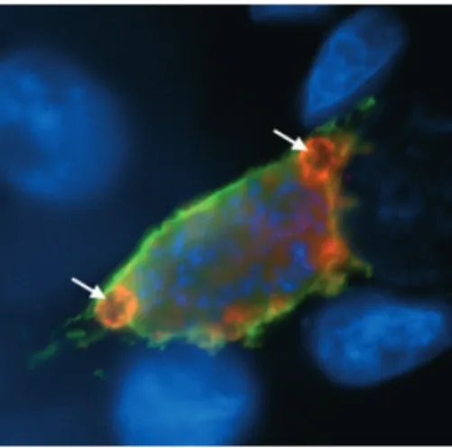
At approximately 20 h after invasion, the parasite nucleus starts dividing repeatedly (Fig. 2) and displays one of the fastest replication rates known for eukaryotes: during the next 35 h, up to 30 000 nuclei are generated. At the same time, the PV expands in size to accommo-date the growing parasite (Movie S7). During blood stage replication, the parasite is known to take up hemoglobin from its host cell through a kind of cell mouth, a so-called cytostome (Elliott et al., 2008) and to digest it in a food vacuole (Rosenthal & Meshnick, 1996). Interestingly, exoerythroytic merozoites of avian-infecting Plasmodium species also possess a cytostome (Aikawa et al., 1966) and we have found a morphologically similar structure in the growing schizont (Fig. 3). Whether these structures have a function, however, remains to be deter-mined. It is therefore still unknown how the parasite manages to obtain the resources necessary for its immense reproduction effort within the hepatocyte. Apart from the host cell ER, which gathers around the parasite and a loose association with the Golgi apparatus, there appears to be no constant association between the PV and host cell organelles (Bano et al., 2007). For intra-cellular Toxoplasma parasites, it has been shown that host cell mitochondria are firmly associated with the PVM and
supply the parasite with the enzyme co-factor lipoic acid (LA) (Crawford et al., 2006). A similarly close association of the mitochondrion was not found in P. berghei-infected hepatocytes, but very careful microscopic exami-nation revealed distinct areas at the PVM that appear to indeed interact with host cell mitochondria (C. Descher-meier, unpublished observations). We could also demon-strate that P. berghei needs to import LA from the host cell, most likely from host cell mitochondria. It is very surprising that liver stage parasites largely rely on LA import as they produce this co-factor themselves in the apicoplast, an essential plastid-like organelle that plays an important role in liver stage development (Stanway et al., 2009a). It seems that LA cannot be easily transferred from the apicoplast to the mitochondria, which is surprising considering that other metabolites are known to be exchanged easily between organelles within the parasite, e.g. during heme detoxification (Sato et al., 2004; van Dooren et al., 2006; Padmanaban et al., 2007). However, as hepatocytes contain many mitochondria producing LA, perhaps there has been no evolutionary pressure for the parasite to independently transport this co-factor from the apicoplast to the mitochondrion.
It has been speculated that for nutrient uptake, the parasite inserts transport channels into the PVM allowing molecules up to 800 Da to freely cross this membrane. (Bano et al., 2007; Sturm et al., 2009). However, larger molecules like lipids and peptides need to be actively imported and again the parasites modifies the PVM to allow this to occur. It exports a number of proteins into the PVM, which most likely interact with host cell proteins to direct nutrients toward the parasite. Fig. 3. Liver stage parasites possess structures resembling a cytostome. The PVM marker protein Exp1 is stained in green and the cysteine protease inhibitor PbICP is stained in red. Parasite and host cell nuclei are visualized by DAPI staining (blue). Round structures (putative cytostomes) labeled by the PVM marker and PbICP are indicated with arrows.
However, so far only few host proteins have been identified that might support liver stage growth. One of them is the protein ApoA1 that was found to localize to the PVM. It is thought to interact with uis4 and is speculated to play a role in the synthesis of additional membrane during the enlargement of the vacuole (Prudencio et al., 2006).
During its extensive growth, use of host cell resources is likely to deplete nutrients from the infected hepatocyte. In response to the resulting starvation conditions, the host cell is expected to induce autophagy and indeed we find increased autophagy in infected cells (N. Eickel, unpublished observation). However, autophagy is also a very potent mechanism to eliminate pathogens and again we have evidence that in vitro parasites can be destroyed in autophagosomes. Host cell autophagy therefore appears to be a double-edged sword for the parasite: on the one hand, it could provide nutrients for its extensive growth and on the other hand, it could result in parasite elimina-tion. Further research on this highly interesting topic is needed to fully understand the function of host cell auto-phagy in regard to parasite survival and elimination. What has been underestimated so far is that the host cell has the capacity to eliminate the parasite. It was an accepted view that the parasite can manipulate the sur-vival of the host cell but now it turns out that the host cell is not helpless and can successfully fight the infection. This topic will be very important to study as it may help to develop new antimalarial strategies.
For the parasite, the supply of nutrients is essential, but it must also dispose of metabolic waste products. In blood stage parasites, for example, the toxic end product of hemoglobin metabolism, hemozoin, is stored in the food vacuole (Goldie et al., 1990). For liver stage para-sites, so far no food vacuole has been described and the question remains how the metabolically highly active liver stage parasite deals with its waste products. It has been suggested that it employs efficient export transporters in its membrane and in the PVM (Sturm et al., 2009). This might partly explain why liver stage parasites are less sus-ceptible to drugs than blood stage parasites: they might be able to actively export these from the PV, therefore preventing them from reaching the parasite. It would be interesting to combine drug treatment with blockers of these putative transporters. Although there is evidence for the expression of such transporters in the blood stage (Valderramos & Fidock, 2006), proof of their existence and characterization in the liver stage is still missing.
When one becomes many: the challenges of replication
As liver stage parasites grow very rapidly, a classical mode of cell division and cytokinesis is most likely not possible
because it requires the expression of numerous proteins in a concerted fashion followed by the removal or inactivation of the entire machinery. It is therefore not surprising that the parasite has developed strategies to streamline proliferation and growth. The most obvious phenomenon is that the parasite avoids cytokinesis until all nuclear division is complete and thus develops into a huge syncytium, the multinucleated schizont. Less obvious is the fact that the nuclear division is not accom-panied by the disappearance of the nuclear membrane. Electron microscopy studies from other parasite stages suggest that the segregating chromosomes and the spindle apparatus remain within the nuclear envelope (Bannister et al., 2000) but this does not explain the highly amor-phous phenotype of the nuclei during division. Could it be that the microtubule organization center is localized in a way that it can associate with the cytoskeleton in the cytoplasm of the parasite? It has indeed been shown very recently for blood stage parasites that the microtubule organization centers are embedded in the nuclear membrane (Gerald et al., 2011) and it might well be pos-sible that they are connected to the spindle apparatus in the nuclei and to the cytoskeleton outside the nucleus. Thus mitosis in Plasmodium parasites appears to differ in many respects from that in the mammalian host (Gerald et al., 2011). This is an interesting aspect because differ-ences in the cell biology of the parasite and its host cell might reveal new strategies for interference with parasite development. Once schizogony is completed, cytokinesis and daughter cell formation take place. As each daughter cell requires a full set of organelles, including a mitochon-drion and an apicoplast, which cannot be synthesized de novo, the existence of a highly organized distribution machinery must be postulated. In eukaryotic cells, cell division is normally preceded by the division of organ-elles, which are then distributed to both daughter cells. Does the same happen in Plasmodium during the times of schizogony when no cytokinesis is taking place? The next section takes a closer look at the replication and dis-tribution of several Plasmodium organelles and will pri-marily focus on the morphological and positional changes that occur for the apicoplast, mitochondrion and nuclei.
During erythrocytic development, the parasite has already been shown to form both a branched apicoplast and mitochondrion, with fission of these organelles occurring only after completion of the multiple rounds of nuclear division and with each nucleus ultimately being paired with a single apicoplast and mitochondrion (van Dooren et al., 2005). During the liver stage of Plasmo-dium development, where not 16–32 but up to 30 000 daughter parasites are formed, the parasite faces an even greater challenge in terms of organelle growth and segregation into merozoites. Until recently, it was unclear
whether the liver stage parasite employs a similar mechanism of apicoplast and mitochondrial growth and segregation into daughter parasites as that in the blood stage.
The trophozoite: calm before the storm
Various transgenic P. berghei parasite lines have been gen-erated that allow the visualization of the nuclei, apicoplast and mitochondrion during liver stage develop-ment (Stanway et al., in press). From previous studies it was known that salivary gland sporozoites contain a sin-gle nucleus, apicoplast and mitochondrion, which do not necessarily have a clear physical connection (Stanway et al., 2009a; Kudryashev et al., 2010). For approximately the first 20 h after the invasion of the hepatocyte, the parasite maintains a single nucleus. During this time, the apicoplast and mitochondrion both elongate (Fig. 4). In an interesting resemblance to the blood stage, at this point the mitochondrion primarily lies at the periphery of the parasite. However, in contrast to the blood stage, where both organelles appear to maintain a continuous interaction (van Dooren et al., 2005), in liver trophozo-ites both organelles are mainly found separated from each other. Contact between apicoplast and mitochondrion seems to be rather accidental. This observation is surpris-ing because these two organelles are thought to share the
heme biosynthesis pathway and their physical connection is expected to be required for metabolite exchange. How-ever, there are other apicomplexan parasites, like Toxo-plasma, in which the connection between the apicoplast and mitochondrion is not continuous (Nishi et al., 2008). Perhaps a physical connection between the organelles is not necessary to maintain a functional heme pathway or the occasional points of interaction provide enough time for metabolite exchange. Another important aspect, which has not been tackled so far, is the question how organel-lar genomes are distributed correctly within the huge developing network. After fission each organelle must contain the genetic material and a highly sophisticated machinery to achieve this can be predicted. Future research will hopefully shed some light on this issue.
The schizont: rapid nuclear division and extensive organelle growth
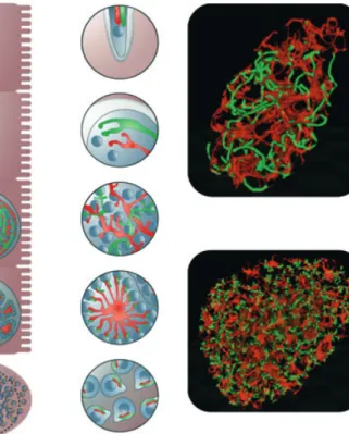
Following the trophozoite stage, the parasite begins repeated rounds of nuclear division. Based on a replica-tion from 1 nucleus to up to 30 000 nuclei in approxi-mately 30 h, this would imply that a round of nuclear division occurs approximately every 2 h. In parallel with this extensive nuclear division, both the apicoplast and mitochondrion become highly intertwined branched struc-tures, but each appears to remain as a single organelle
Fig. 4. Organelle development and distribution into daughter cells: invading sporozoites contain a mitochondrion (red), apicoplast (green) and a nucleus (cyan). During schizogony, the nucleus divides but the mitochondrion and the apicoplast continue to grow and branch extensively. During the cytomere stage, nuclei become attached to the invaginating parasite membrane and the apicoplast and the mitochondrion are directed toward the forming merozoites. Each merozoite finally contains a single apicoplast, mitochondrion and nucleus. On the right are live spinning disc microscopy images of representative parasite stages, where GFP is targeted to the apicoplast and mCherry to the mitochondrion. The upper image shows a schizont with an extensive and intertwined apicoplast and mitochondrion, whereas the lower image shows a cytomere, briefly before fission of the apicoplast and mitochondrion.
(Fig. 4; Stanway et al., 2011). Thus far, it is unclear how the growth of these organelles is controlled but neither branching of the mitochondrion nor the apicoplast appears to involve any clear association with nuclei. This is surprising, because in both Toxoplasma and Sarcocystis parasites, the apicoplast is connected with the nucleus (Striepen et al., 2000; Vaishnava et al., 2005). The obser-vation that the mitochondrion, like the apicoplast, under-goes extensive branching during schizogony contradicts earlier EM studies that described a proliferation of the mitochondrion during schizogony based on the observa-tion of multiple mitochondrial profiles in thin secobserva-tion (Meis et al., 1985a, b, c). However, this observation is not contradictory to a branched network as described by us because in thin sections a highly branched structure would also result in multiple profiles. Furthermore, such branch-ing is consistent with what has been seen in blood stage development and indeed occurs in each of the three stages of asexual replication during the Plasmodium life cycle (van Dooren et al., 2005; Stanway et al., in press).
The extensive growth of the apicoplast and mitochon-drion requires a massive quantity of membrane. Coupled with the extensive growth of the parasite and presumably the replication of other cellular organelles, this may at least in part explain the reliance of the parasite on fatty acid biosynthesis pathways during exo-erythrocytic devel-opment, which stands in contrast to blood stage develop-ment and sporogony (Yu et al., 2008; Pei et al., 2010). Knockout of genes coding for important components of the fatty acid pathway like pyruvate dehydrogenase and FabI allow normal blood stage development but block rapid proliferation of liver stages.
The cytomere: generating order out of chaos
To manage the extensive growth of the apicoplast and mitochondrion during liver stage development is already an impressive feat but the fission of these organelles and correct segregation into forming daughter merozoites appears to be an even greater challenge. How the parasite manages this on a molecular level is not understood, but again double-fluorescent parasites have allowed us to begin to understand the morphological and positional changes in the apicoplast, mitochondrion and nucleus prior to and during merozoite formation. It appears that the processes undertaken by the liver stage parasite paral-lel those in the blood stage, but on a much larger scale. The large size of the liver stage parasite, however, allows a more detailed examination of these processes.
Following the completion of nuclear division, at which point the single parasite can contain many thousands of nuclei, the parasite develops to a stage known as the
cytomere. Here the plasma membrane of the parasite invaginates to form what appear to be spheres of membrane, portioning the cytoplasm into between approximately 5 to 20 units (Figs 2 and 4; Movies S7 and S8). At this stage, the nuclei of the parasite seem to be closely associated with the plasma membrane and so they are in a sphere-like arrangement, which appears like rings of nuclei when examined by two-dimensional microscopy.
The apicoplast primarily lies at the periphery of these spheres of nuclei, with a surprising resemblance to the position of the Sarcocystis apicoplast prior to division of the polyploid nucleus (Vaishnava et al., 2005). The mito-chondrion on the other hand mostly lies within these spheres of nuclei, forming clumped structures (Fig. 4). During the cytomere stage, the individual units of plasma membrane are not fully separated and mitochondrial branches connect the afore-mentioned clumps, confirm-ing earlier EM studies on exo-erythrocytic development of the parasite (Meis et al., 1981, 1985a, b, c). The cyto-mere stage is an intermediary parasite stage and soon after its formation, the apicoplast develops a concertinaed structure, presumably due to the constriction of the orga-nelle at sites where fission will take place. The morphol-ogy of the apicoplast at this stage again resembles that observed in Sarcocystis parasites (Vaishnava et al., 2005). Around this time, the morphology of the mitochondrion also changes dramatically: finger-like structures form that point to the periphery of the membrane units. The api-coplast of the parasite then divides synchronously. At the point of division or close before, an association is clearly visible between the apicoplast and mitochondrion, with each finger-like structure being associated with a divided portion of apicoplast. It remains to be seen whether these organelles are directly associated or are both connected to a third structure, for example, the same cytoskeletal element. One could hypothesize that organelles may enter the forming daughter merozoites via association with and movement along subpellicular microtubules, as has been seen in the case of Toxoplasma parasites (Striepen et al., 2000). The existence of such microtubules, positioned within each forming daughter parasite, could explain how the mitochondrion is able to undergo such a dramatic and synchronous change in morphology. The association between the apicoplast and mitochondrion occurring primarily at the end of the liver stage would suggest that a connection between these organelles might be required to allow correct organelle segregation, as already proposed by others (Sato et al., 2004; van Dooren et al., 2006; Pad-manaban et al., 2007).
Following the change in its morphology, the mitochon-drion divides. It is interesting to note that during both the asexual blood stage and the liver stage, division of the
apicoplast always precedes that of the mitochondrion. This is also true for T. gondii parasites (Nishi et al., 2008), despite the difference in the methods used for cell division. Once mitochondrial fission is complete, the daughter merozoites are formed and released into the host cell by breakdown of the PVM.
Not just different for the sake of being different
For both mitochondrial and apicoplast fission, the under-lying molecular mechanisms are far from clear. Some components of typical organelle fission machineries appear to be conserved in Plasmodium and Toxoplasma parasites, such as dynamin, which was shown to be involved in the fission of the apicoplast in both species (Charneau et al., 2007; van Dooren et al., 2009). However, homologs of other key players in organelle fission appear to be absent. This is particularly true of the Ftsz protein, which in other systems is responsible for the initial constriction of the organelle by allowing dynamin to bind but is reportedly absent in apicomplexan parasites (Vaishnava & Striepen, 2006). It remains to be seen whether in Apicomplexa, proteins related to division have evolved sufficiently to be elusive in homology searches or whether the parasites have developed alternative mechanisms for the division of their organelles. The latter option currently seems the most likely.
When observing the development of the apicoplast, mitochondrion and nuclei of Plasmodium liver stage para-sites, one could be surprised by the methods used by the parasite to achieve segregation of these essential organ-elles. One can speculate as to why Plasmodium parasites at all stages of asexual reproduction develop via schizog-ony and not via daughter cell formation as performed during the lytic stage of the Toxoplasma life cycle. Indeed, it appears that none of the known apicomplexan parasite genera use an identical mechanism of asexual replication (Vaishnava & Striepen, 2006). It can only be speculated why evolution allowed the parasite to develop huge branched organelles, which undergo one fission event at the end of schizogony rather than repeated apicoplast and mitochondrial fissions along with division of the nucleus and the Golgi apparatus (Struck et al., 2005; R.R. Stan-way, unpublished observation). One reason might be that a majority of the proteins functioning within the apicop-last and mitochondrion are encoded in the genome of the parasite. They need to be targeted to these organelles, where import machineries allow their uptake. For a repeated fission of organelles, the parasite would need to constantly express proteins involved in fission and to import them into the relevant organelles, while at the same time maintaining a tight control of protein activity
to prevent unwanted fission events. In contrast, for a synchronous fission of the apicoplast and mitochondrion after the completion of karyokinesis, presumably only one period of protein expression is required. This would have the added advantage that control of fission could be on the level of protein expression, allowing a tight syn-chronicity. It is also conceivable that in the absence of repeated fission events, both the apicoplast and mito-chondrion may be able to function more efficiently. For the extensive parasite growth and replication during the liver stage, the parasite must require a high level of energy and of fatty acid biosynthesis, the latter for the extensive production of membrane that accompanies this growth. Such demands on the mitochondrion and api-coplast may be incompatible with repeated divisions of these organelles.
Breaking out: egress from the parasitophorous vacuole
Once formation of merozoites is complete, they must be transported into the bloodstream where they can infect RBCs to continue the life cycle. Most text books state, probably in analogy to the events at the end of the blood stage, that the host cell ruptures and releases merozoites, which subsequently infect RBCs. However, if the host cell membrane broke down within the liver tissue, the mer-ozoites would be on the wrong side of the endothelium (Fig. 2). In fact, it has recently been shown that the release of merozoites is a well-orchestrated, multi-step process. The first hurdle for the parasite is the PVM and it has been shown that during merozoite formation parasite proteins begin to leak into the host cell, thus demonstrating that the membrane of the PV becomes increasingly permeable before it is completely disrupted (Schmidt-Christensen et al., 2008; Sturm et al., 2009). As another sign of PVM breakdown, several previous in vivo studies had already indicated that parasite and host cell material mix freely in infected cells (Movie S8) (Meis et al., 1985a, b, c; Baer et al., 2007). Very recently, live imaging of a parasite strain expressing a fluorescent PVM marker protein confirmed the breakdown of the PVM toward the end of the liver stage while the host cell mem-brane stays intact (Graewe et al., in press).
Not much is known about the molecular events result-ing in the breakdown of the PVM in Plasmodium liver stages. It takes place within a relatively short time frame (Graewe et al., in press) and therefore must be a highly efficient process. The first class of enzymes one would consider to act on membranes are lipases, but it remains to be shown whether the parasite secretes or activates lipases to destroy the PVM. However, it is known that proteases can destabilize membranes by removing
membrane-integrated proteins. Indeed, PVM breakdown can be inhibited by E64, an inhibitor of cysteine prote-ases, which indicates a role for this class of proteases (Sturm et al., 2006). The only protein that has been iden-tified to be involved in PVM disruption by Plasmodium parasites is LISP-1 in P. berghei (Ishino et al., 2009). It localizes to the PVM and its deletion results in an inabil-ity of the parasite to escape from the PV. LISP-1 itself, however, has no recognizable functional protease domain and is therefore suspected to be either a membrane recep-tor for proteases or to be involved in the processing of proteases for activation (Ishino et al., 2009). A protein that is proteolytically processed at this time is PbSERA3, a putative cysteine protease that is subsequently released into the host cell cytosol (Schmidt-Christensen et al., 2008). The processing of PbSERA3 is E64-sensitive and it would therefore be a likely candidate for the mediation of the PVM breakdown and subsequent changes in the host cell. Despite its prediction to be a protease, so far it has not been possible to demonstrate any catalytic activity for SERA3. Therefore, like LISP-1, it might merely act as an adapter protein for the recruitment of effector proteases.
Further understanding of liver stage PVM breakdown might be reached by examining other life cycle stages of the parasite. During egress from both oocysts and erythrocytes, Plasmodium parasites need to break down several surrounding membranes to continue their development. In blood stage egress especially, the overall situation is similar to the late liver stage: the parasite is separated from new host cells by two membranes: the PVM and the host cell membrane. A number of molecu-lar simimolecu-larities have already been found that make a common mechanism for the disintegration of the PVM feasible. As in the liver stage (Sturm et al., 2006), egress from the PV is blocked by E64 and members of the SERA family are cleaved shortly before the release of parasites (Yeoh et al., 2007). The processing proteases have already been identified as PfSUB1 and DPAP3 (Yeoh et al., 2007; Arastu-Kapur et al., 2008) and it remains to be seen if they also act in the liver stage. An even more interesting question is whether one of the proteases in this cascade is responsible for the host cell death that is induced upon PVM breakdown. The dramatic modifications of the host cell that occur in parallel to the release of merozoites from the PV are discussed in more detail below.
It is clear that in infected hepatocytes, the host cell membrane remains intact after the disintegration of the PVM (Sturm et al., 2006; Graewe et al., in press), but in infected RBCs, both surrounding membranes rupture in quick succession (Blackman, 2008). This difference in parasite release makes perfect sense as hepatocyte-derived merozoites need transport to the blood vessel whereas RBC-derived merozoites can directly infect another RBC.
Still, some controversy has remained about which membrane breaks down first when parasites exit RBCs: the host cell plasma membrane or the PVM? On one hand, live imaging of parasites that express GFP in their PV has shown that the fluorescent protein spreads throughout the entire host cell toward the end of the blood stage (Wickham et al., 2003). This indicates that the PVM disintegrates first. On the other hand, observa-tions have been made of extracellular clusters of mero-zoites surrounded by a PVM (Soni et al., 2005). Despite the uncertainty regarding merozoite release from RBCs, it is clear, however, that the disruption of the PVM and the host cell membrane are differentially inhibited. While PVM breakdown is blocked by E64, the breakdown of the host cell membrane was shown to be inhibited by the broad-spectrum cysteine and serine protease inhibitors leupeptin and chymostatin (Salmon et al., 2001; Wick-ham et al., 2003; Soni et al., 2005). This indicates that the PVM and the RBC membranes are broken down by different sets of proteases (Wickham et al., 2003; Black-man, 2008). As the plasma membrane of infected hepato-cytes does not break down immediately upon PVM disruption, the parasite-derived protease destroying the RBC membrane might either not be synthesized in the liver stage or might be inhibited by specific parasite factors. A potential candidate protein would be the recently identified cysteine protease inhibitor PbICP, which is released into the host cell during the late liver stage (Rennenberg et al., 2010). In conclusion, the data available from the study of blood stage egress indicate the involvement of specific sets of proteases that are activated in cascades (Yeoh et al., 2007; Blackman, 2008). While it is likely that similar proteolytic mechanisms act in the liver stage, it is still unclear whether these activated prote-ases directly destabilize the PVM by cleaving integral membrane proteins, or if they act by initiating other effector molecules like lipases or pore-forming proteins.
For other intracellular parasites, like the related api-complexan parasite T. gondii, it has been shown that a decrease in the intracellular potassium concentration leads to an increase in calcium concentration within the para-site, which seems to trigger egress (Moudy et al., 2001; Nagamune et al., 2008). The lysis of the PVM and the host cell membrane is then caused by a pore-forming per-forin-like parasite protein, TgPLP1 (Kafsack et al., 2009). The exit from vacuoles or host cells via use of pore-form-ing proteins is a common strategy for many intracellular pathogens, including organisms as diverse as Listeria mon-ocytogenes, Trypanosoma cruzi or Leishmania amazoniensis (Gaillard et al., 1987; Andrews, 1990; Andrews et al., 1990; Noronha et al., 2000). A typical feature of pore-forming proteins is the MACPF (membrane attack com-plex perforin) domain (Xu et al., 2010). In Plasmodium,
several proteins containing MACPF domains have been identified and one of them has already been shown to have a role in the transmigration of Kupffer cells (Kaiser et al., 2004). Further investigation will show if these pro-teins are expressed in the late liver stage and if they play a role in the breakdown of the PVM. It is not clear how such pore-forming activity would be limited to the PVM and would be prevented from affecting the merozoite membrane or the host cell membrane. In intracellular pathogens that escape from a phagosome, such as T. cruzi or L. monocytogenes, control is achieved by restricting pore-forming activity to occurring only at low pH (Andrews, 1990; Andrews et al., 1990; Schuerch et al., 2005). As soon as the parasites are liberated from the phagosome, the pH value changes and the host cell mem-brane is not perforated. As the PV environment of Plas-modium liver stage parasites is not expected to be of low pH, other means of regulation must exist. One attractive possibility is that adapter proteins that are only anchored in the PVM are necessary for the recruitment and activa-tion of such a pore-forming protein.
Host cell death: maintaining a calm exterior
While the exact mechanism of PVM breakdown remains to be elucidated, it is obvious that escape from just the PVM does not allow the parasite to reach the blood vessel. To safely cross the endothelium, the parasite uses a trick. Once the PVM is dissolved, a parasite-dependent host cell death is initiated (Sturm et al., 2006). It is characterized by cytochrome c release and nuclear con-densation under retention of an intact cell membrane (Sturm et al., 2006; Graewe et al., in press). It therefore clearly differs from necrosis, which typically includes the swelling and rupture of the cell (Lemasters, 2005). This difference is not surprising as a necrotic host cell death would lead to the release of pro-inflammatory molecules and the attraction of components of the host immune system (Savill, 1998; Scaffidi et al., 2002; Shi et al., 2003), which could then potentially act against the para-site.
Although, at first glance, the host cell death resembles apoptosis, it differs in many respects and appears to be unique. Whether apoptosis is triggered is usually decided by the balance between pro- and anti-apoptotic stimuli. Both the integration process and the initiation of apopto-sis often take place in the mitochondrion, through the formation of a mitochondrial outer membrane permeabi-lization pore (MAC) (Kroemer & Reed, 2000). This ultimately leads to the release of cytochrome c and other apoptotic mediators into the cytosol. The breakdown of the PVM could theoretically induce host cell death in
different ways. Firstly, parasite effector proteins could be released into the host cell after the PVM breaks down and this might cause death as a rather unspecific effect. As we have evidence that premature disruption of the PVM does not lead to death of the host cell, this scenario is less likely. Secondly, the host cell death might be pur-posefully orchestrated by specific parasite proteins secreted into the PV very late in parasite development and then released into the host cell upon PVM break-down. Several bacteria are already known to regulate host cell death by producing factors that compromise mito-chondrial integrity (Braun et al., 2007; Kozjak-Pavlovic et al., 2009) and Plasmodium might have evolved similar strategies. This hypothesis is supported by the live imag-ing of Plasmodium-infected hepatoma cells with fluores-cently labeled mitochondria, which has shown these organelles to disintegrate rapidly after PVM breakdown (Graewe et al., in press).
Upon closer examination, parasite-dependent host cell death lacks important hallmarks of apoptosis such as DNA fragmentation, caspase cascade activation and loss of phosphatidylserine asymmetry (Sturm et al., 2006; Graewe et al., in press). Interestingly, all of these features are events that take place in the mid to late stages of apoptosis. They also all require energy in the form of ATP: the fragmentation of DNA, for one, is carried out by endonucleases, which hydrolyze ATP during cleavage. Likewise, the caspase cascade is initiated through a complex composed of cytochrome c and Apaf-1, which undergoes an ATP-dependent conformational change that activates procaspase 9 (Zou et al., 1999). While the phos-phatidylserine switch itself does not appear to utilize ATP, it is linked to the activation of the caspase cascade as inhibitors of caspases were shown to prevent the phos-phatidylserine asymmetry loss (Castedo et al., 1996; Martin et al., 1996). Based on these observations, the fol-lowing model was suggested (Graewe et al., in press): while the parasite undergoes schizogony, it depletes the host cell of nutrients and ATP. Upon rupture of the PVM, mitochondrial integrity is compromised, probably by activation of proteases other than caspases. This leads to the uncoupling of oxidative phosphorylation and therefore loss of the ability to produce ATP de novo, which aggravates the lack of accessible energy. Simulta-neously, apoptotic factors that are released from the mitochondria initiate the apoptotic program, which pro-ceeds until it reaches a point where major amounts of ATP are required. It then stalls, which leads to an aborted version of apoptosis.
The arrest of the apoptotic program might additionally be aided by inhibitors produced by the parasite. It has already been shown that Plasmodium is capable of suppressing host cell death pathways earlier in liver stage
development (van de Sand et al., 2005) and this may also be true after merozoites are liberated into the host cell cyto-sol. A possible candidate for this inhibition is the previ-ously mentioned cysteine protease inhibitor PbICP, which is not only present in the host cell cytosol early after inva-sion but also floods the host cell upon PVM disruption (Rennenberg et al., 2010). It has been shown to inhibit apoptosis when expressed in CHO (Chinese hamster ovary) cells. As it is effective against cathepsin L-, but not B-type proteases, PbICP could in theory block host cell effec-tor proteases while allowing parasite proteases of the cathepsin B-type to remain functional (Rennenberg et al., 2010).
In addition, findings from T. gondii research indicate that inhibitors might be necessary to prevent rapid host cell lysis after the mass release of proteins from the PV. In infections with an avirulent T. gondii strain, host IRG (interferon-inducible immunity-related GTPases) proteins have been shown to disrupt the PVM prematurely (Zhao et al., 2009). Although this kills the parasite, the concomi-tant discharge of the PV contents into the cytosol resembles Plasmodium PVM egress. As in Plasmodium infections, a caspase-independent host cell death is trig-gered but in contrast to the Plasmodium liver stage, it results in a rapid host cell death, including membrane per-meabilization and the release of inflammatory proteins (Zhao et al., 2009). A rapid breakdown of the host plasma membrane makes perfect sense considering the biology of Toxoplasma parasites. After rupture of their host cell, they immediately infect another cell and thus there is no need for the ordered cell death observed for P. berghei-infected hepatocytes. During liver stage development, Plasmodium merozoites remain within their host cell for a compara-tively long time period after PVM rupture and therefore might have evolved inhibitors that slow down cell death and create a shift toward an immunologically silent out-come.
Despite the similarities between Plasmodium egress from hepatocytes and RBCs, there are also clear differences, most likely because of the different nature of the respective host cells and the different needs of the parasite. Upon PVM breakdown, the exo-erythrocytic merozoites stay for an extended period of time in hepatocytes until they reach their final destination where they can safely infect RBCs. In contrast, egress from RBCs needs less coordination as liberated merozoites can infect another RBC within sec-onds. After invasion of RBCs, P. falciparum activates non-selective cation channels in the erythrocyte membrane, presumably to have easy access to sodium and calcium (Kasinathan et al., 2007). Usually, the activation of these channels leads to a rise in intracellular calcium levels and triggers eryptosis, the programmed cell death of erythro-cytes. In Plasmodium infections, however, this process is
delayed, possibly through the uptake of calcium by the parasite (Kasinathan et al., 2007). Characteristic features of eryptosis such as the activation of calpain do not occur until the late blood stage. Even then they seem to be tailored to support parasite development as it has been shown that in the absence of host cell calpain-I, P. falciparum is incapable of egressing from the erythro-cyte (Chandramohanadas et al., 2009). The exact mecha-nism is still unclear but it appears to involve a remodeling of the cytoskeleton. It has been proposed that calpain-I also becomes activated in the liver stage but this remains to be shown. As the Plasmodium ICPs appear to inhibit calpain-I (Pandey et al., 2006) and PbICP was found in the host cell upon PVM breakdown, calpain-I is not likely to play a major role in parasite egress from hepatocytes. In addition, the increased uptake of calcium by exo-erythro-cytic merozoites would further argue against activation of calpain-I. It has even been demonstrated that calcium uptake by exo-erythrocytic merozoites blocks the switch of phosphatidylserine residues from the inner leaflet to the outer leaflet of the membrane allowing the parasite to interfere with host cell signaling and to avoid attack by phagocytes (Heussler et al., 2010).
Taken together, toward the end of the Plasmodium liver stage, the host cell undergoes an unusual cell death that appears to be an aborted version of apoptosis. It is not yet fully understood how this process occurs, but both specialized parasite inhibitors and a shortage of available energy might play a role. The result is a host cell that has detached from its surroundings and contains free exo-erythrocytic merozoites in its cytosol.
Moving out and taking the blinds: merosome formation and re-entry into the bloodstream
Until recently, it was unclear how liver stage development was ultimately concluded. For a long time, it was believed that the host cell membrane ruptured along with the PVM to release the infectious merozoites. However, it was not known how merozoites would pass through the endo-thelium to reach the bloodstream and infect RBCs. Care-ful electron microscopical analysis revealed already that groups of merozoites are released into the bloodstream (Meis et al., 1985a,b,c), but from this work it was not clear that the parasites are still surrounded by a host cell membrane. In vitro live imaging revealed that the host cell membrane remains intact for an extended period of time after PVM breakdown and that the entire cell detaches from its surroundings toward the end of liver stage devel-opment (Graewe et al., in press). In vivo live imaging showed that subsequently, vesicles (merosomes) bud off, which contain up to several thousand motile merozoites
(Movie S9) (Sturm et al., 2006; Baer et al., 2007). These merosomes are released directly into adjacent blood vessels. The process of merosome formation has not yet been fully understood. It can be blocked by protease inhibitors and is therefore assumed to involve a protease-mediated destabilization of the surrounding membrane (Sturm et al., 2006). Interestingly, there are several other intracellular pathogens such as Chlamydia and L. mono-cytogenes that remain wrapped within their host cell mem-brane during egress. While no definite underlying molecular mechanism has been identified for these either, a remodeling of the actin cytoskeleton and the active movement of the pathogen seem to play a role (Hybiske & Stephens, 2007). As Plasmodium merozoites have been observed to move rapidly within the detached host cell (Stanway et al., 2009b; Graewe et al., in press), it is in theory possible that they could push themselves into cellular extensions to form merosomes.
Apart from the specifics of the formation process, it is also puzzling that although the merosome membrane is made up of host cell membrane, it does not exhibit typi-cal host cell markers such as ASGR1 (Baer et al., 2007). Recently, however, it was shown that even a fluorescent transmembrane reporter protein is rapidly lost from the host cell membrane upon PVM breakdown (Graewe et al., in press). It is proposed that this is due to the arrest of protein biosynthesis within the host cell, likely as a consequence of mitochondrial damage and a subse-quent lack of energy. It appears that at this point, only a husk of the host cell remains, which is nevertheless invaluable to Plasmodium as the exit from the liver via merosomes is a very elegant immune evasion strategy. On its way into the blood vessel, the parasite needs to pass the resident macrophages of the liver, the Kupffer cells, which line the liver sinusoids (Arii & Imamura, 2000). Merozoites themselves would be recognized as for-eign by these immune cells and it is known that phago-cytes can efficiently engulf merozoites (Terzakis et al., 1979). By traveling in a vesicle composed of host cell membrane, Plasmodium avoids recognition and gains safe passage to the bloodstream. Once the merosomes have entered the circulatory system, they are reduced by shear forces to a size of 12–18 µm (Baer et al., 2007). They pass through the right side of the heart before accumulat-ing in the pulmonary capillaries of the lung. Electron and ex vivo microscopy have shown that the merosomes then rupture and release their cargo of infectious merozoites (Baer et al., 2007). As the local environment is rich in erythrocytes, reinvasion can take place quickly and Plas-modium can again take advantage of the protection of an intracellular environment. Like the liver stage, the blood stage of the parasite ends with the formation of merozo-ites and it is therefore not surprising that both stages
share many features and similar transcriptomes (Tarun et al., 2008).
Lessons from the liver stage
Because the liver stage itself does not cause disease, at first glance the basic research discussed above may seem somewhat academic, but some important aspects should be considered. First of all, the pre-erythrocytic stage of Plasmodium is the main target for vaccine development because it is possible to achieve sterile protection by immunization with attenuated sporozoites which are still able to infect cells (Matuschewski, 2006). A better under-standing of the biology of the liver stage may help to improve currently tested vaccination strategies using sub-unit vaccines or genetically attenuated parasites. Further study of liver stage development is also interesting in so far as most of the live attenuated parasites currently used for vaccination are blocked early in development. They invade cells and then degenerate in the host cell soon afterward (Mueller et al., 2005a, b; van Dijk et al., 2005). These parasites strains will express only early parasite antigens. Parasites attenuated late in liver stage develop-ment by, for example, interfering with the lipid biosyn-thesis (Yu et al., 2008; Tarun et al., 2009; Pei et al., 2010) have the advantage of expressing a fuller set of proteins normally produced during the liver stage. Recently pub-lished work (Butler et al., 2011) and our own preliminary data confirm that late attenuated parasites indeed induce a potent protective immune response, potentially allowing administration of much lower numbers of parasites dur-ing immunizations.
It was not expected that genetic manipulation of the parasite to interfere with lipid metabolism would not affect the blood stage at all but would be deleterious for the liver stage. How can it be that both stages express the same set of genes but expression of these genes is essential for only one stage? The most obvious difference between these parasite stages is the number of merozoites pro-duced, up to 32 for the blood stage and up to 30 000 for the liver stage in a relatively short time. It is therefore plausible that the liver stage simply needs to produce more metabolites and other resources than the blood stage. In fact, during the fast formation of the enormous number of merozoites at the end of the liver stage, as well as in the rapid expansion of membranous organelles that precedes this, there is an extraordinary demand for mem-brane synthesis. If this is disturbed by interference with lipid biosynthesis, the parasite may not have enough resources to overcome this block, whereas blood stage para-sites may be able to compensate for the lack by uptake of lipids from the host cell. Thus lipid biosynthesis-deficient knockout parasites are fully virulent in the blood stage


