Exp Brain Res (2004) 154: 176–182 DOI 10.1007/s00221-003-1655-6
R E S E A R C H A RT I C L E
Martina Herrmann . Martin Stern . Florence Vollenweider . Cordula Nitsch
Effect of inherent epileptic seizures on brain injury after transient
cerebral ischemia in Mongolian gerbils
Received: 14 May 2003 / Accepted: 30 July 2003 / Published online: 14 October 2003 # Springer-Verlag 2003
Abstract Subthreshold excitotoxic stimuli such as brief cerebral ischemia or chemically induced seizures modulate brain injury resulting from subsequent transient ischemia. Depending on the delay between the two insults, either tolerance or cumulative damage will develop. We were interested whether non-chemically induced inherent epi-leptic seizures as they occur in Mongolian gerbils have an effect on the outcome of a transient global ischemia, i.e., whether they are an interfering variable in ischemia experiments. Occurrence of spontaneous seizures in adult male gerbils was registered with a video-controlled seizure monitoring system. Bilateral occlusion of common carotid arteries was carried out 2 h or 24 h after the last generalized seizure. After 4 days survival, the extent of ischemia-induced neuronal damage and glial activation were assessed in the hippocampus and striatum. No significant difference in the ischemia induced nerve cell loss was observed in cresyl violet stained sections between the 2-h or 24-h interval gerbils. Neuronal expression of endothelial nitric oxide synthase in CA1 disappeared with neuronal degeneration. Distribution and degree of upre-gulation of glial fibrillary acidic protein as marker for astrocytes did not differ between the two groups. We concluded that non-chemically induced inherent epileptic seizures neither protect the gerbil brain from injury nor augment the degree of damage resulting from transient forebrain ischemia. Thus, inherent epileptic seizures do not influence the outcome of the insult, making the gerbil a reliable model for studies on transient brain ischemia. Keywords Carotid artery occlusion . Neuronal death . Hippocampus . Striatum . Seizures
Introduction
Mongolian gerbils (Meriones unguiculatus) are considered a particularly reliable model for transient forebrain isch-emia because of the absence of a patent circulus arteriosus Willisi (Levy and Brierley 1974). Transient clipping of carotid arteries for 5 min duration results in delayed neuronal death of the hippocampal CA1 pyramidal cells (Kirino 1982) and in ischemic damage in the striatum (Chesselet et al. 1990). Both brain regions are considered to be selectively vulnerable to ischemia (Chesselet et al. 1990; Kato et al. 1994). Time-dependent protective effects of brief cerebral ischemia (Kato et al. 1991; Kitagawa et al. 1990; Tomida et al. 1987), hypoxia (Gidday et al. 1994) or prior chemically induced seizures (Towfighi et al. 1999) on subsequent ischemia have been reported. Depending on the delay between two ischemic insults, e.g., either tolerance (1–7 days delay) or cumulative damage (1–6 h delay) will develop (Kato et al. 1991). Similarly, the protective effect of prior chemically induced seizures in rats against hippocampal injury by subsequent seizures has been described to be time dependent (Sasahira et al. 1995). In immature rats, seizures were reported to confer tolerance not only to a second seizure but also to a subsequent hypoxic-ischemic insult (Towfighi et al. 1999). Adult Mongolian gerbils are seizure-prone animals (Loskota et al. 1974). Generalized tonic-clonic seizures occur spontaneously or in response to environmental stimuli (Loskota et al. 1974; Peterson and Ribak 1987; Scotti et al. 1998) and may occur inadvertently in gerbils destined for ischemia experiments. These seizures are of limbic origin as demonstrated on the basis of EEG recording from hippocampus (Loskota and Lomax 1975) and substantiated with analysis of regional Fos protein expression (Mirzaeian and Ribak 2000). We have shown that ischemia-induced degeneration of CA1 pyramidal cells decreases seizure severity (Winkler et al. 2001), again demonstrating the involvement of the hippocampal CA1 subfield in both ischemia and epileptic seizures.
It has not yet been systematically investigated whether non-chemically induced seizures interfere with the
out-M. Herrmann . out-M. Stern . F. Vollenweider . C. Nitsch (*) Section of Neuroanatomy, Institute of Anatomy, University of Basel, Pestalozzistr. 20, 4056 Basel, Switzerland e-mail: [email protected] Tel.: +41-61-2673954 Fax: +41-61-2673959
come of the ischemic insult. To clarify this, we compared the effect of long versus short intervals between induced inherent seizure and subsequent transient ischemia by measuring the postischemic extent of neuronal damage and glial changes in CA1 of the hippocampus and in the striatum.
Materials and methods Animals
Adult male Mongolian gerbils (Meriones unguiculatus) from our outbred population were included in this study. Previous studies had shown that during the course of a weekly testing schedule, all gerbils of this population exhibited reproducible generalized tonic-clonic seizures in response to sensory stimulation (Scotti et al. 1998). Free access to food and water was provided throughout the study. Breeding, animal care and all experiments were approved by the Local Animal Care Committee and according to present Swiss law.
Seizure monitoring
Gerbils were monitored for 2.5 days before ischemia by an activity analysis system (ActiMot-Activity-and-Motility-Monitoring System, TSE, Bad Homburg, Germany) in combination with video recording using an infrared videocamera and a time-lapse recorder. Each gerbil was gently placed into a 250-mm-square ActiMot acrylic cage closed with a perforated lid. Detection of animal location was performed with infrared sensor pairs on MOTIL frames arranged in three dimensions around the cage. Recorded movement data were analyzed by a standardized and evaluated computer program for event-related recording selecting sequences suspicious for epileptic activity (Stern 2000). To verify the presence of spontaneous tonic-clonic generalized seizures, two independent investigators evaluated the simultaneous video recording. Gerbils having experienced spontaneous seizures during the monitored 2.5-day period were excluded from the study.
Induction of seizures
Gerbils without any detectable spontaneous seizures during 2.5 days before ischemia were randomly attributed into two groups. Seizures were induced by a standardized seizure provoking manipulation as previously described (Scotti et al. 1998). In brief, the animal was placed on a metal grid spanning a sink which was bounced (maximum cumulative duration 90 s) or the animal was patted repeatedly (maximum cumulative duration 30 s) for a maximum of 3 min. As soon as the first seizure symptoms appeared with a typical motor arrest, symmetric spasms of the ears and whiskers, eye blinking and flattening of head and shoulders, the animal was placed back into its acrylic cage for protection from injury because of uncoordinated running or jumping during the seizure. Seizure monitoring was continued until the time of ischemia.
In the long interval group (LI, n=12) the seizure provoking manipulations 48 and again 24 h prior to ischemia resulted each time in a generalized epileptic seizure. In the short interval group (SI, n=11) three seizure provoking manipulations 4, 3 and 2 h prior to ischemia resulted in generalized seizures only 4 and 2 h prior to ischemia. One hour after the onset of an epileptic seizure all gerbils were resistant to an additional manipulation. Two additional animals from the SI group in which no second seizure could be induced after 2 h were excluded from the study.
Induction of transient global ischemia
Animals were anesthetized with 2.5% isoflurane in a mixture of 50% oxygen and 50% air. Both common carotid arteries were clamped with aneurysm clips for 5 min duration. Body temperature was maintained at 36.5–37.5°C with a homeothermic blanket during the whole operative procedure. After closure of the wound, animals were returned to their home cages. Six sham-operated gerbils underwent exactly the same procedure without clamping the carotid arteries.
Histological methods
After 4 days of survival, animals were deeply anesthetized with sodium pentobarbital (Vetanarcol 1.5 mg/g body weight, i.p.) and perfused transcardially with 4% paraformaldehyde and 0.1% glutaraldehyde in 0.1 mol/l phosphate buffer. Brains were removed and kept in the fixative for 24 h. Serial sagittal 50-μm-thick sections were cut from one hemisphere (for consistency, always the right hemisphere was taken) with a vibratome. Consecutive sections were submitted to three different stainings: cresyl violet as cellular staining; immunohistochemistry with a polyclonal rabbit antibody raised against a synthetic peptide derived from bovine eNOS (1:250; Alexis, Switzerland) for evaluation of dendritic processes of pyramidal cells; and with a polyclonal rabbit antibody raised against cow GFAP (1:1,000; DAKO, Denmark) as astrocytic marker. Immunostaining was visualized using the avidin-biotin-peroxidase complex (ABC, Vector) technique. Stained sections were mounted on gelatine-coated slides, dehydrated in graded ethanol and xylene, and coverslipped using Eukitt (Kindler, Freiburg, Germany).
Evaluation
Sections were examined with a light microscope (Nikon, 20× objective) by two independent investigators. Exclusively septal sections (laterality 0.7–1.7 mm from the midline) were selected for evaluation. Semiquantitative analysis of the degree of changes was made for two regions (CA1 subfield of the hippocampus and the striatum) and the three stainings. The following grading was used: grade 0 representing no difference compared to sham-operated animals, grade 1 for moderate changes and grade 2 for severe changes.
Statistical analysis
Statistical analysis of data was performed using the Mann-Whitney U-test for group comparisons. Spearman’s correlation was per-formed using grades for cresyl violet, eNOS and GFAP staining. Statistical significance was assumed at p<0.05.
Results
Neuronal changes in the hippocampus
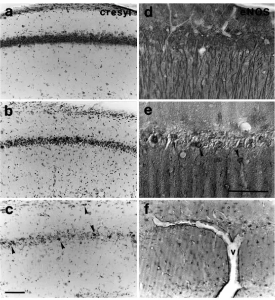
Four days after transient ischemia, 91% (21 of 23) of all gerbils exhibited pyramidal cell loss in CA1 compared to sham-operated controls. Cresyl violet staining of CA1 in control animals was concentrated in the densely packed pyramidal cell layer (grade 0, Fig. 1a). Moderate loss of neuronal staining and an accumulation of apoptotic cell bodies in stratum pyramidale together with an increased staining of glial cells in total CA1 (grade 1, Fig. 1b) was
detected in 17% of all gerbils. The majority of gerbils (74%) showed severe changes with complete loss of pyramidal cell staining (grade 2) and preservation of a few interneurons as identified by their position and shape (Fig. 1c). No significant difference in the ischemia induced pyramidal cell loss was observed in cresyl violet stained sections between gerbils experiencing a short (SI group) or a long interval (LI group) between the last seizure and ischemia (p=n.s., tied z-value=0).
In CA1 of sham-operated animals, eNOS immunoreac-tivity showed intense staining of perikarya as well as dendritic processes (grade 0, Fig. 1d). After ischemia, neuronal expression of eNOS disappeared with pyramidal
cell degeneration. Grade 1 representing significant loss of pyramidal cells together with a marked decrease of dendritic staining (Fig. 1e) was detected in 17% of all gerbils. In 74% of all gerbils, dendritic staining was completely lost whereas glial cell staining became prominent (grade 2; Fig. 1f). In parallel, endothelial cells of cerebral blood vessels started to overexpress eNOS. The expression of eNOS in CA1 did not differ between the SI and the LI group (p=n.s., tied z-value=0). eNOS immu-nostaining in CA1 correlated significantly with cresyl violet staining (p<0.05, rho corrected for ties=0.82). Table 1 summarizes the percentages and numbers of
Fig. 1a–f Neuronal changes in hippocampus. Cresyl violet stained sections show in a densely packed pyramidal cells (grade 0) resembling the control situation, in b nerve cell peri-karya still clearly detectable within a mixture of preserved and apoptotic cell bodies (grade 1), in c absence of pyramidal cells and only a few preserved interneurons (arrowheads) (grade 2). eNOS-stained sec-tions exhibit in d intense stain-ing of perikarya and dendritic processes (grade 0), in e only single surviving neurons (ar-rowheads) with decrease of dendritic staining (grade 1), in f complete loss of perikaryal and dendritic staining of pyramidal cells. Glial cells are immunore-active and walls of larger blood vessels (v) overexpress eNOS (grade 2). Bars 0.1 mm; bar in c applies for a, b, c, f; bar in e applies also for d
Table 1 Histological evaluation of neurodegeneration and glial reactions (LI long interval, 24 h, between last seizure and isch-emia, SI short interval, 2 h, between last seizure and isch-emia, numbers in brackets in-dicate absolute number of ani-mals showing the respective outcome)
Staining region Histological gradings
2: severe changes 1: moderate changes 0: no changes
LI SI LI SI LI SI
Cresyl-CA1 83% (10) 64% (7) 17% (2) 18% (2) - 18% (2)
eNOS-CA1 75% (9) 73% (8) 25% (3) 9% (1) - 18% (2)
GFAP-CA1 50% (6) 36% (4) 50% (6) 64% (7) -
animals of each group regarding the histological grading for cresyl violet and eNOS in CA1.
Glial changes in the hippocampus
Proliferation of glia accompanies ischemia-induced neu-rodegeneration and is evaluated via GFAP labeling. Fifty-seven percent of all gerbils showed hypertrophic astro-cytes in GFAP immunostaining of CA1 (grade 1, Fig. 2b) as compared to only few astrocytes with thin processes in sham-operated gerbils (grade 0, Fig. 2a). In 43% of all gerbils an additional hyperplasia of astrocytes was detected consisting of increased staining and thickening of astrocytic cell bodies and their processes (grade 2, Fig. 1c). The distribution and degree of upregulation of GFAP as specific marker for astrocytes did not differ significantly between the two groups of gerbils in CA1 (p=n.s., tied z-value=0). Percentages and numbers of animals of each group with moderate or severe glial changes are summarized in Table 1.
Neuronal changes in the striatum
Only one animal of the present study exhibited patchy neuronal cell loss and necrotic areas detectable by a loss of cresyl violet staining in the striatum (grade 1, Fig. 3c, d). In contrast, sham-operated gerbils as well as 96% of all investigated animals showed a homogeneous pattern of neurons distributed within the gray matter (grade 0, Fig. 3a, b).
Glial changes in the striatum
Astrocytic reaction in the striatum as revealed in GFAP stained sections of gerbil brains was unchanged in comparison to controls in 22% of all animals (grade 0, Fig. 2d), moderate in 57% (grade 1, Fig. 2e) and severe in 22% (grade 2, Fig. 2f). An accumulation of hyperplastic astrocytes was specifically found in necrotic zones surrounding striatal blood vessels (Fig. 3e, f). GFAP upregulation did not differ significantly between the SI and the LI group in the striatum (p=n.s., tied z-value=0). In Table 1 the percentages and numbers of animals of each
Fig. 2a–f Astrocytic reaction in GFAP-stained sections of CA1 and striatum (CP). a and d show grade 0: few astrocytes with thin processes; b and e grade 1: increased number of astrocytes; c and f grade 2: hypertrophy and hyperplasia of astrocytes. In CP, in addition, accumulation of astrocytes sur-rounding perivascular necrotic zones was noted (p pyramidal cell layer). Bar in c applies for all pictures and equals 0.1 mm
group regarding the histological grading for GFAP in the striatum are shown.
No correlation between neuronal and glial changes Activation of glial cells in CA1 was seen in response to neuronal injury in 18 animals, but also in 2 animals with no or in 4 animals with only moderate neuronal changes. These discrepancies were reflected in non-significant correlations between GFAP and cresyl violet as well as between GFAP and eNOS (both p=n.s., rho corrected for ties=0). In the striatum, GFAP positive astrocytes were detected in 77% (17 of 22) of gerbils with no detectable neuronal cell loss.
Discussion
The major finding of the present study is that the outcome of a 5-min global ischemia in seizure-sensitive Mongolian gerbils is not influenced by prior inherent epileptic
seizures whether they are induced 24 or 2 h before ischemia. Four days after ischemia, neuronal and glial changes in CA1 of the hippocampus and in the striatum of both investigated groups did not differ significantly and were mainly consistent with previous reports of other authors (Chesselet et al. 1990; Kirino 1982) and our own observations (Hoffmann et al. 1992). In particular, neither significant protection nor cumulative damage could be detected. In two animals of the short interval group, representing 9% of all animals, CA1 pyramidal cells were preserved after ischemia. This failure rate (due to misplaced clips, postoperative hypothermia or well-functioning posterior communicating arteries between carotid and vertebrobasilar system) is within the expected range.
Regarding neuronal damage, in hematoxylin-eosin or cresyl violet stained sections of the hippocampus, delayed neuronal death of the pyramidal cells was prominent 4 days after ischemia with an almost complete destruction of the CA1 pyramidal cell layer (Kirino 1982; Suzuki et al. 1983). The selective vulnerability of CA1 that has specifically been shown for ischemia was not different
Fig. 3a–f Neuronal changes in striatum and corresponding glial reactions. Cresyl violet stained section a and at a higher mag-nification b show homogeneous distribution of neurons in gray matter (grade 0); c and at a higher magnification d demon-strate diffuse loss of neurons as well as necrotic zones (grade 1). The adjacent GFAP-stained sections in e and at a higher magnification in f show a patchy loss of astrocytes in neuropil and an accumulation in perivas-cular areas (grade 2 in GFAP stain). All bars 0.1 mm
between the SI and LI group, suggesting that the time point of inherent seizures does not influence the outcome of ischemia.
Immunoreactivity for eNOS which is concentrated in hippocampal neurons and outlines their dendrites (Diner-man et al. 1994) decreased together with pyramidal cell loss. It showed dendritic alterations which were not detectable with cresyl violet stain. Taken together, we would suggest a combined analysis of postischemic hippocampus by cresyl violet as a sensitive marker for the integrity of neuronal somata and eNOS for their dendrites. Further, the constitutive expression of eNOS in cerebral blood vessels was upregulated after ischemia as previously reported (Zhang et al. 1993).
Astrocyte hyperplasia is indicative for regional brain damage (Petito et al. 1990). In the gerbil striatum, glial upregulation was present in the majority of gerbils and was widespread. Only in few cases was it focally enhanced in perivascular zones. The latter changes might suggest a direct relation to microvascular thrombosis in postisch-emic lesions of the striatum (Ames et al. 1968). The more diffuse pattern of hypertrophic and hyperplastic astrocytes in hippocampus and striatum indicates a feature of selective vulnerability in comparison to other brain regions which were spared. Ischemic damage of a subpopulation of selectively vulnerable striatal neurons has been shown by immunohistochemistry with antibodies specific for peptides expressed in efferent neurons (Chesselet et al. 1990). In our study, in the majority of all animals glial changes like an increased GFAP immunoreactivity, hy-pertrophy and hyperplasia of astrocytes were seen in hippocampus and striatum as previously reported (Chen and Simon 1997; Lin et al. 1993; Petito et al. 1990; Tanaka et al. 1992), even in the absence of detectable neuronal damage. This might be due to the upregulation of GFAP in response to only minor neuronal cell death. eNOS and cresyl violet stain, in particular, show only the endpoint of neuronal degeneration. Unlike several studies in the rat that showed increased GFAP concentration and immuno-reactivity in the hippocampus after different modes of hippocampal kindling (Hansen et al. 1991; Khurgel et al. 1995; Stringer 1996), kindled and sham-operated gerbils from our population exhibited only rare and weakly stained astrocytes (compare grade 0 of GFAP staining, Fig. 2a). Therefore, the discrepancy between hypertrophic GFAP-positive glia and no overt perikaryal necrosis could be due to discrete neurodegeneration and/or terminal degeneration of projecting afferences and/or other altera-tions in tissue homeostasis.
A combination of seizures and ischemia was tested by Towfighi and colleagues in immature rats (1999). Seizures induced by kainic acid or flurothyl at 24 and again at 6 h prior to hypoxic ischemia were protective against cerebral hypoxic-ischemic lesions. Our results, however, provide no evidence for a time-dependent effect of inherent seizures on the outcome of transient cerebral ischemia comparing a 24-h and a 2-h interval. Many parameters could be taken into account for these discrepancies, e.g., species, age of the animals, latency between two insults,
and intensity, duration and mechanism of induction of the preconditioning stimulus. It should also be considered that a purely negative control group consisting of animals that have no seizures within 2–3 days cannot be studied in a species such as Mongolian gerbils in which all animals exhibit spontaneous seizures at least twice per week. The advantage of the gerbil is that the application of epilep-togenic substances can be avoided because seizures can be provoked in response to environmental stimuli. In rat experiments seizures necessarily need to be chemically induced and a negative control group would not be exposed to the epileptogenic drug.
Protection against ischemic injury has previously been shown for two ischemic insults. For tolerance induction time intervals of longer than 1 day and up to 7 days have been suggested by Kitagawa and colleagues (1990) and by Kato and colleagues (1994). Two successive seizures with a time interval of 1–3 days were also described to be protective (Sasahira et al. 1995).
Apart from latency between preconditioning and final insult, the intensity of the prior insult may be crucial for the development of ischemic tolerance (Chen and Simon 1997). Compared to 18±1 min bicuculline induced seizures with continuous high-voltage spiking (Sasahira et al. 1995) or 1-h episodes of kainic acid induced seizures (Najm et al. 1998) or at least 60 min status epilepticus induced by kainic acid or flurothyl (Towfighi et al. 1999), non-chemically induced generalized tonic-clonic seizures in gerbils last for a rather short period of 1–5 min (Cutler and Mackintosh 1989; Loskota et al. 1974; Scotti et al. 1998; Seto-Oshima et al. 1992).
In conclusion, non-chemically induced inherent epilep-tic seizures at a short (2 h) or a long (24 h) interval prior to transient forebrain ischemia neither protect the gerbil brain from injury nor augment the degree of damage. Our data suggest that inherent seizures in gerbils do not induce tolerance neither to ischemia nor to seizures. Our results render it unlikely that variations in the outcome after transient ischemia in gerbils are due to prior inherent seizures. Therefore, our present data indicate that future studies in the ischemia model in seizure-prone gerbils do not require preischemic monitoring of spontaneous epi-leptic seizures as they do not influence the outcome after transient ischemia.
Acknowledgements The authors thank Olga Bollag and Gaby Kalt for expert technical assistance and Dr. Kurt Kräuchi for help with the statistical analysis. This study was funded by the University of Basel.
References
Ames ARL 3rd, Wright, Kowada M, Thurston JM, Majno G (1968) Cerebral ischemia. II. The no-reflow-phenomenon. Am J Pathol 52:437–453
Chen J, Simon R (1997) Ischemic tolerance in the brain. Neurology 48:306–311
Chesselet MF, Gonzales C, Lin CS, Polsky K, Jin BK (1990) Ischemic damage in the striatum of adult gerbils: relative sparing of somatostatinergic and cholinergic interneurons contrasts with loss of efferent neurons. Exp Neurol 110:209– 218
Cutler MG, Mackintosh JH (1989) Epilepsy and behaviour of the Mongolian gerbil: an ethological study. Physiol Behav 46:561– 566
Dinerman JL, Dawson TM, Schell MJ, Snowman A, Snyder SH (1994) Endothelial nitric oxide synthase localized to hippo-campal pyramidal cells: implications for synaptic plasticity. Proc Natl Acad Sci U S A 91:4214–4218
Gidday JM, Fitzgibbons JC, Shah AR, Park TS (1994) Neuropro-tection from ischemic brain injury by hypoxic preconditioning in the neonatal rat. Neurosci Lett 168:221–224
Hansen A, Jorgensen OS, Bolwig TG, Barry DI (1991) Hippocam-pal kindling in the rat is associated with time-dependent increases in the concentration of glial fibrillary acidic protein. J Neurochem 57:1716–1720
Hoffmann MC, Nitsch C, Scotti AL, Reinhard E, Monard D (1992) The prolonged presence of glia-derived nexin, an endogenous protease inhibitor, in the hippocampus after ischemia-induced delayed neuronal death. Neuroscience 49:397–408
Kato H, Kogure K, Araki T, Itoyama Y (1994) Astroglial and microglial reactions in the gerbil hippocampus with induced ischemic tolerance. Brain Res 664:69–76
Kato J, Liu Y, Araki T, Kogure K (1991) Temporal profile of the effects of pretreatment with brief cerebral ischemia on the neuronal damage following secondary ischemic insult in the gerbil: cumulative damage and protective effects. Brain Res 553:238–242
Khurgel M, Switzer RC 3rd, Teskey GC, Spiller AE, Racine RJ, Ivy GO (1995) Activation of astrocytes during epileptogenesis in the absence of neuronal degeneration. Neurobiol Dis 2:23–35 Kirino T (1982) Delayed neuronal death in the gerbil hippocampus
following ischemia. Brain Res 239:57–69
Kitagawa K, Matsumoto M, Tagaya M, Hata R, Ueda H, Niinobe M, Handa N, Fukunaga R, Kimura K, Mikoshiba K, Kamada T (1990)‘Ischemic tolerance’ phenomenon found in the brain. Brain Res 528:21–24
Levy DE, Brierley JB (1974) Communications between vertebro-basilar and carotid arterial circulations in the gerbil. Exp Neurol 45:503–508
Lin RC, Polsky K, Matesic DF (1993) Expression of gamma-aminobutyric acid immunoreactivity in reactive astrocytes after ischemia-induced injury in the adult forebrain. Brain Res 600:1–8
Loskota WJ, Lomax P (1975) The Mongolian gerbil (Meriones unguiculatus) as a model for the study of the epilepsies: EEG records of seizures. Electroencephalogr Clin Neurophysiol 38:597–604
Loskota WJ, Lomax P, Rich ST (1974) The gerbil as a model for the study of the epilepsies. Epilepsia 15:109–119
Mirzaeian L, Ribak CE (2000) Immunocytochemical mapping of Fos protein following seizures in gerbils indicates the activation of hippocampal neurons. Hippocampus 10:31–36
Najm IM, Hadam J, Ckakraverty D, Mikuni N, Penrod C, Sopa C, Markarian G, Lüders HO, Babb T, Baudry M (1998) A short episode of seizure activity protects from status epilepticus-induced neuronal damage in rat brain. Brain Res 810:72–75 Peterson GM, Ribak CE (1987) Hippocampus of the
seizure-sensitive gerbil is a specific site for anatomical changes in the GABAergic system. J Comp Neurol 261:405–422
Petito CK, Morello S, Felix JC, Lesser ML (1990) The two patterns of reactive astrocytosis in postischemic rat brain. J Cereb Blood Flow Metab 10:850–859
Sasahira M, Lowry T, Simon RP, Greenberg DA (1995) Epileptic tolerance: prior seizures protect against seizure-induced neuro-nal injury. Neurosci Lett 185:95–98
Scotti AL, Bollag O, Nitsch C (1998) Seizure patterns of Mongolian gerbils subjected to a prolonged weekly test schedule: evidence for a kindling-like phenomenon in the adult population. Epilepsia 39:567–576
Seto-Oshima A, Ito M, Kudo T, Mizutani A (1992) Intrinsic and drug-induced seizures of adult and developing gerbils. Acta Neurol Scand 85:311–317
Stern M (2000) Entwicklung eines rechnergestützten Aktivitätsana-lysesystems zur Überwachung krampfbereiter Kleinnagetiere. Dissertation Medizinische Fakultät, Universität Basel
Stringer JL (1996) Repeated seizures increase GFAP and vimentin in the hippocampus. Brain Res 717:147–153
Suzuki R, Yamaguchi T, Kirino T, Orzi F, Klatzo I (1983) The effects of 5-minute ischemia in Mongolian gerbils: I. Blood-brain barrier, cerebral blood flow, and local cerebral glucose utilization changes. Acta Neuropathol 60:207–216
Tanaka H, Araki M, Masuzawa T (1992) Reaction of astrocytes in the gerbil hippocampus following transient ischemia: immuno-histochemical observations with antibodies against glial fibril-lary acidic protein, glutamine synthetase, and S-100 protein. Exp Neurol 116:264–274
Tomida S, Nowak TS Jr, Vass K, Lohr JM, Klatzo I (1987) Experimental model for repetitive ischemic attacks in the gerbil: the cumulative effect of repeated ischemic insults. J Cereb Blood Flow Metab 7:773–782
Towfighi J, Housman C, Mauger D, Vannucci RC (1999) Effect of seizures on cerebral hypoxic-ischemic lesion in immature rats. Dev Brain Res 113:83–95
Winkler DT, Scotti AL, Nitsch C (2001) Ischemia-induced degen-eration of CA1 pyramidal cells decreases seizure severity in a subgroup of epileptic gerbils and affects parvalbumin immu-noreactivity of CA1 interneurons. Exp Neurol 168:364–372 Zhang ZG, Chopp M, Zaloga C, Pollock JS, Forstermann U (1993)
Cerebral endothelial nitric oxide synthase expression after focal cerebral ischemia in rats. Stroke 24:2016–2021