Profound reduction of mature B cell
numbers, reactivities and serum Ig levels in
mice which simultaneously carry the XID
and CD40 deficiency genes
Yoshihiro Oka
12, Antonius G. Rolink
1, Jan Andersson
1-
3, Masahito Kamanaka
4,
Junji Uchida
4, Teruhito Yasui
4, Tadamitsu Kishimoto
2, Hitoshi Kikutani
4and
Fritz Melchers
11Basel Institute for Immunology, Grenzacherstrasse 487, 4005 Basel, Switzerland
department of Medicine III, Osaka University Medical School, 2-2 Yamada-oka, Suita, Osaka 565, Japan department of Immunology, University of Uppsala, Biomedical Center, S-751 23 Uppsala, Sweden institute for Molecular and Cellular Biology, Osaka University, 1-3 Yamada-oka, Suita, Osaka 565, Japan
Abstract
It has been known for some time that single mutant nude or CD40T mice have apparently normal numbers of cells in the precursor compartments of bone marrow and the mature B cell
compartments of the periphery. X-linked immunodeficiency (XID) mice are deficient only in some of the slgM+slgD+ B cells. We have investigated further the contributions of the xid mutation, of the T
cell deficiency of nude and of the inability of CD40T B cells to cooperate with T cells in the generation of the precursor and the mature B cell compartments in mice. Double mutant XID/nu and XID/CD40T mice have precursor B cell compartments that are no more deficient than the single mutant XID mice. However, the peripheral B cell compartments of both XID/nu and XID/CD40T are even more deficient than those of single mutant XID mice. While 10% of the peripheral B cells of wild-type or CD40T, one-third of XID and half of XID/nu mice turn over rapidly, as many as three-quarters of those in XID/CD40T are short-lived. Total numbers of slgM+slgD+ B cells in the spleen
are at best 10-15% of normal mice at 6-8 weeks of age in XID, XID/nu and XID/CD40T mice. They remain that low at 3 months of age in XID/CD40T mice, while in XID mice these peripheral B cells slowly build up in numbers with age. As expected, double mutant XID/CD40T mice do not respond to the T-dependent antigen keyhole limpet hemocyanin. Only the responses to the T-independent type I antigen, TNP-lipopolysaccharide (LPS), appear to be normal. In vitro, their splenic B cells respond poorly to LPS or to IgM-specific antibody in either the absence or presence of cytokines. Most notably, serum IgM, lgG2b and lgG3 levels are severely depressed, while lgG1, lgG2a and IgA levels are <10 ng/ml. The results suggest a model of mature B cell development in which the peripheral, mature B cell compartments are generated in two parallel, not tandemly organized pathways. They could be selected and/or stimulated at the transition from immature to mature B cells: in btk controlled or in CD40 controlled ways.
Introduction
The discovery of X-linked immunodeficiency (XID) diseases in humans and mice has demonstrated the existence of genetic loci on the X chromosome which influences the growth, differentiation and function of lymphocytes (1-3). Recently, two groups have isolated the gene encoding a cytoplasmic tyrosine kinase, named Bruton's tyrosine kinase (Btk), which in different patients with X-linked
agamma-globulinemia (XLA) appears mutated in different parts of the gene encoding different domains of the Btk protein (4,5). On the other hand, one of the best characterized models of this disease in mice is the CBA/N strain that carries a XID gene (2). The murine btk gene was mapped to the XID locus of the murine X chromosome (6,7). A missense mutation, predicted to alter an amino acid residue (Arg28 to Cys) in the unique
Correspondence ta F. Melchers
1676 Reduction of B cells and serum Ig in double mutant XID/CD40T mice N-terminal region of the Btk protein, was found in the btk
gene of XID mice, suggesting that this mutant btk gene is responsible for the XID defect in CBA/N mice (6).
XID mice do not respond to thymus-independent type II (Tl-ll) antigens (2), respond with a lower number of B cells to lipopolysaccharide (LPS) (8), do respond to inde-pendent type I (Tl-I) antigens and respond normally to thymus-dependent (TD) antigens (2,9). B cells of XID mice also do not respond to cross-linking of surface Ig (slg) in the presence of IL-5 or IL-10 (10,11). Somewhat reduced numbers of peripheral, mature B cells, and reduced levels of IgM and lgG3 are found in the XID mice, indicating that the generation of mature B cells reactive to Tl-I and TD antigens is not severely affected by the btk mutation.
Thymectomy of CBA/N mice abolishes the ability of these mice to populate the peripheral lymphoid compartment with T and B cells, suggesting that at least some of the peripheral B cells need T cells for their development (12).
In previous studies by Wortis and his colleagues (13), the XID mutation has been combined with the nude (nu/nu, called nu herein) defect leading to thymic dysplasia and failure to develop the a(3 TCR-expressing lineage of T cells. The analysis of these double mutant XID/nu mice has indicated a more severe B cell dysplasia, so that B lineage precursor cell compartments in the bone marrow appeared normal at early pro/pre-B stages, but depleted of cells expressing IgM either in the cytoplasm or on their surface. The peripheral, mature B cell compartments were found severely depleted, incapable of responding to either Tl or TD antigens. Concomitantly serum Ig levels were much reduced (12,14-16). The grafting of thymus into the double mutant XID/nu mice restored the peripheral mature B lymphoid compartments with cells capable of responding to Tl and TD antigens (16). Again, this finding was taken as evidence that some peripheral, mature B cells are generated in thymus-dependent ways.
CD40 expressed on B cells and its ligand expressed on T cells control the interactions of helper T cells with B cells and, thus, responses of B cells to TD antigens which lead to Ig isotype switching, germinal center formation and induction of immunological memory (17-24). CD40-deficient (CD40T) mice, generated by targeted mutation, have peripheral mature B cells which respond normally to Tl antigens, do respond to TD antigens with IgM production, but do not respond to TD antigens with IgG, IgA or IgE production, and do not develop germinal centers (21). This indicates that the interaction between CD40 and its ligand is essential for helper T cell-dependent Ig class switching during responses of B cells to TD antigens, but is not important for responses to Tl antigens. Taken together, all these findings suggest that peripheral B cells can be generated in both btk controlled and CD40 controlled ways.
In this report we further investigate the contributions of the
Xid gene to the nu/nu defect in the primary, B cell generating
compartments of bone marrow. We have also combined the XID defect effected by the btk mutation with the CD40 defect in a double mutant mouse strain (called XID/CD40T) to see whether the inability of B cells to respond to some Tl combined with the inability to cooperate with helper T cells controls all of the mature B cell development.
Methods
Mice
CBA/N mice which carry the xid gene were originally obtained from the Jackson Laboratory (Bar Harbor, ME). In addition, we also used XID congenic C57BI/6 mice for some experiments. Athymic C57BI/6 nu/nu mice were originally obtained from BRL (Fullinsdorf, Switzerland), xid/xid x nu/nu double mutant mice (called XID/nu in this paper) were generated as described by Wortis and his colleagues (15). The generation of CD40T mice has been described elsewhere (21).
Generation of double mutant mice with XID and CD40 deficiency
In order to analyze the effect of a simultaneous abnormality of XID and CD40 deficiency on B cell development, we bred double mutant mice from XID and CD40T mice (XID/CD40T mice). Female CBA/N (xid/xid; homozygous for the mutated
btk gene) mice or female XID congenic C57BI/6 mice (here
called XID) were mated with male CD40T mice (homozygous for the mutated CD40 gene), followed by mating of the female F-| mice with male CD40T mice. In the next male offspring, double mutant XID/CD40T, single mutant CD40T, XID and wild-type mice were expected and found in a ratio of 1:4 as described below.
Mice were bled at ages between 10 and 12 weeks to measure serum Ig levels. To detect the defect of CD40, PCR analysis was done on DNA from spleen or tail as described (21). To detect the XID phenotype we analyzed peritoneal cells by FACS for the absence or presence of CD5+ B cells, because lack of CD5+ peritoneal B cells is one of the striking features of XID mice. To confirm the serological typing of XID mice, PCR analyses was done on DNA from spleen or tail to detect the btk mutation, using primers and protocols as described (7).
Antibodies and flow cytometric analysis
FITC-conjugated mAb RA3-6B2 (anti-CD45R, B220), phyco-erythrin (PE)-conjugated mAb 53-7.3 (anti-CD5), biotinylated mAb 7D4 (anti-CD25, IL-2R a-chain, TAC) and mAb B3B4 (anti-CD23, IgE Fc receptor) were purchased from Phar-Mingen (San Diego, CA). mAb ACK4 (anti-c-MQ (25), mAb M41 (anti-IgM) (26), mAb 1.19 (anti-lgD) (27) and mAb 7E9 (anti-CD21) (28) were purified from culture supernatants on Protein G-Sepharose (Pharmacia, Uppsala, Sweden) as recommended by the supplier, followed by conjugation with biotin according to standard protocols.
Bone marrow, spleen and peritoneal cells were cell surface stained with antibodies as described elsewhere (29-31), and stained cells were analyzed by FACScan (Becton-Dickinson, Mountain View, CA).
Ouantitation of serum Ig levels
Serum IgM, lgG1, lgG2a, lgG2b, lgG3 and IgA levels were determined by ELISA as described elsewhere (29,31,32). Goat antibodies specific for mouse Ig classes and subclasses and mouse Ig standards were purchased from Southern Biotechnology Associates (Birmingham, AL).
BrdU labeling of cells
Mice were fed with BrdU (Sigma, St Louis, MO) at 1 mg/ml in drinking water for 6 days, or for 6 days, followed by a 7 day period of feeding without BrdU (chase) before sacrifice. Blood, spleen and bone marrow lymphocytes were prepared in PBS, fixed for 1 h on ice with 0.5% paraformaldehyde in PBS. Cells were collected by centrifugation and resuspended in 1 ml of 3N HCI/0.5% Tween 20 and left for 20 min at room temperature in order to denature DNA. Cells collected by centrifugation were neutralized by resuspending them in 0.2 ml of 0.1 M borate buffer, pH 8.5, and thereafter adding 1 ml PBS-azide. Cells were washed once, resuspended in FACS buffer (PBS-azide/2% FCS) and stained with PE-labeled anti-B220 (PharMingen) and FITC-labeled anti-BrdU (Becton Dickinson Immunocytometry System, San Jose, CA) followed by analyses on a FACScan.
Antigens and immunization of mice
For generation of antibody responses to keyhole limpet hemo-cyanin (KLH), animals were immunized i.p. with 50 ng in complete Freund's adjuvant. The anti-KLH antibody responses were measured in sera on day 14 by utilizing a KLH isotype-specific ELISA (see below).
Primary responses to TNP-LPS were generated by immuniz-ation with 50 ng TNP-LPS in PBS i.p. The IgM, lgG1, lgG2a and lgG3 anti-TNP responses were measured in sera on day 14 by a TNP-specific ELISA (see below).
Antigen-specific ELISA
Microtiter plates for ELISA were coated with 10 ng/ml KLH or 5 ng/ml DNP-BSA in PBS overnight at 4°C. They were washed and blocked with PBS containing 5% FCS and sodium azide. Diluted serum samples were incubated overnight at 4°C. Samples were washed and antigen-specific antibody titers were determined with alkaline phosphatase-conjugated detection antibodies: goat anti-mouse IgM, lgG1, lgG2a, lgG2b, lgG3 and IgA (all obtained from Southern Biotech-nology Associates). After 1 h incubation at 37°C plates were washed and the ELISA was developed with the substrate 4-nitrophenyl phosphate (1 mg/ml; Sigma) in 1 M diethanol-amine buffer, pH 9.8. Plates were analyzed on an ELISA reader (ThermoMax; Molecular Devices, Menlo Park, CA) at 410 and 610 nm. Units represent arbitrary values based on the titration curve of a standard immune serum from normal mice according to the procedure described by Foy etal. (19).
Cell culture and in vitro stimulation of spleen cells
Single cell suspensions of spleen from the various mutant and wild-type mice were prepared and cultured in Iscove's modified DMEM (Gibco/BRL, Life Technologies, Paisley, UK) containing antibiotics, 5 X 1 0 ~5 M 2-mercaptoethanol and 2% FCS as described earlier (29). Cultures of 0.2 ml containing 3X10⁵ total spleen cells/ml were set up in 96-well flat-bottom microtiter ptates and incubated for 3 days at 37°C in a 10% CO2/air atmosphere. At the onset of culture, various mitogens and/or recombinant cytokines were added to probe for B cell proliferative functions. The following mitogens were used: LPS at 20 |ig/ml (from Escherichia co//EH 100, kindly provided by Dr C. Galanos, Max Planck Institute for Immunobiology,
Freiburg, Germany), monoclonal anti-CD40 at 10 ng/ml [clone FGK 45.5 generated as elsewhere (33)], Sepharose-bound anti-IgM at 1% beads in culture [monoclonal rat anti-mouse |i-chain, clone M41 (26) bound at 1 mg/ml of packed CNBr-activated Sepharose 4B (Pharmacia, Uppsala)]. The recom-binant IL-4 and IL-2 were used at 100 and 1000 U/ml respectively. They were obtained from a transfected X63-Ag8.653 cell line (34) generated in our laboratory. Cultures were pulsed with 1 nCi [3H]thymidine (sp. act. 5 Ci/mmol; Amersham Life Science, Buckinghamshire, UK) during the last 4 h of incubation and harvested onto glass fiber paper which was processed and counted in a Rack-Beta plate scintillation counter (LKB Pharmacia, Uppsala). Radioactivity recorded from triplicate cultures was expressed as the mean c.p.m. per culture. SD between triplicates never exceeded 5% of the mean.
Results
8 lineage precursor cells in the bone marrow of homozygous
wild-type, XID, nu, CD40T, XID/nu and XID/CD40T mice
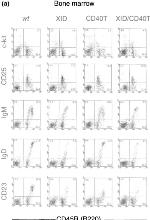
B220(CD45R)+ B lineage precursors in bone marrow of different wild-type, mutant and double mutant mice were analyzed by FACS for the expression of c-kit+, CD25(TAC)+, slgM+, slgD+ and CD23+ cells. The results of these analyses are shown in Fig. 1(a and b). The quantities of the different B-lineage subpopulations are given in Table 1. CD40T and nude mice have c-kit+ pro/pre-B-l cells, CD25(TAC)+ pre-B-II cells, slgM+ and CD23+ immature as well as slgD+ mature B cells in numbers comparable to wild-type mice. XID mice lack a large part of the slgM+B220 high cells, of the slgD+ cells and of the CD23+ cells, indicating a deficit in the more mature subpopulations of B cells. XID/nu and XID/CD40T double mutant mice have normal numbers of c-kit+
pro/pre-B-l cells. The CD25(TAC)+ pre-B-ll cells and slgM+, slgD+ and CD23+ cells are comparable in both phenotype and numbers to that of XID single mutant mice in both types of double mutant mice. The low level of CD23 expression on B cells of CD40T animals is a consistent finding and has been reported earlier (21) (also, see below).
We conclude from these analyses that the double expres-sion of the XID and nu, respectively XID and CD40T mutations, does not impair B cell development in bone marrow beyond the defects seen with the single XID mutation. This is in contrast to previous analyses of XID/nu double mutant mice performed by Wortis and his colleagues (15).
B lineage cells in the spleen
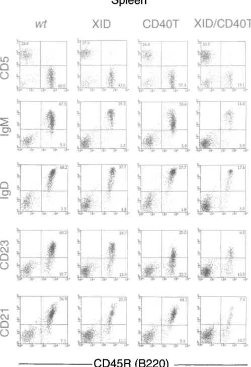
B220(CD45R)+ B lineage wild-type cells and the different mutant and double mutant mice were also analyzed in spleen by FACS for the expression of CD5, slgM, slgD, CD23 and CD21. These analyses were performed with mice 4-6 weeks as well as 10-12 weeks of age. The results of these analyses are shown in Fig. 2, the quantities of the different B-lineage cell subpopulations are given in Table 2. Wild-type, nu and CD40T single mutant mice have comparable numbers of B220+ cells expressing slgM and slgD at both ages. Wild-type and CD40T single mutant mice have comparable numbers of CD5+ and CD21+ cells. Consistently, we find that the level of
1678 Reduction of B cells and serum Ig in double mutant XID/CD40T mice CD23 expression is lower in CD40T B cells and thus the
number of detectable CD23+ cells is underscored in such mice. Spleens of XID/nu and XID/CD40T double mutant mice at 4-6 weeks of age had slgM+ and slgD+ cells in numbers slightly less than those of single mutant XID mice. When the total number of CD45(B220)+ s l g+ cells in spleen was calculated, seven wild-type mice had an average of 39 ± 5X10⁶ cells, 10 XID mice an average of 9 ± 4X10⁶, six
double mutant CD40T/XID mice an average of 6 ± 1X10⁶ cells and five XID/nu mice an average of 4 ± 2x10⁶ cells (Table 2). When the same measurements were done with spleens of 10- to 12-week-old mice, the number of slg+ B cells had more than doubled in XID mice but remained as low as in 4- to 6-week-old mice in XID/CD40T mice (Table 2). We conclude from these data that, in contrast to bone marrow, the B lineage representation in spleen of XID/nu and
(a)
Bone marrow
wt
XID
CD40T XID/CD40T
6
*
i » -I 2.70
• it* 27ft 43,
lTS 1*» t 3.5 3.1 * -1 4.0IP
4.9 3$.' •3t£-'--. its * i\i 1^1 if i IT) C\JQ
O
1 i 0.7If'
17.2 ;A'£&? 32.1 1 * * 1*» 1 i i i . 0 JIP
23.2 M>$-- • 24.0 * i4' i t1 ifc* t V » -r 0.6 # 18.21
* if i • 2.1 13.8 , • > • " - : ' • . • • . + - • « • 'V, */';. 218 a *1l 0.3ii
1 It' 14.5 v f r ' 35.1 S 0.6 * 20.4 '-. Hi.*1' / * ' 18.4 1» *Q
-11 t 26.7 •••;.,«* i a i. i T 0.4 & 1 ii* * 1 6 2"•W-% • • 22.2 1«> I** 1 4.9 •IK"'" 29-1 CO
Q
O
X 1 0 J • I*1 18.60
' • •'.-'' ••CD45R (B220)
Fig. 1. (a) FACS analyses of bone marrow lymphoid cells of wild-type (wt), CD40T, XID and XID/CD40T mice. Horizontal axes represent
fluorescence intensities of CD45R(B220) after surface labeling, while vertical axes represent fluorescence intensities after surface labeling for c-kit and CD25 (TAC), n (IgM), 8 (IgD) and CD23 as described in Methods.
XID/CD40T double mutant mice shows a severe reduction in the numbers of cells expressing different markers associated with mature B cells. With increasing age this defect remains severe in the XID/CD40T mice. Our results suggest that the simultaneous deficiency in the XID and CD40 genes affects the generation of mature, peripheral B cells at the transition from the immature bone marrow compartment profoundly and much more so than does single mutations.
Turnover of peripheral B cells
Mice at 4-6 weeks of age were given BrdU in drinking water for 6 days, to allow incorporation into DNA of dividing cells (pulse period). Thereafter, BrdU was removed from the drinking water for 7 days (chase period) to allow analyses of the fate of labeled cells. At 6 days after the pulse, and after 7 days of the chase period, B220+ cells in the spleen and blood were analyzed for BrdU label. The experimental results
(b)
XID
Bone marrow
nu
Xid/nu
C\JQ
O ) • 0.2 9.6dip'
28
-
5
CD45R (B220)
Fig. 1. (b) FAC5 analyses of bone marrow lymphoid cells of XID, nu and XID/nu mice. Horizontal axes represent fluorescence intensities of
CD45R(B220) after surface labeling, while vertical axes represent fluorescence intensities after surface labeling for c-kit and CD25 (TAC), \i (IgM) and 8 (IgD).
1680 Reduction of B cells and serum Ig in double mutant XID/CD4OT mice
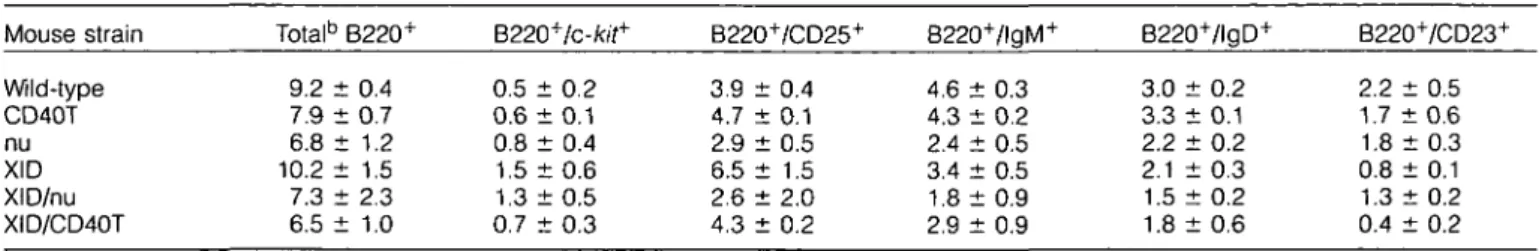
Table 1. Total number of B cells in different subpopulationsa in the bone marrow of indicated wild type, single mutant and double mutant mice
Mouse strain Totalb B220+ B220+/c-/c/f+ B220~7CD25+ B220+/lgM4 B220+/lgD+ B220+/CD23+
Wild-type CD40T nu XID XID/nu XID/CD40T 9.2 i 7.9 i 6.8 i 10.2 J 7.3 i t 0.4 -. 0.7 t 1.2 t 1.5 t 2.3 6.5 ± 1.0 0.5 : 0.6 ; 0.8 : 1.5 : 1.3 : 0.7 : t 0.2 t 0.1 t 0.4 t 0.6 t 0.5 t 0.3 3.9 : 4.7 i 2.9 : 6.5 : 2.6 : 4.3 : t 0.4 t 0.1 t 0.5 t 1.5 t 2.0 t 0.2 4.6 ± 0.3 4.3 ± 0.2 2.4 ± 0.5 3.4 ± 0.5 1.8 ± 0.9 2.9 ± 0.9 3.0 ± 0.2 3.3 ± 0.1 2.2 ± 0.2 2.1 ± 0.3 1.5 ± 0.2 1.8 ± 0.6 2.2 ± 0.5 1.7 ± 0 . 6 1.8 ±0.3 0.8 ± 0.1 1.3 ± 0.2 0.4 ± 0.2
aBone marrow of 4- to 6-week-old mice was prepared for FACS analyses as described in Methods. After gating out dead cells, the
percentages of cells within the lymphoid cell gate of which the surface stained for the indicated markers were calculated. The total numbers of lymphoid cells contained in two femurs from individual mice were counted and they contained 22-26X106 cells. From that number and the
percentage obtained by FACS analyses, the total number of B cells contained in the various fractions was computed. The values given represent the mean ± SD from four to six individual mice analyzed.
bValues represent number of cells (X106) per two femurs.
for peripheral blood B220+ cells are given in Table 3, since analyses on splenic B cells gave identical results. The advant-age of the analyses on blood is that the same animals can be scored both directly after the pulse of BrdU and then continuously monitored during the entire chase period. As expected from previous analyses by other laboratories (35), wild-type and nu mice had -5-10% of their peripheral B cells labeled and most of the labeled cells disappeared upon a chase period of 7 days. This indicates that the labeled cells are short-lived, while the majority of unlabeled cells should be longer lived. The same extent of labeling and disappear-ance upon chase was observed with B lineage cells in CD40T mice. By contrast, XID mice had a much higher (30-40%) portion of short-lived cells, while XID/nu double mutant mice had even higher (-50%) numbers. The highest number of short-lived cells, i.e. almost three-quarters of all peripheral B lineage cells, was found in double mutant XID/CD40T mice. We conclude from these experiments that most of the B cells generated in XID/CD40T double-deficient mice are and remain short lived, indicating that long-lived B cells remain few in the periphery of these mice.
Response of B cells to antigenic stimulation in vivo
KLH was used as a TD antigen and TNP-LPS as a Tl-I antigen to monitor the in vivo responsiveness of B cells in wild-type, single mutant CD40T or XID, and double mutant XID/CD40T mice (Fig. 3a and b). Wild-type and XID mice showed normal IgM and lgG1 responses to KLH, while CD40T mice, as expected (21), were 5-fold lower in their IgM responses, compared to the wild-type mice, and deficient in lgG1 responses. Double mutant XID/CD40T mice showed > 10-fold reduced IgM responses compared to single mutant CD40T mice and showed no lgG1 (Fig. 3a), lgG3 or IgA responses (data not shown) to KLH. Wild-type, single mutant XID as well as double mutant XID/CD40T mice responded equally well to the Tl-I antigen TNP-LPS (Fig. 3b)
We conclude that the double mutant XID/CD40T mice are deficient in TD responses, but do show responses to a Tl-I antigen.
Responses of peripheral B cells to mitogenic stimulation in vitro
Spleen cells of wild-type, single mutant CD40T, XID and double mutant XID/CD40T, varying in B cell content by a factor of 3-5 (see preceding section), were cultured in medium alone, or in the presence of LPS, of CD40-specific mAb FGK45.5 or of mouse |iH-chain specific mAb M41 coupled to Sepharose either alone (Fig. 4a) or in the presence of IL-4 or IL-2 (Fig. 4b). One of a series of representative experiments is shown. Splenic B cells from wild-type mice were stimulated by all three B cell mitogens. The same mitogens also stimulated spleen cells of XID mice, although - 2 - to 4-fold lower responses were consist-ently recorded in such mass cultures, in the presence or absence of the cytokines indicated. However, as reported earlier (8), upon limiting dilution in LPS containing thymus filler cell cultures (36), spleen cells from XID mice showed 10- to 30-fold lower frequencies of LPS-reactive cells (see legend to Fig. 4). Spleen cells of CD40T mice were stimulated by LPS and IgM-specific mAb in the presence and absence of the cytokines to the same extent as wild-type spleen cells except, as expected, that the CD40-specific mAb FGK45 did not stimu-late the CD40-deficient B cells.
In marked contrast, spleen cells of XID/CD40T double mutant mice showed poor responses to these mitogens, either in the absence or presence of cytokines.
These results indicate that while single mutant XID or CD40T B cells retain reactiveness to B cell mitogens, double mutant XID/CD40T B cells show reactiveness to all mitogens tested in this study, which are less than proportional to the number of mature B cells present in their spleens.
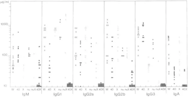
Serum levels of Ig classes and subclasses
The sera of wild-type, single and double mutant XID/nu and XID/CD40T mice at 10-12 weeks of age were analyzed for their content of IgM, lgG1, lgG2a, lgG2b and lgG3, and, except for the XID/nu mice, also for their IgA content by ELISA. The results are summarized in Fig. 5. As expected (37) XID single mutant mice had lower levels of IgM and lgG3, and CD40T mice had decreased levels of lgG1 and IgA; while the other classes and subclasses of Ig were found
Spleen
wt
XID
CD40T XID/CD40T
10
Q
O
% 0 f 26-8 v * " • 1 tit . * 60.0 . ' * 573 .F»K-- -;'••* 19.1 •ift" IS' rt* & I # • • • 39.1 2.3 • ii' if if i 5-» • 14.4 3.5 p"1 i v iS> i i i • iQ
2.5 • i 1 • I*1 • • . S ':' 3.5 it> il> I CO CMQ
O
1 i • i*1 60.2 \ 10.7 » • 33.7Q
O
56.9 7.1 V 1 * 25.9 • • • 3 r ' ' - > • • : • n.i 5.6•CD45R (B220)
7.6±
Fig. 2. FACS analysis of splenic lymphoid cells of wild-type (wt), single mutant CD40T mice, and XID mice and double mutant XID/CD40T
mice. Horizontal axes represent fluorescence intensities of CD45R(B220), while vertical axes represent those of CD5, IgM, IgD, CD23 and CD21.
in concentrations comparable to those of wild-type mice. The double mutant XID/nu mice had depressed levels of IgM, lgG2a and lgG3. Most notably the double mutant XID/CD40T had 5- to 10-fold lower levels of IgM, while in >90% of all mice the lgG1, lgG2a, lgG2b and lgG3 as well as IgA classes and subclasses were below the detection limit of the assay. In particular, lgG1, lgG2a and lgG3 subclasses were below the detection level in all mice tested.
We conclude that the double mutant XID/CD40T mice
show a severe immunodeficiency, manifested in lower or undetectable serum Ig levels.
Discussion
Single mutant XID, nu or CD40T mice show no severe deficiencies in the generation of precursor and immature B cells in the bone marrow, and in the case of CD40T and nu mice have normal numbers of mature B cells in bone marrow
1682 Reduction of B cells and serum Ig in double mutant XID/CD40T mice
Table 2. Total number of B cells in different subpopulationsa in the spleen of indicated wild-type, single mutant and double mutant mice Mouse strain Wild-typec Wild typed CD40T CD40T nu XID XID XID/nu XID/CD40T XID/CD40T Age (weeks) 4-6 10-12 4-6 10-12 4-6 4-6 10-12 4-6 4-6 10-12 Total5 41.2 : 36.7 : 45.8 : 34.1 : 45.1 : 11.0 : 23.3 : 9.0 : 10.9 : 10.2 : B220+ t 4.4 t 11.8 t 22.7 t 14.3 t 6.2 t 6.5 t 7.2 t 2.3 t 1.0 t 14.4 B220+/lgM+ 38.9 ± 5.3 32.1 ± 15.6 44.5 ± 20.2 35.7 ± 12.2 35.7 ± 5.5 8.8 ± 3.5 20.2 ± 6.0 4.5 ± 1.9 6.1 ± 0 . 9 8.7 ± 8.9 B220+/lgD+ 38.0 ± 5.2 30.7 ± 12.7 44.3 ± 23.1 34.6 ± 1 1 . 2 37.8 ± 6.2 8.1 ± 4.3 18.9 ± 5.5 3.8 ± 1.2 5.8 ± 1.1 8.5 ± 11.1 B220+/CD23+ 32.2 + 6.5 36.7 ± 23.1 18 ± 3.2 31.8 ± 17.2 ND 5.1 ± 1.5 15.4 ± 7.0 ND 2.4 ± 0.9 5.9 ± 6.3 B220+/CD21 + 35.9 ± 5.5 ND 40.2 ± 20.6 ND ND 4.6 ± 0.4 ND ND 3.6 ± 0.3 ND
aTotal splenic cells prepared and analyzed by FACS as described in Methods and in the legend of Table 1.
Calculated numbers represent total number of spleen cells (X106). ND, denotes not determined. cSpleen of the same animals (4-6 weeks old) analyzed for bone marrow B cells in Table 1. dSpleen of four to 10 individual animals 10-12 weeks old.
and the periphery. In the case of XID and CD40T mice, the peripheral, mature B cells are not reactive to Tl-ll antigens, respectively TD antigens, but respond normally to TD, respect-ively Tl-ll antigens. It is not clear how the peripheral B cell repertoire of short-lived and long-lived mature, antigen reactive cells is established. If it was established in ways comparable to the establishment of the peripheral T cell repertoire one might expect positive selection of immature cells through signaling via Ig which could mediate a transition without division into the mature B cell pool (reviewed in 38,39). The normal establishment of a peripheral B cell compartment in the single mutant CD40T and nu mice suggests that such antigenic selection of the B cell repertoire could occur without T cells and the interaction of CD40-CD40 ligand, while the lower number of mature B cells, of which a large fraction is short lived, in XID mice indicates the involvement of btk signaling for the generation of long-lived B cells.
Double mutant XID/nu and XID/CD40T mice, again, show no severe deficiencies in the generation of normal numbers of precursor and immature B cells in bone marrow but, in contrast to the single mutant mice, have severe deficiencies in the mature, peripheral B cell compartments, which in the case of XID/CD40T mice are very severe. Wortis and colleagues (15) previously described a severe B cell defect also in bone marrow of XID/nu mice, which is at variance with the results described herein. One possible explanation is the difference in nu/nu strains used for breeding, where we used C57BI/6 and they used C3H nudes.
We find that double mutant XID/CD40T mice have decreased numbers of mature B cells, almost three-quarters of them with a high turnover, and hence show a deficiency in the generation of long-lived B cells. They have depressed levels of IgM in the serum, and lack detectable levels of lgG1, lgG2a, lgG2b, lgG3 and IgA. Furthermore, their B cells cannot be efficiently stimulated in vivo by TD antigens or in vitro by a variety of mitogenic stimuli. However, they show responses to TNP-LPS in vivo, although in vitro responses to LPS are deficient. It, therefore, appears that B cells are made in normal numbers in the bone marrow, arrive in the periphery but cannot be rendered long lived, nor reactive to TD or Tl-ll
Table 3. Percentage of BrdU-labeled peripheral blood B cells
in wild-type, single and double mutant mice Mouse strain Wild-type CD40T nu XID XID/nu XID/CD40T BrdU-labeled cells (%) CD45R(B220)+ cells
After 6 days labeling 10.3 ± 0.4 8.8 ± 0.5 7.0 ± 2.0 38.0 ± 1.7 49.0 ± 3.7 71.5 ± 3.0 of total
After 7 days chase 1.0 ± 0.5 2.0 ± 0.5 2.0 ± 1.2 6.2 ± 6.1 5.2 ± 1.7 4.8 ± 2.0
antigens. The in vivo responses to the Tl TNP-LPS might be explained if we consider that CBA/N mice have been found to react to LPS with a 100-fold lower frequency of peripheral B cells in spleen, but still with a 100-fold higher frequency than splenic B cells of the LPS non-reactive strain C3H/Hej (8). In vivo this low but measurable number of reactive B cells may be sufficient to elicit an antigen-specific response triggered in association with the mitogen, such as TNP-LPS. This may also serve as an explanation for the finding here, that approximately half of the double mutant XID/CD40T mice have IgM levels >10 ^g/ml and in some cases at almost normal levels, approaching 100 ng/ml (Fig. 5).
The number of peripheral, mature B cells differs by factors of 5-10 in individual XID/CD40T mice. Such fluctuations in the number of peripheral B cells might either be due to a difference in the genetic background of the mice or have epigenetic causes. It should be remembered that the XID/ CD40T double mutant mice were generated by mating CBA/N with CD40T mice, the latter being a mix of the strains C57BL/6 and 129. Hence, three different genotypes are mixed in various proportions in individual XID/CD40T mice. It remains to be investigated how such different genetic backgrounds could influence the number of short-lived and long-lived peripheral B cells.
D I9M antl-KLH • IgG, antl-KLH wt XID CD40T XID/ C040T CD40T XID/CD40T Q IgM anti-TNP • lgG1 anti-TNP 0 l g G2 a anti-TNP S lgG3 anti-TNP XID CD40T XID/
Fig. 3. (a) Antibody responses to KLH of wild-type, single mutant
CD40T and XID, and double mutant XID/CD40T mice. Mice were immunized with KLH in complete Freunds adjuvant (50 ng/mouse, i.p.) and the KLH-specific serum antibody titers of IgM (open bars) and lgG1 (filled bars) were determined on day 14 after immunization by a KLH-specific ELISA (see Methods). Units represent arbitrary values based on the titration curve of a standard immune serum according to Foy et al. (19). The means of titers from three mice per group are shown, (b) Single mutant XID and double mutant XID/ CD40T mice were immunized with TNP-LPS (50 u.g/mouse, i.p.). On day 14, mice were bled and the anti-TNP antibody titer of IgM (open bars), lgG1 (filled bars), lgG2a (light shaded bars) and lgG3 (dark shaded bars) type were determined by ELISA as described in Methods. Units represent arbitrary values based on the titration curve of a standard immune serum. The responses in wild-type and CD40T mice were similar to those of the XID and XID/CD40T [ND; data not shown, but given elsewhere (21)].
Anti-lg Sepharose® * IL-2
XID/CD40T
0 2 4 6
3H-TdR uptake on day 2 of culture
(com y 10'4 cer 2 y 105 cu'tu-od cells)
Fig. 4. Proliferative responses of wild-type, single mutant CD40T and
XID, and double mutant XID/CD40T mice stimulated by various B cell mitogens (as described in Methods) in the absence (a) and presence (b) of cytokines. In parallel experiments the frequency of LPS-reactive cells was determined in limiting dilution analyses using rat thymus filler cells (36). The frequency of LPS-reactive cells developing into clones of IgM-secreting cells were 1:12 for wt, 1:3 for CD40T, 1:100 for XID and < 1:1000 for XID/CD40T mice.
On the other hand, it is also possible that epigenetic influences, such as antigenic stimulation, could influence the level of short- and long-lived B cells in the periphery. This could either occur at the mature, resting B cell stage in the periphery or at the transition from immature to mature B cells in the bone marrow. If it were antigenic stimulation, it would have to be of a type which mediates its effects by ways different from those used by Tl-ll or TD antigens used in our experiments. In fact, a Tl-I antigen might be a good candidate for such B cell stimulation. Influence of antigen on the development of peripheral B cells could also occur at an earlier stage. Recent experiments with Ig transgenic mice on a rearrangement-deficient (RAG-1T and RAG-2T) genetic background have shown that B cell development is arrested at the stage of an immature, slg+ B cell in the bone marrow, provided that the transgenic Ig recognizes an autoantigen expressed in bone marrow (40,41). In the case of the TNP-specific Sp6 Ig transgene, arrest is likely to be mediated by double-stranded DNA present in bone marrow. It was found that injection of TNP-Ficoll, a Tl-ll antigen cross-reactive with
DNA, relieved this arrest of differentiation, so that B cells appeared in the periphery and could be stimulated to a response, with the development of transgenic Ig-secreting plasma cells (41).
The severely impaired establishment of the peripheral, mature B cell compartment(s) in the double mutant XID/ CD40T mice suggests that CD40-dependent and W/c-depend-ent ways of selection and/or stimulation of immature and/or mature B cells could control the generation and maintenance of normally-sized mature B cell pools. The fact that XID/nu mice, although deficient in the generation of mature B cells, can generate a peripheral B cell pool of which a considerably larger part is long-lived compared to that of XID/CD40T mice and the finding that slg+ B cells do accumulate with age in the periphery of XID, but not of XID/CD40T mice might indicate that extrathymic components such as y5 TCR-expressing T cells might directly or indirectly contribute to this build up of a peripheral B cell pool. The btk gene and the CD40
ligand-1684 Reduction of B cells and serum Ig in double mutant XID/CD40T mice 1000_ 100 _ IgGi
lgG2a
lgG2b lgG3 40 X 40X IgAFig. 5. Serum Ig levels of wild-type, single mutant CD40T and XID, and double mutant XID/CD40T and XID/nu mice. The serum levels of IgM,
lgG1, lgG2a, lgG2b and lgG3 of individual wild-type (denoted W and crosses), CD40T (denoted 40 and filled circles); XID (denoted X and open circles), nu (denoted nu and filled diamonds), XID/nu (denoted nuX and open diamonds) and XID/CD40T (denoted 40X and filled squares) mice are given. Values <10 ng/ml are placed at the bottom. Serum levels of IgA were not determined in nu and XID/nu mice. Wild-type mice represent values of serum Ig levels in C57BL/6 mice and in mice of other genetic backgrounds which were pooled in this figure, since they did not significantly differ from each other.
CD40 interaction between helper T cells and B cells appear to control all of the detectable mature, long-lived B cell pools, and appear to do so in parallel and not in tandem. It remains remarkable that the toWc-mutation alone is relatively mild (XID) in mice, but severe (XLA) in man. This might suggest that man generate peripheral B cell pools, much more so than mice, in a £>f/<-dependent rather than in a CD40-dependent way.
Acknowledgements
The able technical assistance of Ms S. Crivelli and Ms N. Staube is gratefully acknowledged. We also thank Ekaterina Platoshkina for helping in the typing of the different mouse strains. We thank Dr P. Sideras and Dr T. Winkler for critical review of this manuscript and Ms. C. Geiger for preparation of the manuscript. H. K. and T. K. are supported by grants from The Ministry of Education, Science and Culture, Japan, and J. A. is partly supported by The Swedish Medical Research Council. The Basel Institute for Immunology was founded and is supported by F. Hoffmann-La Roche Ltd, Basel, Switzerland.
Abbreviations Btk KLH PE LPS XID XLA
Bruton's tyrosine kinase keyhole limpet hemacyanin phycoerythrin
lipopolysaccharide X-linked immunodeficiency X-linked agammaglobulinemia
References
1 Bruton, O. C. 1952. Agammaglobulinemia. Pediatrics 9:722. 2 Scher, I. 1982. CBA/N immune defective mice: evidence for the
failure of a B cell subpopulation to be expressed. Immunol. Rev. 64:117.
3 Tsukada, S., Rawlings, D. J. and Witte, O. N. 1994. Role of Bruton's tyrosine kinase in immunodeficiency. Curr. Biol. 6:623. 4 Tsukada, S., Saffran, D. C , Rawlings, D. J., Parolini, O., Allen,
R. C, Klisak, I., Sparkes, R. S., Kubagawa, H., Mohandas, T., Quan, S., Belmont, J., Cooper, M., Conley, M. and Witte, O. N. 1993. Deficient expression of a B cell cytoplasmic tyrosine kinase in human X-linked agammaglobulinemia. Cell 72:279.
5 Vetrie, D., Vorechovsky' , I., Sideras, P., Holland, J., Davies, A., Flinter, F., Hammarstrom, L., Kinnon, C , Levinsky, R., Bobrow, M., Smith, C. I. E. and Bently, D. R. 1993. The gene involved in X-linked agammaglobulinaemia is a member of the src family of protein-tyrosine kinases. Nature 361:226.
6 Rawlings, D. J., Saffran, D. C, Tsukada, S., Largaespada, D. A., Grimaldi, J. C , Cohen, L, Mohr, R. N., Bazan, J. R, Howard, M., Copeland, N. G., Jenkins, N. A. and Witte, O. N. 1993. Mutation of the amino-terminal unique region of Bruton's tyrosine kinase in murine X-linked immunodeficiency. Science 261:358.
7 Thomas, J. D., Sideras, P., Smith, C. I. E., Vorechovsky, I., Chapman, V. and Paul, W. E. 1993. Colocalization of X-linked agammaglobulinemia and X-linked immuno-deficiency genes. Science 261:355.
8 Huber, B. and Melchers, F. 1979. Frequencies of mitogen-reactive B cells in the mouse. Lipopolysaccharide-, lipoprotein- and Nocardia mitogen-reactive B cells in CBA/N mice. Eur. J. Immunol. 9:827.
9 Wicker, L. S. and Scher, I. 1986. X-linked immune deficiency (xid) of CBA/N mice. Curr. Top. Microbiol. Immunol. 124:87.
10 Hitoshi, Y, Sonoda, E., Kikuchi, Y., Yonehara, S., Nakauchi, H. and Takatsu, K. 1993. IL-5 receptor positive B cells, but not eosinophils, are functionally and numerically influenced in mice carrying the X-linked immune defect. Int. Immunol. 5:1183. 11 Go, N. F., Castle, B. E., Barret, R., Kastelein, R., Dang, W.,
Mosmann, T. R., Moore, K. W. and Howard, M. 1990. Interleukin 10, a novel B cell stimulatory factor: Unresponsiveness of X chromosome-linked immunodeficiency B cells. J. Exp. Med.
172:1625.
12 Sprent, J. and Bruce, J. 1984. Physiology of B cells in mice with x-linked immunodeficiency. II. Influence of the thymus and mature T cells on B cell differentiation. J. Exp. Med. 160:335.
13 Karagogeos, D., Rosenberg, N. and Wortis, H. H. 1986. Early arrest of B cell development in nude, X-linked immune-deficient mice. Eur. J. Immunol. 16:1125.
14 Mond, J. J., Scher, I., Cossman, J., Kessler, S., Mongini, P. K. A., Hansen, C , Finkelman, F. D. and Paul, W. E. 1982. Role of the thymus in directing the development of a subset of B lymphocytes. J. Exp. Med. 155:924.
15 Wortis, H. H., Burklu, L, Hughes, D., Roschelle, S. and Wanek, G. 1982. Lack of mature B cells in nude mice with X-linked immune deficiency. J. Exp. Med. 155:903.
16 Karagogeos, D. and Wortis, H. H. 1987. Thymus grafts induce B cell development in nude, X-linked immune deficient mice. Eur. J. Immunol. 17:141.
17 Stamenkovic, I., Clark, E. A. and Seed, B. 1989. A B-lymphocyte activation molecule related to the nerve growth factor receptor and induced by cytokines in carcinomas. EMBO J. 8:1403. 18 Lane, P., Brocker, T., Hubele, S., Padovan, E., Lanzavecchia, A.
and McConnell, F. 1993. Soluble CD40 ligand can replace the normal T derived CD40 ligand signal to B cells in T cell-dependent activation. J. Exp. Med. 177:1209.
19 Foy, T. M., Laman, J. D., Ledbetter, J. A., Aruffo, A., Claassen, E. and Noelle, R. J. 1994. gp39-CD40 interactions are essential for germinal center formation and the development of B cell memory. J. Exp. Med. 180:157.
20 Gray, D., Dullforce, P. and Jainandusing, S. 1994. Memory B cell development but not germinal center formation is impaired by in vivo blockade of CD40-CD40 ligand interaction. J. Exp. Med. 180:141.
21 Kawabe, T., Naka, T., Yoshida, K., Tanaka, T., Fujiwara, H., Suematsu, S., Yoshida, N., Kishimoto, T. and Kikutani, H. 1994. The immune responses in CD40-deficient mice: impaired immunoglobulin class switching and germinal center formation. Immunity 1:167.
22 Xu, J.-C, Foy, T. M., Laman, J. D., Elliott, E. A., Dunn, J. J., Waldschmidt, T. J., Elsemore, J., Noelle, R. J. and Flavell, R. A.
1994. Mice deficient for the CD40 ligand. Immunity 1:423. 23 Renshaw, B. R., Fanslow, W. C , III, Armitage, R. J., Campbell,
K. A., Liggitt, D., Wright, B., Davison, B. L. and Maliszewski, C. R. 1994. Humoral immune responses in CD40 ligand-deficient mice. J. Exp. Med. 180:1889.
24 Durie, F. H., Foy, T. M., Masters, S. R., Laman, J. D. and Noelle, R. J. 1994. The role of CD40 in the regulation of humoral and cell-mediated immunity. Immunol. Today 15:406.
25 Ogawa, M., Matzusaki, Y, Nishikawa, S., Hayashi, S. I., Kunisada, T., Sudo, T., Kina, T, Nakauchi, H. and Nishikawa, S. I. 1991. Expression and function of c-kit in hemopoietic progenitor cells. J. Exp. Med. 174:63.
26 Leptin, M. 1985. Monoclonal antibodies specific for murine IgM. II. Activation of B lymphocytes by monoclonal antibodies specific for the four constant domains of IgM. Eur. J. Immunol. 15:131. 27 Parkhouse, R. M. E., Preece, G., Sutton, R., Cordell, J. L. and
Mason, D. Y. 1992. Relative expression of surface IgM, IgD and the Ig-associating a(mb-1) and P(B-29) polypeptide chains. Immunology 76:535.
28 Kinoshita, 1 , Takeda, J., Hong, K., Kozono, H., Sakai, H. and Inoue, K. 1988. Monoclonal antibodies to mouse complement receptor type 1 (CR1). Their use in a distribution study showing that mouse erythrocytes and platelets are CR1-negative. J. Immunol. 140:3066.
29 Rolink, A. G., Kudo, A., Karasuyama, H., Kikuchi, Y. and Melchers, F. 1991. Long-term proliferating early pre B cell lines and clones with the potential to develop to surface Ig-positive, mitogen reactive B cells in vitro and in vivo. EMBO J. 10:327.
30 Winkler, T. H., Rolink, A. G., Melchers, F. and Karasuyama, H. 1995. Precursor B cells of mouse bone marrow express two different complexes with the surrogate light chain on the surface. Eur. J. Immunol. 25:446.
31 Oka, Y., Rolink, A. G., Suematsu, S., Kishimoto, T. and Melchers, F. 1995. An interleukin-6 transgene expressed in B lymphocyte lineage cells overcomes the T cell-dependent establishment of normal levels of switched immunoglobulin isotypes. Eur. J. Immunol. 25:1332.
32 Reininger, L., Radaszkiewicz, T., Kosco, M., Melchers, F. and Rolink, A. G. 1992. Development of autoimmune disease in scid mice populated with long-term in vitro proliferating (NZBxNZW) F, pre-B cells. J. Exp. Med. 176:1343.
33 Hasbold, J., Johnson-Leger, C , Atkins, C. J., Clark, E. A. and Klaus, G. G. 1994. Properties of mouse CD40: cellular distribution of CD40 and B cell activation by monoclonal anti-mouse CD40 antibodies. Eur J Immunol 24:1835.
34 Karasuyama, H. and Melchers, F. 1988. Establishment of mouse cell lines which constitutively secrete large quantities of interleukin 2, 3, 4 or 5, using a modified cDNA expression vectors. Eur. J. Immunol. 18:97.
35 Forster, I., Vieira, P. and Rajewsky, K. 1989. Flow cytometric analysis of cell proliferation dynamics in the B cell compartment of the mouse. Int. Immunol. I:32I.
36 Andersson, J., Coutinho, A., Lernhardt, W. and Melchers, F. 1977. Clonal growth and maturation to immunoglobulin secretion in vitro of every growth-inducible B lymphocyte. Cell 10:27.
37 Perlmutter, R. M., Nahm, M., Stein, K. E., Slack, J., Zitron, I., Paul, W. E. and Davie, J. M. 1979. Immunoglobulin in subclass-specific immunodeficiency in mice with an X-linked B-lymphocyte defect. J. Exp. Med. 149:993.
38 von Boehmer, H. 1994. Positive selection of lymphocytes. Cell 76:219.
39 Melchers, F., Rolink, A., Grawunder, U., Winkler, T. H., Karasuyama, H., Ghia, P. and Andersson, J. 1995. Positive and negative selection events during B lymphopoiesis. Curr. Opin. Immunol. 7:214.
40 Spanopoulou, E., Roman, C. A. J., Corcoran, L. M., Schlissel, M. S., Silver, D. P., Nemazee, D., Nussenzweig, M. C , Shinton, S. A., Hardy, R. R. and Baltimore, D. 1994. Functional immunoglobulin transgenes guide ordered B-cell differentiation in RAG-1-deficient mice. Genes Dev. 8:1030.
41 Andersson, J., Melchers, F. and Rolink, A. 1995. Stimulation by T cell independent antigens can relieve the arrest of differentiation of immature auto-reactive B cells in the bone marrow. Scand. J. Immunol. 42:21.