1
β-Cyclodextrin PAMAM dendrimer: how to overcome the tumbling
process for getting fully available host cavities
Israel González-Méndez,
[a]Aurélien Hameau,
[b,c]Régis Laurent,
[b,c]Christian Bijani,
[b,c]Valérie
Bourdon,
[d]Anne-Marie Caminade,
[b,c]Ernesto Rivera,
[a]* and Kathleen I. Moineau-Chane Ching
[b,c]*
[a] Instituto de Investigaciones en Materiales, Universidad Nacional Autónoma de México, Circuito Exterior Ciudad Universitaria C.P. 04510, Ciudad deMéxico, México.
E-mail : [email protected] ; http://www.iim.unam.mx/erivera/
[b] CNRS; LCC (Laboratoire de Chimie de Coordination); 205 route de Narbonne, BP 44099, F-31077 Toulouse, Cedex 4, France E-mail : [email protected] ; https://www.lcc-toulouse.fr/auteur744.html
[c] LCC-CNRS, Université de Toulouse, CNRS, Toulouse, France
[d] ICT – Service de spectrométrie de masse – Université Paul Sabatier, 118, Route de Narbonne, 31062 Toulouse Cedex 9, France Supporting information for this article is given via a link at the end of the document.
Keywords: β-cyclodextrin • Click Chemistry • PAMAM • dendrimer • tumbling process
Abstract: The synthesis of a G0 polyamidoamide (PAMAM) dendrimer end-capped with four β-cyclodextrin (βCD) units, named as Tetra-CD was performed by using copper(I)-catalyzed alkyne-azide cycloaddition (CuAAC). This new platform presents a considerable high water-solubility in comparison to native βCD. Tumbling process involving half of the CD cavities in D2O was
evidenced by 1H NMR spectroscopy. Interaction of adamantane with
Tetra-CD has been fully investigated by 1D/2D NMR techniques and mass spectrometry besides a joint study on new βCD monomers (named as A and B) and on a dimer (Di-CD) and a trimer (Tri-CD). The obtained results demonstrate that the tumbling effect can be shifted in the presence of an appropriate hydrophobic guest resulting in the full host-ability of the CD units in water for that guest.
Introduction
Cyclodextrins (CDs) are a series of natural cyclic
oligosaccharides comprising 6, 7 or 8 D-(+)-glucopyranose units linked by α-1,4linkages, named as α-, β- or γ-CD, respectively,[1]
represented as a truncated cones with a hydrophilic exteriorand a hydrophobic inner cavity. Their water-solubility is not correlated with their size but with the ability of their hydroxyl groups to form hydrogen bonds with water. It can be improved by eventual chemical modifications on these OH groups situated on the edges of the conical cylinder. Thanks to its size, hydrophobicity and hydrogen bonding of its inner cavity, βCD can accommodate diverse guest molecules. Its low cost, water-solubility and biocompatible properties[2] make it a molecule of choice for the
construction of supramolecular systems.[3] The interaction of CD
with non-water-soluble molecules operates via a dynamic equilibrium process of host-guest complexation, following which the guest molecules continuously associate and dissociate from the CD cavity. Due to its inner cavity diameter of around 6.5 Å, βCD has been found to be particularly attractive for preparation of drugs delivery systems.[4]
Chemical modification of native βCDs have been performed via reactions on the primary or secondary hydroxyl groups,[5] leading
to decorated βCDs, formation of βCD polymers,[6] or generation
of CD nanosponges.[7] Among the used synthetic techniques, the
copper(I)-catalysed alkyne-azide cycloaddition (CuAAC) “click” reaction[8] has emerged as one of the most valuable synthetic
tools in the development of a wide variety of CD derivatives for biological applications in various fields, due to its simplicity, efficiency, and robustness properties.[9] CuAAC is used for
cycloaddition of the mono azido cyclodextrin (βCD-N3), obtained
from the mono-(O-6)-tosylated βCD,[10] on the appropriate
alkyne.[11] In that way, a hydrophobic moiety can be grafted on
one primary hydroxyl function of βCD for obtaining CD monomers[12] or more complex assemblies[13] with modulated
inclusion and transport properties. Besides, the use of hydrophilic dendritic structures has been proved to be a suitable strategy for obtaining highly soluble active ingredients in water.[14]
In the present work, we used this strategy for designing a new platform, named as Tetra-CD, based on a central G0 PAMAM dendrimer (IV) substituted by 4 βCD units via spacers formed by CuAAC. The presence of 4 peripheral βCD units confers to this compound a high potential of guest accommodation. The PAMAM core was chosen because it could contribute to great solubility in water for Tetra-CD, without being a competitive encapsulating site contrarily to what was observed with higher generations of
PAMAM.[15] We used 2D ROESY NMR experiments to fully
characterize the pristine platform and its inclusion complexes formed with adamantane sodium carboxylate (AdCOONa) and the poorly water-soluble adamantane carboxylic acid (AdCOOH). Adamantane was selected as a model guest molecule because it presents a high association constant with βCD in water, of around 5.104 M-1 at room temperature.[16] Besides, diverse
molecules that are too voluminous to fit the βCD cavity can be functionalized by an adamantyl moiety, offering them the possibility to be connected to inclusion complexes.[17] Interaction
of adamantyl unit with Tetra-CD was discussed in light of results obtained with previously reported trimer and dimer,[18] named here
as Tri-CD and Di-CD, respectively, as well as with new mono-CDs called A and B, used as assessment systems. The CD substituents of A and B are of similar nature to that of the spacers used for Tri-CD and Di-CD, and for Tetra-CD, respectively (see scheme 1). In that way, comparisons were made independently of the nature of the spacer.
Results and Discussion
Synthesis and characterization of βCD-derivatives
The synthetic routes used for the synthesis of the βCD derivatives are summarized in Scheme 1. For the construction of
2
Tetra-CD, the tetra-alkyne precursor (III) was preparedvia amide bond formation between the amino groups of PAMAM G0 and carboxylic functions of II, using a mixture of EDC·HCl/HOBt as coupling reagents.[19] The target molecules Tetra-CD, A and Bwere obtained using CuAAC in mild conditions with Cu(I) as catalyst. Catalytic Cu(I) species can be generated from Cu(0) as the source,[20] but also in situ from a mixture of CuSO
4 and sodium
ascorbate.[21] This last method has been preferred because it
excludes the formation of any by-products such as diacetylene and may be even applied in aqueous systems in presence of oxygen. Each compound was isolated in high yield (90 to 98%) after purification by size exclusion chromatography (SEC). Elemental analyses confirmed the high degree of purity for the obtained compounds, except for an extra-amount of water, namely about 6 water molecules per βCD unit for Tetra-CD, and up to 10 for A and B. All compounds were characterized by electrospray ionisation time-of-flight mass spectrometry (ESI- TOF-MS).
Scheme 1. Top: synthetic routes to the target molecules. a. EDC·HCl/HOBt/DMF, b. βCD-N3, CuSO4: sodium ascorbate 1:3 / DMSO:H2O
5:2 ; Bottom: structures of Di-CD and Tri-CD.
The measured masses of the synthesized compounds were found to be in excellent agreement with the calculated ones (see “Experimental Section” part and spectra in ESI). The characterization of the new βCD derivatives by NMR techniques (1H, 13C, HMQC, COSY, NOESY) were carried out in detail in
DMSO-d6 (see text pp. SI-6,7 and corresponding figures). It
resulted that a singularity was detected for some protons of the functionalized glucopyranose sub-unit of the βCD cavity (see Scheme 2): protons H-1', H-5' and H-6' appeared at lower field than their respective correspondents H-1, H-5 and H-6 of non-functionalized sub-units (Figure 1 top). This deshielding effect was particularly marked for H-6’ protons. There was also a strong impact of the substitution on one -CH2-OH fragment of the primary
hydroxyl rim, since the signals of protons named as H-6" (two diastereotopic protons resonating at around 3.15 and 2.92 ppm) and OH-6" (around 4.30 ppm) appeared to be significantly up-shielded in comparison to their respective analogues H-6 and OH-6. Those protons were attributed to a glucopyranose sub-unit contiguous to the substituted one, their up-shielding might result from interaction with the vicinious branch of the molecule. These observations were common to the 3 new compounds for which the CD cavities behave similarly.
Scheme 2. Representations of the CD cavity with atoms numbering (H-3 and H-5 are inner protons, H-2 and H-4 are outer protons).
Behaviour of β-CD derivatives in water
The new molecules were then investigated in water, which is the medium of choice for many applications or studies of CD derivatives.[4,22] It is worth to notice that the isolated compounds
A and B present good solubilities in water, of 37.5 mg/mL and 97.4 mg/mL, respectively, in comparison to 18.3 mg/mL for β-CD. Moreover, Tetra-CD exhibits remarkable high solubility in water (> 2580 mg/mL), which is of about 140 fold higher than that of β-CD.
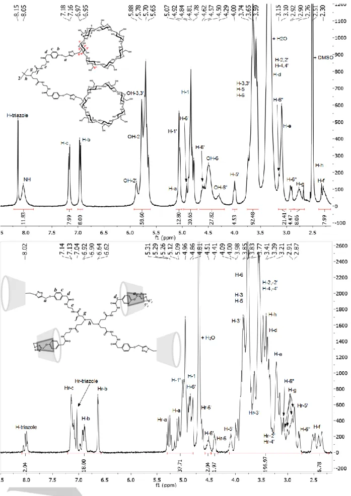
The 1H NMR spectrum of the compound Tetra-CD in D
2O showed
a drastic change in the aromatic region, as compared to that obtained from DMSO-d6 solution (Figure 1): the integration of the
protons signal at around 8 ppm, the region where the triazole protons were identified in DMSO, matched only with 2 out of the 20 protons resonating in the 6.5 to 8.2 ppm region. This resulted from the previously observed tumbling process for CD tetramers and dimers in water,[23-25] which affected the triazole protons that
now appeared as a multiplet at around 8 ppm (assigned to H-triazole of branches wearing a non-reversed CD cavity) and also as a singlet centred on 7.04 ppm (assigned to the so-called
3
Figure 1. 1H NMR spectrum of compound Tetra-CD. Top: in DMSO-d6 with schematic representation of the molecule giving the atoms numbering for CD cavity.
4
Hr-triazole of branches wearing a reversed CD cavity). The complexity of the signal at around 8 ppm was explained by the possibility of 7 conformations for compound Tetra-CD in water as sketched below.
Scheme 3. The 7 possible forms for Tetra-CD with 0 to 4 reversed cavities
The phenyl protons were also disturbed since they did not appear anymore as two doublets but as i) a first massif between 7.05 and 7.2 ppm originating from H-c and Hr-c aromatic protons of branches wearing a non-reversed and a reversed CD cavity, respectively, ii) asecond massif between 6.8 and 7.0 ppm due to the H-b aromatic protons of branches wearing a non-reversed CD, and iii) a doublet (J = 7.3 Hz) at 6.63 ppm due to the Hr-b aromatic protons of branches wearing a reversed CD cavity. Integration of the signals indicated that, on average, half of the CD cavities underwent tumbling. Besides these changes observed in the aromatic region when going from DMSO to water, the rest of the spectrum was also affected by the tumbling effect. Assignments of the protons signals were made on the basis of the analysis previously performed in DMSO-d6 (see SI), with the help of 1D
and 2D NMR experiments in D2O. Firstly, the 2D COSY spectrum
(see Figure SI-25) gave the confirmation for chemical shifts values of aromatic protons and allowed also to assign some other
CD proton signals. For example, the non-equivalent
diastereotopic protons H-6' of the modified glucopyranose ring of β-CD appeared in water as two sets of two signals, one set originating from the non-inverted cavity (H-6’ centred at around 4.9 and 4.6 ppm, respectively), the other from the reversed form (Hr-6’ centred at around 4.7 and partially hidden by the water peak, and 4.4 ppm, respectively). The tumbling of the CD cavity resulted in a blending effect for those protons, as it was the case for the Hr-5’ protons, detected at around 2.9 ppm. Secondly, the HSQC and HMBC spectra (see Figures SI-26 and -27) allowed us to identify most of the protons and carbon signals and also to raise some ambiguities. For example, the H-a protons appeared as two multiplets centred at around 5.3 and 5.1 ppm, respectively, correlated with two carbon signals at 62.4 and 61.0 ppm, respectively. The comparison with the data for C-a/H-a in
DMSO-d6 led us to assign the 5.1/61.0 and 5.3/62.4 ppm correlations to
H-a/C-aand Hr-a/Cr-a, respectively. The C-d signal was identified at around 41.7 ppm through HMBC with aromatic H-c protons, thus H-d protons were identified at around 3.4 ppm. Thirdly, 1D TOCSY experiments (see Figures SI-28 to -30) gave rise to the chemical shifts values of the H-f and H-g protons of the branches and to those of the protons of the substituted glucopyranose units
for reversed and non-reversed CD cavities, as well as to the precise value of the chemical shift of Hr-c, found to be 7.15 ppm. Lastly, 2D ROESY experiment (see Figure SI-31) highlighted specific interactions of Hr-a, Hr-b, Hr-c and Hr-triazole protons with the inner protons of the reversed CD cavity. This fine analysis work led us to propose detailed assignments of the resonance domains of the CD protons (see Figure 1 and Figure SI-32). The
13C NMR spectrum in D
2O of Tetra-CD evidenced also some
changes in comparison to the situation in DMSO-d6 since the
carbon signals are splitted in water as the result of tumbling. This is particularly apparent for the phenyl and triazolyl carbon signals, which are particularly sensitive to CD tumbling effect (Figure SI-33).
The situation was simpler for compounds A and B for whose no tumbling was detected in D2O. Their respective 1H and 13C NMR
spectra in D2O (see Figures SI-34 and 35) exhibited the same
features as in DMSO-d6. Nevertheless, their 2D NOESY spectra
(see Figure SI-36) revealed correlations between phenyl protons and inner protons of CD cavities, indicating interaction between the extremity of the arm of molecule and the CD cavity, as
sketched in Figure SI-36, leading to supramolecular
assemblies.[26,27]
Interaction with adamantane carboxylic acid and its sodium salt
According to literature, head-to-tail assemblies similar to those observed with compounds A and B can be altered by the addition of AdCOOH that helps in releasing the CD cavity.[26] Besides, the
tumbling observed for Tetra-CD could result in an inclusion ability of an average of two molecules of guest per Tetra-CD platform, thus limited accessibility to the CD cavities,[24,25,28] which is of
major importance for considering its potential applications in aqueous medium. Due to the high affinity of adamantane for βCD,[16,29] the adamantyl unit (Ad) could be convenient to
overcome the tumbling of the CD cavity of Tetra-CD in water. To clarify this point, we have examined changes in 1H NMR
spectra recorded in D2O with increasing amounts of AdCOONa
relative to Tetra-CD. The results were numbered using the molar fraction XCD = [β-CD]/([β-CD]+[AdCOONa]), for simplicity reasons
(see Figure 2). For a 0.9 XCD molar fraction, the spectrum shape
was similar to that obtained for pure Tetra-CD in D2O, unless a
slight increase in integration for H-triazole protons (centred on 8 ppm) relatively to the total 20 protons resonating from 6.5 to 8.2 ppm. As XCD decreased, the spectrum shape became simpler with
the decrease of the multiplicity for the triazole and H-b protons, concomitant with the progressive disappearance of the Hr-b signal. As soon as XCD reached 0.5, the signal of triazole proton
looked as a singlet at 7.96 ppm, and that of phenyl protons as two doublets (J= 8.8 Hz) at 7.1 and 6.9 ppm, respectively, in a similar
way to what was observed for pure Tetra-CD in DMSO-d6. This
was interpreted as the total disappearance of reversed form of cyclodextrin.
Analyses of those mixtures by 2D ROESY NMR spectroscopy revealed also drastic changes upon addition of AdCOONa. The reference ROESY spectrum of pristine Tetra-CD in D2O, given in
Figure SI-37, displayed interactions of Hr-triazole, Hr-a, Hr-b, and Hr-c, with inner protons of reversed CD cavity. Conversely, when XCD was set at 0.1, i.e. in the presence of a large excess of
AdCOONa, only cross-peaks between the protons of CD cavity (H-3,3’; H5,5’) and those of AdCOONa (H-α; H-β; H-γ) were observed, which indicates that the CD cavities hosted only
5
AdCOONa molecules (see Figure SI-38). The absence of NOE effect involving triazole and aromatic protons suggests that no CD cavity remained in reversed conformation. Intermediate cases for which XCD ranged from 0.8 to 0.4 are shown in Figures SI-39 to42. For XCD = 0.8, the internal protons of the CD cavities displayed
NOE with triazole, aromatic and Hr-a protons, as well as with adamantyl protons: the interaction with inner CD protons occurred for both inverted and non-inverted CD rings. For a molar fraction of 0.6, inclusion occurrence was more pronounced, with intense correlation peaks in the 1.5-2 ppm region, in comparison to what was observed in the aromatic region. Correlation with Hr-a protons was no more seen. As XCD reached 0.5, correlations
involving inner CD protons were exclusively seen with adamantyl H-α, H-β, and H-γ protons; the same observation was made for
XCD = 0.4 (Figure SI-40). The use of AdCOOH as the guest led
to same observations (see Figure SI-43). The apparent diffusion coefficients of adamantane carboxylate and adamantane carboxylic acid in the examined mixtureswere evaluated through
1H DOSY NMR experiments. The results, gathered in Table 1,
indicated that as long as adamantyl unit was in default with respect to cyclodextrin, i.e. until a XCD value of 0.5, its diffusion
coefficient (D) value was low and rather constant, due to its inclusion into the CD cavity. Upon decreasing XCD, a progressive
increase in D value was observed, due to increasing amounts of free adamantane species in solution, until a maximum corresponding to these pure compounds. To summarize, beyond an equimolar ratio of adamantyl unit and cyclodextrin, no tumbling was detected by NMR spectroscopy and an increase in apparent value of D was measured. This could be indicative of formation of a [1:4] [Tetra-CD:Ad] complex.
Figure 2. Evolution from top to bottom of 1H NMR spectra in the 6.5-8.3 ppm
region of mixtures of Tetra-CD and AdCOONa in D2O with decreasing molar
fraction, expressed with respect to the concentration in β-CD cavities (XCD = [β-CD]/([β-CD]+[AdCOONa]). Italic: protons integration over the red
segments domains. Total concentration [β-CD]+[AdCOONa] = 8 mmol.L-1.
For evaluating the complex stoichiometry with adamantane carboxylate salt, Job’s plot analyses were performed on compounds A and Tetra-CD. The cases of the so-called Di-CD and Tri-CD were also examined. These last molecules were chosen as comparison compounds, because the structure of their spacers was similar to that of compound A, and they constituted intermediate cases in between A and Tetra-CD: Di-CD is a dimer for which it was found that the tumbling process occurred for 90% of molecules in water,[24] whereas Tri-CD underwent no reversion
process for steric reasons as demonstrated by the presence of 1H
NMR singlets in D2O at 8.15 and 6.42 ppm, originating from 3
triazolyl and 3 phenyl protons, respectively.[18] Figure SI-44
shows the continuous-variation plots of the 1H NMR chemical shift
change for the four compounds. For each case, a maximum value was obtained at XCD = 0.5, which indicated the formation of a [1:1]
complex, as expressed with regards to CD cavities. It followed that compounds A and Tri-CD lead in water, to [A:AdCOONa] and [Tri-CD:AdCOONa] complexes which stoichiometries are [1:1] and [1:3], respectively, as logically awaited, and
[Di-CD:AdCOONa] and [Tetra-CD:AdCOONa] complexes
exhibited respective stoichiometries of [1:2] and [1:4]. This set of analyses, based on NMR experiments, led us to the assumption that, even in presence of reversed CD cavities in the starting water solutions, it was possible to make them available for inclusion phenomena, thanks to shifting of the tumbling process by using the strongly affine host adamantyl unit.
In order to confirm the possibility for those two molecules to involve all CD cavities in inclusion phenomena, we have examined their isolated inclusion complexes (see Experimental part) by mass spectrometry (MS) with electrospray ionization (ESI) from MeOH/H2O solutions. Compounds A and B led to
complexes which weights were compatible with MS-ESI detection in positive mode. The molecular peak for A was detected at m/z = 1308.4 (55%, [MH]+), along with its sodium adduct at m/z =
1330.4 (100%, [M+Na]+) (see Figure SI-45). The mass spectrum
revealed also the signal of the [1:1] inclusion complex at m/z = 1510.5 (73%, [M+AdCOOH+Na]+), and others corresponding to
multi-charged species resulting to higher inclusion compounds, based on 2 up to 3 molecules A supposed to be included in cascade. Similar species were observed with complex formed from B, i.e. molecules B itself, [1:1] complex of [B:AdCOOH], and related molecules included in cascade (see Figure SI-46). Note for B complex analysis the 100 % abundance for the peak at m/z = 1552.6 corresponding to the [M+AdCOOH+Na]+ complex.
Note also that, except for pristine A and B molecules, peaks of significant abundance always correspond to species for whose the CD cavity is hosting an adamantane molecule. Thus, as
previously reported in literature,[27] the formation of
supramolecular assemblies due to head-to-tail interactions is not a major inconvenience for getting [1:1] complexes for compounds A and B. We also examined inclusion complex formed from Di-CD in the same conditions. MS-ESI analysis in the positive mode (see Figure SI-47) afforded the molecular peak for Di-CD at m/z =
1275.9 [M+2Na]2+. The presence of [1:2] complex was detected
by zooming in the m/z 1400-1500 zone as multicharged species forming adducts with sodium or potassium cations. Interestingly, MS-ESI in the negative mode revealed to be an efficient techniques for detecting more obviously this [1:2] complex as showed in Figure SI-48. The peak at m/z = 1432 (92% abundant) was identified as the di-charged [M-2H+2AdCOOH]2- complex
6
tri-charged complex. [Tetra-CD:AdCOOH] revealed also suitable for MS-ESI analysis from water solution in the negative mode: the peak at m/z = 1640.6 (12% abundant) was attributed to the [1:4]
complex appearing as tetra-charged species [M-4H+4AdCOOH]
4-[see Figure SI-49]. These last results suggest the possibility for the CD cavities to be fully available for hosting adamantane derivatives, despite their initial reversed conformation in water.
Conclusion
There are several reports on βCD-based platforms used as drug carriers, particularly those obtained from grafting high molecular-weight polymers on primary/secondary face of the βCD.[30 One
drawback of this strategy is that the polymer chains may compete with other guests and occupy the inner space of the βCD cavity,[31] limiting the host-guest interaction that causes a lower
drug load. Therefore, we synthesized a highly water-soluble PAMAM βCD derivative, Tetra-CD, initially thought to eliminate such host-host competition. We demonstrated that, despite the occurrence of tumbling in water, it was possible to form [1:4] inclusion complexes with adamantyl species in that medium. It results that the tumbling effect reported in literature[23-25] can be
efficiently shifted in the presence of an appropriate hydrophobic guest, resulting in the full host-ability of the CD units in water. This strategy is an encouraging alternative for the modification of PAMAM with carbohydrates[32] which can help in mitigating the
toxicity already known for low PAMAM generations.[33] This
tetramer combines thus high solubility, multiple functions surface of PAMAM dendrimer, and inclusion properties of βCD, making it promising for drug loading purpose.[34] Works in that aim are in
progress.
Experimental Section
General: 4-(Propargyloxy)phenol (I),[35] 2-[4-(propargyloxy)phenyl]acetic
acid (II),[36] 6-O-monotosyl-β-cyclodextrin (β-CD-OTs)[37],
6-O-monoazido-β-cyclodextrin (β-CD-N3),[11] and the two platforms Di-CD and Tri-CD, were synthesized according to literature.[18] All starting materials were
purchased from Aldrich, Acros, Fluka and Strem. 1-Ethyl-3-(3-dimethylaminopropyl) carbodiimide hydrochloride (EDC·HCl),
N-hydroxybenzotriazole (HOBt), PAMAM G0 dendrimer, copper sulfate (CuSO4) and solvents were used as received. Sodium-ascorbate was
freshly recrystallized from a water:ethanol mixture. Distilled water was used in all experiments. Reactions were performed under argon atmosphere and monitored by analytical TLC on pre-coated silica gel 60 F254 plates (Aldrich) with detection carried out under UV light. Purifications by column chromatography were performed on silica gel 60-200 μm VWR CHEMICALS. Size Exclusion Chromatography (SEC) was performed using water as the eluent with resins Bio-Gel® P-6 medium
(BIO-RAD Laboratory) and Sephadex G-15 (Aldrich). H, C NMR and 2D HMQC, COSY, NOESY, ROESY, TOCSY experiments were obtained at 298 K on Bruker spectrometers (AV 400 MHz, Bruker Avance 500 spectrometer equipped with a 5 mm triple resonance inverse Z-gradient probe (TBI 1H, 31P, BB) and Bruker NEO 600 spectrometer equipped with
a 5 mm triple resonance inverse Z-gradient probe (TBI 1H, 31P, BB)). All
spectra were recorded at 25°C in the indicated deuterated solvents. Chemical shifts are reported in ppm (δ) and coupling constants (J) are reported in Hz. Multiplicities are reported by using the following abbreviations: s = singlet, d = doublet, t = triplet, br = broad, and m = multiplet. Protons and carbons signal assignments are indicated on the spectra reported in SI. Mass spectrometry was performed by the Mass Spectrometry Core Facility of ICT; ESI-TOF experiments have been made on a Q-TOF Premier spectrometer (Waters in positive and negative ionisation modes, using direct introduction. HR-ESI-TOF experiments have been made on a Q-TOF Premier (Waters) and on a Xevo G2 QTOF (Waters).
Synthesis
PAMAM G0-alkyne (III): According to a slightly modified general protocol,[19] a mixture of compound II (515 mg, 2.7 mmol), EDC·HCl (727
mg, 3.79 mmol), and HOBt (366 mg, 2.7 mmol) in DMF (14 mL) was stirred for 4h at room temperature. Then, a solution of PAMAM G0 (250 mg, 0.483 mmol) in DMF (2 mL) was added dropwise. The resulting mixture was kept under stirring for 3 days at 35°C. After evaporation of the solvent under vacuum, the resulting solid was recrystallized in methanol, affording PAMAM G0-alkyne as a white powder (571 mg, 0.473 mmol, yield 98%).
1H NMR (400 MHz, DMSO-d
6, δ ppm): 8.03 (br, 4H, NH), 7.96 (br, 4H, NH), 7.16 (d, J = 8.7 Hz, 8H, H-c), 6.90 (d, J = 8.7 Hz, 8H, H-b), 4.75 (d, J = 2.4 Hz, 8H, H-a), 3.53 (tr, J = 2.4 Hz, 4H, C≡H), 3.33 (8H, H-d overlapped with H2O), 3.08 (br, 16H, H-e), 2.65 (m, 8H, H-g), 2.45 (br, 4H, H-h), 2.19 (m,
8H, H-f); 13C {1H} NMR (101 MHz, DMSO-d
6, δ ppm): 171.95, 171.02, 156.26, 130.41, 129.49, 115.03, 79.84, 78.54, 55.80, 51.25, 50.04, 41.92, 38.98, 38.73, 33.60. ESI-TOF-MS m/z = 1205.5 (calcd. 1205.6 for [MH]+);
1227.5 (calcd. 1227.6 for [MNa]+). Elemental analysis: calculated for
C66H80N10O12 +1 H2O: C: 64.80; H: 6.76; N: 11.45; O: 17.00; found C:
64.63; H: 6.77; N: 11.72; O: 16.22.
Compound Tetra-CD: PAMAM-G0 alkyne (130 mg, 0.107 mmol) and β-CD-N3 (550 mg, 0.474 mmol) were dissolved in DMSO (5 mL) previously degassed by argon bubbling for 15 min. A solution of CuSO4 (8 mg, 0.047
mmol) in 0.5 mL of H2O, followed by a solution of sodium ascorbate (28
mg, 0.142 mmol) in H2O (0.5 mL) added dropwise over 5 min. The mixture
immediately turned yellow, and then was heated at 80 °C under vigorous stirring for 48h under argon atmosphere. After cooling down to room temperature, the mixture was precipitated in an excess of acetone (130 mL), giving a yellow powder recovered by filtration on a glass filter. The powder was dissolved in water and purified by SEC (Bio-Gel® P-6 medium). Evaporation of the solvent gave a pale yellow powder (579 mg, 0.099 mmol, yield 92.5%). 1H NMR (400 MHz, DMSO-d
6, δ ppm): 8.15 (s, 4H, H triazole), 8.05 (s, 8H, NH), 7.17 (d, J = 8.1 Hz, 8H, H-c), 6.96 (d, J = 8.1 Hz, 8H, H-b), 5.88 (s, 4H, OH-2'), 5.78 (s, 24H, OH-2), 5.74-5.60 (m, 28H, O3,3'), 5.18-5.00 (m, 12H, 1'; a), 4.95-4.75 (m, 28H, 6'; H-1),4.67- 4.20 (m, 28H, H-6', OH-6, OH-6''), 4.00 (m, 4H, H-5'), 3.83-3.50 (m, 92H, H-6, H-3,3', H-5), 3.50-3.20 (m, 56 H, H4,4', H-2,2' overlapped with H2O), 3.20-3.05 (m, 20H, H-6''; H-e), 2.91 (m, 4H, H-6''), 2.76 (s, 8H,
H-g), 2.55-2.46 (m, 12H, H-d, H-h overlapped with DMSO), 2.30 (m, 8H, H-f); 13C {1H} NMR (101 MHz, DMSO-d
6, δ ppm): 171.93, 171.14, 157.22,
143.00, 130.48, 128.98, 125.94, 114.86, 102.38, 102.04, 101.74, 83.43, 81.99, 73.50, 72.86, 70.64, 61.39, 60.21, 51.54, 51.52, 50.76, 41.92, 38.74, 33.43. ESI-TOF-MS m/z = 1963.8 (calcd. 1964.0 for [MHNa2]3+; 1478.6
(calcd. 1478.8 for [MHNa3]4+. Elemental analysis calculated for
C234H356N22O148 + 25H2O C: 44.64; H: 6.50; N: 4.89; O: 43.96; found C:
43.65; H: 6.31; N: 4.80; O: 43.30.
Compound A: Compound I (70.2 mg, 0.47 mmol) and β-CD-N3 (500 mg, 0.43 mmol) was dissolved in DMSO (5 mL) previously degassed by argon bubbling for 15 min. To a solution of compound I (70.2 mg, 0.47 mmol) and
Table 1. Diffusion coefficients in D2O of AdCOONa and AdCOOH, pure or
in mixture with Tetra-CD, measured by 1H DOSY NMR technique. The
composition of the mixtures is given by XCD. Total concentration
[β-CD]+[Ad] = 8 mML-1
XCD 0.8 0.6 0.5 0.4 0.1 0
DAdCOONa (10-10 m²/s) 1.0 1.0 1.3 2.4 3.8 4.2
7
β-CD-N3 (500 mg, 0.43 mmol) dissolved in 5 mL of DMSO degassed by argon bubbling for 15 min, solutions of CuSO4 (15 mg, 0.094 mmol) in 1
mL of H2O, then sodium ascorbate (56 mg, 0.282 mmol) in 1 mL of H2O,
were added dropwise. The mixture turned rapidly yellow and was heated at 50 °C with vigorous stirring for 24 h. After cooling down to room temperature, the mixture was precipitated in an excess of acetone (80 mL). A green powder was recovered by filtration and then purified by SEC (Sephadex-G15). Evaporation of the solvent gave a pale yellow powder (507 mg, 0.39 mmol, yield 90%). 1H NMR (400 MHz, DMSO-d
6, δ ppm): 8.94 (s, 1H, phenol), 8.12 (s, 1H, triazole), 6.85 (d, J = 9.0 Hz, 2H, H-b), 6.68 (d, J = 9.0 Hz, 2H, H-c), 5.90-5.60 (m, 14H, OH-2-2'; OH-3,3'), 5.04 (s, 1H, H-1'), 4.97 (s, 2H, H-a), 4.99-4.75 (m, 7H, H-6'; H-1), 4.65-4.55 (m, 1H, H-6'), 4.65-4.55-4.40 (m, 5H, OH-6), 4.28 (m, 1H, OH-6''), 4.00 (m, 1H, H-5'), 3.75-3.50 (m, 23H, H-6; H-3,3’; H-5), 3.48-3.20 (m, 14H, H-4,4', H-2,2' overlapped with H2O), 3.11 (m, 1H, H-6''), 2.93 (m, 1H, H-6''); 13C
{1H} NMR (101 MHz, DMSO-d
6, δ ppm): 152.08, 151.33, 143.35, 125.68,
116.21, 116.14, 102.67, 102.49, 102.45, 102.36, 101.72, 83.90, 82.52, 81.97, 81.82, 81.45, 73.67, 73.45, 73.10, 72.95, 72.82, 72.50, 72.21, 70.42, 60.39, 60.30, 61.98, 60.61, 60.42, 59.44, 50.79. HR-ESI-TOF-MS m/z = 1308.4429 [MH]+, 1330.4265 [MNa]+. Elemental analysis: calculated for
C51H77N3O36 + 9 H2O: C: 41.66; H: 6.51; N: 2.86; O: 48.97; found C: 41.55;
H: 6.50; N: 2.49; O: 43.21.
Compound B: The same procedure for the obtainment of compound A was used, starting from Compound II (90 mg, 0.47 mmol) and β-CD-N3 (500 mg, 0.43 mmol). The obtained powder was purified by SEC (Bio-Gel® P-6 medium). Evaporation of the solvent gave a pale yellow powder (569 mg, 42 mmol, yield 98%).1H NMR (400 MHz, DMSO-d
6, δ ppm): 12.03 (s, 1H, COOH), 8.16 (s, 1H, H triazole), 7.18 (d, J = 8.0 Hz, 2H, Hc), 6.98 (d, J = 8.0 Hz, 2H, Hb), 5.88 (d, J = 8.0 Hz, 1H, OH-2'), 5.78 (s, 6H, OH-2), 5.75-5.55 (m, 7H, OH-3,3'), 5.08 (s, 2H, Ha), 5.05 (s, 1H, H-1'), 5.00-4.70 (m, 7H, H-6'; H-1), 4.61 (m, 1H, H-6'), 4.55-4.40 (m, 5H, OH-6), 4.29 (s, 1H, OH-6''), 4.00 (m, 1H, H-5'), 3.80-3.45 (m, 23H, H-6, H-3,3', H-5), 3.45-3.25 (m, 14H, H-4,4', H-2,2' overlapped with H2O), 3.25-3.10 (m, 1H,
H-6''), 2.91 (m, 1H, H-H-6''), 2.55 (s, 2H, Hd overlapped with H2O); 13C {1H}
NMR (101 MHz, DMSO-d6, δ ppm): 173.45, 157.35, 143.02, 130.85, 127.75, 125.92, 114.90, 102.66, 102.35, 101.73, 83.90, 82.51, 81.98, 81.82, 81.47, 73.66, 73.45, 73.29, 73.10, 72.87, 72.81, 72.50, 72.20, 70.44, 61.37, 60.29, 59.44, 50.82, 40.00 (overlap with DMSO). HR-ESI-TOF-MS m/z 1350.4503 [MH]+, 1372.4316 [MNa]+. Elemental analysis calculated
for C53H79N3O37 + 10 H2O: C: 41.60; H: 6.52; N: 2.75; O: 43.14; found C:
41.62; H: 5.90; N: 2.30; O: 43.85.
Solubility tests in water
The method reported by Jozwiakowski and Connors was used for the determination of the solubility of all platforms.[38] Appropriate amounts of
solid were placed in vials with 1 mL of H2O. The samples were stirred in a
constant temperature bath at 25.00 ± 0.01°C for 48 hrs. The supernatants were then separated from the solid phase at 25°C and filtered with Milli-Q membrane filters (0.45 µm pore size) upon injection from 3 mL disposable plastic syringes and the solutions were analysed for dissolved solid. Supernatant of each sample was placed in at least three different vials. The samples were dried by freeze drying for 2 days and weighted to within ± 0.1 mg.
Formation of inclusion complexes.
Inclusion complexes with AdCOOH were prepared according to the freeze-drying method:[39] 300 µL of a 0.185 mmol.L-1 solution of AdCOOH
in MeOH was added to 3 mL of an aqueous solution of βCD compounds (0.0185 mmolL-1 in CD cavity) under vigorous stirring at 40°C for 24h. The
resulting solution was freeze-dried to give inclusion complexes. Job’s plot method.
Two stock solutions, (Sol1) of βCD compound 8 mmol.L-1 in βCD cavities,
and (Sol2) of AdCOONa[40] 8 mmol.L-1, were prepared in D
2O. A series of
nine samples in NMR tubes containing both βCD compound and AdCOONa with a total concentration ([AdCOONa]+[βCD]) fixed at 8 mmol.L-1. This was accomplished by introducing 50; 100; 150; … 450 µL
of Sol1 in the 1 ; 2 ; 3 ; …; 9 NMR tube, and then 450; 400; 350; …; 50 µL of Sol2 in the 1st; 2nd; 3rd; …; 9th tube.Thus, solutions
with constant volume at varying β-CD molar fractions (XCD = [βCD]/([βCD]+[AdCOONa])) in a complete range (0.1 < r < 0.9)
were obtained. The tubes were submitted to NMR analysis, with D2O as
the internal standard. The continuous variation of the 1H NMR
chemical-shift change “Δδ x XCD” (Δδ taken for the adamantyl H-γ, see Figure
SI-38), was plotted against “XCD”.[26]
Acknowledgements
We thank Gerardo Cedillo and Miguel Canseco for their assistance in the characterization of the compounds. We are grateful to CONACyT (Projects 253155 and 279380) and PAPIIT-DGAPA (IN101119) for financial support. I. González-Méndez thanks Posgrado en Ciencias Químicas UNAM and CONACYT for scholarship and financial support, respectively. We are also grateful to the Framework of the French-Mexican International Laboratory (LIA-LCMMC) supported by CNRS and CONACyT, for travel financial support.
Keywords: β-cyclodextrin • Click Chemistry • PAMAM • dendrimer • tumbling process
[1] J. Szejtli, Chem. Rev. 1998, 98, 1743-1753.
[2] E.M.M. Del Valle, Process Biochem. 2004, 39, 1033-1046. [3] a) D.A. Uhlenheuer, K.Petkau, L. Brunsveld, Basic Chem. Soc. Rev.
2010, 39, 2817-2826; b) C. Muankaew, T. Loftsson, Basic Clin.
Pharmacol. Toxicol. 2018,122, 46-55.
[4] a) R. A. Rasheed, C.K.A. Kumar, V.V.N.S.S. Sravanthi, Sci. pharm. 2008, 76, 567-598; b) T. Loftsson, M.E. Brewster, J. Pharm.
Pharmacol. 2010, 62, 1607-1621; c) B. Gidwani, A. Vyas, Biomed
Res. Int. 2015, ID 198268, 15 pages; d) D. Duchêne, A. Bochot, Int.
J. Pharm. 2016, 514, 58-72.
[5] a) A. Martinez, C. Ortiz Mellet, J.M. Garcia Fernandez, Chem Soc.
Rev. 2013, 42, 4746-4773; b) Y. Dinga, V.N.S. Chamakura, V.
Prasad, C. Ding, B. Wang, Carbohydr. Polym. 2018, 181, 957-963; c) L.J. López-Méndez, I. González-Méndez, R. Aguayo-Ortiz, L. Dominguez, S.L. Alcaraz-Estrada, Y. Rojas-Aguirre, P. Guadarrama,
Carbohydr. Polym. 2018, 184, 20-29.
[6] a) J. Li, H. Xiao, J. Li, Y.P. Zhong, Y., Int J Pharm. 2004, 278, 329-342; b) R. W.M. Krause, B.B. Mamba, F.M. Bambo, T. Malefetse in
Cyclodextrins: Chemistry and Physics, Chap.9 (Ed.: J. Hu),
Transworld Research Network, 2010, 185-210; c) C. Folch-Cano, M. Yazdani-Pedram, C. Olea-Azar, Molecules 2014, 19, 14066-14079. [7] P. Atul, S. Bhushan, R. Dravyakar, D. Kadam, M. Jadhav,
Carbohydr. Polym. 2017, 173, 37-49.
[8] L. Liang, D. Astruc, Coord. Chem. Rev. 2011, 255, 2933-2945. [9] a) V.K. Tiwari, B.B. Mishra, K.B. Mishra, N. Mishra, S. Anoop, A.S.
Singh, X. Chen, Chem. Rev. 2016, 116, 3086-3240; b) P. Thirumurugan, D. Matosiuk, K. Jozwiak, Chem. Rev. 2013, 113, 4905-4979
[10] K. Hattori, H. Ikeda in Cyclodextrins and their complexes, Chap.2 (Ed.: H. Dodziuk), Wiley-VCH, Weinheim, 2006, pp. 31-64. [11] H. Liu, Y. Zhang, J. Hu, C. Li, S. Liu, Macromol. Chem. Phys. 2009,
210, 2125-2137
[12] a) C. Cézard, X. Trivelli, F. Aubry, F. Djedaïni-Pilard, F.Y. Dupradeau, Phys. Chem. Chem. Phys. 2011, 13, 15103-15121; b) V. Legros, F. Hamon, B. Violeau, F. Turpin, F. Djedaini-Pilard, J. Désiré, C. Len, Synthesis 2011, 2, 235-242.
[13] a) D. N. Tran, C. Blaszkiewicz, S. Menuel, A. Roucoux, K. Philippot, F. Hapiot, E. Monflier, Carbohydr. Res. 2011, 346, 210-218; b) K. Tungala, P. Adhikary, S. Krishnamoorthi, Carbohydr. Polym. 2013,
95, 295-298; c) L. Chen, X. Zhao, Y. Lin, Y. Huanga, Q. Wang, Chem. Commun. 2013, 49, 9678-9680.
8
[14] a) H. Namazi, A. Heydari, Polym. Int. 2014, 63, 1447-1455; b) Y.Toomari, H. Namazi, Int. J. Polym. Mater. PO 2016, 10, 487-496; c) D.M. Shadrack, H.S. Swai, J.J.E. Munissi, E.B. Mubofu, S.S. Nyandoro, Molecules 2018, 23, 1419-1439.
[15] H. Wang, N. Shao, S. Qiao, Y. Cheng, J. Phys. Chem. B 2012, 116, 11217-11224.
[16] D. Harries, D.C. Rau, V.A. Parsegian, J. Am. Chem. Soc. 2005,127, 2184-2190.
[17] a) D. Granadero, J. Bordello, M.J. Perez-Alvite, M. Novo, W. Al-Soufi, Int. J. Mol. Sci. 2010, 11, 173-188; b) L. Wang, L.-l. Li, Y.-s. Fan, H. Wang, Adv. Mater. 2013, 25, 3888–3898; c) J. Sheng, Y. Wang, L. Xiong, Q. Luo, X. Li, Z. Shen, W. Zhu, Polym. Chem. 2017,
8, 1680-1688.
[18] a) M. Mourer, F.Hapiot, E. Monflier, S. Menuel, Tetrahedron 2008,
64, 7159-7163; b) M. Mourer, F. Hapiot, S. Tilloy, E. Monflier, S.
Menuel, Eur. J. Org. Chem. 2008, 5723-5730.
[19] Y. Zeng, P. Li, X. Liu, T. Yu, J. Chen, G. Yang, Y. Li, Polym. Chem., 2014, 5, 5978-5984.
[20] H.A. Orgueira, D. Fokas, Y. Isome, P.C.M. Chan, C.M. Baldino, Tet.
Lett. 2005, 46, 2911-2914.
[21] V.V. Rostovtsev, L.G. Green, V.V. Fokin, K.B. Sharpless, Angew.
Chem. Int. Ed. 2002, 41, 2596-2599.
[22] C. Senac, S. Desgranges, C. Contino-Pépin, W. Urbach, P.F.J. Fuchs, N. Taulier, ACS Omega 2018, 3, 1014-1021.
[23] a) Y. Liu, C.-F. Ke, H.-Y. Zhang, J. Cui, F. Ding, J. Am. Chem. Soc. 2008, 130, 600–605; b) K. Yamauchi, A. Miyawaki, Y. Takashima, H. Yamaguchi H., Harada A., Org. Lett. 2010, 12, 1284-1286; c) K. Yamauchi, A. Miyawaki, Y. Takashima, H. Yamaguchi, A. Harada,
J. Org. Chem. 2010, 75, 1040–1046; d) Y. Takashima, Y. Fukui, M. Otsubo, N. Hamada, H. Yamaguchi, H.i Yamamoto, A. Harada,
Polym. J. 2012, 44, 278-285.
[24] S. Menuel, N. Azaroual, D. Landy, N. Six, F. Hapiot, E. Monflier,
Chem. Eur. J. 2011, 17, 3949-3955.
[25] V. Legros, C. Vanhaverbeke, F. Souard, C. Len, J. Désiré, Eur. J.
Org. Chem. 2013, 2583-2590.
[26] W. Deng, H. Yamaguchi, Y. Takashima, A. Harada, Chem. Asian J. 2008, 3, 687-695.
[27] N. Six, S.Menuel, H. Bricout, F. Hapiot, E. Monflier, Adv. Synth.
Catal. 2010, 352, 1467-1475.
[28] J. Potier, S. Menuel, N. Azaroual, E. Monflier, F. Hapiot, Eur. J. Org.
Chem. 2014, 1547-1556.
[29] C.B. Rodell, J.E. Mealy, J.A. Burdick, Bioconjug. Chem. 2015, 26, 2279-2289.
[30] G. Varan, C. Varan, N. Erdogar, A.A. Hincal, E. Bilensoy Int. J.
Pharm. 2017, 531, 457-469.
[31] M. Rekharsky, Y. Inoue in Supramolecular Chemistry: From
Molecules to Nanomaterials, Vol. 1 (Eds.: P. A. Gale, J. W. Steed),
John Wiley & Sons, Ltd, Chichester, UK, 2012, pp.117-133. [32] a) H. Arima, F. Kihara, F. Hirayama, K. Uekama, Bioconjugate
Chem. 2001, 12, 476-484; b) B.J. Roessler, A.U. Bielinska, K.
Janczak, I. Lee, J.R. Jr Baker, Biochem. Biophys. Res. Commun. 2001, 283, 124-129; c) J. Martinelli, K. Thangavel, L. Tei, M. Botta,
Chem.: Eur. J. 2014, 20, 10944-10952.
[33] D. Luong, P. Kesharwani, R. Deshmukh, M.C.I. Mohd Amin, U. Gupta, K. Greish, A.K. Iyer, Acta Biomater. 2016, 43, 14-29. [34] J.-J. Yin, S. Sharma, S.P. Shumyak, Z.-X. Wang, Z.-W. Zhou, Y.
Zhang, P. Guo, C.-Z. Li, J. R. Kanwar, T. Yang, S.S. Mohapatra, W. Liu, W. Duan, J.-C. Wang, Q. Li, X. Zhang, J. Tan, L. Jia, J. Liang, M.Q. Wei, X. Li, S.-F. Zhou, PLoS ONE 2013, 8, e62289 [35] E. Folgado, M. Guerre, C. Bijani, V. Ladmiral, A.-M. Caminade, B.
Ameduri, A. Ouali, Polym. Chem. 2016, 7, 5625-5629.
[36] S. Rahmani, A.S.Z. Mahani, Macromol. Res. 2015, 23, 1018-1025. [37] N. Zhong, H.-S. Byun, R. Bittrnan, Tet. Lett. 1998, 39, 2919-2920. [38] M.J. Jozwiakowski, K.A. Connors, Carbohydr. Res. 1985, 143,
51-59.
[39] a) R. Gharib, H. Greige-Gerges, S. Fourmentin, C. Charcosset, L. Auezova, Carbohydr. Polym. 2015, 129, 175-186; b) J. Sheng, Y. Wang, L. Xiong, Q. Luo, X. Li, Z. Shen, W. Zhu, Polym. Chem. 2017,
8, 1680-1688.
9
Entry for the Table of Contents
A G0 PAMAM dendrimer bearing four β-cyclodextrin cavities is synthesized through click chemistry. From results of 1H NMR studies
and mass spectrometry analyses, we demonstrate that the complexation strength of adamantyl species with cyclodextrin is favourable to the full availability of the cavities in water, despite of a spontaneous tumbling process in that medium for the pristine platform.
Tetra-CD in DMSO Tetra-CD in water
Tumbling Tumbling Tetra-CD:Guest in water Tumbling Host-Guest interaction [1:4] Complex
![Figure 2. Evolution from top to bottom of 1 H NMR spectra in the 6.5-8.3 ppm region of mixtures of Tetra-CD and AdCOONa in D 2 O with decreasing molar fraction, expressed with respect to the concentration in β-CD cavities (X CD = [β-CD]/](https://thumb-eu.123doks.com/thumbv2/123doknet/13657395.429128/5.892.69.436.643.1042/evolution-mixtures-adcoona-decreasing-fraction-expressed-concentration-cavities.webp)
