HAL Id: inserm-00768700
https://www.hal.inserm.fr/inserm-00768700
Submitted on 23 Dec 2012
HAL is a multi-disciplinary open access
archive for the deposit and dissemination of
sci-entific research documents, whether they are
pub-lished or not. The documents may come from
teaching and research institutions in France or
abroad, or from public or private research centers.
L’archive ouverte pluridisciplinaire HAL, est
destinée au dépôt et à la diffusion de documents
scientifiques de niveau recherche, publiés ou non,
émanant des établissements d’enseignement et de
recherche français ou étrangers, des laboratoires
publics ou privés.
patients through vitamin D supplementation.
Benjamin Terrier, Nicolas Derian, Yoland Schoindre, Wahiba Chaara,
Guillaume Geri, Noël Zahr, Kubéraka Mariampillai, Michelle Rosenzwajg,
Wassila Carpentier, Lucile Musset, et al.
To cite this version:
Benjamin Terrier, Nicolas Derian, Yoland Schoindre, Wahiba Chaara, Guillaume Geri, et al..
Restora-tion of regulatory and effector T cell balance and B cell homeostasis in systemic lupus erythematosus
patients through vitamin D supplementation.. Arthritis Research and Therapy, BioMed Central,
2012, 14 (5), pp.R221. �10.1186/ar4060�. �inserm-00768700�
R E S E A R C H A R T I C L E
Open Access
Restoration of regulatory and effector T cell
balance and B cell homeostasis in systemic lupus
erythematosus patients through vitamin D
supplementation
Benjamin Terrier
1,2,3, Nicolas Derian
1,2, Yoland Schoindre
4, Wahiba Chaara
1,2,5, Guillaume Geri
1,2,3, Noël Zahr
6,
Kubéraka Mariampillai
7, Michelle Rosenzwajg
1,2,3,5, Wassila Carpentier
8, Lucile Musset
9, Jean-Charles Piette
7,
Adrien Six
1,2,5, David Klatzmann
1,2,3,5, David Saadoun
1,2,3,7, Cacoub Patrice
1,2,3,7and
Nathalie Costedoat-Chalumeau
1,2,3,7*†Abstract
Introduction: Systemic lupus erythematosus (SLE) is a T and B cell-dependent autoimmune disease characterized by the appearance of autoantibodies, a global regulatory T cells (Tregs) depletion and an increase in Th17 cells. Recent studies have shown the multifaceted immunomodulatory effects of vitamin D, notably the expansion of Tregs and the decrease of Th1 and Th17 cells. A significant correlation between higher disease activity and lower serum 25-hydroxyvitamin D levels [25(OH)D] was also shown.
Methods: In this prospective study, we evaluated the safety and the immunological effects of vitamin D
supplementation (100 000 IU of cholecalciferol per week for 4 weeks, followed by 100 000 IU of cholecalciferol per month for 6 months.) in 20 SLE patients with hypovitaminosis D.
Results: Serum 25(OH)D levels dramatically increased under vitamin D supplementation from 18.7±6.7 at day 0 to 51.4±14.1 (p<0.001) at 2 months and 41.5±10.1 ng/mL (p<0.001) at 6 months. Vitamin D was well tolerated and induced a preferential increase of naïve CD4+T cells, an increase of regulatory T cells and a decrease of effector Th1 and Th17 cells. Vitamin D also induced a decrease of memory B cells and anti-DNA antibodies. No
modification of the prednisone dosage or initiation of new immunosuppressant agents was needed in all patients. We did not observe SLE flare during the 6 months follow-up period.
Conclusions: This preliminary study suggests the beneficial role of vitamin D in SLE patients and needs to be confirmed in randomized controlled trials.
Introduction
Systemic lupus erythematosus (SLE) is a systemic autoim-mune disease characterized by skin, joint, neurological and kidney involvement and serositis. Therapeutic manage-ment depends on the type and severity of organ involve-ment and includes nonsteroidal anti-inflammatory drugs, hydroxychloroquine, corticosteroids, and immunosuppres-sive agents [1]. Nevertheless, long-term suppresimmunosuppres-sive
corticosteroids and/or immunosuppressive agents remain associated with morbidity and mortality [2]. SLE is a T and B cell-dependent disease characterized by the appearance of a variety of autoantibodies, some of which are pathogenic [1,3]. T cells are needed to initiate and sus-tain the secretion of antibodies by B cells, in particular to histones and double-stranded DNA [4]. SLE is also associated with global depletion of regulatory T cells (Tregs) [5], an increase in T helper lymphocytes producing IL-17 (Th17 cells) [6,7] and an increased expression of IFN-inducible genes [8].
* Correspondence: nathalie.costedoat@gmail.com †Contributed equally
1UPMC Université Paris 6, UMR 7211, F-75013 Paris, France
Full list of author information is available at the end of the article Terrier et al. Arthritis Research & Therapy 2012, 14:R221 http://arthritis-research.com/content/14/5/R221
© 2012 Terrier et al.; licensee BioMed Central Ltd. This is an open access article distributed under the terms of the Creative Commons Attribution License (http://creativecommons.org/licenses/by/2.0), which permits unrestricted use, distribution, and reproduction in any medium, provided the original work is properly cited.
Vitamin D from the skin and diet is metabolized in the liver to 25-hydroxyvitamin D (25(OH)D), which is used to determine a patient’s vitamin D status; 25(OH)D is metabolized in the kidneys by the enzyme 25-hydroxyvi-tamin D-1a-hydroxylase (CYP27B1) to its active form, 1,25-dihydroxyvitamin D. Recent studies have shown the multifaceted immunomodulatory effects in vitro of active vitamin D (calcitriol or 1,25-dihydroxyvitamin D), which is the ligand of the vitamin D receptor, notably the expansion of Tregs, which are able to suppress prolifera-tion of effector T cells [9], and the decrease of Th1 and Th17 cells [9,10]. Active vitamin D inhibits B cell activa-tion and differentiaactiva-tion into plasmablasts and immuno-globulin production [10-12]. Active vitamin D has also been shown to inhibit the activation and maturation of dendritic cells [13]. In addition, studies have shown a significant correlation between higher SLE activity and lower serum 25(OH)D levels [13,14]. These findings provide a rationale for considering vitamin D supplemen-tation as an immunomodulatory agent for SLE.
We report here on the findings of a preliminary pro-spective monocenter open-label study designed to assess the safety and immunological effects of oral vitamin D supplementation in patients with SLE. We showed that vitamin D supplementation modulates Tregs and effector T cell balance by increasing Tregs and decreasing the Th17 and Th1 cells, and it decreases memory B cells and anti-dsDNA levels.
Materials and methods
Patients
This prospective study included consecutive SLE patients from the Department of Internal Medicine at Pitié-Salpêtrière Hospital (http://www.clinicaltrials.gov NCT01413230). Patients were eligible for the study when they met at least four of the 1997 American Col-lege of Rheumatology criteria for SLE [15]. Inclusion cri-teria for the study were as follows: 1) inactive disease or mild to moderate active disease indicated by a score ≤ 8 in the Safety of Estrogens in Lupus Erythematosus National Assessment-Systemic Lupus Erythematosus Disease Activity Index (SELENA-SLEDAI), and 2) stable dosage of prednisone and/or immunosuppressive agents for at least 1 and/or 3 months, respectively. Pregnant patients and those planning pregnancy, and patients who had previously received B cell-targeted therapy were excluded. Disease activity was assessed using SELENA-SLEDAI [16].
Study design
Between 1 September and 31 November 2010, we assessed 24 SLE patients for eligibility (twenty-two women and two men, mean age ± SD, 31 ± 8 years). Their serum 25(OH)D level was measured. Hypovitaminosis D was defined as
serum 25(OH)D < 30 ng/mL, while vitamin D sufficiency was defined as serum levels between 30 and 100 ng/mL [17]. Those with hypovitaminosis D (< 30 ng/mL) were placed on the following schedule of oral vitamin D supple-mentation: 100,000 IU of cholecalciferol per week for 4 weeks, followed by 100,000 IU of cholecalciferol per month for 6 months. All supplemented patients were screened before vitamin D supplementation (Day 0, or D0), and 2 and 6 months (M2 and M6) after the beginning of vitamin D supplementation. All but four patients received hydroxychloroquine (200 or 400 mg daily) and/or oral prednisone (≤ 15 mg/day, median dosage 5 mg/day). Three patients received a stable dosage of immunosup-pressive agents. The study was approved by the institu-tional ethics committee, the Comité de protection des personnes Ile-de-France VI, in the Pitié-Salpêtrière Hospi-tal (Paris, France) and informed consent was obtained from all patients.
At each visit, the SELENA-SLEDAI was recorded. Routine measurements were made of 25(OH)D levels, antinuclear antibodies by indirect immunofluorescence on HEp2 cells (Immunoconcepts, Sacramento CA, USA), anti-dsDNA antibodies levels by ELISA (DiaSorin, Saluggia, Italy), complement C3 and C4 levels, complete blood count, serum creatinine, proteinuria and hema-turia. Serum 25(OH)D was measured in serum samples by means of a radioimmunoassay after simple extraction with acetonitrile, as described previously [18].
The primary endpoint of this study was safety. Safety endpoints included the occurrence of hypercalcemia, hyperphosphoremia or lithiasis. Secondary outcomes included changes from baseline in T cell and B cell homeostasis, and cytokines and gene expression profiles, in peripheral blood mononuclear cells (PBMCs), and clinical efficacy assessment using the SELENA-SLEDAI. Flow cytometry
PBMC subsets (CD3+, CD4+, CD8+T lymphocytes, CD19+ B lymphocytes and CD3-CD56+NK cells) counts (cells/μL) were established from fresh blood samples using CYTO-STAT tetraCHROME kits with Flowcount fluorescents beads as an internal standard and tetra CXP software with a Navios cytometer according to the manufacturer’s instructions (Beckman Coulter, Villepinte, France). Subsets of these cells were analyzed using multicolor flow cytome-try and monoclonal antibodies (mAbs) directly conjugated to various fluorescent markers. PBMCs were also stained with the following conjugated mAbs at predetermined opti-mal dilutions for 30 minutes at 4°C: CD3-ECD, CD4-PCy7, CD4-ECD, CD8-PCy7, CD8-APC, CD10-APC, CD16-FITC, CD19-ECD, CD27-PE, CD28-CD16-FITC, CD45RO-CD16-FITC, CD45RA-APC, CD56-PE, HLA-DR-PCy7 (Beckman Coul-ter), CD25-PE, CD38-PCy7, CD56-FITC, CD62L-FITC, IgD-FITC (BD Pharmingen, Le-Pont-de-Claix, France),
CCR7-PE and LAP-PE (R&D systems, Lille, France), CD127-FITC (eBioscience, Paris, France) and GITR-PE and CD25-APC (Miltenyi Biotec, Paris, France). Intracellular detection of FoxP3 and CD152 (CTLA4) was performed on fixed and permeabilized cells using appropriate buffer (eBioscience for FoxP3, BD Pharmingen for CD152). Cell acquisition and analysis by flow cytometry were performed using a Navios Cytometer (Beckman Coulter). Instrument setting parameters (gains, compensations, and threshold) were set with machine software (CXP Software; Beckman Coulter) in conjunction with calibration beads (Flow-set beads, Cytocomp kit, and CYTO-TROL Control Cells). Machine reproducibility was verified with standardized beads (Flow-check). Data were analyzed with CXP analysis software and Kaluza software (Beckman Coulter)
For detection of intracellular cytokine production, PBMCs were stimulated with 50 ng/mL phorbol myristate acetate (PMA) and 1 mM ionomycin in the presence of Golgi-Plug (BD Pharmingen) for 4 hours and then stained with anti-IL-4-FITC, anti-IFN-g-FITC, anti-IL-10-PE (BD Pharmingen), or anti-IL-17A-Alexa Fluor 647 (eBioscience) after fixation and permeabilization, accord-ing to the manufacturer’s instructions. Culture super-natants from PBMCs stimulated in the absence of Golgi-Plug were harvested and immediately frozen at -80°C. Statistical analyses
We compared measures taken at baseline (before the vitamin D supplementation) with those taken at M2 and M6 after the initiation of vitamin D supplementation, using the Wilcoxon signed-rank test. All tests were two-sided with a 0.05 significance level. Graphing and statis-tical analyses were performed using Prism software (GraphPad Software, Inc.).
Results
Study design and participants
Out of the 24 patients screened for serum 25(OH)D levels, 20 patients (83%) had hypovitaminosis D and were included in this prospective study. The baseline character-istics of the 20 patients are listed in Table 1. All patients completed the 6 months follow-up period.
Safety and clinical and biological efficacy of vitamin D supplementation
Serum 25(OH)D levels dramatically increased under vita-min D supplementation from 18.7 ± 6.7 at D0 to 51.4 ± 14.1 (P < 0.001) at M2 and to 41.5 ± 10.1 ng/mL (P < 0.001) at M6 (Figure 1A). Treatment was safe, with no sig-nificant increase of serum calcium and phosphorous and no occurrence of lithiasis. Although not statistically signifi-cant, disease activity assessed by the SELENA-SLEDAI slightly decreased from 2.9 ± 2.5 at D0 to 2.6 ± 2.5 at M2
(P = 0.67) and to 1.9 ± 1.8 at M6 (P = 0.16) (Figure 1B). C3 complement fraction levels remained stable during fol-low-up, while anti-dsDNA levels decreased from 177 ± 63 at D0 to 124 ± 67 at M2 (P < 0.05) and to 103 ± 36 IU/mL at M6 (P < 0.01) (Figures 1C). No patients required modification of the prednisone dosage or initiation of new immunosuppressant agents. We did not observe SLE flare during the 6 months follow-up period.
Peripheral blood lymphocytes with vitamin D supplementation
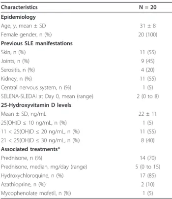
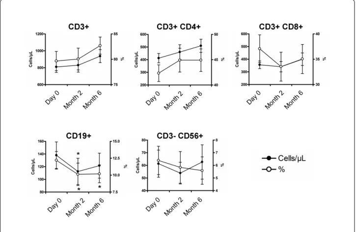
The impact of vitamin D supplementation on the propor-tion and the absolute number of CD3+T cells, CD4+and CD8+T cells, CD19+B cells and CD3-CD56+NK cells is shown in Figure 2. The mean proportions and absolute numbers at baseline were 80 ± 8% and 810 ± 274/mm3 for CD3+T cells, 42 ± 7% and 412 ± 157/mm3for CD4+ T cells, 37 ± 12% and 356 ± 128/mm3 for CD8+T cells, 12 ± 6% and 138 ± 89/mm3for CD19+B cells and 6 ± 5% and 61 ± 46/mm3for CD3-CD56+NK cells. At M2 and M6, the proportion and absolute number of CD3+ T cells, CD4+ and CD8+ T cells and CD3-CD56+ NK cells remained stable, while CD19+B cell frequency and counts significantly decreased at M2 (P < 0.05 for both) (Figure 2).
Table 1 Patient characteristics
Characteristics N = 20 Epidemiology
Age, y, mean ± SD 31 ± 8 Female gender, n (%) 20 (100) Previous SLE manifestations
Skin, n (%) 11 (55) Joints, n (%) 9 (45) Serositis, n (%) 4 (20) Kidney, n (%) 11 (55) Central nervous system, n (%) 1 (5) SELENA-SLEDAI at Day 0, mean (range) 2 (0 to 8) 25-Hydroxyvitamin D levels Mean ± SD, ng/mL 22 ± 11 25(OH)D ≤ 10 ng/mL, n (%) 1 (5) 11 < 25(OH)D ≤ 20 ng/mL, n (%) 11 (55) 21 < 25(OH)D ≤ 30 ng/mL, n (%) 8 (40) Associated treatments* Prednisone, n (%) 14 (70) Prednisone, median, mg/day (range) 5 (0 to 15) Hydroxychloroquine, n (%) 17 (85) Azathioprine, n (%) 2 (10) Mycophenolate mofetil, n (%) 1 (5)
*At the time of the study. SLE: systemic lupus erythematosus; SELENA-SLEDAI: Safety of Estrogens in Lupus Erythematosus National Assessment-Systemic Lupus Erythematosus Disease Activity Index; 25(OH)D: 25-hydroxyvitamin D.
Terrier et al. Arthritis Research & Therapy 2012, 14:R221 http://arthritis-research.com/content/14/5/R221
Vitamin D supplementation induces a preferential increase of naïve CD4+T cells and a decrease of effector memory CD8+T cells and memory B cells
Naïve CD4+ T cells increased in frequency without reaching the significance level (P = 0.09 and P = 0.10, respectively) and in absolute number (P = 0.05 and P < 0.01, respectively) at M2 and M6 with vitamin D supple-mentation, while other CD4+ T cell subsets remained stable. Effector memory CD8+T cells decreased in fre-quency (P = 0.02 and P = 0.01, respectively) but not in absolute number (P = 0.11 and P = 0.55, respectively) at M2 and M6 with vitamin D supplementation (Figure 3A). Using IgD and CD27 surface markers, we also observed a decrease in IgD-CD27+ class-switched
mem-ory B cell frequency (P < 0.05) and absolute number (P = 0.06) at M2 and in IgD-CD27- memory B cell fre-quency (P = 0.11 and P < 0.001, respectively) and abso-lute number (P < 0.05 and P < 0.01, respectively) at M2 and M6, while IgD+CD27+marginal zone-like B cell fre-quency increased at M6 (P < 0.001) (Figure 3B).
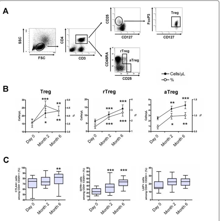
Vitamin D supplementation induces a significant increase of regulatory T cells
The impact of vitamin D supplementation on CD3+CD4+ CD25hiCD127-FoxP3+ Tregs and Treg subsets was
evaluated (Figure 4A). The percentage and absolute count of Tregs at baseline was 3.5 ± 1.2% and 15 ± 8 cells/μL. The percentage of Tregs was increased at 4.6 ± 1.3% at M2 (P <0.001) and 4.3 ± 1.4% at M6 (P <0.01), and the absolute count of Tregs was increased at 23 ± 14 cells/μL (P <0.05) and 25 ± 14 cells/μL (P <0.01), respectively (Figure 4B). Analysis of CD45RA and CD25 expression on CD4+ T cells revealed that this increase concerned both resting and activated memory Tregs (Figure 4B). The increase in Tregs was associated with an increased expres-sion of molecules associated with Treg suppresexpres-sion (that is, CTLA4, GITR and LAP) (Figure 4C).
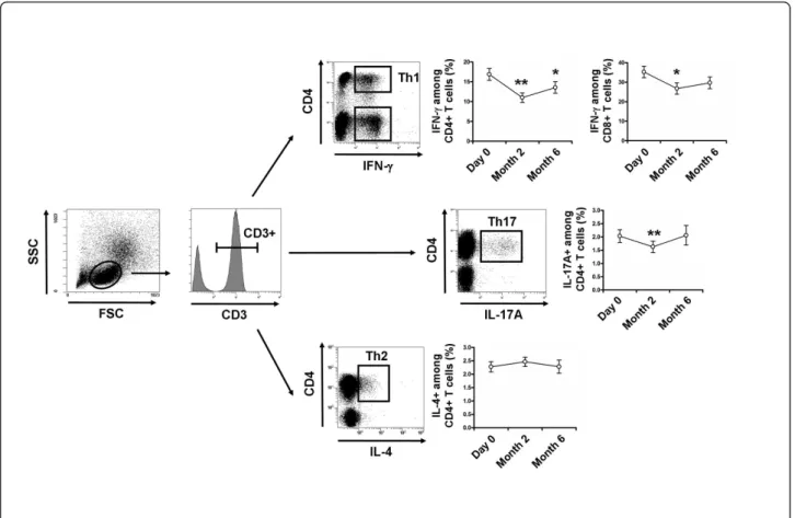
Vitamin D supplementation induces a significant decrease of effector Th1 and Th17 cells
Decreases were observed in Th1 cells from 16.9 ± 6.7% at D0 to 11.0 ± 5.1% at M2, and to 13.6 ± 6.5% at M6 (P < 0.01 and P < 0.05, respectively), in IFN-producing CD8+T cells from 35.3 ± 13.2% at D0 to 26.8 ± 12.5%
at M2, and to 29.7 ± 13.4% at M6 (P < 0.05 and P = 0.06, respectively), and in Th17 from 2.0 ± 1.1% at D0 to 1.6 ± 0.9% at M2 and to 2.0 ± 1.3% at M6 (P < 0.01 and P = 0.81, respectively) after vitamin D supplementa-tion. The Th2 cells remained stable during follow-up (Figure 5).
Figure 1Safety and clinico-biological efficacy of vitamin D supplementation in SLE patients. Time evolution of serum 25(OH)D levels (A disease activity assessed by the SLEDAI (B), and anti-dsDNA levels (C). Serum 25(OH)D levels dramatically increased with vitamin D supplementation, while anti-dsDNA levels decreased. The dotted line in panel A indicates normal values. Box plots indicate median, interquartile ranges, minimum and maximum values. Histograms indicate mean ± standard error of the mean (SEM). *P < 0.05, **P < 0.01, ***P < 0.001 versus Day 0; Wilcoxon test.
Discussion
No study to date has assessed the in vivo benefit of vitamin D supplementation in SLE patients. For the first time, we assessed the safety and the immunological effects of vita-min D supplementation in patients with SLE. We demon-strated that vitamin D was safe and induced a decrease of memory B cells, an increase of Tregs and a decrease of effector Th1 and Th17 cells.
First, we confirmed the high frequency of hypovitami-nosis D in SLE patients. Recent studies have highlighted vitamin D insufficiency in SLE patients, with approxi-mately 65% of the patients showing serum 25(OH)D levels < 30 ng/mL [19-21]. In the present study, we aimed to reach the recommended serum 25(OH)D ranges for bone metabolism, that is, 30 to 80 ng/mL, within the first 2 months. The schedule of vitamin D administration that we choose included a first phase of intensive supplementation for one month, followed by a maintenance phase. Regarding the evolution of serum levels of 25(OH)D, calcium and phosphorous and the occurrence of clinical events, this regimen was effective
and safe. We included only SLE patients with low disease activity (SLEDAI ≤ 8) because we wanted to determine the specific effects of vitamin D in the absence of initia-tion or modificainitia-tion of associated therapy, such as an increase of the prednisone dosage or the use of immuno-suppressive agents.
We demonstrated that vitamin D supplementation induced a beneficial effect in vivo on the perturbations of B cell and T cell homeostasis associated with SLE, by increasing Tregs and decreasing Th17 and Th1 cells, and memory B cells. Interestingly, the increase of Tregs con-cerned both resting Tregs and activated memory Tregs according to CD45RA and CD25 expression [22]. Our findings are supported by recently reported in vitro stu-dies. 1,25(OH)2vitamin D3, the active form of vitamin D,
was shown to exert a marked inhibitory effect on adap-tive immune cells, by inhibiting the T cell proliferation [23,24], the expression of IL-2 [24,25] and IFN-g mRNA and protein in T cells [9,26,27], while promoting Th2-cell responses in vitro [28]. In our in vivo study, we did not observe a significant increase of Th2 cells under vitamin
Figure 2Evolution of peripheral blood lymphocytes with vitamin D supplementation. Time evolution of the proportion and the absolute number of CD3+T cells, CD4+and CD8+T cells, CD19+B cells and CD3-CD56+NK cells. At 2 and 6 months, the proportion and absolute number of CD3+T cells, CD4+and CD8+T cells and CD3-CD56+NK cells remained stable, while the CD19+B cell frequency and count significantly decreased at 2 months. Mean ± standard error of the mean (SEM) is shown. *P < 0.05 versus Day 0; Wilcoxon test. Terrier et al. Arthritis Research & Therapy 2012, 14:R221
http://arthritis-research.com/content/14/5/R221
D supplementation, since this T cell subset remains stable during follow-up. 1,25(OH)2vitamin D3 was also
shown to inhibit Th17 responses, probably owing to its capacity to inhibit IL-23 production [9,29], and to induce the differentiation and/or expansion of FoxP3+Tregs and an increased expression of CTLA4 [9,30-32]. Finally, we observed a decrease of IgD-CD27+ and IgD-CD27
-memory B cells and a decrease of anti-dsDNA autoanti-body levels in vitamin D-treated patients. Consistent with these findings, 1,25(OH)2 vitamin D3 was shown to
decrease B cell proliferation, plasma-cell differentiation and IgG secretion in vitro [12,23]. The mechanisms by which 1,25(OH)2vitamin D3 exerts its
immunomodula-tory effect on B cells remains unclear.
Figure 3Vitamin D supplementation induces a preferential increase of naïve CD4+T cells and a decrease of memory B cells. Peripheral blood mononuclear cells (PBMCs) in the lymphocyte light-scatter gate were analyzed for T cell subsets using CD3, CD4, CD45RA and CD62L staining, and for B cell subsets using CD19, CD27 and IgD staining. (A) Time evolution of CD4+and CD8+T cell subsets. The T cell population includes naive (N; CD45RA+CD62L+), central memory (CM; CD45RA-CD62L+), effector memory and effector (EM; CD45RA-CD62L-), and terminally differentiated effector (TE; CD45RA+CD62L-). (B) Time evolution of CD19+B cell subsets. The B cell population includes naive (N; IgD+CD27-), marginal zone-like (MZ; IgD +CD27+), class-switched memory (CS IgD-CD27+), and IgD-CD27- B cells. Peripheral blood naïve CD4+T cells increased and IgD-CD27- memory B cells decreased under vitamin D supplementation. Mean ± standard error of the mean (SEM) is shown. *P < 0.05, **P < 0.01, ***P < 0.001 versus Day 0; Wilcoxon test.
Although too preliminary to be presented in the present study, we have performed a transcriptome analysis of PBMCs at D0 versus M2 after vitamin D supplementation on ten randomly selected patients, in
order to provide additional insights into the immuno-logical effects of vitamin D in SLE (Additional file 1). Using independent component analysis (ICA) [33] and gene set enrichment analysis (GSEA) dataset [34,35],
Figure 4Vitamin D supplementation induces a significant increase of regulatory T cells. (A) Peripheral blood mononuclear cells (PBMCs) in the lymphocyte light-scatter gate were analyzed for CD3, CD4, CD25, CD127, CD45RA and FoxP3 staining. Regulatory T cells (Tregs), resting Tregs (rTregs) and activated memory Tregs (aTregs) were defined as CD3+CD4+CD25hiCD127-FoxP3+cells, CD3+CD4+CD25++CD45RA+cells and CD3+CD4
+CD25+++CD45RA-cells, respectively. (B) Time evolution of peripheral blood Tregs, rTregs and aTregs in percentage and absolute number. (C) Time
evolution of CTLA4, GITR and LAP expression by Tregs. Peripheral blood Tregs, rTregs and aTregs increased under vitamin D supplementation, as did the expression of molecules associated with suppression of Tregs. Mean ± standard error of the mean (SEM) is shown in panel B. Box plots in panel C indicate median, interquartile ranges, minimum and maximum values. *P < 0.05, **P < 0.01, ***P < 0.001 versus Day 0; Wilcoxon test.
Terrier et al. Arthritis Research & Therapy 2012, 14:R221 http://arthritis-research.com/content/14/5/R221
we identified 48 molecular signatures that were differ-entially expressed between D0 and M2, with 34 up-and 14 down-regulated signatures (see Figure S1 inAdditional file 1. This preliminary finding suggests an effect of vitamin D supplementation on the immune system. Among the identified signatures, we observed the down-regulation of RNA polymerase functions and histone expression and the up-regula-tion of the TP53/CDKN1A-related pathway that repre-sent interesting pathways to explore in the future, owing to their possible involvement with a decrease in the accu-mulation of autoantigens and the activation and prolifera-tion of autoreactive T and B lymphocytes. Also, the up-regulation of the TP53/CDKN1A-related pathway is inter-esting because CDKN1A is a potent cyclin-dependent kinase inhibitor that functions as a regulator of cell cycle progression [36].
Conclusions
Vitamin D supplementation provides beneficial immu-nological effects in patients with SLE, with a decrease of
memory B cells and effector T cells and an increase of regulatory T cells. Our results must be interpreted with caution in the absence of well-designed randomized controlled trials, particularly for a relevant clinical end-point. In addition, we focused on the immunological effects of vitamin D after two months and six months of supplementation, but earlier time points should bring additional information, particularly for gene expression analysis, calling for further studies.
Significance and innovation
Vitamin D supplementation using 100,000 IU of cholecal-ciferol per week for 4 weeks, followed by 100,000 IU of cholecalciferol per month for 6 months, was well tolerated. Vitamin D induced a preferential increase of naïve CD4+ T cells, an increase of regulatory T cells, a decrease of effector Th1 and Th17 cells, and a decrease of memory B cells and anti-DNA antibodies.
This preliminary study suggests a beneficial role of vitamin D in SLE patients and needs to be confirmed in randomized controlled trials.
Figure 5Vitamin D supplementation induces a significant decrease in Th1 and Th17 cells. Peripheral blood mononuclear cells (PBMCs) were stimulated for 4 hours with Phorbol myristate acetate (PMA) and ionomycin. After gating on CD3+T cells, frequencies of IFN-g-producing CD4+(Th1) and CD8+T cells, IL-17A-producing CD4+T cells (Th17) and IL-4-producing CD4+T cells (Th2) were analyzed with vitamin D supplementation. Th1, IFN-g-producing CD8+T cells and Th17 cells decreased after 2 months of vitamin D supplementation. Mean ± standard error of the mean (SEM) is shown. *P < 0.05, **P < 0.01, ***P < 0.001 versus Day 0; Wilcoxon test.
Additional material
Additional file 1: Microarray gene expression profile analysis. This file contains additional material and a methods section that describes microarray gene expression profile analysis, and Figure S1 that illustrates clustering analysis of significant modulated signatures in patients at month 2 compared to baseline.
Abbreviations
APC: allophycocyanin; CDKN1A: cyclin-dependent kinase inhibitor 1A; ECD: Phycoerythrin-Texas Red; ELISA: enzyme-linked immunosorbent assay; FITC: fluorescein isothiocyanate; FoxP3: forkhead box P3; GSEA: gene set enrichment analysis; ICA: independent component analysis; IFN: interferon; IL: interleukin; 25(OH)D: 25-hydroxyvitamin D; PBMC: peripheral blood mononuclear cell; PCy7: Phycoerythrin-Cyanin 7; PE: Phycoerythrin; PMA: phorbol myristate acetate; SELENA-SLEDAI: Safety of Estrogens in Lupus Erythematosus National Assessment-Systemic Lupus Erythematosus Disease Activity Index; SLE: systemic lupus erythematosus; Th17: T helper lymphocytes producing IL-17; Tregs: regulatory T cells. Acknowledgements
BT was supported by the Fondation pour la Recherche Médicale (FRM), the Agence Nationale pour la Recherche sur le Sida et les Hépatites(ANRS) and the Société Nationale Française de Médecine Interne(SNFMI).
Author details
1UPMC Université Paris 6, UMR 7211, F-75013 Paris, France.2Centre National
de la Recherche Scientifique (CNRS), UMR 7211, Paris, France.3INSERM, UMRS959; Groupe Hospitalier Pitié-Salpétrière, 47 boulevard de l’Hôpital, 75013 Paris, France.4Department of Internal Medicine, Hôpital Foch, 40 rue Worth, 92150 Suresnes, France.5Department of Biotherapy, Groupe
Hospitalier Pitié-Salpétrière, 47 boulevard de l’Hôpital, 75013 Paris, France.
6Department of Pharmacology, Groupe Hospitalier Pitié-Salpétrière, 47
boulevard de l’Hôpital, 75013 Paris, France.7Department of Internal
Medicine, Groupe Hospitalier Pitié-Salpétrière, 47 boulevard de l’Hôpital, 75013 Paris, France.8P3S post-genomic platform, Groupe Hospitalier
Pitié-Salpétrière, 47 boulevard de l’Hôpital, 75013 Paris, France.9Department of Immunology, Groupe Hospitalier Pitié-Salpétrière, 47 boulevard de l’Hôpital, 75013 Paris, France.
Authors’ contributions
BT, ND, YS, GG, PC and NCC devised and performed experiments; BT, PC and NCC provided patient referrals; BT, ND, YS, WC, GG, NZ, KM, MR, WC, LM, JCP, AS, DK, DS, PC and NCC interpreted results; and BT, PC and NCC designed the research and wrote the paper. All authors have read and approved the manuscript for publication.
Competing interests
The authors declare that they have no competing interests. Received: 10 March 2012 Revised: 10 July 2012 Accepted: 5 September 2012 Published: 17 October 2012 References
1. Rahman A, Isenberg DA: Systemic lupus erythematosus. N Engl J Med 2008, 358:929-939.
2. Cervera R, Khamashta MA, Font J, Sebastiani GD, Gil A, Lavilla P, Mejia JC, Aydintug AO, Chwalinska-Sadowska H, de Ramon E, Fernandez-Nebro A, Galeazzi M, Valen M, Mathieu A, Houssiau F, Caro N, Alba P, Ramos-Casals M, Ingelmo M, Hughes GR: Morbidity and mortality in systemic lupus erythematosus during a 10-year period: a comparison of early and late manifestations in a cohort of 1,000 patients. Medicine (Baltimore) 2003, 82:299-308.
3. Dorner T, Jacobi AM, Lee J, Lipsky PE: Abnormalities of B cell subsets in patients with systemic lupus erythematosus. J Immunol Methods 2011, 363:187-197.
4. Eilat D, Naparstek Y: Anti-DNA autoantibodies: a puzzle of autoimmune phenomena. Immunol Today 1999, 20:339-342.
5. Miyara M, Amoura Z, Parizot C, Badoual C, Dorgham K, Trad S, Nochy D, Debre P, Piette JC, Gorochov G: Global natural regulatory T cell depletion in active systemic lupus erythematosus. J Immunol 2005, 175:8392-8400. 6. Crispin JC, Tsokos GC: Interleukin-17-producing T cells in lupus. Curr Opin
Rheumatol2010, 22:499-503.
7. Apostolidis SA, Crispin JC, Tsokos GC: IL-17-producing T cells in lupus nephritis. Lupus 2011, 20:120-124.
8. Bennett L, Palucka AK, Arce E, Cantrell V, Borvak J, Banchereau J, Pascual V: Interferon and granulopoiesis signatures in systemic lupus
erythematosus blood. J Exp Med 2003, 197:711-723.
9. Jeffery LE, Burke F, Mura M, Zheng Y, Qureshi OS, Hewison M, Walker LS, Lammas DA, Raza K, Sansom DM: 1,25-Dihydroxyvitamin D3 and IL-2 combine to inhibit T cell production of inflammatory cytokines and promote development of regulatory T cells expressing CTLA-4 and FoxP3. J Immunol 2009, 183:5458-5467.
10. Mora JR, Iwata M, von Andrian UH: Vitamin effects on the immune system: vitamins A and D take centre stage. Nat Rev Immunol 2008, 8:685-698.
11. Linker-Israeli M, Elstner E, Klinenberg JR, Wallace DJ, Koeffler HP: Vitamin D (3) and its synthetic analogs inhibit the spontaneous in vitro immunoglobulin production by SLE-derived PBMC. Clin Immunol 2001, 99:82-93.
12. Chen S, Sims GP, Chen XX, Gu YY, Lipsky PE: Modulatory effects of 1,25-dihydroxyvitamin D3 on human B cell differentiation. J Immunol 2007, 179:1634-1647.
13. Ben-Zvi I, Aranow C, Mackay M, Stanevsky A, Kamen DL, Marinescu LM, Collins CE, Gilkeson GS, Diamond B, Hardin JA: The impact of vitamin D on dendritic cell function in patients with systemic lupus erythematosus. PLoS One2010, 5:e9193.
14. Amital H, Szekanecz Z, Szucs G, Danko K, Nagy E, Csepany T, Kiss E, Rovensky J, Tuchynova A, Kozakova D, Doria A, Corocher N, Agmon-Levin N, Barak V, Orbach H, Zandman-Goddard G, Shoenfeld Y: Serum concentrations of 25-OH vitamin D in patients with systemic lupus erythematosus (SLE) are inversely related to disease activity: is it time to routinely supplement patients with SLE with vitamin D? Ann Rheum Dis 2010, 69:1155-1157.
15. Hochberg MC: Updating the American College of Rheumatology revised criteria for the classification of systemic lupus erythematosus. Arthritis Rheum1997, 40:1725.
16. Petri M, Kim MY, Kalunian KC, Grossman J, Hahn BH, Sammaritano LR, Lockshin M, Merrill JT, Belmont HM, Askanase AD, McCune WJ, Hearth-Holmes M, Dooley MA, Von Feldt J, Friedman A, Tan M, Davis J, Cronin M, Diamond B, Mackay M, Sigler L, Fillius M, Rupel A, Licciardi F, Buyon JP: Combined oral contraceptives in women with systemic lupus erythematosus. N Engl J Med 2005, 353:2550-2558.
17. Souberbielle JC, Body JJ, Lappe JM, Plebani M, Shoenfeld Y, Wang TJ, Bischoff-Ferrari HA, Cavalier E, Ebeling PR, Fardellone P, Gandini S, Gruson D, Guerin AP, Heickendorff L, Hollis BW, Ish-Shalom S, Jean G, von Landenberg P, Largura A, Olsson T, Pierrot-Deseilligny C, Pilz S, Tincani A, Valcour A, Zittermann A: Vitamin D and musculoskeletal health, cardiovascular disease, autoimmunity and cancer: Recommendations for clinical practice. Autoimmun Rev 2010, 9:709-715.
18. Hollis BW, Kamerud JQ, Selvaag SR, Lorenz JD, Napoli JL: Determination of vitamin D status by radioimmunoassay with an 125I-labeled tracer. Clin Chem1993, 39:529-533.
19. Ritterhouse LL, Crowe SR, Niewold TB, Kamen DL, Macwana SR, Roberts VC, Dedeke AB, Harley JB, Scofield RH, Guthridge JM, James JA: Vitamin D deficiency is associated with an increased autoimmune response in healthy individuals and in patients with systemic lupus erythematosus. Ann Rheum Dis2011, 70:1569-1574.
20. Wu PW, Rhew EY, Dyer AR, Dunlop DD, Langman CB, Price H, Sutton-Tyrrell K, McPherson DD, Edmundowicz D, Kondos GT, Ramsey-Goldman R: 25-hydroxyvitamin D and cardiovascular risk factors in women with systemic lupus erythematosus. Arthritis Rheum 2009, 61:1387-1395. 21. Kamen DL, Cooper GS, Bouali H, Shaftman SR, Hollis BW, Gilkeson GS:
Vitamin D deficiency in systemic lupus erythematosus. Autoimmun Rev 2006, 5:114-117.
22. Miyara M, Yoshioka Y, Kitoh A, Shima T, Wing K, Niwa A, Parizot C, Taflin C, Heike T, Valeyre D, Mathian A, Nakahata T, Yamaguchi T, Nomura T, Ono M, Amoura Z, Gorochov G, Sakaguchi S: Functional delineation and differentiation dynamics of human CD4+ T cells expressing the FoxP3 transcription factor. Immunity 2009, 30:899-911.
Terrier et al. Arthritis Research & Therapy 2012, 14:R221 http://arthritis-research.com/content/14/5/R221
23. Lemire JM, Adams JS, Sakai R, Jordan SC: 1 alpha,25-dihydroxyvitamin D3 suppresses proliferation and immunoglobulin production by normal human peripheral blood mononuclear cells. J Clin Invest 1984, 74:657-661. 24. Rigby WF, Stacy T, Fanger MW: Inhibition of T lymphocyte mitogenesis by
1,25-dihydroxyvitamin D3 (calcitriol). J Clin Invest 1984, 74:1451-1455. 25. Lemire JM, Adams JS, Kermani-Arab V, Bakke AC, Sakai R, Jordan SC:
1,25-Dihydroxyvitamin D3 suppresses human T helper/inducer lymphocyte activity in vitro. J Immunol 1985, 134:3032-3035.
26. Reichel H, Koeffler HP, Tobler A, Norman AW: 1 alpha,25-Dihydroxyvitamin D3 inhibits gamma-interferon synthesis by normal human peripheral blood lymphocytes. Proc Natl Acad Sci USA 1987, 84:3385-3389. 27. Rigby WF, Yirinec B, Oldershaw RL, Fanger MW: Comparison of the effects
of 1,25-dihydroxyvitamin D3 on T lymphocyte subpopulations. Eur J Immunol1987, 17:563-566.
28. van Etten E, Mathieu C: Immunoregulation by 1,25-dihydroxyvitamin D3: basic concepts. J Steroid Biochem Mol Biol 2005, 97:93-101.
29. Daniel C, Sartory NA, Zahn N, Radeke HH, Stein JM: Immune modulatory treatment of trinitrobenzene sulfonic acid colitis with calcitriol is associated with a change of a T helper (Th) 1/Th17 to a Th2 and regulatory T cell profile. J Pharmacol Exp Ther 2008, 324:23-33.
30. Gorman S, Kuritzky LA, Judge MA, Dixon KM, McGlade JP, Mason RS, Finlay-Jones JJ, Hart PH: Topically applied 1,25-dihydroxyvitamin D3 enhances the suppressive activity of CD4+CD25+ cells in the draining lymph nodes. J Immunol 2007, 179:6273-6283.
31. Penna G, Roncari A, Amuchastegui S, Daniel KC, Berti E, Colonna M, Adorini L: Expression of the inhibitory receptor ILT3 on dendritic cells is dispensable for induction of CD4+Foxp3+ regulatory T cells by 1,25-dihydroxyvitamin D3. Blood 2005, 106:3490-3497.
32. Baeke F, Korf H, Overbergh L, Verstuyf A, Thorrez L, Van Lommel L, Waer M, Schuit F, Gysemans C, Mathieu C: The vitamin D analog, TX527, promotes a human CD4+CD25highCD127low regulatory T cell profile and induces a migratory signature specific for homing to sites of inflammation. J Immunol2011, 186:132-142.
33. Chiappetta P, Roubaud MC, Torresani B: Blind source separation and the analysis of microarray data. J Comput Biol 2004, 11:1090-1109. 34. Subramanian A, Tamayo P, Mootha VK, Mukherjee S, Ebert BL, Gillette MA,
Paulovich A, Pomeroy SL, Golub TR, Lander ES, Mesirov JP: Gene set enrichment analysis: a knowledge-based approach for interpreting genome-wide expression profiles. Proc Natl Acad Sci USA 2005, 102:15545-15550.
35. Mootha VK, Lindgren CM, Eriksson KF, Subramanian A, Sihag S, Lehar J, Puigserver P, Carlsson E, Ridderstrale M, Laurila E, Houstis N, Daly MJ, Patterson N, Mesirov JP, Golub TR, Tamayo P, Spiegelman B, Lander ES, Hirschhorn JN, Altshuler D, Groop LC: PGC-1alpha-responsive genes involved in oxidative phosphorylation are coordinately downregulated in human diabetes. Nat Genet 2003, 34:267-273.
36. Goulvestre C, Chereau C, Nicco C, Mouthon L, Weill B, Batteux F: A mimic of p21WAF1/CIP1 ameliorates murine lupus. J Immunol 2005, 175:6959-6967.
doi:10.1186/ar4060
Cite this article as: Terrier et al.: Restoration of regulatory and effector T cell balance and B cell homeostasis in systemic lupus erythematosus patients through vitamin D supplementation. Arthritis Research & Therapy 2012 14:R221.
Submit your next manuscript to BioMed Central and take full advantage of:
• Convenient online submission
• Thorough peer review
• No space constraints or color figure charges
• Immediate publication on acceptance
• Inclusion in PubMed, CAS, Scopus and Google Scholar
• Research which is freely available for redistribution
Submit your manuscript at www.biomedcentral.com/submit