UNCLASSIFIED
MASSACHUSETTS
INSTITUTE OF TECHNOLOGY
RESEARCH LABORATORY OF ELECTRONICS
AND
LINCOLN
LABORATORY
COMPUTER TECHNIQUES FOR THE STUDY
OF PATTERNS
IN THE ELECTROENCEPHALOGRAM
B. G. FARLEY
W. A. CLARK, JR.
J. T. GILMORE, JR.
Group 63
L. S. FRISHKOPF
Research Laboratory of Electronics
RESEARCH LABORATORY OF ELECTRONICS
TECHNICAL REPORT NO.337
LINCOLN LABORATORY
TECHNICAL REPORT NO. 165
6 NOVEMBER 1957
LEXINGTON
MASSACHUSETTS
UNCLASSIFIED
ABSTRACT
A process has been explored, using the Lincoln TX-O computer, for detecting patterns in the electroencephalogram and for recognizing the characteristics of the EEG corresponding to individual subjects. Preliminary results indicate that
a number of different subjects and statesof the same subject can be distinguished with excellent probability.
iii
V
COMPUTER TECHNIQUES FOR THE STUDY OF PATTERNS IN THE ELECTROENCEPHALOGRAM
I. INTRODUCTION
The electroencephalogram (EEG), popularly known as "brain waves," is the time-varying voltage observed between two electrodes contacting separate points of the head, typically two points of the scalp. Contact resistances between such electrodes are usually of the order of a few thousand ohms, and 20 to 100 livolts of signal are usually obtained. The predominant frequency range is from zero to thirty cycles/sec, although some energy is present at considerably higher values. In the case of an awake subject with closed eyes there is often a well-defined peak in the spectrum near 10 cps which disappears when the eyes are open. This frequency component is the well-known "alpha-rhythm." It is particularly prominent if the electrodes are near the back of the head. Considerable variation in the character of the signal may be observed under differing experimental conditions for a given subject, and between individual subjects. For ex-ample, some individuals show almost continuous trains of alpha activity; some exhibit frequent bursts of such activity, some infrequent bursts and some exhibit no discernible alpha activity at all. The waves during sleep are considerably different and some changes due to drugs are observable. No universally accepted theory of the neurophysiological origin of the waves exists. Many diseased conditions of the brain show abnormal characteristics. Various types of epilepsy and some brain tumors fall in this class. As a result of these facts, electroencephalography has become widely used in clinical neurophysiological work as an aid to the diagnosis and treat-ment of such disorders. In this work, as yet, the main tool of EEG classification is the eye of the experienced clinician observing the data directly as recorded by an ink writer. Some use is made of spectral density analysis and its allied computation, the autocorrelation function, but these have been substantially the only parametric aids in use. References 1 - 6 comprise a representative survey of work in this field.
Situations of similar nature are, of course, common in the behavioral and other complex sciences because of the quantity and complexity of the data encountered, and the lack of relevant techniques for its analysis. Even though the clinical electroencephalographer has made remark-able progress, more parametric aids of a quantitative objective nature would be welcome; this is even more true in the case of the researcher, who needs means of objectively testing his scientific conjectures. It was with these considerations in mind that the present study was under-taken to explore the utility of digital computers as aids in developing techniques of data processing for the electroencephalographer. The main problem to be faced here is one of classification of complex data in accordance with the specific conditions pertaining to its origin. Evidently this is essentially the same as what is often called "pattern" or "percept" recognition and we have approached the problem from this conceptual standpoint. In this case, the "patterns" exist on more than one level. One level roughly comprises those characteristics of the wave which the experienced eye uses as primary data for classification. From this level of pattern recognition several descriptive terms arise, such as "alpha," "beta" and "delta" waves, "spindles," "slow
1
or fast" activity, "dysrhythmia," "spike and wave," etc., which convey certain concepts to the initiated.
On a more complex level, combinations of these simpler patterns may characterize an in-dividual under specified conditions. These combinations may then be considered patterns of a more sophisticated nature. In this study we have dealt with both types of patterns.
The conceptual scheme of "pattern" or "percept" recognition which we have utilized infor-mally in this work is essentially that a percept is determined by a group of common properties which it possesses. To express this more fully, suppose a pattern recognition system has the means to compute the values of a number of parameters (called properties) from incoming data. It is understood that parameters having both discrete and continuous values may be present. Suppose further that the system has already stored various groups of parameters and their values. The incoming values of groups of parameters are compared continually with the values of the
stored groups. If agreement occurs between one of these incoming groups and one of the stored groups, an instance of the percept corresponding to that property group may be said to exist. A more realistic system would store statistical distributions of the parameters in each group, and comparison would be on a correlation basis. This is the type of recognition used in our EEG study. If the system still further had the ability to build up its own property groups as a function of their occurrence in its "data environment," the system would exhibit learned perception. Systems of this kind have not been used in the present work, but the considerations involved have been kept in mind, with a view to exploration of these questions at a later date.
II. EQUIPMENT AND DATA
The digital computer used in this work is the Lincoln TX-0 (Ref. 7). This computer was originally built to test the practicability of transistor computer circuits and a large-scale
magnetic-core memory. Since it is primarily a test computer it is small, having an 18-bit word length and only three instructions that refer to memory. Flexibility in programming is obtained by means of a fourth instruction whose address bits designate operations to be performed instead of memory words. By this means, any desired operation on the arithmetic element may be car-ried out, though at a cost in time since several instructions may be necessary. This handicap is mitigated to a considerable extent by the large memory available (65,000 words) and by its speed (6 tsec per memory cycle). Original data on seven-channel FM-carrier magnetic tape is fed from an Ampex tape playback unit to an Epsco Datrac analog-to-digital converter. A com-puter instruction is arranged to read the digital word output into the comcom-puter and cause the con-verter to begin conversion of another sample. The digital word read at this time is transferred to a register of the computer, and is then stored in its proper place in the memory by further coding. Thus the whole process is under computer control. Rates of sampling vary up to about
10,000/sec depending on recording and playback speeds and the subject-time sampling rate de-sired. The data on which our results have been based were sampled at a rate of 300/sec, sub-ject time. After 53,428 eight-bit samples (nearly 3 minutes of data) have been read in at this rate, various preliminary observations and calculations may be carried out by means of a pro-gram known as the "moving window." This propro-gram enables the operator to observe a chosen section of the data on an oscilloscope screen similar to an ordinary oscilloscope, except that the
data is displayed point-by-point instead of continuously. Various time expansions are available, and the "sweep" may be made to stop or to move at a selected rate of speed through the whole of the 53,428 data points in either direction. The memory address of the word in the center of the
sweep at any time may be recorded automatically on a typewriter, and provision is made to re-turn to that position at will. Controls for these modes of operation are toggle switches on the computer console. Because the eight-bit data number occupies less than half of a memory word, room remains for inclusion of time-dependent computations, which may be displayed simultane-ously with the original data if desired. At present, this space is used to store temporarily the results of a moving-average computation, and also to mark the parts and patterns of the wave by the process to be described later. The convenience afforded by the large memory and the moving-window display for immediate observation of data and computed results has, in our opin-ion, contributed very materially to the progress of the work. A further aid has been a useful set of "utility" programs which make it possible to communicate directly with the computer by typewriter, to look at or change instructions of a program, insert parameters, search for
speci-fied addresses and perform a number of other tasks of technical benefit to the programmer. These utility routines and the moving-window program are left constantly in the memory to be called upon at any time. They occupy about 5000 words of memory storage.
III. THE RHYTHMIC BURST ACTIVITY DETECTION PROCESS
Alpha-rhythm in EEG waves has a tendency to appear in amplitude-modulated bursts. Such bursts are of widely varying lengths from a few cycles to hundreds of cycles per second. In many
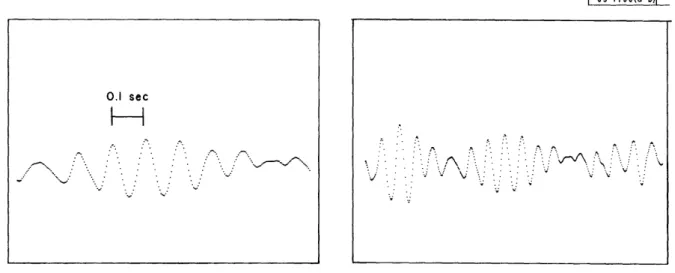
cases the envelope of the burst gradually rises to a broad maximum and falls back toward zero. The photograph of the TX-0 oscilloscope display in Fig. 1 shows what might be termed a "classic" example of this type of activity. Such patterns are particularly striking to the eye and consider-able attention has been paid by electroencephalographers to their frequency and duration. As a
result, it was decided to attempt to detect this type of structure in the wave. Since, however,
1-63- 198(-b)I
Fig. (a). A classical" rhythmic burst. Fig. (b). Same burst on compressed time scale.
3 0.1 sec
F-A
.^. '. ;".. .. ":d.. ... A... ../-. ....:on ... · . .... v.j . .. . -. . C Z :?. " :'? " I i\i v v I : v u j· . 1 __ ___the envelopes may in some instances be quite irregular (especially in particular records), it was not considered desirable to include an envelope specification, but to detect what may be termed "rhythmic bursts" of activity consisting of trains of consecutive waves meeting specifi-cations of amplitude, frequency and duration. It is evidently important that the detection process be independent of channel gain, DC level and frequency components that lie outside the range of interest, as well as of the exact frequency of the alpha rhythm.
These considerations suggest the use of a threshold criterion applied to peak-to-peak am-plitudes, and an admissible range of time intervals related to the alpha frequency. If the am-plitude threshold and the interval range are made to depend on the frequency distributions of amplitudes and intervals obtained from the data, the resulting definition of rhythmic activity will be relative to the particular time function under analysis, as is desired. The low-frequency components may be eliminated by suitable criteria of wave peaks and intervals.
High-frequency components are eliminated by a moving average which corresponds to a filter of sin (x/x) amplitude characteristic with the first zero at 30 cps. This filtering is applied primarily to standardize the EEG records, which were obtained under a variety of conditions (tape speed, filtering, noise and 60-cps pickup) and from two laboratories; in addition, such smoothing eliminates certain sampling and detection difficulties introduced by large energies in the high-frequency range. Standard practice is to sample at a rate corresponding to 300/sec in subject time, so that the moving average is taken over 10 samples. Figure Z(b) shows the effect of this smoothing on the original data of Fig. 2(a). After some experimentation, definitions of peaks and time intervals were found which satisfied the requirements set forth above. Peaks and valleys are detected as follows: A maximum of the data is called a peak if it is followed by at least k decreases in amplitude (k = 3 in all results to be shown). Similarly, a minimum is called a valley if it is followed by at least k increases. Note that the parameter k performs an additional function of small-amplitude high-frequency rejection. Next, the time intervals to be tested must be determined. It is possible to use the peak-to-valley or peak-to-peak times as
1-63-
1799( -b)IFig. 2(a). Sampled data before smoothing. Fig. 2(b). Sampled data after smoothing.
· . . ' . . .. . · ' .~~~~~~K_ '*. *¢~~~~~~~~~. .'l s s".*
~~~~~~~'
.' ' ..: · ",,~^... L· . -.. 1. 1:I ~/ ,.. :
-.; "... itest intervals, but this procedure suffers from the fact that the peaks and valleys are broad and not well-defined in time. This indeterminacy results in large variations in the time intervals. However, the peaks and valleys are well-defined in amplitude, so that if the point on the curve
just halfway in amplitude between a peak and valley is selected, it will generally be well-defined in time, due to the non-zero slope of the curve at that point. These are the points that are used to define the time intervals. They are referred to as the "midvalue" points. If no low-frequency
fluctuations are present, these midvalue points correspond to the zero-crossings, roughly speaking.
Briefly, detection of rhythmic activity proceeds as follows: Peak-and-valley data points are detected as explained above. Each such point is marked by storing a number designating peak or valley in the spare part of the memory word containing the data. Likewise, midvalue points are found and marked. An amplitude threshold is set as a specified fraction of the median
of the peak-to-valley amplitude frequency distribution compiled over the complete data.
Now, every midvalue of a peak-to-valley amplitude that is greater than the calculated am-plitude threshold is "doubly" marked, and a frequency distribution is made of time intervals de-fined by just these points. The admissible time interval is now calculated as the median of this
distribution plus or minus a specified fraction (called the interval threshold factor) of the same median.
We are now ready for the final step - detection and marking of rhythmic burst activity. A rhythmic burst is defined by a successive string of at least n midvalue points all of which must be doubly marked and each of which must be preceded by an interval that satisfies the time in-terval criteria as defined above (in the results to follow n = 5). If there are fewer than five such
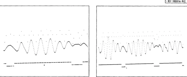
successive points, they are ignored; if there are p such successive points (that is, p > 5), a burst is defined consisting of the p intervals preceding these p-points. The burst is continued until a point fails to meet one of the criteria, whereupon the burst is terminated on the previous point. Every point of the data within a rhythmic burst, so defined, is now marked by inserting a number in the spare part of its memory word. When the data are displayed by the moving win-dow program, these marks may also be shown, and detected burst activity is indicated by an upward step in a base line. The marks designating peaks, valleys and midvalues may also be observed at the same time. This display is illustrated by Fig. 3(a) and (b). These photographs show the same wave as in Fig. l(a) and (b) after marking with both threshold criteria set at 50 per cent of their respective medians. The break in rhythmic activity on the left occurs be-cause of a midvalue time interval too large for admissibility, while the break on the right is caused by an interval that fails to meet both amplitude and time-interval criteria. It is possible to see above the data many of the marks that designate the time positions of peaks, midvalues (doubly and singly marked) and valleys. These marks have amplitudes that decrease in the order listed, but since they "float" on the burst activity marks, they are displaced whenever a step in the latter occurs. Figures 4 through 6 show various samples of the result of the rhythmic burst detection process on typical waves. It will be noted that the mark appears somewhat early in Fig. 4(a). Figure 5 shows examples of rather irregular amplitude envelopes that are accepted as rhythmic, showing that the process is independent of changes in level. Figure 6 shows examples
5
of irregular waves that are not accepted. In this case, most of the peak-to-valley amplitudes do not pass the amplitude threshold test, although they would do so at a lower threshold.
In general, when there are clear-cut rhythmic bursts, the detection process as described, for reasonable values of amplitude and interval parameters (say, both equal to 0.5), agrees rather well with the verdict of experienced workers. On more irregular records, there may not be complete agreement, but the human observer is often uncertain in such cases. The final verdict on such a process must in any case lie in whether the detected pattern has meaning in terms of the "environment" of controllable conditions. The most obvious controllable condition in this case is the identity of the subject. In order to investigate the results of the process, programs were written to sum up and tabulate the total number of points detected in rhythmic
activity, the total number of rhythmic bursts, the first, second and third quartiles of the am-plitude and interval distributions, and also of the distribution of burst lengths, over the whole 3 minutes of data (or any portion of it). After some experimentation, it was found desirable to compute these data for a range of values of either the amplitude threshold factor or the interval range factor, the other meanwhile being held constant. Up to the present, varying the amplitude threshold factor has been found most useful, and the results to be presented show the dependent variables (total activity, or number of bursts) as a function of amplitude threshold factor. The latter takes on values from zero to two in steps of 0.25. It was first established that these re-sults could be reproduced satisfactorily on different read-ins of the same data, despite minor differences not easily avoided, such as failure to start sampling in precisely the same place
(sampling was started manually), and possible gain changes. Variability of this kind proved to be so small, in general, that it does not show on the plot scales used. Sampling variability in the records of a single individual taken at different times is the next consideration. Figure 7(a) and (b) shows curves obtained from the data of subject A. L.; brain waves were recorded on four successive days under fairly constant conditions. The subject was awake and sat with closed eyes in an anechoic chamber. An attempt was made to standardize the time of recording.
Figure 7(a) shows total activity vs amplitude threshold, and Fig. 7(b) shows number of bursts vs amplitude threshold; the interval range factor was held constant at 0.5 (admissible range from 0.5 to 1.5 times the median interval). The mean of the four runs is shown as a heavy solid line. In order to get a reasonable idea of the expected statistical variation of the data, the stand-ard deviation of the population was estimated from the average range of the samples of four at each abscissa by standard small sampling techniques (0.486 x the average range is an unbiased estimate of the population standard deviation for samples of 4). This, of course, involves the assumptions that the samples of four all come from the same normal distribution. More data will be required to verify these assumptions. When this estimate of the standard deviation had been made, confidence limits of three times the standard deviation on either side of the mean were determined in accordance with established practice and plotted as the dashed lines and the included region is shaded. More sophisticated techniques involving nonlinear regression might be used, but it is felt that the extensive calculation involved is not justified by the amount of data available at present. The method used was adapted from standard control chart techniques. Under the assumptions made, roughly one point in 300 should lie outside the limits shown. Of course, the successive points on a curve cannot be considered independent, but if two or more
points of a curve lie outside the band, it is relatively certain that this curve does not belong to the original population of curves.
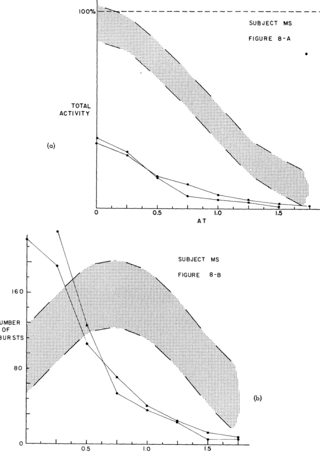
When the variability for an individual has been estimated, we are now in a position to com-pare with it curves derived from the data of other subjects, all of whom at the time of recording were awake and had their eyes closed. In Fig. 8(a) and (b) the confidence limits for A. L. are plotted, together with two runs on subject M. S. It is very clear that these individuals are dis-tinguishable on the basis of these functions. Figure 9(a) and (b) show the same comparison for
subject H. K. Although there is less difference than before, it is clear from the discussion above that a significant difference exists.
Figure 10(a) and (b) show still another case of reasonably clear differentiability. In Fig.11(a) and (b) we have plotted the same functions obtained from four tests of subject R. C. These func-tions lie within or near one boundary of the confidence limits for A. L.; the difference between these subjects is therefore marginal at the level of confidence defined by these limits. It is
in-teresting to note, however, that another parameter, the median midvalue interval, is very stable and significantly different for these two subjects, so the subjects can still be separated by our analysis.
In comparing curves by this one-sided method, it is, of course, clear that no exact infor-mation has been presented concerning the possible overlap between two subjects, that is, what percentage of curves from the population of one subject will be statistically indistinguishable
from a percentage of the population of another. To do this it would be necessary to draw confi-dence limits for each subject, but not enough data, taken under satisfactorily constant conditions, are available at the present time t make this meaningful. However, this consideration does not invalidate comparison between the distribution for one subject, and particular curves of another, as we have been doing. Even when considerable overlap exists by the present method, it does not seem unreasonable to expect, on the basis of the present data, that more powerful statistical methods operating on more abundant data will be able to divide subjects into a number of separa-ble categories. Figure 12(a) and (b) shows functions obtained from a number of other subjects on each of whom one run is available, to give an idea of other types of curves. Figure 13(a) and (b) compares the functions obtained from filtered thermal noise and the confidence limits of sub-ject A. L. The noise was passed through a filter centered at 100 cps with 36 db/octave skirts,
giving a 6-db width of about 25 cps. It was then sampled at a rate of 3000/sec, thereby providing 30 samples/cycle at the center frequency; thus about the same quantization per cycle was ob-tained for the filtered noise as had been obob-tained for the alpha rhythm in the EEG. It is
interest-ing that the filtered noise is distinterest-inguishable from subject A. L. on the basis of one function but not on the basis of the other. The variability observed in the functions obtained from different samples of the noise was somewhat smaller than that obtained from subject A. L.'s data.
Figure 14(a) and (b) shows an example of the change that occurs in these functions as a sub-ject goes to sleep. The two top curves were obtained from data taken when the subject was awake; the bottom curve was obtained from data taken in deep sleep, and the intermediate curve refers to data intermediate in time. This is an example of the same subject under differing conditions.
Other conditions such as the effect of drugs and repetitive light flashes are under investigation.
7
[- 63-1800(a-b)]
Fig. 3(a). la after marking.
Fig. 4(a). Marked rhythmic burst.
Fig. 3(b). b after marking.
1-65- 1801(o-b)L
Fig. 4(b). Interval between marked regions. -~
~
~
. 2 -/ V * L' V l - -__ _____________________ I:-, i, i 1.
:
-:
:v
V ..-63- -102(o-b)
Fig. 5(a). Irregular marked burst.
Fig. 6(a). Irregular unmarked region.
Fig. 5(b). Irregular marked burst.
Fig. 6(b). Irregular unmarked region.
9 I. . ' - .. ,. : ..,... * *. .1 --1 - . :,- "" I c. v v r,'· I- 6 3 -18 0 3 (-b)|
(a)
0 0.5 1.0 1.5
AMPLITUDE THRESHOLD (AT)
SUBJECT A. L. FIGURE 7-B .. .... . .. . .: ;::::::::::::::: . ....
~
... ... ::::::::::... ''.:'.' . iiiiiiii~i... ' . .'...: .,.,,... ... .... /'~~~~~~~~~.
... ... ... .::--.. ,,,,.::--..., .im _'.,.,. ..,... - : '"' X, e _.YP.5LO. .' ...~~~~...
...'. ': .:- g :::~~~~~~~~~~ ... ... .... ... , :: ...,... ... ,',.',,, ''-j ',..,,,- ... . . ... ... .. .. .... ,, ...,, ,.,, ...,A
_wN...
... ...
....
,s
.. ... ... .
.. ...
. .. ...
~~~~~~~~~~~~~~~~~~~~~~~.
... .... s... N... N.,... . N...,., .'.'''..,.',,,... ... . .. ...,j I I I I , I I I I I I 0 0,5 1.0 1.5 AT 100% TOTAL ACTIVITY 160 NUMBER OF BURSTS 80, (b) 0 t:TO ACTIV (a) AT 160 SUBJECT MS .. ::i!!i ii~i!!!i! i . ... . .:.
i<ii. .i ..i i . .i .i{{iiiiiii.{ii i{{{iiiii~i~:GUR:E 8
-iiiiiliiiiii~ iii... .,.,,,.,.,,.-.-..-.... ... .... ,.,..., .. . .-. ' . ' ' ' ... ... . ... -... ...--. '. ... ... ..\ ... -..,, , ,, - - ,.....-., ... ... NUMBER .: .:: OF BURSTS (b) ... .0.5 .1.0 1.5 AT
Fig. 8. Subject M.S. compare with confidence limits o... subect A.L.
.,,.,,.,./ . \ < '::'.'' '''
~~~~~~~~(b
11
----··Ip----I---ql I
-(a) AT 160 NUMBER OF BURSTS 80
(b)
e% SUBJECT HK .. ... . . ,.,. ..: .. .. . . ... .. ... ..... :.FIUE9 , , , ,,,, ,,,,,,,..,,, iiiiii jji~,,., FIGURE 9-B
,...,..,.,,, ... ,,...,. ,- . '..'.-'' .- ' ., ... ,,..,., . ,., .,.,....,, · .... '-8· i;.... ... .. ,,, .. - - . .. . ::... X - .. ... ... ... ... .. .... .... .... .. ... ... ... ... ... , ,,,,.. .. ,... I I i IEI... I
X~~~~~~~~
I I ,...
I I.... ... ...,y\\~~~~~~~~~~~~~~~~~~~~~~~~~~~~~~...,...
~~~~~~~~~~~~~~~~~~~~~~~~~...
~~~~~~~~~~~~~~~~~~~~~~~~~~~~~~~~~~~~~~~...
..,,.,,.,.,., N~~~~~~~~~~~~~~~~~~~~~~~~~~~~~~~~~~~...
..'....'.' ....~~
.. ,,. , ...~~~~~~~~~~~~~
-...~~~~~~~~~~~~~~~i:
~i
~
.... ... .. . . '.','.E. .iiii~~; "~
~ ~ ~ ~ ~ ~ ~ ~ ~~~~~....
~
~
' r~~~~~~~~~~~~~~~~~~~~~~~~~~~~~:::>::::: ...,,,,.... _ \ \.r ll l v0 1 0.5 1.0 1.5 AT 10 TOTAL ACTIVITY1
TOTi ACTIVI (a) AT SUBJECT FR ..,,... ,:. :/ ... ...
:...
,i ... !..i FIGURE10-
B ::::::::::::::::::::::::::::::: 180 [iiii[:'I
.. .I , , .. ..I
I
I (b...( 0 0.5 1.0 1.5 ATFig. 10. Subject F.R. compared with confidence limits of subject A.L.
Fig. I10. Subject F. R. compared with conf idence Ilimi ts of subj ect A. L.
13 --- ·---- · 11111111I- 11 , -s dI II1
t
CO
Ir (a) AT SUBJECT RC FIGURE lI-B 160 .:::::::::::::: ... NUMBER OF BURSTS 80 (b) 0 AT TO' ACTI)I00% TOTAL ACTIVITY (a) RMB AT AT
Fig. 12. Single 3-minute runs on various subjects.
15 160 NUMBER OF BURSTS 80 0 (b) _ ___ ___ .___ __I_
--(a) 160 NUMBER OF BURSTS 80
(b)
n1
t, I i I i I SUBJECT FTN r . :::X:i~i /~~~~~~~~~~~~~~~...,
.,, ,.,,... , , , ... ,,,,,, ,, , ,,N, ... ...... , , .r GJRE 13-B / .:-. /~~~~~~~~~...
-. ,.,.,.,-., ... -...,,...,. ,,.,\ , ,, ,,,.slli... /~~~~~~~~~~~~~~~~~~.
.. .... ... .... ... .... ... ... ... t M~~~~~~~~~~~~~~~~~~~~~~...
... ...~~~~~~~~~~~~~~~~~~~~~~~~~~~~~~~~~~~... ..
... ...
...
..
~~~~~~~~~~~~~~~~~~~~~~~~~~~~~~...
...
...
x
, ,.,., ~~~~~~~~~~~~~~~~~~~~~~~~~~~...
.. ...
... . , , \iiiiiiiiiii~~~iiiiiiiiiiiiiiiiiiiiiiiiiii~~~~~~~~~~~~~~~~~~~~
... ... .. ,jr l~ii~lii s \lii~.ii~ . .. /~~~~~~~~'~'iiiii5,Uiii'ili~
ji~
~~~~iiiiiio
... I 1 1 1 1 1 1 1111 ) 1 1~~~~~~~~~~~~~~~~... Vo 0.5 1.0 1.5 AT TOTA ACTIVI160 NUMBER OF BURSTS 80 (a) AT (b) AT
Fig. 14. Two runs on subject J.B. awake, and two in different sleep states.
17 100% TOTAL ACTIVITY _ I __
l
IV. DISCUSSION AND CONCLUSION
This work has been presented as an example of what may be termed a "practical" problem in pattern or percept recognition. It was desired initially to recognize the rhythmic burst pat-tern, and then to use the resulting process as part of a more complex process to recognize dif-ferent subjects, as well as to distinguish difdif-ferent states of the same subject. Two parameters were introduced as a basis for recognizing the burst pattern, namely, the amplitude and interval
characteristics of the waves. In addition, a minimum burst length (relative to the median in-terval) was specified. The value of this pattern was initially suggested by previous experience, and the amplitude and interval parameters were suggested heuristically, although it has been mentioned that there was some trial and error experimentation to find the most satisfactory of several heuristic possibilities. The results of this detection process were initially compared with the verdict of several human observers; however, the final test in evaluating a pattern is whether it helps solve a problem posed by the environment, such as the subject recognitionprob-lem. A pattern may be perfectly unrecognizable to the eye and still pass this test. As we have seen, parameters built upon the rhythmic burst recognition process, namely the total activity in bursts, and number of bursts per unit time, turn out to be useful in recognizing individual sub-jects, after suitable statistical distributions and tests are made. The resulting "pattern" cor-responding to a subject is not necessarily visually discernible in the EEG itself. These facts emphasize that it would be highly desirable, in scientific fields producing such complex data, to develop automatic techniques for assembling groups of parameters as percepts having useful meaning with respect to the controllable environmental conditions. Since enough difficulties were presented by the problem at hand, such an automatic process has not yet been tried in the EEG field, but the experience we have had in an informal but conscious application of the rec-ognition principles we have discussed leads us to expect that a successful process for at least part of the task can be devised. For example, the process might be initially furnished with a list of parameters to measure together with data from various controlled conditions. Arrange-ment could then be made for automatic seeking of those parameter groups that jointly display statistical reproducibility. These groups, in turn, may be used to generate new parameters, etc. Naturally such a system is complex, and, after it has been given specific properties, it will undoubtedly require computer techniques to determine its operating characteristics.
An important objective of this work has been to develop improved methods of recognizing in-dividual EEG subjects, and of characterizing waves of different kinds. It is desired that the methods lead to objective, quantitative results for comparison. Not enough data have yet been analyzed to achieve this aim in full, but in view of the results presented, it does not seem too optimistic to conclude that subjects can be separated into a number of statistically distinguish-able classes, although it will probably not be possible to distinguish all subjects because of vari-ations that occur in data obtained from a given individual, even under controlled conditions.
The most useful features of the equipment used have already been described. It is perhaps worthwhile to emphasize the utility of the large high-speed memory. Having the data immedi-ately available in long, unbroken blocks provides very great advantages in convenience and time. There is another point about the method of operation which we feel must be strongly emphasized.
In this study, the computer was used as an immediately available, flexible tool for analysis. The researchers wrote the programs, examined the results of their operations by the moving window display, and altered them accordingly on the spot, so that new ideas could be tried out for comparison with the old while fresh in mind. Such computer operation contrasts strongly with usual methods of operating at "second-hand" through a large organization, with no oppor-tunity to change ideas during actual operation. The more formal method is perhaps adequate when the object is simply to solve a well-defined mathematical problem, but when the computer must be used as a trial-and-error tool for "feeling the way in unknown country," the formalities lead to time-consuming frustrations of almost unsurmountable nature. It may be argued that this trial-and-error type of operation is wasteful of computer time. However, it is no more wasteful than the trial-and-error thought processes of any scientific worker. In fact, it should be viewed as an extension of these processes, without which scientific progress is not possible.
ACKNOWLEDGMENT
Some of the original data used in this study were recorded in the Communications
Biophysics Laboratory, M.I.T., but the larger portion was recorded byDr. MaryA.B.
Brazier at Massachusetts General Hospital in connection with research projects being run in her laboratory under grants from the United States Air Force (A.F. 49(638)-98) and from the United States Public Health Service, National Institutes of Health (Neuro (2) B-369). Grateful acknowledgment is made to Dr. Brazier and to Dr. John S.
Barlow for their interest and cooperation in allowing us access to their records.
Substantial assistance in collection and interpretation of the data and in preparation
of the paper has been given the authors by Margaret Freeman and Charles Molnar. Some programming assistance was given by H.P. Peterson.
We are especially grateful to Professor Walter A. Rosenblith of the Communication
Biophysics Laboratory, M.I.T. and William N. Papian of Lincoln Laboratory, M. I.T.
for their continued interest and encouragement in this research.
REFERENCES
1. M.A.B. Brazier and J.S. Barlow, "Some Applications of Correlation Analysis to Clinical Problems in Electroencephalography," EEG Clinical Neurophysiology, 8, 325 (1956). 2. P.A. Davis, "Technique and Evaluation of the Electroencephalogram," J. Neurophysiol.
4, 92 (1941).
3. F.A. Gibbs and E.L. Gibbs, Atlas of Electroencephalography (Cummings, Cambridge,
Mass., 1941).
4. D. Hill and G. Parr (Ed.), Electroencephalography (Macmillan, New York, ca. 1948). 5. M.A. McLennan and E.G. Correll, "Instrumentation for a Periodic Analysis of the EEG,"
presented at the I.R.E. conference held at Houston, Texas, 10 April 1957; to be published in the proceedings of that conference.
6. Third International Congress of Electroencephalography and Clinical Neurophysiology,
Cambridge, Massachusetts, 16-21 August 1953; in Electroencephalography and Clinical Neurophysiology Supplement 4.
7. J.L. Mitchell and K.H. Olsen, "TX-0, A Transistor Computer with a 256 x 256 Memory," Proceedings of the Eastern Joint Computer Conference, December 1956.
19


