HAL Id: hal-02983610
https://hal.archives-ouvertes.fr/hal-02983610
Submitted on 20 Nov 2020
HAL is a multi-disciplinary open access
archive for the deposit and dissemination of
sci-entific research documents, whether they are
pub-lished or not. The documents may come from
teaching and research institutions in France or
abroad, or from public or private research centers.
L’archive ouverte pluridisciplinaire HAL, est
destinée au dépôt et à la diffusion de documents
scientifiques de niveau recherche, publiés ou non,
émanant des établissements d’enseignement et de
recherche français ou étrangers, des laboratoires
publics ou privés.
Plis de passage in the Superior Temporal Sulcus:
Morphology and local connectivity
C. Bodin, A. Pron, M. Le Mao, J. Régis, P. Belin, O. Coulon
To cite this version:
C. Bodin, A. Pron, M. Le Mao, J. Régis, P. Belin, et al.. Plis de passage in the Superior
Tem-poral Sulcus: Morphology and local connectivity.
NeuroImage, Elsevier, 2020, 225, pp.117513.
NeuroImage225(2021)117513
Contents lists available at ScienceDirect
NeuroImage
journal homepage: www.elsevier.com/locate/neuroimage
Plis
de
passage
in
the
superior
temporal
sulcus:
Morphology
and
local
connectivity
C.
Bodin
a, d, ∗,
A.
Pron
a,
M.
Le
Mao
a,
J.
Régis
b,
P.
Belin
a, c, d,
O.
Coulon
a, d a CNRS, UMR 7289, Institut de Neurosciences de la Timone, Aix-Marseille Université, Marseille, Franceb INSERM U1106, Institut de Neurosciences des Systèmes, Aix-Marseille Université, Marseille, France c Département de Psychologie, Université de Montréal, Montréal, Canada
d Institute for Language, Communication, and the Brain, Aix-Marseille University, Marseille, France
a
r
t
i
c
l
e
i
n
f
o
Keywords:Plis de passage Superior temporal sulcus Structural connectivity Cortical anatomy
a
b
s
t
r
a
c
t
Whilethereisaprofusionoffunctionalinvestigationsinvolvingthesuperiortemporalsulcus(STS),our knowl-edgeoftheanatomyofthissulcusisstilllimitedbyalargeindividualvariability.Inparticular,anaccurate characterizationofthe“plisdepassage” (PPs),annectantgyriinsidethefold,islackingtoexplainthis variabil-ity.Performedon90subjectsoftheHCPdatabase,ourstudyrevealedthatPPsconstitutelandmarksthatcanbe identifiedfromthegeometryoftheSTSwalls.TheywerefoundassociatedwithaspecificU-shapewhite-matter connectivitybetweenthetwobanksofthesulcus,theamountofconnectivitybeingrelatedtothedepthofthe PPs.ThesefindingsraisenewhypothesesregardingthespatialorganizationofPPs,therelationbetweencortical anatomyandstructuralconnectivity,aswellasthepossibleroleofPPsintheregionalfunctionalorganization.
1. Introduction
Neuroanatomists of the 19th century were closely interested in the origin but also the individual variability of cortical folding. They noticed in this context that folds could be subdivided into sub-units separated by specific morphological landmarks. The term "pli de passage" (PPs) was first introduced by the anatomist Gratiolet(1854)in reference to inter- connecting gyri buried inside the main furrows and causing a protrusion at the bottom. In his book comparing the cerebral sulci of primates, Gra- tiolet pointed out that PPs could share similar patterns of organization across species and therefore constituted markers of interest. Later on, Broca(1888)re-used this term and reported the existence of three trans- verse gyri along the central sulcus (CS) connecting the pre- and post- central gyri: the PPs frontal superior, middle and inferior. Such subdi- vision of sulci into several pieces was made clearer by Cunningham’s pioneering work on cortical development: he described how folds ap- pear first as distinct segments that merge at a later stage in parallel with cortical expansion ( Cunningham,1890, 1890c, 1897). The central sul- cus, in particular, would originate from two pieces separated by what he called a “deep annectant gyrus ” ( Cunningham,1897) characterized either by a clear elevation of the fundus or a thickening of its two inter- locking extremities which can unite at the bottom ( Cunningham,1892; Whiteetal.,1997). This landmark, that would correspond to Broca’s “PPs fronto-parietal moyen ” (PPFM), can persist until adulthood leading to a segmented aspect of the sulcus ( Cunningham,1897; Cykowskietal.,
∗Correspondingauthorat:CNRS,UMR7289,InstitutdeNeurosciencesdelaTimone,Aix-MarseilleUniversité,Marseille,France. E-mailaddress:[email protected](C.Bodin).
2008; Régisetal.,2005). More recently, it was shown to reflect the po- sition of the hand motor area ( Bolingetal.,1999; Cykowskietal.,2008; Manginetal.,2019) also co-localized with the omega-shaped bending of the surface or “hand knob ” ( Yousryetal.,1997).
PPs can constitute landmarks for a better understanding of the inter- individual variability. Two complementary approaches had provided evidence in this regard. While the first approach tended to separate the population into distinct anatomical patterns, the second one tended to identify what are the common features to all individuals. In re- lation to the former, Ono et al. (1990)used the term “sulcal inter- ruptions ” to describe the various folding patterns and their relative proportion across individuals. Based on their depth level, they were classified into “true ” interruptions causing a clear discontinuity and “pseudo interruptions ” generating a slight deformation of the fundus. This qualitative description of PPs can be found in more recent stud- ies, sometimes under other names such as “submerged gyri ” or “gy- ral/cortical bridges ”. Especially, PPs were shown relevant to describe the anatomo-functional organization across individuals in the intra- parietal ( ZlatkinaandPetrides,2014), cingulate ( Amiezetal.,2013; Paus et al., 1996), collateral ( Huntgeburthand Petrides,2012) and post-central ( Zlatkinaetal.,2016; ZlatkinaandPetrides,2010) sulci. This first approach provided a direct insight into the variability of cortical folding, but was limited by manual identification procedures. The second approach was well illustrated by the “sulcal roots ” model
https://doi.org/10.1016/j.neuroimage.2020.117513
Received3August2020;Receivedinrevisedform21October2020;Accepted23October2020 Availableonline29October2020
( Régis etal., 1995, 2005), which advocated a common organization scheme of folds across individuals. Like a map, all sulcal roots can be arranged on a flat representation of the cortical surface ( Auziasetal., 2013), each of them being delimited by two parallel and two orthogo- nal gyri (corresponding to transverse PPs locations). This putative or- ganization may be visible at the fetal stage when folding starts to ap- pear and then changes during gyrification to eventually lead to inter- individual variability as observed after birth, in line with Cunningham’s early ideas. This model also gave rise to the study of the anatomical landmarks dual to the PPs, the ‘‘sulcal pits’’, which are points of max- imum depth within folds showing strong inter-subject reproducibility ( Imetal.,2010; Auziasetal.,2015).
What emerges from this literature however is a lack of a clear def- inition of PPs. It is not yet clear whether they are inherently present in a stable pattern as suggested by the root-based emergence of sulci ( Cunningham,1890, 1890c, 1897; Régisetal.,2005) or whether differ- ent patterns exist in individuals ( Onoetal.,1990). One major question that arises here is whether or not PPs should be selected on the basis of the sulcal depth. In other words, can we consider them to vary from being completely apparent at the surface to being completely buried such that they are not related to any decrease of sulcal depth. In order to investigate this, we need to be able to provide a novel anatomical characterization that does not discard the deepest PPs. Such approach, less conservative, was already shown to increase the number of PPs de- tected while improving the correspondence with motor functions in the post-central sulcus ( Zlatkinaetal.,2016). In this case, the authors were able to identify smaller PPs raising the fundus by only 1 mm and char- acterized by an unusual curvature of the sulcus.
The case of the STS is interesting regarding the complexity of its anatomo-functional organization. It endorses a rich set of functions, mainly in the perception and processing of social stimuli derived from multiple sensory modalities ( Deenetal.,2015; HeinandKnight,2008; Lahnakoski et al., 2012), distributed along its antero-posterior axis ( Beauchamp,2015). According to the sulcal roots model, the STS axis would be interrupted by six highly reproducible PPs albeit with a vary- ing depth ( Ochiaietal.,2004). This fixed number is less obvious through visual observation, where the number of PPs was shown to vary from 0 to 4 with a greater amount in the left STS ( Onoetal.,1990). This asym- metric distribution was shown to interfere with a global asymmetry of the STS depth, referred to as the “STAP ” (Superior Temporal Asymmet- rical Pit) ( LeGuenetal.,2018b; Leroyetal.,2015). In these last studies, PPs were identified as local minima on the two-dimensional depth pro- file of the sulcus and only those below a certain threshold on their ab- solute depth value were selected as true PPs. This method appears more restrictive and arbitrary than previous descriptions of the STS patterns ( Onoetal.,1990) or root-based organization ( Ochiaietal.,2004) and potentially lose information by discarding the most buried PPs.
Finally, some indicators in the literature suggest that PPs could be related to the underlying structural connectivity. Notably, Leroyetal.(2015)reported no STS depth asymmetry (STAP) in peo- ple with agenesis of the corpus callosum, for which inter-hemispheric connectivity is severely reduced. In addition, the frequency of PPs was reduced in this group and more symmetrically distributed compared to other groups. In the case of the central sulcus (CS), dense “U-shape ” fibers were found at the location of the hand-knob ( Catanietal.,2012; Magroetal.,2012; Pronetal.,2018) which contains the “pli-de-passage fronto-parietal moyen ” (PPFM) ( Boling et al., 1999; Broca, 1888). Hence, PPs could be associated with specific structural connectivity patterns and particularly with short-range bundles connecting the two banks of sulci ( Catanietal.,2012; Guevaraetal.,2012; Pronetal.,2018; Románetal.,2017; Zhangetal.,2014). By applying clustering methods to whole-brain tractograms generated from diffusion MR data, two stud- ies in particular have described the U-shape connectivity ( Guevaraetal., 2017; Zhangetal.,2014). They revealed several distinct bundles join- ing the pre- and post-central gyri at several locations along the central sulcus. Concerning the STS, only few bundles were found to connect its
two adjacent gyri, mostly restricted to the posterior portion. Improve- ments could however be expected from techniques such as Diffusion Spectrum Imaging (DSI), as for instance shown in ( Zhangetal.,2014). Knowing the extreme variability of the STS region, methods that take into account the local anatomy may be more appropriate than whole- brain approaches. In Pronetal.(2018), for example, the tractograms were filtered to extract only the short fibers joining the two CS adjacent gyri.
From a theoretical point of view, linking PPs and structural connec- tivity would be a step forward in our understanding of their nature and function, but also regarding the overall cortical anatomy. Especially, how gyrification and white matter interact during development is still debated ( Borrell,2018; KroenkeandBayly,2018).
In this context, the goal of the present study is twofold: First, to provide a morphological characterization of the STS plis de passage, using the geometry of the surrounding surface, such that it reveals both superficial and very deep PPs. We assume here that PPs are distributed on a continuum of representations of the cortical relief, from a clear emergent interruption of the sulcus to a completely buried configuration with no depth variation at the fundus. The literature already suggested several morphological clues to identify them, mostly an elevation of the fundus but also an unusual curvature ( Zlatkinaetal.,2016) and a close interlocking of the two surrounding gyri ( Cunningham,1892) for deep PPs. Second, to demonstrate that this morphological characterization is associated with a specific short-range structural connectivity of the STS. We make the hypothesis that PPs constitute places of particular U-shape connectivity connecting the two banks of the STS, similar to what has been found for the central sulcus. Because we consider deep and superficial PPs as part of the same continuum, we expect to find this specific U-shape connectivity under both types of PPs.
2. Methods
2.1. Subjects
We used a subset of the Human Connectome Project (HCP) S900 release, for which detailed information is avail- able here: https://www.humanconnectome.org/study/hcp-young-adult/document/900-subjects-data-release. We selected subjects having completed the full diffusion and structural acquisitions, no twins, being right-handed and between 22 and 40 years old. From these criteria, 100 subjects (50 females; 50 males) were randomly sampled in order to obtain an identical age distribution between gender groups. Ten subjects presented potential anatomical abnormalities as noticed in the QC_Issue file of the HCP 1200S release and were then replaced by 10 new subjects meeting the same selection criteria. This subset was then split into two parts: 10 subjects were used for training in the manual identification of PPs in a consistent manner from one individual to another. After the training phase, structural and diffusion data of the remaining 90 subjects (44 males, mean age 28.9 yo) were analyzed and are presented in this paper. The complete list of subjects is provided in the Supplementary Fig. S6.
2.2. Imageacquisition
Data taken from the HCP database were acquired as follows: struc- tural images were acquired using a modified version of Siemens Skyra 3T scanner (Siemens, Erlangen, Germany) with a maximum gradient strength of 100mT/m, slew rate of 200 T/m/s (reduced to 91T/m/s for diffusion due to peripheral nerve stimulation limits) and a 32-channel head coil. T1-weighted images were acquired using 3D MPRAGE se- quence (TR/TE = 2400/2.14 ms, flip angle = 8, FOV = 224 ×224 mm 2,
resolution = 0.7 mm isotropic).
Diffusion-weighted images were acquired with a spin-echo EPI sequence consisting of 3 shells of 90 diffusion- weighted vol- umes each ( b= 1000, 2000 and 3000 s/mm 2) and 6 interleaved b0
C. Bodin, A. Pron, M. Le Mao et al. NeuroImage 225 (2021) 117513
Fig.1. AnalysispipelineforU-shapefibersextractionillustratedforonesubjectinthelefthemisphere.DelimitedinlengthbytheSTSandinwidthbytheadjacent gyri(1),allPPsaremanuallyidentifiedanddrawn(2)ontheindividual’ssurface.Theirextremitiesareusedasseeds(3)toextracttheunderlyingshort-range connectivity(7).Eachcoloredbundlecorrespondstothestreamlinesextractedfromonepairofseeds(onePP).
volumes (TR/TE = 5520/89.5 ms, resolution: 1.25 mm isotropic, FOV = 210 ×180 mm 2, 111 axial slices, multiband factor = 3, partial
Fourier = 6/8, echo spacing = 0.78 ms). Gradients directions were sam- pled over the entire sphere, using the electrostatic repulsion method. The entire diffusion sequence was repeated twice with RPE (L- >R, R- >L).
Structural and diffusion data served as input to our main analysis pipeline (Fig.1) designed to extract the connectivity associated with the STS PPs and detailed in the next sections.
2.3. Imagepreprocessing
2.3.1. Anatomicalimagesandrelatedmaps
Individual T1-images were first segmented using Freesurfer [ https://fsl.fmrib.ox.ac.uk/fsl/fslwiki] then imported into the Morphol-
ogist pipeline of the BrainVisa (BV) software [ http://brainvisa.info] ( Mangin et al., 2004) in order to produce triangular meshes of the grey/white matter interface for both hemispheres of all subjects (exam- ple subject Fig.1). These surfaces will be further referred to as “white mesh ”.
Then, we generated depth maps for each individual surface by using the depth potential function (DPF) ( Boucheretal., 2009), as already done in ( Auziasetal.,2015). It is known to provide a regularized esti- mation of the sulcal depth that takes into account information from both convexity and curvature. Importantly, it was also shown independent of brain size and therefore does not require a normalization procedure ( Auziasetal.,2015). DPF measure can be either negative or positive depending on whether the vertex is superficial or located in the depth of a sulcus ( topofFig.3).
Finally, we generated mean curvature maps for each individual white mesh using a finite element method as implemented in BrainVisa. Vertices in the STS fundus appear with minimal curvature while gyri crowns appear with maximal curvature ( topofFig.3).
2.3.2. DiffusionMRimages
Diffusion MRI scans preprocessed by the HCP, i.e. corrected for subject movement, susceptibility induced artifacts, eddy-current dis- tortions and diffusion gradients non linearities ( Glasseret al., 2013; Jenkinson etal., 2012) were used to build whole brain tractograms with the Mrtrix software ( www.mrtrix.com) ( Tournier et al., 2012). Preprocessed scans were first corrected for non-uniform intensity us- ing the ANTS ( https://github.com/ANTsX/ANTs) N4 bias correction algorithm ( Tustison et al., 2010). For each subject, a multiple shell multiple tissue (MSMT) (cerebrospinal fluid, grey matter, white mat- ter) response function was then derived from the FreeSurfer tissue seg- mentation using the dwi2response command with default parameters. The obtained response was used to fit a constrained MSMT spheri- cal deconvolution model ( Jeurissenet al., 2014) on the brain diffu- sion signal. Whole brain probabilistic tractography ( Tournier etal., 2010) was performed using the tckgen command (algorithm = iFOD2, step = 0.625 mm, angle = 45°, nb_streamlines = 5 ×10 6) of the Mr-
trix3 software. Streamlines were imposed to respect anatomical con- straints relying on the -act option of the tckgen command as described in Smithetal.(2012). This procedure enforces streamline endings in the cortical grey matter, impedes cortical spinal fluid crossing (ventricles) and prevents streamlines to exit the brain mask. In addition, we initi- ated whole brain tractography by settling seeds at the grey matter/white matter interface according to the process also described in Smith et al. These seeds are drawn randomly in a brain mask then pushed to- ward a volumic grey matter/white matter interface derived from tis- sue segmentation. The resulting tractograms were filtered within the Convex Optimization Modeling for Microstructure Informed Tractog- raphy (COMMIT) framework ( https://github.com/daducci/COMMIT) ( Daduccietal.,2015) to remove spurious or overrepresented stream- lines and insure the tractogram fitted the diffusion signal. Stick Zep- pelin Ball ( Panagiotakietal.,2012) with default diffusivity parameters (parallel diffusivity = 1.7 ×10 –3mm 2s −1, intracellular fraction = 0 .7 ,
isotropic diffusities = 1.7 ×10 –3and 3.0 ×10 –3mm 2s −1) was used as
a forward model. Filtered tractograms ( Fig.1.4-) contained on average one million streamlines. Endpoints of the remaining streamlines were then projected onto the vertices of the GMWM mesh of both hemispheres by minimizing the Euclidean distance.
2.4. Manualidentificationoflandmarks 2.4.1. STSandsurroundinggyri
The superior temporal sulcus (STS) separates the superior (STG) from the middle temporal gyrus (MTG) in the temporal lobe. The STS fundi and STG/MTG crests of each subject were drawn semi-automatically on their white meshes ( Fig.1.1-) using the SurfPaint module of the Anatomist visualization software ( LeTroteretal.,2011). We determined manually the anterior and posterior extremities based on anatomical landmarks identifiable in each subject as described in a previous study ( Bodinetal.,2017). The anterior extremity was chosen at the tip of the temporal lobe excluding the last polar sulcus that is often oriented transversally to the STS ( Ochiaietal.,2004). The posterior extremity was chosen at the intersection between the STS horizontal main branch and its posterior ascending branches ( SegalandPetrides,2012). The three lines corresponding to the STS fundus, STG and MTG crests were then drawn automatically, following the deepest (STS) or shallowest (Gyri) path between their respective extremities ( LeTroteretal.,2012). 2.4.2. Plisdepassage
We identified individually all PPs connecting the two gyri crests (STG, MTG) and crossing the STS fundus line. This identification was
first carried out on an independent pool of ten subjects for training. After being able to consistently identify PP locations in a reproducible manner, we drew them on the 90 subjects described in this study. Their extremities were defined as the intersection between the PP apparent crest and the two adjacent gyri lines in SurfPaint. PP lines ( Fig.1.2-) were then automatically drawn between these two extremities as the shortest path maximizing the DPF ( LeTroteretal.,2012). We carried out the identification of PPs according to several morphological criteria that are illustrated in the following Figs.2and3.
2.4.3. Morphologicalcriteria
As mentioned above, the binary classification of PPs as “present ” or “absent ”, based on STS depth variations alone (as in Leroyetal., 2015; LeGuenetal.,2018b) is insufficient to detect PPs, and to map their topography, namely the spatial arrangement of the different PPs and their respective depth levels ( Ochiaietal.,2004; Onoetal.,1990). Indeed, identification based on a two-dimensional depth profile is not appropriate to capture all PPs as it discards the deepest ones. Crucially, PPs are three-dimensional structures whose cortical deformations can be located on the walls of the STS and thus “missed ” by the depth profile trajectory. This last point is illustrated in the Fig.2, showing that all PPs can be characterized by a local deformation, a “pinching ” of the STS walls, which is clearly observable for superficial PPs but also true for the deepest ones although to a lesser degree ( Cunningham,1890; Manginetal.,2019; Whiteetal.,1997), and establishes a continuum from a superficial apparent transverse gyrus to a completely buried PP with only wall deformations left.
From these considerations, we further identify all potential PPs char- acterized by this local deformation and using precise morphological cri- teria that are described below.
In order to detect PPs, we used DPF, curvature and the white matter mesh previously generated for each hemisphere (topofFig.3). These individual maps were carefully examined while varying the DPF and curvature thresholds to highlight distortions of the cortical sheet. Sev- eral criteria were used to characterize PPs, as illustrated in one example subject in Fig.3.
We observed numerous PPs that clearly interrupt the STS trans- versely, characterized by a strong decrease of the DPF within the sulcus and a high mean curvature forming a thick and continuous line on the surface. We refer to them as “superficial PPs ” (in red). A majority of PPs, however, were less visible at first glance and needed more thorough ob- servations based on variable thresholds of the DPF and curvature maps. We refer to them as “deep PPs ” (in blue). We selected those that delim- itate DPF basins and for which the mean curvature forms a continuous line between the opposite banks of the STS. An additional morpholog- ical criterion was the shape of the white mesh at these locations. The presence of a PP was always associated with a pinching of the adjacent sulcal walls, forming a more or less prominent angle on the wall. This can be also observed on the DPF and curvature maps in the form of a su- perficial crest and a convex angle respectively. Subsequently, a PP was defined as delimited by two opposite “wall pinches ” (WPs) facing each other or presenting a slight offset to each other. We hypothesize that WPs of deep PPs are of the same nature than those observed for super- ficial PPs and that the former constitute a less connected variant of the latter ( Fig.2). If the two WPs are well connected each other they consti- tute a superficial PPs, whereas if they are not (or too deeply) connected they constitute a deep PPs.
2.4.4. Bifurcations
In some cases we observed two PPs sharing one of their wall ’pinch- ing’. Instead of a single transverse interruption characterized by 2 op- posite points, this lead to a triangular “V ” (if they join on the MTG) or “Ʌ ” (if they join on the STG) shape that we call a “bifurcation ”. These structures were not observed in all subjects and often involved one deep and one superficial PP.
C. Bodin, A. Pron, M. Le Mao et al. NeuroImage 225 (2021) 117513
Fig.2. LocalcorticalmorphologyobservedforsuperficialanddeepPPs(coloredareas)illustratedintwoexamplesubjects.OnlythesuperficialPPcausesaclear separationofthemainSTSfurrow.However,athoroughobservation(fromtop:a,corlaterally:b,d)revealsapinchingoftheadjacentwallsinbothcases,whose visibilityisamatterofdegree.
2.4.5. Controls
In order to test our hypothesis of a short-range connectivity specific to the PPs as we define them, we finally generated “control PPs ” as ran- dom pairs of points located in the sections separating true PPs (in green
Fig.3). We wanted controls to have a random orientation and location, while being processed like true PPs afterwards. Therefore, when a sec- tion separating two consecutive true PPs was at least 15 vertices long, we drew random points on each of the two gyral lines, with the con- straint that each of the two points was at least 5 vertices away from the two PPs. A geodesic shortest path was then computed between the two points and served as a control PP. Up to 3 control PPs were randomly drawn between two true PPs if the above mentioned conditions could be fulfilled. Control PPs were then post-processed exactly like the true PPs.
2.5. U-shapefibersextraction
The main question of the study is whether the morphological defini- tion that we propose for PPs is associated with the location of specific local short-range connectivity, connecting the two banks of the STS. To test this hypothesis, the two extremities of each PP, i.e. intersections between PP lines and gyri crests, were used to define pairs of seeds that were in turn used to extract short-range streamlines (see Fig.1). The seeds ( 3-) were built as geodesic circles with a given radius around each intersection. A range of radius was used, from 8 to 16 mm. Each indi- vidual tractogram was then filtered to extract only the streamlines that connect pairs of PPs seeds ( 5-). The selected bundles were then filtered by length to avoid unrealistic streamlines and discard false positives (
6-). To be kept in a bundle, a streamline had to be longer that half of the associated PP length, and be less than three times that length. The re- sulting short-range connectivity is illustrated for one example subject in ( 7-). Control PPs of each individual were analyzed with the exact same procedures.
We performed an additional analysis extracting the overall short range connectivity between the superior and middle temporal gyrus
( SupplementaryFig.S1). This allowed us to qualitatively assess the ro- bustness of our method by extracting the STS connectivity without any assumption on the location of PPs and the size of seeds. Here, the entire length of the STG and MTG lines were used to define seeds, similar to the method used for the central sulcus in Pronetal.(2018). Two seeds per hemisphere were generated by dilating the gyri lines up to 80% of the local sulcal depth in order to cover most of the gyrus walls while avoiding overlap of the two seeds. We then applied the same procedure than before to extract U-shape fibers linking the STG and MTG pair of seeds for each individual. The minimal and maximal length of stream- lines were set to 20 and 80 mm respectively ( Avilaetal.,2019) although we tested shorter intervals into this range to observe how connectivity is affected by this parameter.
2.6. Statisticalanalysis
We reported the number of PPs per individual and compared the distribution between hemispheres taking into account all PPs first, then superficial and deep PPs separately. These comparisons were performed using a non-parametric Wilcoxon signed rank test that assumes a possi- ble dependency of PP numbers across hemispheres in a same individual. We then tested our hypothesis of PPs being associated with a specific short-range connectivity. To this aim, we counted the number of stream- lines extracted from each pair of superficial, deep or control PP seeds, for all seed sizes. We compared these three categories two-by-two in terms of their number of streamlines using a Mann–Whitney rank test. This test was performed for all size of seeds within each hemisphere, then across hemispheres.
3. Results
3.1. Plisdepassage
We identified 2 to 8 PPs in the left STS (mean: 4.5) against 1 to 7 in the right STS (mean: 4.3) with bifurcations frequency of 20% in both. We
Fig.3.MethodsforPPsidentification,illustratedforonesubjectinthelefthemisphere.Toppanel:SurfacemapsgeneratedinBrainVisaandprojectedonthewhite mattersurface.DPFmapshowsanincreasingdepthfrombluetoredcolor.Curvature(finiteelementmethod)mapshowsanincreasingcurvaturefromblueto redcolor.Squaredpanels:MorphologicalcriteriausedtocharacterizePPs(representedbywhitedottedlines).Zoomwindowsillustratethemanualidentification ofoneexamplesuperficial(PPS,firstrow)anddeepPP(PPD,secondrow)aswellascontrols(thirdrow)generatedautomatically.Oncedetectedbasedonthe morphologicalcriteria(writtenontheleft),PPsaredrawnbyselectingtheintersections(whitedots)betweentheirtwoextremitiesandtheadjacentgyricrests(in black).AlineisthenautomaticallygeneratedbetweenthispairofverticespassingbytheshortestpathandconstrainedbyminimalDPFvalues(LeTroteretal., 2012).ControlsaregeneratedusingasimilarprocedureexceptthattheirpositionisgeneratedautomaticallyintothesectionsseparatingPPslocations.Ifthereis enoughspace,severalcontrolsaregeneratedwithdifferentorientations.Here,twocontrols(ingreen)weregeneratedbetweentwotruePPs(whitedottedlines). Numbersunderthesquaredpanelsindicatetherangeoftheblue-to-redcolormapusedtogeneratetherespectivefigures.DPF:DepthPotentialFunction;PP:plisde passage;WP:wallpinches.
found in total more PPs in the left hemisphere (total of 408) compared to the right (total of 388) but this difference was not significant across individuals (no difference between left and right distributions p= 0.42 Wilcoxon rank sum test; median of the left-right distribution not differ- ent from zero Wilcoxon signed rank test p= 0.11). Between these true PPs, we were able to generate 144 control PPs in the left and 117 in the right hemisphere.
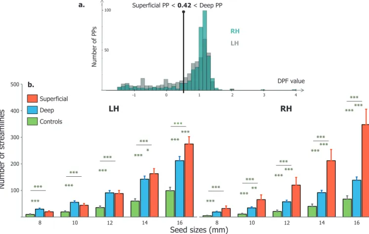
From the DPF maps we extracted one maximal DPF value per PP to plot their DPF distribution as done in LeGuenetal.(2018b). What stands out from this graph ( Fig.4a) is the difference of DPF distribution between hemispheres. We can observe a greater proportion of PPs with
negative or low positive DPF (i.e. superficial) in the left STS (light grey), and a higher number of PPs with high positive DPF (buried in the depth) in the right STS (dark grey). This distribution is very close to what has been reported in LeGuenetal.(2018b). However, in this paper the authors chose to discard PPs that have a DPF values greater than 0.42 considering them as noise. Here, we use this threshold to classify our PPs into “superficial ” (DPF < 0.42) or “deep ” (DPF > 0.42). Based on this classification, there were five times more deep ( n= 327) than superficial ( n= 61) PPs in the right hemisphere but only twice as much in the left ( n= 283 compared to n= 125). These proportions differed significantly in both hemispheres ( p<0.05, Wilcoxon). Between hemispheres, we also
C. Bodin, A. Pron, M. Le Mao et al. NeuroImage 225 (2021) 117513
Fig.4. a.ProportionofPPsaccordingtotheirDPFvaluesfortheleft(lightgrey)andrighthemisphere(lightblue).PPswereclassifiedas“superficial” or“deep” iftheirDPFlevelwasbeloworabove0.42respectively(verticaldashedline),basedonthethresholdusedinLeGuenetal.(2018b).b.Proportionsofstreamlines foundbelowsuperficialPPs(red),deepPPs(blue)andcontrols(green)intheleft(LH)andright(RH)hemisphere.X-axisindicatesthedifferentsurfacicseedsizes usedtoextractstreamlines(inmm).ThenumberofstreamlinesincreasesfromcontrolstodeepandtosuperficialPPsforallseedsizes.Errorsbarsillustratethe standarderrorofthemean(SEM).Significantdifferencesbetweencategoriesareindicatedbythestars(Mann–Whitneyranktest∗p<0.05∗∗p<0.001∗∗∗p<0.0001).
found that individual proportions of both deep PPs and superficial PPs differed significantly ( p<0.05, Wilcoxon). Thus, while the absolute num- ber of PPs does not seem to vary much from one hemisphere to another, their depth level is asymmetrically distributed with a greater proportion of superficial PPs in the left STS.
Some outliers emerged in the right tail of the DPF distribution as deep PPs with maximal DPF value. This is explained by our criteria used for deep PPs identification, for which an elevation of the STS fundus is not required and instead local deformations (‘pinching’) of the adjacent walls is used. Hence, outliers here can correspond to deeply buried PPs causing no STS fundus elevation ( seeFigs.2and3).
We reported in more detail the individual patterns concerning the deep and superficial PPs ( SupplementarymaterialFig.S2). The most represented pattern in our population was “1 superficial and 4 deep PPs ” in both hemispheres, with a frequency of 16% and 20% in the left and right STS respectively.
3.2. U-shapeconnectivity
The main analysis of the paper examined the short-range connec- tivity connecting the pairs of PPs seeds. Quantitative analysis revealed an increasing number of extracted streamlines according to seed size, with significant differences between PPs and controls for all sizes ( Fig. 4). The DPF-based classification into superficial and deep PPs described earlier was applied to extract the connectivity in these two categories separately. Both deep (blue) and superficial PPs (red) exhibited a higher short range connectivity (higher number of streamlines) than control re- gions (green) ( p<0.0001, Mann–Whitney test). The quantity of stream-
lines obtained for superficial PPs was also significantly higher than for deep PPs, from 14 to 16 mm seeds in the left STS and from 10 to 16 mm seeds in the right STS. These results suggest that PPs are at specific loca- tions within the STS where a majority of U-shape fibers cross the sulcus. Because we extracted the connectivity from PP seeds, we cannot re- late directly the number of PPs to that of streamlines due to circularity issues. However, we previously found that the total number of PPs in the left hemisphere did not differ significantly from that of the right hemi- sphere, making this question possible to assess. We found in general more streamlines in the left hemisphere compared to the right across individuals ( p<0.05, Wilcoxon) for all seed sizes except 8 mm ( Supple-mentaryFig.S2). In the precedent section, we showed that the left STS contained more superficial PPs ( n= 125) than the right hemisphere ( n= 61). However, we can see in the Fig.4that superficial PPs exhibited more streamlines in the right than in the left sulcus. Hence, the short- range connectivity appears to be more generally distributed among su- perficial and deep PPs in the left hemisphere, but concentrated mainly below superficial PPs in the right hemisphere.
Visual observation of the short range connectivity revealed clear short bundles that are transverse to the antero-posterior axis of the STS. Example subjects are represented on the Fig.5for which we can com- pare the position of previously identified PPs and the streamlines ex- tracted from the correspondent pairs of seeds. What emerged from the figure is that streamlines seem to co-localize with PPs positions but also that they follow the same orientation. In addition, the structural con- nectivity extracted from whole gyri seeds ( Fig.5right) and without assumptions on PPs’ location exhibit streamlines that are not randomly organized along the STS. Instead, short bundles seem to cross the STS
Fig.5.TheSTSshort-range(U-shape)structuralconnectivityillustratedonthreeindividualcorticalsurfaces(eachrow).Zoomedsagittalviewsofthetemporallobe arerepresented.Left:IndividualPPspreviouslydrawnonthesurface(bluelines).Middle:Short-rangeconnectivityextractedfromPPsseeds.Eachcolorrepresents thestreamlinesextractedfromonedistinctPP.Thetransparencyofthemeshwasincreasedforvisualization.Right:Short-rangeconnectivityextractedfromthe wholegyriseeds.AcolorcodewasattributedtoeachstreamlinedependingonitsorientationinrelationtotheSTS.Theblacklinesrepresentthegyricrestsadjacent totheSTS.
at the level of restricted portions distributed along the antero-posterior axis and corresponding to the identified position of PPs. On average, a greater amount of streamlines were extracted with the gyri-based method compared to those obtained from PPs seeds, across individuals and hemispheres ( Fig.S3).
We made two additional observations from the reported connectiv- ity patterns. First, the start and end points of the streamlines appeared generally distributed in a fan shape on the gyri walls and tighten in the middle, below the fundus of the STS like a bottleneck, as illustrated on the SupplementaryFig.S4for two example PPs. Second, we observed an apparent increase in the number of streamlines from the anterior to the posterior part of the STS. This was particularly true in the case of the gyri-based connectivity ( Fig.5right), for which streamlines are ex- tracted further in the temporo-parietal junction. Because we stopped our identification of PPs before the intersection of the STS horizontal and posterior branches, this can also explain the greater amount of stream- lines extracted from gyri seeds compared to PP seeds ( Fig.S3).
4. Discussion
4.1. PPsaslocalandgradualcorticaldeformations
The results presented in this study reinforce the idea that PPs are important landmarks in the STS organization. They can be associated with specific local geometrical features, “wall pinches ” from surround- ing gyri (here STG and MTG), an increase in the curvature of the STS
fundus and a local decrease of its depth. These criteria are in agreement with the earlier anatomical studies ( Cunningham,1892; Gratiolet,1854; Onoetal.,1990; Zlatkinaetal.,2016). In particular, our observations in the STS fitted particularly well with those of Cunningham for the central sulcus: “hereisgenerallyashallowingofthefissureandadeep in-terlockingofitsadjacentwalls.Twooftheinterdigitatinggyri,oneprojecting backwardsfromtheanteriorcentralconvolution[here downward from the STG] ,andtheotherforwardsfromtheposteriorcentralconvolution [here upward from the MTG] arealwayslargerandmorepronouncedthanthe others,andinaconsiderablenumberofcasestheyuniteatthebottomofthe sulcusintheformofadistinctdeepgyrus,whichconstitutesamarked inter-ruptioninitsfloor.” (Cunningham,1892; Whiteetal.,1997). In addition, instead of a binary categorization of the presence or absence of PPs that eliminates the most buried ones based on their depth ( LeGuenetal., 2018b; Leroyetal.,2015), we used a method that assumed a contin- uum from the deepest to the most superficial PPs. Cunningham’s words are in line again with this idea: “All gradationsbetweenamereshallowing withaninterlockingoftheadjacentwallsofthefissureandthepresenceof adistinctdeepannectantgyrusaremetwith”.
We believe that PPs constitute good candidates for a finer descrip- tion of the STS spatial organization and morphology ( Ochiaietal.,2004; Régis etal., 2005). Indeed the “sulcal roots ” model provides a tem- plate organization of folding to address inter-suject correspondences ( Régisetal.,2005) but still lacks flexibility, especially in the case of very complex sulci such as the STS. Based on this model, another method con- sists in the detection of sulcal pits ( Auziasetal.,2015; Imetal.,2010;
C. Bodin, A. Pron, M. Le Mao et al. NeuroImage 225 (2021) 117513 Lohmannetal.,2008) which relies on reproducible and highly local de-
pressions of the surface. Although they were shown relevant for studies on development ( Brun etal.,2016; ImandGrant,2019; Mengetal., 2014) and heritability ( LeGuenetal.,2018a) we still do not know their exact relationship with morphological patterns of sulci. For this pur- pose, PPs may be more appropriate, as they exhibit a complex geometry dependent on the shape of surrounding gyri ( Figs.2and3) and closely related to the underlying white-matter ( Figs.4and5).
4.2. PPsasshort-rangeconnectivitypathways
We found that PPs as we defined them morphologically constitute specific local pathways for the U-shape structural connectivity linking the two banks of the STS ( Figs.4and5). All PPs were considered as gradual forms of a depth continuum and identified based on specific morphological criteria ( Figs.2and3). In a second step, we classified them into “deep ” (DPF > 0.42) and “superficial ” PPs (DPF < 0.42) and both exhibited a higher connectivity than control regions were no PPs has been found ( Fig.4). This result suggests that PPs, regardless of their depth, are anatomical features of interest and that restrictive identifica- tion from the depth profile ( LeGuenetal.,2018b; Leroyetal.,2015) can miss crucial information. Comparison of the results obtained for PPs- and gyri-seeds reinforced our hypothesis of a dense U-shape connectivity below PPs locations. Instead of a random distribution of fibers between the STG and the MTG, we clearly observed several bundles along the antero-posterior axis of the STS whose location appears to correspond to that of PPs ( Fig.5).
The results we obtained are in agreement with the U-shape connec- tivity found along the central sulcus ( Catanietal.,2012; Magroetal., 2012; Pron et al., 2018), but differ from recent investigations of the short-range connectivity in the STS ( Abouzahr et al., 2019; Guevaraetal.,2017). Indeed, these authors reported only one bilat- eral bundle in the posterior STS that was orthogonal to the sulcus and connecting the STG and MTG. This discrepancy could be attributed to the fact that these studies aimed to extract only the most reproducible bundles across individuals whereas we have considered all the stream- lines under PPs here. Nevertheless, this could suggest that this posterior bundle is more genetically constrained, or at least more systematically present, compared to the others. Recently, a higher heritability was also reported in this region regarding the STS depth and the presence of PPs in the left hemisphere ( LeGuenetal.,2019). This posterior PP would also appear first during the development in-utero, followed by the others until the most anterior one between the 5th and 7th month ( Ochiaietal., 2004).
Further work is required to confirm the link between short-range connectivity and PPs through their respective reproducibility in the pop- ulation. In particular, it is certainly closely related to the location of the sulcal pits and must therefore be evaluated in relation to the heritabil- ity of these landmarks ( LeGuenetal.,2018a, 2019). Another approach would be to compare the occurrence of PPs and short range bundles across species. In Zhangetal.(2014), they did not find U-shape con- nections along the chimpanzee STS but some of them in the posterior STG of macaque monkeys, similar to what has been reported in a hu- man atlas ( Guevaraetal.,2017). Although very few studies investigated U-fibers in primates, the presence of PPs in these species has been docu- mented since the seminal studies ( Cunningham,1890b; Gratiolet,1854). Leroyetal.(2015)noted the presence of PPs in the STS of chimpanzees but they were less numerous and more symmetrically distributed com- pared to humans (again this result should be modulated by the PPs de- tection method). Inter-species comparison raises interesting questions about the functional role of PPs but also about their relationship to brain size and the level of gyrification. In light of the present results, less gyrified species may constitute ideal models for testing the associa- tion between PPs, gyrification and connectivity.
4.3. Anatomicalimplications
The relation suggested here between local cortical morphology and the underlying white matter can be related to recent advances in corti- cal development research ( Llinares-BenaderoandBorrell,2019). Fold- ing would be initially constrained by genetic factors that regulate cel- lular assemblies to differentiate sulci versus gyri as well as regional patterns. This process could also determine some crucial tissue proper- ties of the future cortex such as the stiffness and thickness of its layers. These tissue properties have been shown to interfere with the tangen- tial growth of the cortex by determining the final aspect of folding such as the wavelength of the folds at the regional scale ( Baylyetal.,2013; Buddayetal., 2014). The sulcal roots of the STS were shown to ap- pear separately along a caudal to rostral gradient ( Ochiaietal.,2004), each of them characterized by one pit separated by two transverse plis de passage ( Régisetal.,2005). We can imagine that tangential expan- sion of the surrounding STG and MTG portions follow the same timeline, gradually revealing the STS valley. Orthogonal to the sulcus, this expan- sion would lead to a digging of the STS excepted at the location of PPs that are also expanding in the opposite direction. Parallel to the sulcus, this expansion would then induce a deformation of the PPs depending on the surrounding growing pattern. This hypothetical order of events comes from the apparent deformation of superficial PPs, often form- ing “S ” or “C ” shapes with a slight shift of the extremities on the gyri walls.
The first question that appears here is how deep and superficial PPs are formed. Especially, what are the factors that could lead to a clear interruption of the sulcus or to “wall pinching ” configurations with no real depth variation? We propose here a three-step hypothesis following the developmental time scale: first, tangential growth during cortical development in utero would favor the transition from initial superficial PPs to more buried forms. This corroborates the findings of an increasing sulcal depth ( Glaseletal.,2011) and fusion phenomenon of sub-parts of the folds during this period ( Cunningham,1892). The early maturation of the right STS ( Glaseletal.,2011; Rajagopalanetal.,2011) would result in prolonged erosion of PPs giving rise to a deeper, less interrupted sulcus compare to its left counterpart ( Glaseletal.,2011; Leroyetal., 2015; Ochiaietal.,2004). Then, a second step would involve the gradual introduction of U-shape fibers joining the STS walls, passing in larger proportions through the most direct trajectories provided by superficial PPs, as previously suggested ( Manginetal.,2019). We have shown in this respect fewer streamlines under deep PPs and a generally lower proportion in the right (and deeper) STS ( Fig.4). Superficial PPs would constitute more direct routes since they are generally shorter and less curved than deep PPs (Supplementary Fig. S5).
It is important to note here that the aforementioned short-range con- nections seem to appear late in development, around birth and up to 6 months after ( Kostović et al., 2019). Hence, the third step would occur postnatally and involve in particular the later expansion of the cortex. The U-shape connectivity would serve here as a holding scaf- fold maintaining the pre-established PPs. More precisely, the weak (or strong) connectivity under the preexisting deep (or superficial) PPs mak- ing them more (or less) vulnerable to the surrounding expansion and hence lead to a “wall pinching ” remnant (or superficial bridge). This sec- ondary interaction between connectivity and PPs may be more exposed to environmental factors and potentially explain the large variability ob- served across individuals. Indeed in some subjects we identified more PPs (up to 8 in the left STS) than assumed by the sulcal roots model ( Ochiaietal.,2004; Régisetal.,2005). The functional properties inher- ent to each PP regions could be an important factor as well, especially in the determination of individual patterns ( Manginetal.,2019).
Together, these considerations raise the need for novel longitudinal studies using in-utero imaging that would specifically follow the evolu- tion of each PP, as already done for the sulcal pits ( Mengetal.,2014) or folding patterns ( Duanetal.,2018).
Fig.6. Potentialuseofplisdepassage(PP).a.Examplesulci(red)labelledandextractedautomaticallyinBrainVISAwithaclearPPvisibleatthepialsurface.b.
TwoexamplesubjectsofthepresentstudyshowingasimilarorganizationoftheirPP-associatedconnectivity.c.Curvaturemapsofthreeprimatespeciesprojected ontheirrespectivewhitemesh(generatedinBrainVISA).ThewhiterectangleindicatesapossiblePPcharacterizedbyalocalincreaseofthecurvatureandwall pinching.d.Individualfunctionalmapsofthehandmotorareainthecentralsulcus(up,seepanela.)orvoiceareasintheSTS(below)suggestatightrelationship withthelocationofPPsinthesesulci(whitestars).Redcolorindicatesastrongerfunctionalactivity.
4.4. Functionalimplications
There are two main functional implications of PPs, which are related to their asymmetry and spatial organization. On one hand, the asymmet- ric distribution of PPs could be related to functional asymmetry of the temporal region. We found that the left STS exhibited a stronger U-shape connectivity than the right while their respective number of PPs did not differ significantly. There are at least two possible explanations for this result. The most obvious concerns the greater proportion of superficial PPs in the left STS, which confirms earlier observations ( Ochiaietal., 2004). As we found generally more streamlines under superficial than deep PPs, the connectivity in left STS would be consequently higher. However, although the difference in number of streamlines is large be- tween deep and superficial PPs in the right STS, it is less so in the left STS ( Fig.4). Another possible explanation could be linked to other asymmet- ric properties of the STS. It is well know that language related functions are strongly lateralized in the brain, however, this mostly involves the fronto-temporal networks ( Hickok,2012). Nevertheless, some special- izations exist between the two temporal lobes such as temporal pro- cessing in the left and spectral processing in the right auditory cortex ( ZatorreandBelin,2001).
On the other hand, several studies attested to the relevance of PPs for functional localization. The position of sulcal interruptions was used to explain those of functional activity in various regions such as the intra- parietal ( Zlatkina andPetrides,2014), cingulate ( Amiezetal., 2013; Pausetal.,1996) and post-central ( Zlatkinaetal.,2016; Zlatkinaand Petrides,2010) sulci. In the fusiform gyrus, the presence of one PP in the visual word form area was recently associated to better reading skills, and length of the interruption correlated positively with these ability ( Cachiaetal.,2018). In the central sulcus, the PPFM co-localize with the hand motor area ( Fig.6d) ( Bolingetal.,1999; Cykowskietal.,2008;
Manginetal., 2019) and with U-shaped fibers bundle ( Magroetal., 2012; Pronetal.,2018). This correspondence is sufficiently robust that a variation in the position of the hand-knob is followed by that of the motor activation along the central sulcus ( Sunetal.,2015). Such re- lation was never investigated for the STS PPs, mainly because of the complex functional organization of the STS, also because interruptions vary to a greater extent across individuals. However, functional studies can help to identify candidate functional areas that could be associated with the STS PPs.
As previously noticed, we observed a dense connectivity in the pSTS that is often constituted of several bundles connecting close territories. This could be related to the complex functional organization in the re- gion encompassing the pSTS and the temporo-parietal junction (TPJ) ( Pateletal.,2019). On the right hemisphere, this region was shown to hold high-level social functions such as the representation of identity by integrating both facial and vocal information ( Davies-Thompsonetal., 2018; Hasanetal.,2016; Tsantanietal.,2019; Watsonetal.,2014). In the left hemisphere, this region was mainly associated to the dorsal pathway of language implicated in articulatory and production mech- anisms ( Hickok andPoeppel, 2004). In the middle temporal region, Upadhyayetal.(2008)evidenced two streams going from the primary auditory cortex toward the anterior and the posterior STG using effec- tive connectivity, and these areas where also found connected by struc- tural fiber pathways. Here, we observed several cases of bifurcations (see methods) crossing the middle STS that could correspond to this streams, as on line 2 of the Fig.5.
In a previous study ( Bodinetal.,2017), we found a correspondence between the local depth maxima in the STAP region ( Leroyetal.,2015) and the voice areas’ ( Belinetal.2000) maximal activity. At the indi- vidual level, this relation was less systematic notably because of the variable position of voice-related activity and depth pits along the STS.
C. Bodin, A. Pron, M. Le Mao et al. NeuroImage 225 (2021) 117513 Interestingly, in many cases we observed a maximal activity not in the
depth pit but on one of the PPs adjacent to it ( Fig.6d), which were already identified using the depth profile method on both sides of the STAP ( LeGuenetal.,2018a, 2018b; Leroyetal.,2015). The fact that voice-related activity can be clustered into three areas from the anterior STG to posterior STS ( Pernetetal.,2015) and that these areas are func- tionally inter-connected with each other’s ( Aglierietal.,2018) opens numerous questions on their link with PPs and therefore with the U- shape structural connectivity.
4.5. Limitationsofthestudy
This study was based on the manual identification of PPs based on the cumulated expertise of C.B. and from a previous investigation on the STS ( Bodinetal.,2017). Although more individuals of the HCP database could have met the selection criteria (see methods), we limited our study to 90 for time constraints while ensuring a high quality of PPs identi- fication. Additionally, we assume that this method may have included a proportion of noise in the PP identification procedure. In particular, the fact that deep PPs are more difficult to identify and were more nu- merous in the right STS could have participated to the difference in the number of streamlines found between superficial and deep PP in the right hemisphere ( Fig.4b).
New detection tools are needed to test the strength of the associa- tion between local morphology and connectivity while avoiding time- consuming and operator dependent manual steps. In addition, stream- lines extraction could be improved to prevent the overlap of seed re- gions. Indeed, in the case of two close neighboring seeds A and B (be- cause of close PPs), it happened that streamlines extracted from A ac- tually passed through the PPs corresponding to B. As mentioned above, although the streamlines were tightened in the STS, their extremities were more spatially dispersed on the gyri walls ( Fig.S4). Thus, future studies should also take into account the trajectory of streamlines ac- cording to that of PPs crests.
One general limitation concerns the plausibility of the results ob- tained with diffusion tractography. A recent report showed that the tractograms available to date contain more invalid than valid bundles in comparison to ground truth (post-mortem based) studies ( Maier-Heinetal., 2017). In particular, regions that are exhibiting multiple bundles (called “bottlenecks ”) such as the temporal lobe can lead to spu- rious tractographic reconstructions. Nevertheless, in this report they did not describe U-shape short-range connectivity. Here, we used a similar method to Pronetal.(2018)in which U-shape fibers were found under the PPFM location in the central sulcus, and already describes in another study relying on post-mortem blunt dissections ( Catanietal.,2012). The method is based on state-of-the art tractography methods followed by a filtering step (COMMIT, Daduccietal.,2015) that reduced the number of streamlines by 80%, securing those that explains best the signal in dMRI images. We also filtered streamlines based on their length to add some anatomical plausibility.
Geometric variations between deep and superficial PPs regions, such as the maximum curvature or the length of the path between the STG and MTG (Supplementary Fig. S5a), could potentially affect the perfor- mance of the tractography algorithm differently and thus explain the results observed in Fig.4. In order to control for this, we carried out a covariance analysis and found that the relation between the quan- tity of streamlines and the nature of PPs (superficial, deep or control) remains highly significant when taking into account their length or max- imum curvature as a covariate (Supplementary Fig. S5b). The impact of streamlines length was also controlled because we used a surface-based method to extract them, therefore avoiding the usual bias of voxel-based seeding for which longer tracts get more seeds and are therefore over- represented ( Girardetal.,2014).
Taken together, these arguments advocate in favor of a probable U- shaped connectivity under the STS PPs, although further work is needed to assess its robustness.
4.6. ConclusionandpotentialuseofPPs
This study showed that morphological criteria identifiable from in- dividual cortical surfaces can be used to detect and characterize the plis de passage. This lead us to suggest that the previously described asym- metry in the number of PPs between the left and right STS might in fact be an asymmetry of their depth but that their number is similar between both hemispheres. We demonstrated, for the first time in the superior temporal region, the nature of PPs as key places where short white-matter fibers converge to cross the STS. This heterogeneous fibers distribution appears to be modulated by the depth level of the PPs, with higher connectivity observed below the shallowest ones. Importantly, we do not claim to provide a definitive definition of PPs, rather, we suggest reconsidering these structures as continuous variations of mor- phological features and the U-shaped local connectivity as a new marker for the presence of PPs in the STS ( topofFig.6). These landmarks open up new avenues of research for a more detailed description of cortical anatomy across different cerebral regions and a new index to investi- gate individual variability. Further research might also explore whether PPs are present in other species, indeed, the cortical surface in monkeys and chimpanzees seems to present morphological deformations similar to those of deep PPs in humans ( Fig.6c). New investigations are needed to characterize them in light of the current results on U-shape connec- tivity. Finally, PPs could provide landmarks to localize the functional activity as done in the central sulcus or to reduce the individual vari- ability typical of high-level functions ( Fig.6d).
CRediTauthorshipcontributionstatement
C.Bodin:Writing - review & editing, Methodology, Conceptualiza- tion. A.Pron:Software. M.LeMao:Software. J.Régis:Conceptualiza- tion. P.Belin:Conceptualization. O.Coulon:Writing - review & editing, Software, Conceptualization, Supervision.
Acknowledgments
This work was supported by grants ANR-16-CONV-0002 (ILCB), ANR-11-LABX-0036(BLRI) and the Excellence Initiative of Aix-Marseille University (A ∗MIDEX). Data were provided [in part] by the Human Con-
nectome Project, WU-Minn Consortium (Principal Investigators: David Van Essen and Kamil Ugurbil; 1U54MH091657) funded by the 16 NIH Institutes and Centers that support the NIH Blueprint for Neuroscience Research; and by the McDonnell Center for Systems Neuroscience at Washington University.
Supplementarymaterials
Supplementary material associated with this article can be found, in the online version, at doi:10.1016/j.neuroimage.2020.117513.
References
Abouzahr, H. , Beyh, A. , Dell’Acqua, F. , Catani, M. , 2019. Longitudinal and vertical fibre systems in the human temporal lobe revealed by tractography. In: Proceedings of the Organization for Human Brain Mapping Conference. Rome .
Aglieri, V. , Chaminade, T. , Takerkart, S. , Belin, P. , 2018. Functional connectivity within the voice perception network and its behavioural relevance. NeuroImage 183, 356–365 .
Amiez, C. , Neveu, R. , Warrot, D. , Petrides, M. , Knoblauch, K. , Procyk, E. , 2013. The lo- cation of feedback-related activity in the midcingulate cortex is predicted by local morphology. J. Neurosci. 33 (5), 2217–2228 .
Auzias, G. , Brun, L. , Deruelle, C. , Coulon, O. , 2015. Deep sulcal landmarks: algorithmic and conceptual improvements in the definition and extraction of sulcal pits. NeuroImage 111, 12–25 .
Auzias, Guillaume , Lefevre, J. , Le Troter, A. , Fischer, C. , Perrot, M. , Régis, J. , Coulon, O. , 2013. Model-driven harmonic parameterization of the cortical surface: HIP-HOP. IEEE Trans. Med. Imaging 32 (5), 873–887 .
Avila, N.L. , Lebenberg, J. , Rivière, D. , Auzias, G. , Fischer, C. , Poupon, F. , Guevara, P. , Poupon, C. , Mangin, J.-F. , 2019. Inference of an extended short fiber bundle atlas using sulcus-based constraints for a diffeomorphic inter-subject alignment. In: Pro- ceedings of the Workshop on Computational Diffusion MRI, pp. 323–333 .