HAL Id: hal-02632476
https://hal.inrae.fr/hal-02632476
Submitted on 27 May 2020
HAL is a multi-disciplinary open access
archive for the deposit and dissemination of
sci-entific research documents, whether they are
pub-lished or not. The documents may come from
teaching and research institutions in France or
abroad, or from public or private research centers.
L’archive ouverte pluridisciplinaire HAL, est
destinée au dépôt et à la diffusion de documents
scientifiques de niveau recherche, publiés ou non,
émanant des établissements d’enseignement et de
recherche français ou étrangers, des laboratoires
publics ou privés.
Distributed under a Creative Commons Attribution - NonCommercial| 4.0 International
License
A trilogy of glucocorticoid receptor actions
Chek Kun Tan, Walter Wahli
To cite this version:
Chek Kun Tan, Walter Wahli. A trilogy of glucocorticoid receptor actions. Proceedings of the National
Academy of Sciences of the United States of America , National Academy of Sciences, 2016, 113 (5),
pp.1115-1117. �10.1073/pnas.1524215113�. �hal-02632476�
COMMENTARY
A trilogy of glucocorticoid receptor actions
Chek Kun Tanaand Walter Wahlia,b,c,1
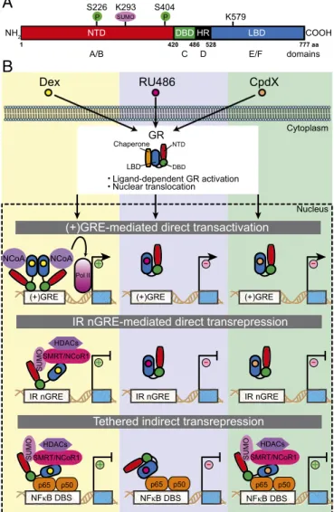
Glucocorticoids (GCs) belong to a class of endoge-nous, stress-stimulated steroid hormones found in vertebrates (e.g., cortisol in humans and corticoste-rone in rodents); they have wide ranging physiologic effects capable of impacting metabolism, immunity, development, stress, cognition, and arousal. GCs exert their cellular effects by binding to the GC receptor (GR), one of a 48-member (in humans) nuclear receptor (NR) superfamily of ligand-activated transcription factors (1). As the first human NR to be cloned (2), GR has provided an invaluable template with which to under-stand how the structurally related NRs exert their com-plex cellular effects. Its activity also underscores the importance of small lipophilic ligands in regulating multiple biologic pathways. Like other NR family members, the GR comprises three major functional domains: (i) an N-terminal domain (NTD), which con-tains a constitutive activation function 1 (AF-1); (ii) a DNA-binding domain (DBD), containing two zinc fin-ger motifs; and (iii) a C-terminal, ligand-binding do-main (LBD), with its ligand-dependent AF-2 (Fig. 1A). The human and mouse GRs are encoded by a single NR3C1 gene, which product can be differentially spliced into two major isoforms, GRα and GRβ; the former is responsible for the majority of GR-mediated transcriptional activity (3). Additional variants, gener-ated via translational regulatory mechanisms, together with posttranslational modifications (PTMs), contrib-ute to the complexity and diversification of GR-medi-ated action (3, 4). These PTMs can dial up, or down, GR-mediated transcriptional activities, to confer dis-tinct biologic functions. Relevant PTMs include phos-phorylation, acetylation, methylation, ubiquitination, and SUMOylation (4). The first of these, phosphor-ylation, has been shown to modulate dimerization and DNA binding, coregulator interaction, and ligand-binding affinity, all of which alter transcriptional activ-ity. A total of nine phosphorylation sites within the human GR NTD has been reported, some of which influence nuclear export and coregulator recruitment (5). Interestingly, NR SUMOylation, which involves the covalent conjugation of SUMO moieties at specific
lysines, triggers molecular transrepression pathways that link metabolism and inflammation (6). In two back-to-back publications in PNAS (7, 8), Hua et al. now report the detailed molecular mechanisms by which GR SUMOylation provokes GC-dependent gene repression.
In the absence of GC hormone, cytoplasmic GR is rendered inactive by bound chaperones (e.g., Hsp90). A conformational change in the GR LBD that accom-panies GC binding causes GR activation and nuclear translocation (9, 10). Once in the nucleus, GR binds to the so-called positive GC response element [i.e., (+)GRE DNA-binding sequence (DBS)]. (+)GRE DBSs lie in the regulatory regions of target genes, and stimu-late their expression via GR-dependent recruitment of a transcription initiation complex (Fig. 1B). While the molecular mechanism involved in GC-induced, GR-stimulated gene activity, has been extensively studied (11), agonist-activated GR also has gene-repressive activities conferred by two quite distinct mechanisms (Fig. 1B). The first of these, termed“tethered indirect transrepression,” arises when ligand-activated GR as-sociates with transcription factors [NFκB (p65), AP1 (c-jun), or STAT3] bound to their cognate DBS (12). The second, more recently described repression mech-anism, involves a direct binding of ligand-activated GR to an evolutionarily conserved negative GRE [inverted repeat (IR) nGRE DBS]; the result is GC-induced direct transrepression (13–15). The IR nGRE is unrelated to the (+)GRE DBSs described earlier, or a variety of nGREs (13). Until now, the molecular mechanisms governing IR nGRE-mediated direct and tethered indirect transre-pression were poorly understood.
In their articles, Hua et al. now decipher these mechanisms (7, 8) (Fig. 1B). The authors demonstrate in vitro and in vivo that SUMOylation of the human and mouse GR [at lysine (K) 293 of human GR] within a conserved region of its NTD, is indispensable for either form of transrepression. For IR nGRE-mediated direct repression, SUMOylation of the GR is manda-tory for the assembly of a repressive complex at the IR nGRE DBS; this complex comprises either one, or
CO
MMENT
ARY
a
Lee Kong Chian School of Medicine, Nanyang Technological University, Singapore 637553;b
Center for Integrative Genomics, University of Lausanne, CH-1015 Lausanne, Switzerland; andcInstitut National de la Recherche Agronomique, Université de Toulouse, UMR1331, ToxAlim,
Research Center in Food Toxicology, 31027 Toulouse, France Author contributions: C.K.T. and W.W. wrote the paper. The authors declare no conflict of interest.
See companion articles on pages E626 and E635.
1To whom correspondence should be addressed. Email: walter.wahli@ntu.edu.sg.
both, of the corepressors SMRT (silencing mediator for retinoid or thyroid-hormone receptors) and NCoR1 (nulear receptor corepres-sor 1), as well as histone deacetylase HDAC3. Furthermore, phos-phorylation at two serine residues (i.e., S226 and S404 in the human GR NTD) could boost SUMOylation. Depending on the identity of the IR nGRE-containing genes, either SMRT or NCoR1 was required for GR binding to the IR nGREs, and direct transrepression. The authors also found that lysine K579, within the human GR LBD, was necessary for NCoR1 binding to SUMOylated GR and IR nGRE-mediated repression; no such dependency was found for SMRT binding and repression. However, whether SMRT may also directly interact with the GR LBD remains to be determined. Impor-tantly, the repressive complexes of SUMOylated GR and SMRT/ NCoR1 corepressors associated with HDAC3 neither bound to a (+)GRE nor inhibited (+)GRE-mediated transactivation. This indicates that formation of the repressive complexes was spe-cific to IR nGREs. Although it is known that the GR DBD can
interact in vitro with both the (+)GRE and IR nGRE DBS, it binds much more strongly to the former (16, 17). Seemingly, SUMOylation of the GR, as well as the recruitment and assem-bly of a SMRT/NCoR1-HDAC3 repressing complex, is required for its efficient binding to IR nGREs. This mode of regulation is in keeping with the observation that only a small fraction of cellular GR is SUMOylated, and would also indicate that there is no absolute need to maintain a dedicated nuclear pool of SUMOylated GR.
Hua et al. (8) also reveal in vitro and in vivo a similar depen-dence on a SUMOylated GR NTD for corepressor recruitment (SMRT/NCoR1) in tethered indirect transrepression, which is relevant to the GC-induced anti-inflammatory effects. However, SUMOylation was dispensable for the binding of GR to DNA-bound transcription factors (NF-κB/AP-1/STAT3); instead, the GR LBD was needed to interact with NF-κB or AP1, as proven by the loss of DNA-bound p65, or c-Jun, to a LBD-deleted GR. Furthermore, in vivo data provide some clues as to the chronology of events. The recruitment of the SMRT/NCoR1 repressor com-plexes appeared to precede HDAC3’s binding to the GR-repressive complex bound to NF-κB and AP1. Interestingly, as for IR nGRE-mediated direct transrepression, phosphorylation of the GR NTD dialed-up SUMOylation of NF-κB/AP1-bound GR and increased binding of the SMRT and NCoR1 corepressors.
Despite their side effects, the potent anti-inflammatory effects of GCs have led to their widespread use in treating inflammatory and allergic disorders. Clearly any property of GCs that could discriminate between harmful and beneficial effects would be a substantial step forward in understanding the GR functions. That step came when it was found that many of the clinically de-bilitating effects caused by prolonged GC therapy were related to its transcriptional activities and specifically (+)GRE-mediated di-rect transactivation (15) and IR nGRE-mediated didi-rect repression (13). On the other hand, the process of GR-mediated tethered indirect transrepression segregated with therapeutically benefi-cial, anti-inflammatory effects (15). Not surprisingly, this distinc-tion led to the search for“dissociated” ligands (SEGRAs, SElective GR Agonists): agonists that would fit the indirect transrepressive profile but avoid any direct effects. Thus far, the search for these elusive compounds has been unsuccessful; compounds lacking (+)GRE-mediated transactivation have been identified, but their IR nGRE-mediated direct transrepression activity remains largely intact (13). Until now, the search for the beneficial SEGRA has suffered from our lack of understanding about the relevant mo-lecular mechanisms. In this respect, the findings now reported by Hua et al. (7, 8) should pave the way to educated designs and screens for such compounds; in short, they tell us what to look for. Better still, encouraging data to support the notion that these compounds exist come from the preliminary characterization of a putative SEGRA, CpdX, which selectively induces GR-mediated NF-κB/AP1 indirect transrepression activity yet maintains the anti-inflammatory properties (8).
There are still intriguing questions to be solved regarding the molecular mechanisms involving GR-induced transrepression. For example, can SMRT directly interact with the GR LBD like NCoR1? Is the retention of an active repressive complex that contains SMRT (but not NCoR1) by a GR construct lacking a LBD, indicative of SMRT recruitment to other regions of the GR, such as the NTD or DBD? Future in vivo studies are required to establish the most beneficial pharmacokinetic profile of CpdX (e.g., duration and dosage of treatment), and determine whether its administra-tion could interfere with the regulatory activities mediated by
Dex
Cytoplasm
B
(+)GRE-mediated direct transactivation
A
Tethered indirect transrepression IR nGRE-mediated direct transrepression
NCoA Pol II (+)GRE NCoA IR nGRE SUMO SMRT/NCoR1 HDACs NFκB DBS SUMO SMRT/NCoR1 HDACs p65 p50 RU486 (+)GRE CpdX Nucleus NTD DBD HR LBD a a 7 7 7 8 2 5 6 8 4 0 2 4 1
A/B C D E/F domains K579 P S226 S404 SUMO K293 ChaperoneGR DBD NTD LBD
+
COOH NH2 p65 p50 NFκB DBS −+
IR nGRE −+
− (+)GRE − IR nGRE − NFκB DBS+
Ligand-dependent GR activation Nuclear translocation P SUMO SMRT/NCoR1 HDACs p65 p50Fig. 1. Schematic illustration (A) of the structural domains within the human GR, indicating relevant phosphorylation (P), and SUMOylation sites, and (B) the three molecular mechanisms by which GR regulates expression of its target genes upon binding an agonist (e.g., dexamethasone, Dex), an antagonist (e.g., RU486), or a SEGRA (e.g., CpdX).“+” indicates stimulation of gene activation or repression, whereas“−” indicates no effect on stimulation or repression. DBD, DNA-binding domain; HR, hinge region; LBD, ligand-binding domain; NTD, N-terminal domain.
endogenous GC. Last but not least, it is also important to find out whether administration of CpdX at the time point of lowest diurnal levels of endogenous GC (i.e., during the resting phase of the circadian cycle) could be beneficial.
The isolation of the GR gene and that of other NR family members, followed by their functional characterization, revolu-tionized our understanding of the signaling pathways triggered by small lipophilic molecules. These data have led to some largely unanticipated discoveries as to how these molecules, and their
associated regulatory apparatuses, have evolved to become such master regulators of our physiology. The demonstration of distinct direct and indirect transcriptional repression mechanisms for GR function illustrates this point. Given the latest discoveries, and those made since the initial cloning of the NRs, we anticipate a further unraveling of the GR’s functional complexity that could help us to discover agents with clinically useful anti-inflammatory functions, which are free of the drawbacks that we currently asso-ciate with GCs.
1 Mangelsdorf DJ, et al. (1995) The nuclear receptor superfamily: The second decade. Cell 83(6):835–839.
2 Hollenberg SM, et al. (1985) Primary structure and expression of a functional human glucocorticoid receptor cDNA. Nature 318(6047):635–641.
3 Lu NZ, Cidlowski JA (2005) Translational regulatory mechanisms generate N-terminal glucocorticoid receptor isoforms with unique transcriptional target genes. Mol Cell 18(3):331–342.
4 Anbalagan M, Huderson B, Murphy L, Rowan BG (2012) Post-translational modifications of nuclear receptors and human disease. Nucl Recept Signal 10:e001. 5 Galliher-Beckley AJ, Cidlowski JA (2009) Emerging roles of glucocorticoid receptor phosphorylation in modulating glucocorticoid hormone action in health and
disease. IUBMB Life 61(10):979–986.
6 Treuter E, Venteclef N (2011) Transcriptional control of metabolic and inflammatory pathways by nuclear receptor SUMOylation. Biochim Biophys Acta 1812(8): 909–918.
7 Hua G, Paulen L, Chambon P (2016) GR SUMOylation and formation of a SUMO-SMRT/NCoR1-HDAC3 repressing complex is mandatory for GC-induced IR nGRE-mediated transrepression. Proc Natl Acad Sci USA 113:E626–E634.
8 Hua G, Ganti KP, Chambon P (2016) Glucocorticoid-induced tethered transrepression requires SUMOylation of GR and formation of a SUMO-SMRT/NCoR1-HDAC3 repressing complex. Proc Natl Acad Sci USA 113:E635–E643.
9 Ricketson D, Hostick U, Fang L, Yamamoto KR, Darimont BD (2007) A conformational switch in the ligand-binding domain regulates the dependence of the glucocorticoid receptor on Hsp90. J Mol Biol 368(3):729–741.
10 Vandevyver S, Dejager L, Libert C (2012) On the trail of the glucocorticoid receptor: into the nucleus and back. Traffic 13(3):364–374. 11 Meijsing SH, et al. (2009) DNA binding site sequence directs glucocorticoid receptor structure and activity. Science 324(5925):407–410.
12 Uhlenhaut NH, et al. (2013) Insights into negative regulation by the glucocorticoid receptor from genome-wide profiling of inflammatory cistromes. Mol Cell 49(1): 158–171.
13 Surjit M, et al. (2011) Widespread negative response elements mediate direct repression by agonist-liganded glucocorticoid receptor. Cell 145(2):224–241. 14 Cruz-Topete D, Cidlowski JA (2015) One hormone, two actions: Anti- and pro-inflammatory effects of glucocorticoids. Neuroimmunomodulation 22(1-2):20–32. 15 Clark AR, Belvisi MG (2012) Maps and legends: the quest for dissociated ligands of the glucocorticoid receptor. Pharmacol Ther 134(1):54–67.
16 Luisi BF, et al. (1991) Crystallographic analysis of the interaction of the glucocorticoid receptor with DNA. Nature 352(6335):497–505. 17 Hudson WH, Youn C, Ortlund EA (2013) The structural basis of direct glucocorticoid-mediated transrepression. Nat Struct Mol Biol 20(1):53–58.
![The characterization of human [gamma]D-crystallin mutants and their differential interactions with the lens chaperone [alpha]B-crystallin](data:image/gif;base64,R0lGODlhAQABAIAAAP///wAAACH5BAEAAAAALAAAAAABAAEAAAICRAEAOw==)