HAL Id: hal-03004373
https://hal.archives-ouvertes.fr/hal-03004373
Submitted on 13 Nov 2020
HAL is a multi-disciplinary open access
archive for the deposit and dissemination of
sci-entific research documents, whether they are
pub-lished or not. The documents may come from
teaching and research institutions in France or
abroad, or from public or private research centers.
L’archive ouverte pluridisciplinaire HAL, est
destinée au dépôt et à la diffusion de documents
scientifiques de niveau recherche, publiés ou non,
émanant des établissements d’enseignement et de
recherche français ou étrangers, des laboratoires
publics ou privés.
atypical striated muscles involved in maintaining the
position of internal organs
Laetitia Bataillé, Nathalie Colombié, Aurore Pelletier, Achim Paululat, Gaëlle
Lebreton, Yannick Carrier, Jean-Louis Frendo, Alain Vincent
To cite this version:
Laetitia Bataillé, Nathalie Colombié, Aurore Pelletier, Achim Paululat, Gaëlle Lebreton, et al.. Alary
muscles and thoracic alary-related muscles are atypical striated muscles involved in maintaining the
position of internal organs. Development (Cambridge, England), Company of Biologists, 2020, 147
(8), pp.dev185645. �10.1242/dev.185645�. �hal-03004373�
RESEARCH ARTICLE
Alary muscles and thoracic alary-related muscles are atypical
striated muscles involved in maintaining the position of internal
organs
Laetitia Bataillé1,*, Nathalie Colombié1, Aurore Pelletier1, Achim Paululat2, Gaë lle Lebreton1, Yannick Carrier1,
Jean-Louis Frendo1and Alain Vincent1
ABSTRACT
Alary muscles (AMs) have been described as a component of the cardiac system in various arthropods. Lineage-related thoracic muscles (TARMs), linking the exoskeleton to specific gut regions, have recently been discovered in Drosophila. Asymmetrical attachments of AMs and TARMs, to the exoskeleton on one side and internal organs on the other, suggested an architectural function in moving larvae. Here, we analysed the shape and sarcomeric organisation of AMs and TARMs, and imaged their atypical deformability in crawling larvae. We then selectively eliminated AMs and TARMs by targeted apoptosis. Elimination of AMs revealed that AMs are required for suspending the heart in proper intra-haemocelic position and for opening of the heart lumen, and that AMs constrain the curvature of the respiratory tracheal system during crawling; TARMs are required for proper positioning of visceral organs and efficient food transit. AM/TARM cardiac versus visceral attachment depends on Hox control, with visceral attachment being the ground state. TARMs and AMs are the first example of multinucleate striated muscles connecting the skeleton to the cardiac and visceral systems in bilaterians, with multiple physiological functions.
KEY WORDS: Alary muscles, Striated muscles, Cardiac system, Respiratory system, Visceral system,Drosophila
INTRODUCTION
Three types of muscles are classically distinguished in mammals: skeletal, cardiac and smooth muscle. The cardiac and skeletal muscles are striated, with striation reflecting the reiteration of sarcomeres, which are the basic contractile units assembled from alternating antiparallel rows of myosin-based thick and actin-based thin filaments. Skeletal muscle fibres, which underlie body movements, are multinucleated syncytia formed by fusion of myocytes. They attach to the skeleton via tendons at both ends, except for some facial muscles and extra-ocular muscles, which either attach to the epidermis or to other muscles (Pélissier et al., 2000; Noden and Francis-West, 2006; Ziermann et al., 2018). Holometabolous insects display two successive muscle patterns, which underlie larval and adult locomotion. Locomotion of the soft-bodied Drosophila larva includes linear crawling, head turns and rolling movements (Hwang et al., 2007;
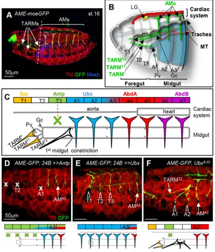
Heckscher et al., 2012). About 30 distinct body wall muscles per hemi-segment are attached at both ends to the larval exoskeleton via tendon cells (Bate and Rushton, 1993; Armand et al., 1994). Each muscle displays a specific morphology, which is linked to the expression of muscle identity transcription factors (iTFs), including many orthologues of mammalian myogenic TFs, such as MyoD/MRF, Islet1, Six and Tbx1 (de Joussineau et al., 2012; Dubois et al., 2016; Buckingham, 2017). Beside the muscles required for locomotion, one alary muscle (AM) in each abdominal hemi-segment connects the lateral exoskeleton to the dorsal vessel, the Drosophila cardiac system which comprises the aorta and the heart. At larval hatching, seven pairs of AMs adhere to the extracellular matrix (ECM) surrounding the pericardial cells (PCs) located along the aorta and heart proper (Rizki, 1978; LaBeau et al., 2009; Lehmacher et al., 2012). Org-1 (Optomotor-blind-related-gene-1), the Drosophila Tbx1 orthologue, and Tailup (Tup)/Islet1 are iTFs expressed in AMs (Tao et al., 2007; Schaub et al., 2012; Boukhatmi et al., 2012). Characterisation of Org-1 and tup mesodermal enhancers led us to discover the existence of three pairs of thoracic alary-related muscles (TARMs), each attaching to a specific midgut region (Fig. 1A,B and Boukhatmi et al., 2014).
AMs have been described in many arthropods and proposed to connect epi/pericardial cells around the adult heart and to control ostia opening and heart beating (Jones, 1954; Curtis et al., 1999; Ejaz and Lange, 2008; Buechling et al., 2009; Glenn et al., 2010; League et al., 2015), although neither role has been formally established. Moreover, the finding that adhesion of one AM to the distal tip cell of Malpighian tubules (MTs) is required for proper MT bending during embryogenesis (Weavers and Skaer, 2013) and that AMs and TARMs loop around main branches of the respiratory tracheal system (Boukhatmi et al., 2014) led us to postulate a function of AMs and TARMs in internal organ positioning in crawling larvae (Bataillé et al., 2015).
Here, we analysed in detail the striated organisation of AMs/ TARMs and their attachments to multiple internal organs, and imaged their deformability in crawling larvae. We then ablated AMs and TARMs by transcription enhancer-targeted apoptosis. Loss of AMs induces a collapse of the cardiac system and relieves topological constraints on the curvature of the respiratory system. Loss of TARMs interferes with positioning of the gastric caeca and visceral mass and affects food transit. Our characterisation of TARMs/AMs, a new type of muscle connecting the skeleton to internal organs with multiple physiological functions, brings a new viewpoint on animal anatomy.
RESULTS
Morphological diversification of AMs and TARMs: Hox control
Pan-mesodermic expression of the homeotic protein Ubx induces transformation of TARMs into AM-like muscles connecting to
Received 15 October 2019; Accepted 4 March 2020
1Centre de Biologie du Développement (CBD), Centre de Biologie Intégrative (CBI),
Université de Toulouse 3, CNRS, UPS, 118 route de Narbonne, 31062 Toulouse, France.2University of Osnabrü ck, Department of Biology/Chemistry, Zoology and
Developmental Biology, Barbarastraße 11, 49076 Osnabrü ck, Germany. *Author for correspondence (laetitia.bataille@univ-tlse3.fr; laetitia.bataille@inserm.fr)
L.B., 0000-0002-3897-9520; N.C., 0000-0002-6239-7109; J.-L.F., 0000-0003-0118-5556; A.V., 0000-0002-2769-7501
DEVEL
O
the aorta (LaBeau et al., 2009; Weavers and Skaer, 2013; Bataillé et al., 2015), the first indication that Hox activity distinguishes between AM and TARM morphology and their attachment to either the circulatory or the visceral system. To investigate this in depth, we surveyed Hox expression in TARMs
and AMs (Fig. 1C; Fig. S1). TARM* and TARMT1, which
connect the proventriculus (Pv) and dorsal gastric caeca (Gc), respectively, originate from progenitor cells specified in T1 and
express Scr. No Hox expression could be detected in TARMT2,
which connects to the first midgut constriction, and Antp is
expressed in the abortive TARMT3, which undergoes apoptosis
during embryogenesis (Fig. S1). Abdominal AMs express the Bithorax complex proteins Ubx in A1-A3, AbdA in A3-A7 (LaBeau et al., 2009) and AbdB in A5-A7 segments.
The TARM/AM Hox code raised the question of whether Antp expression induces TARM apoptosis. Ectopic Antp expression in the entire mesoderm results in loss of all TARMs plus, in most embryos,
AMA1(Fig. 1D). Conversely, as previously reported (Bataillé et al.,
2015), Ubx pan-mesodermal expression induces formation of an
AM-like muscle in T3 (Fig. 1E). Thus, Antp promotes TARMT3
apoptosis in the absence of posterior Hox function. The absence of
Hox expression in TARMT2is reminiscent of the situation reported
for somatic muscles by Roy et al. (1997), who proposed that the T2
muscle pattern was a ‘ground state’. To analyse this further, we
examined Ubx loss-of function embryos. Absence of Hox
information leads to AMA1and AMA2transformation into
TARM-like muscles connecting to the gut (21/34 hemi-embryos, 62%) at the
same position as TARMT2(Fig. 1F). Together, Hox expression,
loss-of-function and gain-loss-of-function phenotypes show both that the number of TARMs and attachment of TARMs and AMs to either
the visceral or circulatory systems are under Hox control, and that the background state is visceral attachment.
AMs and TARMs connect to multiple internal organs in larvae
AMs have been described as a structural component of the adult insect heart, but their physiological role has never been analysed. One essential step towards addressing TARM/AM functions in larvae is examining their morphology. One plausible reason why TARMs were only recently discovered is their unusual thinness and internal position (Boukhatmi et al., 2014). To circumvent any possible damage to TARMs/AMs during dissections, we engineered fluorescent
transgenes AMER-cd4-tandem(td)Tomato or -cd4-tdGFP in order to
visualise AMs and TARMs in intact larvae (Fig. 2; Fig. S2). These tags revealed that TARMs and AMs maintain their connections to specific internal organs throughout larval development. However, although all AMs display a similar morphology in embryos (Fig. 1A), the anterior AMs connecting to the aorta (A1-A4 segments) adopt
a conspicuous‘T’ shape in larvae (Fig. 2A); the shaft runs from
lateral tendon cells to the aorta, whereas lateral branches run orthogonally along the aorta. Each A1-A4 AM thus connects to a
specific number of PCs [AMA1: 1 PC; AMA2: 1.7±0.7; AMA3: 3.3±
0.8; AMA4: 4.4±0.5 (n=24)]. AMA1also connects to the primary lobe
of the lymph gland (LG), the larval haematopoietic organ, and vesicles originating from AMs are observed in the aorta in this region
(Fig. S3). AMA2 and AMA3 connect to secondary, posterior LG
lobes (Fig. S3). Posterior AMs (A5-A7 segments) maintain
delta-shaped connections to PCs distributed along the heart, with AMA5
connecting to three PCs (3.2±0.5), AMA6 to four (4.1±0.5) and
AMA7to one PC (1.1±0.3) (n=24). Quadruple labelling of semi-intact
larvae, including Pericardin (Prc), a cardiac ECM component
Fig. 1. Hox control of TARM and AM internal organ connections. (A) Stage 16 AME-moeGFP embryo stained for GFP (green), Tropomyosin (TM, red) and Mesh (blue) to visualise the AMs and TARMs, somatic muscles, and gut, respectively. (B) Schematic of the anterior region of a stage 16 embryo, showing AMs (green) connections to the cardiac system (red), lymph gland (LG, yellow) and tip cell of the Malpighian tubule (MT, orange). AMs run internal to the dorsal tracheal trunk (grey). TARM*, TARMT1and TARMT2span
several segments and connect to the proventriculus (Pv), dorsal gastric caeca (Gc) andfirst midgut constriction, respectively. Scheme modified from Boukhatmi et al. (2014). (C) Colour-coded representation of Scr, Antp, Ubx, AbdA and AbdB expression in TARMs/AMs and of TARM and AM connections to specific midgut regions and the cardiac system in wild-type embryos. The green cross indicates muscle death induced by Antp expression. (D-F) Anterior region of moeGFP; 24B-Gal4/UAS-Antp (D), AME-moeGFP; 24B-Gal4/UAS-Ubx (E) and AME-moeGFP, Ubx9.22(F)
embryos stained for GFP and TM (see A). AMA3position, and ablated
(x) or transformed (arrowhead) muscles are indicated. Below are schematic representations of the observed AM/TARM
transformations (n>30). Scale bars: 50 µm.
DEVEL
O
(Lehmacher et al., 2012), shows that AM attachment is mediated by cardiac ECM that surrounds PCs (Fig. 2B). 3D reconstructions reveal that, in addition to attaching AMs to the heart, ECM also ties the heart
to the dorsal wall (Fig. 2B′). Along their path from the exoskeleton
to the heart, AMs loop around the dorsal tracheal branch (Fig. 2B′).
Moreover, cytoplasmic protrusions at intermediate positions show
that AMs adhere to other internal organs: AMA3 to the anterior
MT (Weavers and Skaer, 2013), AMA1 to the anterior fat body,
and AMA4and AMA5to the fat body surrounding the male gonad
(Fig. S3).
TARM connections to internal organs were originally mapped in the embryo by immunostainings (Boukhatmi et al., 2014). Here, we
visualised these connections in larvae by using AMER
-cd4-tdTomato labelling of AMs/TARMs and A142-GFP expression in visceral organs (Buchon et al., 2013). It shows that TARM* remains
connected to the Pv, TARMT1to the tip of dorsal Gcs (Fig. 2C) and
TARMT2 to the first midgut loop, which shows a characteristic
U-shaped bending in larvae (Fig. 2C′,D). This bending occurs
where enteroendocrine cells, which express the diuretic hormone DH31, marked by ChAT-Gal4 expression, are located, which is at the junction between the anterior and acidic portions of the midgut (LaJeunesse et al., 2010).
Close examination of AM/TARM connections to the lateral epidermis revealed an unexpected morphological diversity of these attachment sites (Fig. S4). Triple staining for AMs/TARMs, tendon cells and the integrin adhesion complex (Ilk-GFP fusion protein), shows that the discrete adhesion zone of AM/TARMs to one tendon
cell is extended by a web of ‘filopodia’ reaching beyond the
contacted tendon cell. The role of these projections remains to be
explored. Moreover, whereas AMA1-AMA5attach to dorsal tendon
cells, AMA6and AMA7attach to lateral tendon cells, i.e. at a more
ventral position than AMA1-AMA5, possibly relating to different
functions of anterior and posterior AMs.
In summary, AMs and TARMs connect the larval epidermis to many multiple internal organs, reinforcing the hypothesis that AMs
and TARMs could contribute to maintenance of the internal anatomy of moving larvae.
AMs and TARMs are multinucleated, sarcomeric muscles displaying unique deformability
In somatic muscle fibres, nuclei are uniformly spaced within the fibre (Volk, 2013). To examine myonuclei number and repartition in AMs and TARMs, we expressed simultaneously nuclear RFP-labelled and membrane-associated GFP proteins (Fig. 3A). TARMs
contain between three and four nuclei (TARM*: 2.9±0.3; TARMT1:
4.1±0.9; TARMT2: 4.4±0.5; n=10 larvae), evenly distributed along
the fibre length. AMA1to AMA7contain five or six nuclei (5.5±1.1;
n=20). In anterior AMs with a ‘T’ shape, nuclei are distributed
between the shaft and lateral branches. In posterior AMs, nuclei are distributed in a single row, up to the position where the muscle widens to adopt a delta-like morphology. At this point, nuclei are laterally scattered among the myofibrils (Fig. 3A). Thus, repartition of nuclei in TARMs and AMs adapts to their peculiar asymmetric geometry.
The atypical shapes of TARMs and AMs raised the question of how myofibrils are organised in these muscles. We used cd4-Tomato decoration of AM/TARM contours together with MHC-GFP (Sarov et al., 2016) and Phalloidin staining of thick and thin filaments, respectively, to visualise the sarcomeric
myofibrils. In the anterior AMs (Fig. 3B,B′), myofibrils align
with the shaft, before bending 90° to form lateral branches aligning with the aorta. This bending occurs without rupture in the sarcomeric organisation, such that anterior AMs display two perpendicular sarcomeric contraction lines. In posterior AMs
(Fig. 3C,C′), myofibrils scatter in a pattern similar to a fan frame
to connect the ECM surrounding the heart (see also Fig. 2B and Lehmacher et al., 2012). The sarcomeric region is extended dorsally
by thin cellular extensions reaching cardiomyocytes and,
sometimes, the opposite AM proteins (Fig. 3C′). The AM
sarcomeres were also visualised by the expression of a titin-like
Fig. 2. AMs and TARMs connect to multiple larval internal organs. (A) Dorsal view of an intact AMER-Gal4;
UAS-cd4-tdTomato, HandC-GFP L3 larva, showing AMs and TARMs in green and PCs, cardioblasts and valve cells in red. Grey dashed lines indicate the position of the dorsal tracheal trunks. (B) AMA4-A5region
of an AMER-cd4-tdTomato; HandC-GFP L3 larva stained for Prc
and F-Actin, showing PCs and cardioblasts (red), cardiac ECM and AMs (green) and sarcomeres of AMs, somatic muscles and the heart (blue). (B′) 3D reconstructed transversal view of the heart at the position of the dashed line in B, showing the positions of AMs, ECM (green), heart ( purple/blue), PCs (red) and dorsal somatic muscles (SM, blue). Dashed circles delineate the tracheal dorsal trunks. ECM connects the heart to AMs (arrow) and to the dorsal epidermis (arrowhead). (C,C′) Dorsal view of the anterior region of an intact AMER-cd4-tdTomato; A142-GFP L3 larva showing the gut
(cyan) and TARM*, TARMT1and TARMT2(green). TARM* and
TARMT1are connected to the Pv and the Gc, respectively (C);
TARMT2are connected to the first loop of the midgut (C′). D shows
magnification of boxed area in C′. (D) AMER-cd4-tdTomato;
Cha-Gal4, UAS-cd4-tdGFP larva showing TARMT2connection to
DH31-expressing enteroendocrine cells. Scale bars: 500 µm in A; 100 µm in B-D.
DEVEL
O
protein, the M-line Unc-89-GFP protein; Kettin/Sallimus (Kettin-GFP), a short form of titin linking the Z-disc to myosin filaments (Sarov et al., 2016); and Zasp66-GFP, a Z band protein (Hudson et al., 2008). Zasp66-GFP expression also indicates the striated structure of TARMs (Fig. S5).
We then imaged TARMs and AMs deformations in living larvae
(Fig. 3D-E″; Movies 1 and 2). This revealed the extensibility of
TARMs, which are >3-fold longer when extended than when
contracted, and are often curved (Fig. 3D,D′), indicating elastic
properties. Deformability of AMs is even more peculiar, with three successive shapes along each crawling stride cycle, which comprises a visceral piston phase during which internal organs move asynchronously with surrounding abdominal body wall (Heckscher
et al., 2012) (Fig. 3E-E″; Movie 2). AMs (1) are roughly straight,
oriented ventral to dorsal in fully elongated larvae, at the start and end of each cycle (Fig. 3E); (2) form an anterior vertex angle during
the visceral piston phase, when the gut and tail move forward
together (Fig. 3E′); and (3) adopt a reverse, posterior vertex angle, as
the peristaltic wave travels from posterior to anterior, during which
the abdominal body wall advances (Fig. 3E″). Along this sequence,
AMs length varies by about 1.5 times. Detection of a fluorescent calcium indicator, GCaMP3 (Tian et al., 2009), when AMs shorten
(Fig. 3F-G′; Movie 3) suggests that they contract upon intracellular
calcium release. Whether calcium release drives AM contraction or is a response to mechanical stretching of AMs remains, however, to be deciphered.
Taken together, our data show that TARMs and AMs are both contractile and highly deformable. These unique deformation properties could be linked to their dual attachment to the exoskeleton and internal organs during larval movements. It was thus very important to understand the specific functions of AMs and TARMs.
Fig. 3. AMs and TARMs: multinucleated, sarcomeric, deformable muscles. (A) Dorsal view of an AMER-cd4-tdGFP; AMER-Gal4,
UAS-H2bRFP L3 larva showing the nuclei distribution (green) in AMs and TARMs (red). The image corresponds to a mosaic reconstruction of confocal acquisitions of an entire larva. (B,C) Dissected AMER
-cd4-tdTomato; MHC-GFP L3 larva, showing the striated structure (MHC-GFP, green; F-Actin, blue) of AMA1(B) and AMA5(C). The
heart and somatic muscles (SM) are visible in C. (B′) Magnification of the image shown in B, showing green and blue channels only. (C′) The same image as shown in C but with the red channel only, showing AMA5cytoplasmic protrusions. (D-E″) Snapshots of AME
R
-Gal4, UAS-cd4-tdGFP/HandC-GFP L3 crawling larva, illustrating the deformability of TARMs (D,D′) and AMs (E-E″). Yellow lines indicate TARMT1and TARMT2sizes in contracted (D) and elongated (D′)
phases, at two different time points (t0 and t1). The arrowhead in D indicates TARMT2deformation. In E-E″, yellow lines underline AMA5
deformations at three successive phases of larval crawling (three time points, t0-t2). (F-G′) Snapshots at two different time points (t0 and t1) of AMER-cd4-tdTomato; AMER-Gal4; 20xUAS-GCaMP3
larva showing intracellular calcium rise in shortening posterior AMs (n=12). F and G show Tomato staining of AM shape; F′ and G′ show GCaMP fluorescence level in rainbow false colours. Yellow lines indicate AMA6size in its elongated (F) and contracted (G) forms.
Scale bars: 500 µm in A, D-G′; 100 µm in B-C′.
DEVEL
O
AM ablation induces a collapse of the dorsal vessel
To address TARMs and AMs functions in larvae, we devised strategies to eliminate these muscles specifically. Expression of the pro-apoptotic gene rpr under control of the AM/TARM
AMER-Gal4 driver (Figs S2 and S6) generates larvae lacking both
AMs and TARMs, whereas Antp expression ablates TARMs only (Fig. S6). Either ablation condition led to strong lethality,
with only 7.5% AMER>>rpr and 14.6% AMER>>Antp embryos
reaching the end of L3 (Fig. S6; Table S1). Because AMs and TARMs are connected to several internal organs, lethality following their elimination could reflect cumulative defects of larval physiology.
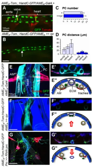
HandC-GFP expression in PCs and cardiomyocytes showed that the number and repartition of PCs is not significantly modified upon rpr-induced AM ablation (Fig. 4A-C). However, the left and right PCs rows become juxtaposed along the midline (Fig. 4A-D). We analysed the relative topology of the PCs and
heart in semi-intact quadruple-labelled larvae (Fig. 4E-F″).
In control larvae, AMs adhere to the ECM surrounding PCs lateral to the heart, and loop around the ventral aspect of the
tracheal trunk (Fig. 4E-E″). Upon AM ablation, PCs become
pressed against the heart, which itself shifts dorsally. In addition,
the heart lumen collapses (Fig. 4F-F″). Prc accumulation in
wild-type larvae (Figs 4E′,E″ and 2B′) suggested anchoring of the
heart to the dorsal epidermis. Upon Prc depletion, AMs and PCs are no longer tied and come apart from the heart and, at the same time,
the heart lumen collapses (Fig. 4G-G″) (Drechsler et al., 2013), as
we observed upon AM elimination. However, rather than being shifted dorsally, the heart adopts a more ventral position. Together, the loss-of-AMs and loss-of-ECM phenotypes indicate that forces
exerted by AMs and dorsal anchoring‘suspend’ the heart within
the body.
We analysed the heart beating in HandC-GFP white pupae (Fig. S7; Movies 4 and 5), a pre-metamorphosis stage when skeletal muscles contractions do not interfere with recording. Upon AM elimination, the heart systolic and diastolic phases strongly decrease. Yet the heart beating rhythm is not affected, indicating that cardiomyocytes retain their full contractility in the absence of AMs, similar to other conditions that prevent haemolymph flux (Choma et al., 2011; Drechsler et al., 2013). We conclude that AMs are not required for larval heart beating per se, but are essential for maintaining proper heart anatomy and, thereby, physiological functions necessitating haemolymph circulation.
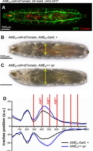
AM ablation affects the position of the tracheal trunk
AMs loop around the ventral aspect of the dorsal tracheal trunk
(Fig. 4E-E″). When AMs are either eliminated or detached from PCs
(Fig. 4F,G), the position of the dorsal tracheal trunk fluctuates in the body cavity. This suggests that AMs sterically constrain the tracheal trunk. To test this hypothesis, we first measured the dorsal tracheal trunk position relative to the midline in wild-type larvae (Fig. 5; Fig. S8). The larval length varies from fully elongated at the beginning of a stride cycle to compacted during the somatic peristaltic wave and visceral piston phase. We observed that dorsal tracheal trunks are more curved at the end of each peristaltic wave, with two conspicuous curves located in the thoracic and abdominal regions, respectively (Fig. S8). AMs A2-A5 roughly frame the major, abdominal curvature (Fig. 5D). Upon AM elimination, the double tracheal curvature is still observed. However, the abdominal curve is larger and the dorsal trachea more distant from the midline (Fig. 5B-D; Fig. S8). These results indicate that AMs constrain the tracheal position during larval crawling.
TARMs are essential for gut and gastric caeca positioning and food transit
A142-GFP expression allows visualisation of the Pv, Gc, anterior loop of the gut and positioning of the visceral mass. Upon removal
of either TARMs (AMER>>Antp) or TARMs plus AMs
(AMER>>rpr), there is a posterior shift in the position of the first
gut loop and visceral mass (Fig. 6A-C′; Table S2), suggesting that
TARMT2connection to the midgut is required for maintaining the
proper position of the visceral system in the body cavity. In addition, the dorsal Gcs, which normally extend anterior-wards by attaching
to TARMT1 (9/10 larvae), now coil around the proventriculus
(14/14 larvae) (Fig. 6D-E′). We then tested whether removal of
Fig. 4. AMs maintain heart intra-celomic position and lumen opening. (A,B) Dorsal views of the AMA4-A6
region of intact AMER-cd4-tdTomato; HandC-GFP,
AMER-Gal4 (A) and >>UAS-rpr (B) larvae. AMs and TARMs are in red and PCs,
cardioblasts and valve cells in green. Arrow in A and B indicates the valve position. (C) The number of PCs is unchanged in control and following AM ablation. (D) Distance separating left and right PCs at three positions (yellow bars in A), aorta, valve and heart chamber, in control and following AM ablation. Bar graphs show mean±s.d. (n=10 larvae). (E-G″) Semi-intact AMER
-cd4-tdTomato; HandC-GFP, AMER-Gal4 (E), >>UAS-rpr (F) and AMER
-cd4-tdTomato; HandC-GFP/prc-Gal4, UAS-RNAi-prc (G) larvae stained for F-Actin and Prc. PCs and cardioblasts are green, AMs red, somatic muscles and heart blue, the ECM and trachea cyan. E′-G′ show transversal views. (E″-G″) Schematic representations adapted from the images in E′-G′ showing the relative positions of AMs, ECM, heart, PCs and the tracheal trunks. Red arrows indicate the shift in dorsoventral position of the heart. Scale bars: 100 µm.
DEVEL
O
AMs had a global impact on food transit through the midgut, using a Bromophenol Blue food ingestion assay. Absence of TARMs significantly reduces the amount of ingested food (Fig. 6F,G). We conclude that TARMs are required for maintaining visceral organ positioning in the larva, insuring efficient food transit inside the gut.
DISCUSSION
Drosophila organogenesis, along with morphogenesis during embryonic development, ends with larval hatching. In most textbooks, development of each larval organ is considered separately, even though interactions between different germ layers, tissues or cell types are referenced. Here, we show that, during larval life, multiple internal organs remain physically connected to the exoskeleton by a web of thin and deformable muscles, the AMs and TARMs (Fig. 7). These atypical, asymmetrically attached muscles are required to maintain proper position of cardiac, visceral and tracheal organs within the coelomic cavity, and heart lumen opening, and, thus, are important physiological regulators.
AM/TARM targeting to different organs: Hox input
Establishment of the muscle pattern, i.e. positions and orientations of each skeletal muscle relative to the other, involves positional
values in the epidermis in each body segment. Targeting specific epidermal attachment sites and muscle-muscle matching are intrinsic properties of developing muscle, under the control of iTFs (de Joussineau et al., 2012). We show here that attachment of AMs and TARMs to either the circulatory or the visceral system is
under intrinsic Hox control. TARMT2 does not express any
homeotic gene of the Antp or bithorax complex, extending to AMs the previous conclusion that the T2 muscle pattern could represent a ground state for the somatic mesoderm (Roy et al.,
1997). However, TARMT2specifically connects to the first midgut
constriction, where Antp-expressing cells are specified and required for this constriction to form (Reuter and Scott, 1990). In homeotic
Ubx mutants, AMA1and AMA2are transformed into TARMT2-like
muscles, which connect to the same specific gut constriction as
normal TARMT2. We thus infer that the background state of AM/
TARM attachment is the gut and that targeting information is provided by midgut regionalisation. The guidance cues remain to be
identified. MT anchoring of AMA3 is also driven by MT tip cell
properties rather than homeotic identity of the AM (Weavers and Skaer, 2013). Altogether, these findings show that the unusual morphology and connections of TARMs and AMs both depend upon iTF and Hox intrinsic information, and guidance cues from internal organs.
Specific morphologies and internal organ connections of AMs
Posterior AMs (A5-A7) connected to the heart chamber display a similar fan-like morphology in larvae and adults. Literature on the physiology of the insect adult heart pointed to the absence of a consistent link between AM contraction and heart-beating rates, and severing of AMs could result in a heart chamber collapse (Jones, 1954; Bullock and Horridge, 1965). More recent studies on adult mosquitos suggested that tension exerted by AMs causes an expansion of the heart cross-sectional area (Glenn et al., 2010). Genetic ablations show that AMs are indeed both required for maintaining the heart in the proper haemocelic position and for opening of the heart chamber, but not for heart beating. AMs do not directly bind to cardiomyocytes but to the ECM surrounding PCs, reminiscent of attachment of the diaphragm to the pericardium, a mesothelial monolayer that equalises gravitational, hydrostatic and inertial forces over the surface of the vertebrate heart (Hoit, 2017). During metamorphosis, anterior AMs dedifferentiate into mononucleate myoblasts, which later form
ventral longitudinal muscles, a component of the ‘dorsal
diaphragm’ covering the ventral surface of the adult dorsal vessel
(Schaub et al., 2015).
Anterior AMs (segments A1-A4) adopt a conspicuous‘T’ shape
during larval development, with myofibres oriented ventrodorsally from the exoskeleton to the aorta, then laterally along the aorta. The
functional role of this ‘T’-shape morphology remains unknown.
Lateral branches of AMA1-A3run along LG lobes (Fig. 7A). This
geometry and presence in the aorta of vesicles originating from AMs raises the possibility that, in addition to a mechanical support function, AMs could play a signalling role in haematopoiesis.
AMs contact different organs in their trajectory from the
exoskeleton to the heart (Fig. 7A). AMA3adhesion to the anterior
MT tip cell and positioning of AMA4 and AMA5 anterior and
posterior to the gonad, respectively, are established during
embryogenesis. Whereas AMA3 is required to anchor the MT
(Weavers and Skaer, 2013), the germ cell/niche assembly is independent of AMs (Anllo et al., 2019). Several AMs send
projections to fat body, including AMA4and AMA5, which project
Fig. 5. AMs constrain the tracheal curvature in larvae. (A) Dorsal view of a fully elongated AMER-cd4-tdTomato; btl-Gal4, UAS-GFP L3 larva. AMs and
TARMs are in red and trachea in green. (B,C) Dorsal views of AMER
-cd4-tdTomato; AMER-Gal4 (B) and >>rpr (C) larvae. The tracheal trunk, seen in
brightfield, is outlined in white, and spacing indicated by yellow double arrows. (D) Tracheal curvatures in AMER-Gal4 (black lines) and >>rpr (blue lines) L3
larvae at the end of a peristaltic wave. Mean±s.e.m. are given (n=15 larvae). AMA1to AMA7positions are indicated by red lines. Scale bars: 500 µm.
DEVEL
O
to the fat body surrounding the male gonad. Whether AM segment-specific projections play architectural and/or signalling roles via the release of diffusible signals similar to myokines in vertebrates (Das et al., 2019) remains to be fully assessed. The multiple shapes adopted by AMs suggest that they could be passively deformed by the steric volume of internal organs during larval crawling.
Conversely, lateral ‘floating’ of the dorsal tracheal branches and
dorsal shifting of the heart in the absence of AMs shows that the internal architecture of the larva is constrained by AMs.
TARM-midgut connections are required for efficient visceral system activity
TARMs (Fig. 7) were only recently discovered in the Drosophila embryo and both their phylogenic spread and fate in adults remain to be explored. Ablation of TARMs prevents unfolding of the dorsal Gc and provokes a posterior shift of the midgut position. Little is known about the precise role of the Gc, which could act as an exocrine organ in synthesising and secreting digestive enzymes in Drosophila (Matsumoto et al., 1995; Grönke et al., 2005) and osmoregulation in Aedes and Anopheles larvae (Volkmann and
Peters, 1989; D’Silva and O’Donnell, 2018). TARMT1attachment
raises further questions, of whether dorsal caeca play specific roles,
and which roles require extended shape and/or TARMT1
connection. TARMT2 attaches to the midgut where
DH31-expressing enteroendocrine cells are located. DH31 cells are required for peristalsis of the anterior midgut (LaJeunesse et al., 2010). The finding that TARM ablation reduces the transit of food through the midgut suggests two, not mutually exclusive,
interpretations: the loss of the first midgut loop impairs peristalsis
and/or loss of the TARMT2connection to DH31-expressing cells
leads to dysregulation of enteroendocrine functions.
Neither cardiac, nor visceral or skeletal: a novel type of Tbx1/Islet1-expressing muscles
The recent discovery of TARMs was an unanticipated outcome of the characterisation of Org-1 and Islet1 transcription enhancers (Schaub et al., 2012; Boukhatmi et al., 2012, 2014), that allowed live imaging of AMs and TARMs in larvae. Being multinucleated and striated, Drosophila AMs and TARMs resemble skeletal muscles, correlating with a related mode of embryonic development (Boukhatmi et al., 2014). However, they exhibit features that distinguish them from body wall muscles. One is their morphology and deformation properties. It will be interesting to determine whether specific MHC isoforms (George et al., 1989) and/or proteins of Z-discs, the sarcomere anchors in striated muscles (Luther, 2009; Steinmetz et al., 2012), are expressed in either TARMs or AMs.
Another AM/TARM-specific feature is their asymmetric
attachment, to the exoskeleton and soft tissues at the other. In primates, asymmetric attachment has been observed for facial subcutaneous muscles, which insert into the skin on one side and to facial bones or other muscles on the other (Ziermann et al., 2018). Facial muscles derive in part from cardiopharyngeal mesoderm, also at the origin of the oesophagus striated muscle (ESM) (Gopalakrishnan et al., 2015; Heude et al., 2018). ESM development is regulated by Tbx1 and Islet1, Tbx1 acting genetically upstream of Isl1 in ESM
Fig. 6. TARMs are required for proper topology of visceral organs and food transit. (A-B′) Dorsal views of intact A142-GFP; AMER-cd4-tdTomato; AMER-Gal4 (A) and >>UAS-Antp (B) larvae.
AMs and TARMs are red, visceral organs green. A′ and B′ show only the visceral system. (C,C′) Relative positions (percentage of larva length) of the Pv, first midgut loop and visceral mass in controls and following TARM ablation. Bar graphs show mean±s.d. (n=10 larvae). (D-E′) Magnified views of the anterior region of A142-GFP; AMER-cd4-tdTomato; AMER-Gal4 (D) and >>Antp (E) larvae. D′ and
E′ show the GFP channel only. (D,D′) TARMT1maintains Gc
extension (asterisk; n≥10); TARMT2connects to the first loop of the
gut. Upon TARM ablation (E,E′), Gcs coil around the Pv, and the first midgut loop is shifted posteriorly. (F,G) Ventral views of AMER
-cd4-tdTomato; AMER-Gal4 (F) and >>Antp (G) larvae fed 45 min with
blue yeast. Anterior at the top (n>25). Scale bars: 500 µm in A,B; 100 µm in D,E.
DEVEL
O
progenitors (Gopalakrishnan et al., 2015; Comai et al., 2019). The Tbx1/Islet genetic hierarchy controls Drosophila AM/TARM development (Boukhatmi et al., 2014) and is redeployed during metamorphosis to initiate transdifferentiation of some AMs into the ventral longitudinal muscles of the adult heart (Schaub et al., 2015). Whether this genetic cascade has been independently recruited in mammals and insects for diversification and specific adaptations of striated muscles is an open question.
The diaphragm, which separates lung and heart from visceral organs in mammals, is also an asymmetric muscle. Its C-shape results from insertion of muscular fibres to, on one side, a sheet of fibrous tissue, the central tendon, which surrounds the oesophageal hiatus, and, on the other, tendinous tissue surrounding ribs and vertebrae (Merrell and Kardon, 2013). Although highly speculative, whether the mammalian diaphragm and the insect AMs/TARMs could represent two specific adaptations of an ancestral demarcation between circulatory, respiratory and visceral organs, is a possibility.
Conclusions and perspectives
To the best of our knowledge, TARMs and AMs are the first example of multinucleate striated muscles connecting the (exo)skeleton to internal organs in bilaterians. Characterisation of a new type of striated muscles raises novel anatomical, physiological and evolutionary questions. Innervation of the heart and AMs in adult arthropods remains controversial (Dulcis and Levine, 2003). Extant reports of embryonic and larval AM innervation concluded that AMs are innervated by a peripheral neuron, the lateral-bidendritic neuron or a motoneuron, or both (Gorczyca et al., 1994; Landgraf and Thor, 2006). Innervation by sensory neurons would raise the exciting possibility that AMs/TARMs are part of internal proprioceptive circuits. Integrating neuromuscular connectivity into physiological roles of TARMs and AMs is the next challenge.
MATERIALS AND METHODS
Drosophila strains
All Drosophila melanogaster stocks and crosses were grown on standard medium at 25°C. Strains used were: AME-moeGFP (Boukhatmi et al., 2012), A142-GFP (Buchon et al., 2013), HandC-GFP (Sellin et al., 2006), UAS-H2bRFP (Zobeck et al., 2010), fTRG500-MHC-GFP, fTRG569-Kettin/sls-GFP, fTRG1046-Unc-89-GFP (Sarov et al., 2016), Prc-Gal4 (Chartier et al., 2002), UAS-Ubx (Michelson, 1994), org-1-LacZ (Schaub et al., 2012), Pcol85-Gal4 (Krzemień et al., 2007), domeMESO-GFP (Oyallon et al., 2016), FB-Gal4 (gift from M. Meister, Institut de Biologie Moléculaire et Cellulaire, UPR 9022 CNRS, Strasbourg, France; Grönke et al., 2003), btl-Gal4 (gift from J. Casanova, Institute for Research in Biomedicine, IBMB-CSIC, Barcelona, Spain) and sr-Gal4 (obtained from T. Volk, Weizmann Institute of Science, Rehovot, Israel). The following strains were obtained from the Bloomington Drosophila Stock Center: cd4-tdTomato (BDSC:35841), cd4-tdGFP (BDSC:35836), UAS-mcd8GFP (BDSC:5137), UAS-mIFP (BDSC:64183), 20xUAS-GCaMP3 (BDSC:32235), Cha-Gal4 (BDSC:6793), Ilk-GFPZCL3111 (BDSC:6831),
how24B-Gal4 (BDSC:1767), UAS-Antp (BDSC:7301), Ubx9.22(BDSC:3474),
UAS-rpr (BDSC:5824) and UAS-RNAi-prc (BDSC:65898). Zasp66ZCL0663was
obtained from the Kyoto Stock Center.
Lethal stocks were balanced using CyO,{dfd-YFP}, TM6,{dfd-YFP} or TM3, Ser,{twi-lacZ} and mutant embryos or larvae identified by absence of lacZ or YFP expression, respectively.
Constructions of transgenic reporter lines
For constructing AM/TARM-specific reporter and Gal4 lines, we shortened the tup-AME enhancer (Boukhatmi et al., 2014), based on the position of Org-1 binding sites and conserved sequence blocks. tup-AMER
DNA, corresponding to the tup upstream region, chr2L positions: 18900307..18899855 (FlyBase; release R6.32), was amplified from yw genomic DNA using the primers 5′-ATCCGCTGCTGCTGCATC-3′ and 5′-GCCGAGCAAGTACTAAGTACC-3′. The amplified 452 bp fragment was cloned into pENTR4 (Gateway cloning system; Invitrogen) to give the donor plasmid pDONR-AMER, which was recombined, using
Fig. 7. TARMs and AMs maintain the positions of internal organs in larva. (A-C) Schematics of a fully extended L3 larva (adapted from Hartenstein, 1993). (A) AM connections to the cardiac system (red), LG (yellow), anterior MTs (orange) and fat body (beige), and surrounding of trachea (grey). (B) TARM connections to specific midgut regions (blue). (C) Mis-positioning of multiple internal organs following TARM/AM elimination, as indicated by arrows: Gc and visceral mass (blue arrows), heart (red arrow) and dorsal trachea (dashed grey arrow). The heart lumen is collapsed.
DEVEL
O
LR recombinase (Invitrogen), with pDEST-Gal4, pDEST-HemmarG (Addgene-plasmid #31221) and pDEST-HemmarR (Addgene-plasmid #31222), to give pEXP-AMER-Gal4, pEXP-AMER-tdGFP and
pEXP-AMER-cd4-tdTomato. Transgenes were inserted at position 25C6 on the
second chromosome (AMER-cd4-tdTomato), or 99F8 (AMER-cd4-tdTomato
and GFP) or 68A4 (AMER-Gal4) on the third chromosome, by injection into
attP embryos (BDSC: 25709, BDSC: 24867, nos-phiC31-NLS; attP2, respectively) (Bischof et al., 2007; Markstein et al., 2008).
Immunohistochemistry of whole-mount embryos and semi-intact L3 preparation
Immunostainings of whole-mount embryos were performed using standard techniques, as described previously (Dubois et al., 2016); third instar larvae dissection and staining were as described (Drechsler et al., 2013).
The following primary antibodies were used: chicken anti-GFP (1:500; Abcam, ab13970) and anti-β-galactosidase (1:2000; Abcam, 9361); rabbit anti-Scr (1:400; courtesy of David Cribbs, Centre de Biologie Intégrative, Toulouse, France), cleaved Dcp1 (1:200; Cell Signaling Technologies, #9578), anti-β3-Tub (1:5000; Renate Renkawitz-Pohl, Philipps-Universität Marburg, Fachbereich Biologie, Entwicklungsbiologie, Marburg, Germany), anti-Mesh (1:1000) (Izumi et al., 2012); mouse anti-Antp [1:100; Developmental Studies Hybridoma Bank (DSHB), 8C11], Ubx (1:50; DSHB, FP3.38), anti-AbdB (1:50; DSHB, 1A2E9), anti-FasIII (1:20; DSHB, 7G10); anti-GFP (1:100; Roche, 11 814 460 001), Prc (1:200; DSHB, EC11); and rat anti-TM (1:500; Abcam, ab50567). Phalloidin-647 (1/200) and Texas RedX (1/ 500) were obtained from Thermo Fisher Scientific (A22287 and T7471, respectively). Secondary antibodies were Alexa Fluor 405-, 488-, 647-, 555-conjugated antibodies (1:300; Thermo Fisher Scientific, A-31553, A-11001, A-11034, A-11039, A-21434, A-21437, A-21235 and A-21244).
Optimised confocal sections were acquired using Zeiss710 or Leica SPE microscopes. Projections and 3D reconstructions were created using ImageJ, Fiji and Imaris software.
Imaging of entire immobilised larva
To image fully extended, translucent, immobilised larvae, larvae were drowned in water for 1 h. Larvae placed between the slide and coverslip were imaged using a Leica SPE microscope, a Nikon AZ100 466 Macroscope at 2× or 5× objective plus optical zoom, or a Nikon SMZ18 Macroscope for brightfield colour images. Mosaic acquisitions and image reconstructions of some entire larvae were performed to improve focus and resolution.
Time-lapse imaging of larva
For time-lapse imaging, late L2 or early L3 GFP larva were mounted between the slide and coverslip with a small drop of water. Image series were acquired using a Nikon AZ100 466 Macroscope, with the following settings: objective ×5, zoom ×1.25, time exposure: 30 ms, 30 fps (Movie 1) or 150 ms, 5 fps (Movie 3); objective ×2, zoom ×1.25; time exposure: 100 ms, 10 fps (Movie 2). Ca2+waves in AMs (Fig. 3F-G′, Movie 3) were imaged in AME
R
-cd4-tdTomato; AMER-Gal4; 20xUAS-GCaMP3 early L3 larvae immobilised
by pressure to prevent crawling. The filters were manually changed during the mono-colour acquisition to successively visualise the muscle shape with Tomato and intracellular calcium rise with GCaMP-GFP. Movies were processed using HCImage Live and Fiji software.
Feeding assay
Feeding L3 larvae were collected and transferred to a 1% agarose plate covered with a thin layer of blue yeast paste (0.5 g/ml baker’s yeast, 0.005 g/ml Bromophenol Blue in water) for 45 min at a density of 20 larvae/ plate. Larvae were then washed and fixed by heat (5 s at 65°C) and imaged from the ventral side using a Nikon SMZ18 Macroscope.
Quantification of embryonic, larval and pupal viability
The numbers of embryos laid for 8 h on an agar plate, and unhatched embryos 36 h later, were counted to determine embryonic viability (two independent counts for each genotype; nmin=509, nmax=725 embryos). For
larval viability, L1 larvae collected 24 h after egg deposition (8 h egg collections) were gently transferred into fly food tubes (30 L1/tube;
nmin=240, nmax=510 L1) and the number of resulting pupae was counted.
Pupal viability corresponds to the percentage of pupae developing to adults (nmin=37; nmax=347 pupa).
Quantification of PC number and distance between PCs
Entire immobilised HandC-GFP larvae were imaged using a Nikon AZ100 466 Macroscope. The number of PCs per hemi-larva, visualised with GFP, were manually counted (nmin=20 hemi-larvae). For each genetic condition,
the mean number of PCs±s.d. is given. The distance between left and right PCs was measured at three distinct positions– aorta, valve and heart – using Fiji software. For each genetic condition, the mean distance between PCs ±s.d. is given (nmin=10 larva).
Quantification of heart beating in white pupa
PCs were visualised in HandC-GFP white pupae. A Nikon AZ100 466 Macroscope was used to record heart beating (objective ×5, zoom ×1.25; time exposure: 100 ms, 10 fps, 20 s, two repetitions for each pupa). Heart pulses were then manually counted (n=12 pupa for each genotype).
Quantification of trachea position in entire larva
L3 feeding larvae were collected and transferred onto 1% agarose plates. Larva were filmed using a Nikon SMZ18 Macroscope (brightfield; zoom ×0.75; 10 fps) and for each larva, the image corresponding to the tracheal distortion step was extracted using Fiji. The left and right dorsal trunk paths from anterior to posterior spiracles and the midline were manually drawn. To standardise the data, the three lines were normalised to a fixed value of 1000 from anterior to posterior spiracles. A homemade C program (available on request) using MagickWand API (ImageMagick) allowed us to extract xy coordinates. Relative y positions of left and right trachea, for y midline position=0, were calculated. Excel was used to calculate the mean tracheal position and the s.d. (n=15 larva for each genotype).
Quantification of visceral system position in entire larva
The visceral system was visualised using A142-GFP on entire immobilised larvae. The larvae size and positions of the proventriculus (Pv), first loop of the gut and visceral mass, respectively, were measured using Fiji software. The relative positions of Pv, first loop and visceral mass, were calculated (%) relative to the larva size normalised to 100%. For each genetic condition, the mean larva size and the mean relative positions±s.d. are given (n=10 larva).
Statistical analysis
For all experiments, data plots and statistical analyses were performed with Prism 5.0. Mean values are shown, error bars represent s.d. or s.e.m. as indicated in figure legends. For statistical comparisons, unpaired t-test or one-way analysis of variance and Tukey post-tests were used, where appropriate. Asterisks show the significance of variation (ns, not significant; ***P<0.001).
Acknowledgements
We thank the Bloomington and Kyoto Stock Centers and colleagues for Drosophila strains and antibodies. We thank Michèle Crozatier, Alice Davy and Cédric Polesello for helpful discussion and critical reading of the manuscript. We acknowledge the help from the Toulouse RIO Imaging platform, Marine Mercier, Ismael Morin-Poulard and Julien Favier for fly stocks and Benoît Condoumy for computer image analysis.
Competing interests
The authors declare no competing or financial interests.
Author contributions
Conceptualization: L.B., A.V.; Methodology: L.B., A.V.; Validation: L.B., A.V.; Formal analysis: L.B.; Investigation: L.B., N.C., A. Pelletier, J.-L.F., A.V.; Resources: A. Paululat, G.L., Y.C., J.-L.F.; Writing - original draft: L.B., A.V.; Writing - review & editing: L.B., A.V.; Visualization: L.B., A.V.; Project administration: L.B., A.V.; Funding acquisition: L.B., A. Paululat, A.V.
Funding
This work was supported by the Centre National de la Recherche Scientifique, the Institut National de la Santé et de la Recherche Médicale, the Ministère de
l’Enseignement supérieur, de la Recherche et de l’Innovation, the Association
DEVEL
O
Française contre les Myopathies (AFM) (21887), the Agence Nationale de la Recherche (13-BSVE2-0010-0 to A.V.), Campus France (PHC PROCOPE 2017 to L.B. and A. Paululat), and the Deutsche Forschungsgemeinschaft (SFB944, PA 14-1 to A. Paululat).
Supplementary information
Supplementary information available online at
http://dev.biologists.org/lookup/doi/10.1242/dev.185645.supplemental
References
Anllo, L., Plasschaert, L. W., Sui, J. and DiNardo, S. (2019). Live imaging reveals hub cell assembly and compaction dynamics during morphogenesis of the Drosophila testis niche. Dev. Biol. 446, 102-118. doi:10.1016/j.ydbio.2018.12.014 Armand, P., Knapp, A. C., Hirsch, A. J., Wieschaus, E. F. and Cole, M. D. (1994). A novel basic helix-loop-helix protein is expressed in muscle attachment sites of the Drosophila epidermis. Mol. Cell. Biol. 14, 4145-4154. doi:10.1128/MCB.14.6. 4145
Bataillé, L., Frendo, J.-L. and Vincent, A. (2015). Hox control of Drosophila larval anatomy; the alary and thoracic alary-related muscles. Mech. Dev. 138, 170-176. doi:10.1016/j.mod.2015.07.005
Bate, M. and Rushton, E. (1993). Myogenesis and muscle patterning in Drosophila. C. R. Acad. Sci. III 316, 1047-1061.
Bischof, J., Maeda, R. K., Hediger, M., Karch, F. and Basler, K. (2007). An optimized transgenesis system for Drosophila using germ-line-specific phiC31 integrases. Proc. Natl. Acad. Sci. USA 104, 3312-3317. doi:10.1073/pnas. 0611511104
Boukhatmi, H., Frendo, J. L., Enriquez, J., Crozatier, M., Dubois, L. and Vincent, A. (2012). Tup/Islet1 integrates time and position to specify muscle identity in Drosophila. Development 139, 3572-3582. doi:10.1242/dev.083410
Boukhatmi, H., Schaub, C., Bataillé, L., Reim, I., Frendo, J.-L., Frasch, M. and Vincent, A. (2014). An Org-1-Tup transcriptional cascade reveals different types of alary muscles connecting internal organs in Drosophila. Development 141, 3761-3771. doi:10.1242/dev.111005
Buchon, N., Osman, D., David, F. P. A., Fang, H. Y., Boquete, J.-P., Deplancke, B. and Lemaitre, B. (2013). Morphological and molecular characterization of adult midgut compartmentalization in Drosophila. Cell Rep. 3, 1725-1738. doi:10. 1016/j.celrep.2013.04.001
Buckingham, M. (2017). Gene regulatory networks and cell lineages that underlie the formation of skeletal muscle. Proc. Natl. Acad. Sci. USA 114, 5830-5837. doi:10.1073/pnas.1610605114
Buechling, T., Akasaka, T., Vogler, G., Ruiz-Lozano, P., Ocorr, K. and Bodmer, R. (2009). Non-autonomous modulation of heart rhythm, contractility and morphology in adult fruit flies. Dev. Biol. 328, 483-492. doi:10.1016/j.ydbio. 2009.02.013
Bullock, T. H. and Horridge, G. A. (1965). Structure and Function in the Nervous Systems of Invertebrates. San Francisco: W.H. Freeman.
Chartier, A., Zaffran, S., Astier, M., Sémériva, M. and Gratecos, D. (2002). Pericardin, a Drosophila type IV collagen-like protein is involved in the morphogenesis and maintenance of the heart epithelium during dorsal ectoderm closure. Development 129, 3241-3253.
Choma, M. A., Suter, M. J., Vakoc, B. J., Bouma, B. E. and Tearney, G. J. (2011). Physiological homology between Drosophila melanogaster and vertebrate cardiovascular systems. Dis. Model. Mech. 4, 411-420. doi:10.1242/dmm.005231 Comai, G., Heude, E., Mella, S., Paisant, S., Pala, F., Gallardo, M., Langa, F., Kardon, G., Gopalakrishnan, S. and Tajbakhsh, S. (2019). A distinct cardiopharyngeal mesoderm genetic hierarchy establishes antero-posterior patterning of esophagus striated muscle. eLife 8, e47460. doi:10.7554/eLife. 47460
Curtis, N. J., Ringo, J. M. and Dowse, H. B. (1999). Morphology of the pupal heart, adult heart, and associated tissues in the fruit fly, Drosophila melanogaster. J. Morphol. 240, 225-235. doi:10.1002/(SICI)1097-4687(199906)240:3<225:: AID-JMOR2>3.0.CO;2-V
Das, D. K., Graham, Z. A. and Cardozo, C. P. (2019). Myokines in skeletal muscle physiology and metabolism: recent advances and future perspectives. Acta Physiol. (Oxf.) 228, e13367. doi:10.1111/apha.13367
de Joussineau, C., Bataillé, L., Jagla, T. and Jagla, K. (2012). Diversification of muscle types in Drosophila: upstream and downstream of identity genes. Curr. Top. Dev. Biol. 98, 277-301. doi:10.1016/B978-0-12-386499-4.00011-2 Drechsler, M., Schmidt, A. C., Meyer, H. and Paululat, A. (2013). The conserved
ADAMTS-like protein lonely heart mediates matrix formation and cardiac tissue integrity. PLoS Genet. 9, e1003616. doi:10.1371/journal.pgen.1003616 D’Silva, N. M. and O’Donnell, M. J. (2018). The gastric caecum of larval. J. Exp.
Biol. 221, jeb172866. doi:10.1242/jeb.172866
Dubois, L., Frendo, J.-L., Chanut-Delalande, H., Crozatier, M. and Vincent, A. (2016). Genetic dissection of the Transcription Factor code controlling serial specification of muscle identities in Drosophila. eLife 5, e14979. doi:10.7554/ eLife.14979
Dulcis, D. and Levine, R. B. (2003). Innervation of the heart of the adult fruit fly, Drosophila melanogaster. J. Comp. Neurol. 465, 560-578. doi:10.1002/cne. 10869
Ejaz, A. and Lange, A. B. (2008). Peptidergic control of the heart of the stick insect, Baculum extradentatum. Peptides 29, 214-225. doi:10.1016/j.peptides.2007.07. 036
George, E. L., Ober, M. B. and Emerson, C. P. (1989). Functional domains of the Drosophila melanogaster muscle myosin heavy-chain gene are encoded by alternatively spliced exons. Mol. Cell. Biol. 9, 2957-2974. doi:10.1128/MCB.9.7. 2957
Glenn, J. D., King, J. G. and Hillyer, J. F. (2010). Structural mechanics of the mosquito heart and its function in bidirectional hemolymph transport. J. Exp. Biol. 213, 541-550. doi:10.1242/jeb.035014
Gopalakrishnan, S., Comai, G., Sambasivan, R., Francou, A., Kelly, R. G. and Tajbakhsh, S. (2015). A cranial mesoderm origin for esophagus striated muscles. Dev. Cell 34, 694-704. doi:10.1016/j.devcel.2015.07.003
Gorczyca, M. G., Phillis, R. W. and Budnik, V. (1994). The role of tinman, a mesodermal cell fate gene, in axon pathfinding during the development of the transverse nerve in Drosophila. Development 120, 2143-2152.
Grö nke, S., Beller, M., Fellert, S., Ramakrishnan, H., Jäckle, H. and Kühnlein, R. P. (2003). Control of fat storage by a Drosophila PAT domain protein. Curr. Biol. 13, 603-606. doi:10.1016/S0960-9822(03)00175-1
Grö nke, S., Mildner, A., Fellert, S., Tennagels, N., Petry, S., Müller, G., Jäckle, H. and Kü hnlein, R. P. (2005). Brummer lipase is an evolutionary conserved fat storage regulator in Drosophila. Cell Metab. 1, 323-330. doi:10.1016/j.cmet.2005. 04.003
Hartenstein V. (1993). Atlas of Drosophila Development, in The Development of Drosophila melanogaster, (ed. M. Bate and A. Martinez-Arias). Cold Spring Harbor, NY: Cold Spring Harbor Laboratory Press.
Heckscher, E. S., Lockery, S. R. and Doe, C. Q. (2012). Characterization of Drosophila larval crawling at the level of organism, segment, and somatic body wall musculature. J. Neurosci. 32, 12460-12471. doi:10.1523/JNEUROSCI.0222-12.2012
Heude, E., Tesarova, M., Sefton, E. M., Jullian, E., Adachi, N., Grimaldi, A., Zikmund, T., Kaiser, J., Kardon, G., Kelly, R. G. et al. (2018). Unique morphogenetic signatures define mammalian neck muscles and associated connective tissues. eLife 7, e40179. doi:10.7554/eLife.40179
Hoit, B. D. (2017). Anatomy and Physiology of the Pericardium. Cardiol. Clin. 35, 481-490. doi:10.1016/j.ccl.2017.07.002
Hudson, A. M., Petrella, L. N., Tanaka, A. J. and Cooley, L. (2008). Mononuclear muscle cells in Drosophila ovaries revealed by GFP protein traps. Dev. Biol. 314, 329-340. doi:10.1016/j.ydbio.2007.11.029
Hwang, R. Y., Zhong, L., Xu, Y., Johnson, T., Zhang, F., Deisseroth, K. and Tracey, W. D. (2007). Nociceptive neurons protect Drosophila larvae from parasitoid wasps. Curr. Biol. 17, 2105-2116. doi:10.1016/j.cub.2007.11.029 Izumi, Y., Yanagihashi, Y. and Furuse, M. (2012). A novel protein complex,
Mesh-Ssk, is required for septate junction formation in the Drosophila midgut. J. Cell Sci. 125, 4923-4933. doi:10.1242/jcs.112243
Jones, J. C. (1954). The heart and associated tissues of Anopheles quadrimaculatus Say (Diptera: Culicidae). J. Morphol. 94, 71-123. doi:10.1002/ jmor.1050940104
Krzemień, J., Dubois, L., Makki, R., Meister, M., Vincent, A. and Crozatier, M. (2007). Control of blood cell homeostasis in Drosophila larvae by the posterior signalling centre. Nature 446, 325-328. doi:10.1038/nature05650
LaBeau, E. M., Trujillo, D. L. and Cripps, R. M. (2009). Bithorax complex genes control alary muscle patterning along the cardiac tube of Drosophila. Mech. Dev. 126, 478-486. doi:10.1016/j.mod.2009.01.001
LaJeunesse, D. R., Johnson, B., Presnell, J. S., Catignas, K. K. and Zapotoczny, G. (2010). Peristalsis in the junction region of the Drosophila larval midgut is modulated by DH31 expressing enteroendocrine cells. BMC Physiol. 10, 14. doi:10.1186/1472-6793-10-14
Landgraf, M. and Thor, S. (2006). Development of Drosophila motoneurons: specification and morphology. Semin. Cell Dev. Biol. 17, 3-11. doi:10.1016/j. semcdb.2005.11.007
League, G. P., Onuh, O. C. and Hillyer, J. F. (2015). Comparative structural and functional analysis of the larval and adult dorsal vessel and its role in hemolymph circulation in the mosquito Anopheles gambiae. J. Exp. Biol. 218, 370-380. doi:10. 1242/jeb.114942
Lehmacher, C., Abeln, B. and Paululat, A. (2012). The ultrastructure of Drosophila heart cells. Arthropod. Struct. Dev. 41, 459-474. doi:10.1016/j.asd.2012.02.002 Luther, P. K. (2009). The vertebrate muscle Z-disc: sarcomere anchor for structure
and signalling. J. Muscle Res. Cell Motil. 30, 171-185. doi:10.1007/s10974-009-9189-6
Markstein, M., Pitsouli, C., Villalta, C., Celniker, S. E. and Perrimon, N. (2008). Exploiting position effects and the gypsy retrovirus insulator to engineer precisely expressed transgenes. Nat. Genet. 40, 476-483. doi:10.1038/ng.101
Matsumoto, I., Watanabe, H., Abe, K., Arai, S. and Emori, Y. (1995). A putative digestive cysteine proteinase from Drosophila melanogaster is predominantly expressed in the embryonic and larval midgut. Eur. J. Biochem. 227, 582-587. doi:10.1111/j.1432-1033.1995.tb20428.x
DEVEL
O
Merrell, A. J. and Kardon, G. (2013). Development of the diaphragm– a skeletal muscle essential for mammalian respiration. FEBS J. 280, 4026-4035. doi:10. 1111/febs.12274
Michelson, A. M. (1994). Muscle pattern diversification in Drosophila is determined by the autonomous function of homeotic genes in the embryonic mesoderm. Development 120, 755-768.
Noden, D. M. and Francis-West, P. (2006). The differentiation and morphogenesis of craniofacial muscles. Dev. Dyn. 235, 1194-1218. doi:10.1002/dvdy.20697 Oyallon, J., Vanzo, N., Krzemień, J., Morin-Poulard, I., Vincent, A. and
Crozatier, M. (2016). Two independent functions of collier/early B cell factor in the control of Drosophila blood cell homeostasis. PLoS ONE 11, e0148978. doi:10.1371/journal.pone.0148978
Pélissier, P., Pistre, V., Bustamante, K., Martin, D. and Baudet, J. (2000). [The modiolus. Comparative anatomy, embryological and physiological review, surgical importance]. Ann. Chir. Plast. Esthet. 45, 41-47.
Reuter, R. and Scott, M. P. (1990). Expression and function of the homoeotic genes Antennapedia and Sex combs reduced in the embryonic midgut of Drosophila. Development 109, 289-303.
Rizki, T. M. (1978). The Circulatory System and Associated Cells and Tissues. New York: Academic Press.
Roy, S., Shashidhara, L. S. and VijayRaghavan, K. (1997). Muscles in the Drosophila second thoracic segment are patterned independently of autonomous homeotic gene function. Curr. Biol. 7, 222-227. doi:10.1016/S0960-9822(06)00117-5
Sarov, M., Barz, C., Jambor, H., Hein, M. Y., Schmied, C., Suchold, D., Stender, B., Janosch, S., KJ, V. V., Krishnan, R. T. et al. (2016). A genome-wide resource for the analysis of protein localisation in Drosophila. eLife 5, e12068. doi:10.7554/ eLife.12068
Schaub, C., Nagaso, H., Jin, H. and Frasch, M. (2012). Org-1, the Drosophila ortholog of Tbx1, is a direct activator of known identity genes during muscle specification. Development 139, 1001-1012. doi:10.1242/dev.073890 Schaub, C., Mä rz, J., Reim, I. and Frasch, M. (2015). Org-1-dependent lineage
reprogramming generates the ventral longitudinal musculature of the Drosophila heart. Curr. Biol. 25, 488-494. doi:10.1016/j.cub.2014.12.029
Sellin, J., Albrecht, S., Kö lsch, V. and Paululat, A. (2006). Dynamics of heart differentiation, visualized utilizing heart enhancer elements of the Drosophila melanogaster bHLH transcription factor Hand. Gene Expr. Patterns 6, 360-375. doi:10.1016/j.modgep.2005.09.012
Steinmetz, P. R. H., Kraus, J. E. M., Larroux, C., Hammel, J. U., Amon-Hassenzahl, A., Houliston, E., Wö rheide, G., Nickel, M., Degnan, B. M. and Technau, U. (2012). Independent evolution of striated muscles in cnidarians and bilaterians. Nature 487, 231-234. doi:10.1038/nature11180
Tao, Y., Wang, J., Tokusumi, T., Gajewski, K. and Schulz, R. A. (2007). Requirement of the LIM homeodomain transcription factor tailup for normal heart and hematopoietic organ formation in Drosophila melanogaster.Mol. Cell. Biol. 27, 3962-3969. doi:10.1128/MCB.00093-07
Tian, L., Hires, S. A., Mao, T., Huber, D., Chiappe, M. E., Chalasani, S. H., Petreanu, L., Akerboom, J., McKinney, S. A., Schreiter, E. R. et al. (2009). Imaging neural activity in worms, flies and mice with improved GCaMP calcium indicators. Nat. Methods 6, 875-881. doi:10.1038/nmeth.1398
Volk, T. (2013). Positioning nuclei within the cytoplasm of striated muscle fiber: cooperation between microtubules and KASH proteins. Nucleus 4, 18-22. doi:10. 4161/nucl.23086
Volkmann, A. and Peters, W. (1989). Investigations on the midgut caeca of mosquito larvae-II. Functional aspects. Tissue Cell 21, 253-261. doi:10.1016/ 0040-8166(89)90070-0
Weavers, H. and Skaer, H. (2013). Tip cells act as dynamic cellular anchors in the morphogenesis of looped renal tubules in Drosophila. Dev. Cell 27, 331-344. doi:10.1016/j.devcel.2013.09.020
Ziermann, J. M., Diogo, R. and Noden, D. M. (2018). Neural crest and the patterning of vertebrate craniofacial muscles. Genesis 56, e23097. doi:10.1002/ dvg.23097
Zobeck, K. L., Buckley, M. S., Zipfel, W. R. and Lis, J. T. (2010). Recruitment timing and dynamics of transcription factors at the Hsp70 loci in living cells. Mol. Cell 40, 965-975. doi:10.1016/j.molcel.2010.11.022