HAL Id: hal-00016691
https://hal.archives-ouvertes.fr/hal-00016691
Submitted on 31 May 2020
HAL is a multi-disciplinary open access
archive for the deposit and dissemination of
sci-entific research documents, whether they are
pub-lished or not. The documents may come from
teaching and research institutions in France or
abroad, or from public or private research centers.
L’archive ouverte pluridisciplinaire HAL, est
destinée au dépôt et à la diffusion de documents
scientifiques de niveau recherche, publiés ou non,
émanant des établissements d’enseignement et de
recherche français ou étrangers, des laboratoires
publics ou privés.
Higher plant plastids and cyanobacteria have folate
carriers related to those of trypanosomatids
Sebastian M.J. Klaus, Edmund R.S. Kunji, Gale G. Bozzo, Alexandre Noiriel,
Rocio Diaz de la Garza, Gilles J.C. Basset, Stéphane Ravanel, Fabrice
Rébeillé, Jesse F. Gregory, Andrew D. Hanson
To cite this version:
Sebastian M.J. Klaus, Edmund R.S. Kunji, Gale G. Bozzo, Alexandre Noiriel, Rocio Diaz de la Garza,
et al.. Higher plant plastids and cyanobacteria have folate carriers related to those of trypanosomatids.
Journal of Biological Chemistry, American Society for Biochemistry and Molecular Biology, 2005,
280(46) (46), pp.38457-38463. �10.1074/jbc.M507432200�. �hal-00016691�
Higher Plant Plastids and Cyanobacteria Have Folate Carriers
Related to Those of Trypanosomatids
*
□SReceived for publication, July 8, 2005, and in revised form, August 19, 2005 Published, JBC Papers in Press, September 14, 2005, DOI 10.1074/jbc.M507432200
Sebastian M. J. Klaus
‡, Edmund R. S. Kunji
§, Gale G. Bozzo
‡, Alexandre Noiriel
‡, Rocı´o Dı´az de la Garza
‡,
Gilles J. C. Basset
‡, Ste´phane Ravanel
¶, Fabrice Re´beille´
¶, Jesse F. Gregory, III
储, and Andrew D. Hanson
‡1From the
‡Horticultural Sciences and
储Food Science and Human Nutrition Departments, University of Florida, Gainesville, Florida
32611, the
§Medical Research Council, Dunn Human Nutrition Unit, Hills Road, Cambridge CB2 2XY, United Kingdom, and the
¶
Laboratoire de Physiologie Cellulaire Ve´ge´tale, CNRS-Commissariat a` l’Energie Atomique (Saclay, France) (CEA)-Institut National
de la Recherche Agronomique-Universite´ Joseph Fourier, CEA-Grenoble, F-38054 Grenoble Cedex 9, France
Cyanobacterial and plant genomes encode proteins with some similarity to the folate and biopterin transporters of the trypanoso-matid parasite Leishmania. The Synechocystis slr0642 gene product and its closest Arabidopsis homolog, the At2g32040 gene product, are representative examples. Both have 12 probable transmem-brane domains, and the At2g32040 protein has a predicted chloro-plast transit peptide. When expressed in Escherichia coli pabA pabB or folE, mutants, which are unable to produce or take up folates, the slr0642 protein and a modified At2g32040 protein (truncated and fused to the N terminus of slr0642) enabled growth on 5-formyltet-rahydrofolate or folic acid but not on 5-formyltet5-formyltet-rahydrofolate tri-glutamate, demonstrating that both proteins mediate folate mono-glutamate transport. Both proteins also mediate transport of the antifolate analogs methotrexate and aminopterin, as evidenced by their ability to greatly increase the sensitivity of E. coli to these inhibitors. The full-length At2g32040 polypeptide was translocated into isolated pea chloroplasts and, when fused to green fluorescent protein, directed the passenger protein to the envelope of Arabi-dopsis chloroplasts in transient expression experiments. At2g32040 transcripts were present at similar levels in roots and aerial organs, indicating that the protein occurs in non-green plastids as well as chloroplasts. Insertional inactivation of At2g32040 significantly raised the total folate content of chloroplasts and lowered the pro-portion of 5-methyltetrahydrofolate but did not discernibly affect growth. These findings establish conservation of function among folate and biopterin transporter family proteins from three king-doms of life.
Tetrahydrofolate (THF)2 and its derivatives (collectively termed
folates) are essential cofactors for one-carbon transfer reactions in almost all organisms (1, 2). Bacteria and plants synthesize folates de
novo, but higher animals do not and so need a dietary supply (1, 3, 4). Folates consist of a pteridine, p-aminobenzoate (pABA) and one or
more glutamate residues (Fig. 1A). All these moieties are synthesized and assembled into folates in the cytosol of bacteria, but in plants the pathway is split among three subcellular compartments. The pteridine is made in the cytosol, and the pABA is made in plastids; both then enter mitochondria, where they are coupled together and glutamylated to yield folate (5–12) (Fig. 1B). Much of the folate produced exits the mito-chondria to supply folate pools in the cytosol, plastids, and vacuoles (13–17). This compartmentation of the synthesis pathway and its end products entails various transmembrane fluxes of folates and their pre-cursors (Fig. 1B). Although pABA can cross membranes by simple dif-fusion (18), folate and pteridine fluxes are likely to be carrier-mediated, as in animals (19 –22) and bacteria (23–25). Plants, therefore, presum-ably have several as yet undiscovered organellar folate or pteridine transporters (Fig. 1B).
Multiple transporters that act on folates as well as antifolates such as methotrexate or aminopterin have been characterized in mammals. These transporters include the reduced folate carrier, which catalyzes exchange with organic anions, folate receptors, which mediate folate entry via endocytosis, folate efflux pumps of the ATP binding cassette family, and a protein from the mitochondrial carrier family (19 –21).
High affinity folate and pteridine transporters are also known from the trypanosomatid Leishmania, a parasitic protist that is auxotrophic for folate and pteridine (26). These carriers are a biopterin transporter (BT1) and 2 folate transporters (FT1, FT5), all 3 being similar proteins with about 12 putative transmembrane helices (27–29). Together, they define a small family (the FBT family) that is a distant member of the major facilitator superfamily (30). BT1 is the only carrier yet known to act on pteridines; it also has low affinity folate transport activity (22).
Besides proteins from trypanosomatids, the FBT family (also known as the BT1 family, Pfam03092) includes a highly conserved protein from Cyanobacteria and nine Arabidopsis proteins. In this study we used heterologous expression in Escherichia coli mutants to probe the func-tions of a representative cyanobacterial FBT protein and its closest
Ara-bidopsishomolog. Having demonstrated that both have folate and anti-folate transport activity, we investigated the subcellular location of the
Arabidopsisprotein, the expression of its gene, and the effect of ablating this gene.
EXPERIMENTAL PROCEDURES
Chemicals and Reagents—Folates and pteridines were from Schircks Laboratories (Jona, Switzerland). p-Aminobenzoylglutamate (pABA-Glu) was HPLC-purified before use. Percoll and [5,6-3H]UTP (35
Ci/mmol) were from Amersham Biosciences. [3H]Leucine (59
Ci/mmol) was from PerkinElmer Life Sciences.
E. coli Strains—Strain BN1163 (pabA1, pabB::Kan, rpsL704, ilvG-,
rfb-50, rph-1) was supplied by B. Nichols (University of Chicago, Chi-*This work was supported in part by the Florida Agricultural Experimental Station, by an
endowment from the C. V. Griffin, Sr. Foundation, and by National Institutes of Health Grant R01 GM071382-01 and approved for publication as Journal Series no. R-10984. The costs of publication of this article were defrayed in part by the payment of page charges. This article must therefore be hereby marked “advertisement” in accordance with 18 U.S.C. Section 1734 solely to indicate this fact.
□SThe on-line version of this article (available at http://www.jbc.org) contains
supple-mental Fig. 1 and Table I.
1To whom correspondence should be addressed: Horticultural Sciences Dept.,
Univer-sity of Florida, Gainesville, FL 32611. Tel.: 352-392-1928 (ext. 334); Fax: 352-392-5653; E-mail: adha@mail.ifas.ufl.edu.
2The abbreviations used are: THF, tetrahydrofolate; 5-CHO-THF, 5-formyltetrahydrofolate;
pABA, p-aminobenzoic acid; pABA-Glu, p-aminobenzoylglutamate; FBT, folate-biopterin
transporter; GFP, green fluorescent protein; RT, reverse transcription; IPTG, isopropyl 1-thio--D-galactopyranoside; HPLC, high performance liquid chromatography.
THE JOURNAL OF BIOLOGICAL CHEMISTRY VOL. 280, NO. 46, pp. 38457–38463, November 18, 2005 © 2005 by The American Society for Biochemistry and Molecular Biology, Inc. Printed in the U.S.A.
at INRA Institut National de la Recherche Agronomique, on November 8, 2010
www.jbc.org
cago, IL), and strain NM1200 was supplied by N. Majdalani (NCBI, Bethesda, MD). The NM1200 strain was constructed by P1 transduc-tion of the Mini- CmRprophage carrying the Red gam, beta, and exo
genes under the PL promoter (31) in MG1655 (E. coli K12, E. coli
Genetic Stock Center). The folE gene was deleted by targeted homolo-gous recombination using electroporated linear DNA and strain NM1200 as described (32). Briefly, the kanamycin cassette was ampli-fied from plasmid pUC4K with primers FolE-5⬘ and FolE-3⬘ (see Sup-plemental Table I), which respectively included the start and stop codon of folE and 47-bp 5⬘- and 3⬘-flanking sequences. After inducing the recombination system at 42 °C for 15 min, strain NM1200 was trans-formed with the PCR product. Clones with the desired replacement of the folE gene were selected on LB plates containing 300Mthymidine and 30g/ml kanamycin. The kanamycin resistance locus was then transferred by P1 transduction to strain K12, to give strain P1–7B. Dele-tion of the folE gene was confirmed by PCR and sequencing. P1–7B was maintained on LB medium containing 300Mthymidine and 30g/ml kanamycin.
Plants and Growth Conditions—Arabidopsis (ecotype Columbia) plants were grown at 23–28 °C in 12-h days (photosynthetic photon flux density 80 microeinsteins m⫺2s⫺1) in potting soil irrigated with water. When roots were required, plants were grown hydroponically (33).
Expression Constructs—Synechocystis slr0642 and Arabidopsis At2g32040 amplicons preceded by a Shine-Dalgarno sequence were cloned between the NotI and SstI sites of pLOI707HE (34), which con-tains the tac promoter, the lacIqrepressor gene, and the tet marker.
Templates were Synechocystis sp. PCC 6803 genomic DNA (from W. Vermaas, Arizona State University, Tempe, AZ) and an At2g32040 cDNA (GenBankTM BT003363, from the Arabidopsis Biological
Resource Center). The following PCR primers were used (see Supple-mental Table I): for Synechocystis slr0642, Slr1–5⬘ and Slr1–3⬘; for the mature At2g32040 construct, At-t1–5⬘ and At-t1–3⬘; for the chimeric At2g32040 construct (with codons 1–128 replaced by codons 1–37 of slr0642), Slr1–5⬘ and Slr-c1–3⬘ to amplify the slr0642 fragment, At-c1–5⬘ and At-t1–3⬘ to amplify the At2g32040 fragment, and Slr1–5⬘ and At-t1–3⬘ to splice the fragments. The complete slr0642 amplicon was cloned into pGEM威-T Easy (Promega), excised with NotI and SstI, and then cloned into pLOI707HE; other amplicons were cut with NotI and SstI and cloned straight into pLOI707HE. For the vector minus insert control, pLOI707HE was digested with NotI and SstI, end-pol-ished with T4 polymerase, and religated. Constructs were made in
E. coli strain DH5␣, sequence-verified, then introduced into E. coli
strains BN1163 and P1–7B, both harboring pACYC-RIL (Stratagene), which contains three rare E. coli tRNA genes and the cat marker. Trans-formants were grown on LB plates containing tetracycline 10g/ml, kanamycin 30 –50g/ml, chloramphenicol 20 g/ml and (for P1–7B only) thymidine 300M.
Folate Uptake Tests—BN1163 cells harboring pACYC-RIL and a pLOI707HE construct were streaked on minimal medium plates con-taining M9 salts (35), 0.1 mMCaCl2, 0.5 mMMgSO4, trace elements (36),
and 0.4% (w/v) glucose supplemented with antibiotics as above and 0.5 mMIPTG. To this medium was added 3.6MpABA or 11M (6R,6S)-5-formyl-THF (5-CHO-THF), folic acid, or (6R,6S)-(6R,6S)-5-formyl-THF tri-glutamate. P1–7B cells harboring pACYC-RIL and a pLOI707HE con-struct were streaked on LB medium containing 0.1% (w/v) sodium ascorbate and 1 mMdithiothreitol, appropriate antibiotics and IPTG minus or plus 11M5-CHO-THF, dihydropteroate, hydroxymethyl-dihydropterin, or dihydropterin-6-aldehyde. Tests with the two latter compounds were made in a 10-l vessel flushed with N2at 25 ml/h.
Antifolate Uptake Tests—Solid or liquid minimal media were supple-mented with antibiotics, IPTG, and pABA as described above. For tests on solid medium, cells were streaked on plates containing a central 1-cm disk of Whatman No. 1 paper soaked with 10l of dimethyl sulfoxide alone or containing 100 mMmethotrexate or 80 mMaminopterin. For tests in liquid medium, cells from an overnight culture were inoculated to give an A600reading of 0.05 in 1.5 ml of fresh medium supplemented
with various amounts of methotrexate or aminopterin. Growth was evaluated by measuring A600after incubating at 37 °C for 9.5 h with
shaking at 220 rpm.
Transient Expression of Green Fluorescent Fusion Protein in Arabi-dopsis Protoplasts—The entire At2g32040 sequence was amplified using primers At-gfp-5⬘ and At-gfp-3⬘, digested with SalI and NcoI, and cloned in-frame upstream of the green fluorescent protein (GFP) gene in pTH2 (37). Protoplasts were prepared from cell cultures, trans-formed, and cultured as described at 22 °C for up to 48 h (7, 38). Trans-formation efficiencies were typically 5–20%. Samples were analyzed by epifluorescence microscopy as described (11).
Chloroplast Import Assays—The full-length At2g32040 sequence and a version lacking the first 88 residues were, respectively, amplified using forward primer At-ifl-5⬘ or At-it-5⬘ and reverse primer At-i-3⬘. The amplicons were cloned as EcoRI-HindIII fragments into pGEM®-4Z
(Promega). Coupled in vitro transcription-translation was performed using a wheat germ TNT威 kit (Promega) and [3H]leucine according to
the manufacturer’s protocol. Translation product import into pea chlo-roplasts was assayed as described (39). Briefly, translation product (50 l) was added to 50 l of 2⫻ import buffer (39) containing 100 mM
FIGURE 1. Structure of folates and compartmentation of folate biosynthesis in
plants. A, folic acid. Aminopterin and methotrexate differ from folic acid in having an
amino instead of an oxo group at C4
, and N10
is methylated in methotrexate. The pteri-dine ring of folic acid is fully oxidized; in natural folates it is reduced to dihydro or tetra-hydro forms. A␥-linked polyglutamyl tail of up to about six residues is usually attached to the first glutamate. B, distribution of folate biosynthesis steps between cytosol, mito-chondria, and plastids and inferred carrier-mediated transport steps for folates (open
symbols) and pteridines (gray symbol). Note that polyglutamylation of folates also occurs
in the cytosol and plastids (7). DHN, dihydroneopterin; DHNTP, dihydroneopterin triphosphate; DHM, dihydromonapterin; HMDHP, hydroxymethyldihydropterin; DHP, dihydropteroate; DHF, dihydrofolate; ADC, aminodeoxychorismate; PP, pyrophosphate.
FBT Family Transporters
at INRA Institut National de la Recherche Agronomique, on November 8, 2010
www.jbc.org
unlabeled leucine; 100l of 1⫻ import buffer containing 30 mMMg2⫹ -ATP and 200l of purified chloroplasts (0.2 mg chlorophyll) were added, and samples were incubated for 15 min at 25 °C in the light. Chloroplasts were re-purified as described (39), resuspended in SDS loading buffer containing 4Murea, and held for 10 min at 37 °C before loading.
Real-time Quantitative RT-PCR—Total RNA was extracted using RNeasy plant mini kits (Qiagen) and treated with DNase (DNA-freeTM
kit, Ambion, Austin, TX). RT-PCR was performed on 250 ng of RNA in 25-l reactions using Taq-Man One-Step RT-PCR Master Mix reagents and a GeneAmp 5700 system (Applied Biosystems, Foster City, CA). The primers (At-rt-5⬘, At-rt-3⬘) and Taq-Man probe (At-rt-probe) were designed with Applied Biosystems Primer Express software. The amplicon length was 74 bp. Primer At-rt-5⬘ spanned two exons to avoid amplifying contaminating genomic DNA, and controls without reverse transcriptase were run to verify that no such amplification occurred. RT-PCR conditions were 48 °C for 30 min and 95 °C for 10 min followed by 40 cycles of 95 °C for 15 s and 60 °C for 1 min. An At2g32040 RNA standard was synthesized from a cDNA template using primers At-SP6std-5⬘ and At-std-3⬘ and labeled using [5,6-3H]UTP as described
(10). An internal standard RNA was added to each sample before RT-PCR to estimate recoveries, which were 34 –100%. Data were corrected for recovery.
Insertional Mutant—An At2g32040 mutant (Salk 109024) was iden-tified in the Salk Arabidopsis (ecotype Columbia) T-DNA insertion col-lection (40). Wild type or homozygous mutant segregants were identi-fied by PCR using gene-specific primers located 5⬘ or 3⬘ of the T-DNA insertion (At-s-5⬘ and At-s-3⬘, respectively) and the T-DNA-specific primer SalkLBb1. Genomic DNA was extracted as described (41). The insertion site was confirmed by sequencing the amplicon from mutant homozygotes. Homozygous mutant and wild type segregants were selfed, and their progeny were used for experiments. Kanamycin resist-ance was shown to co-segregate with the At2g32040 mutation, indicat-ing the absence of T-DNA insertions at other loci.
Chloroplast Folate Analyses—Chloroplasts were prepared from the leaves of 4-week-old Arabidopsis plants as described (42, 43). Folates were extracted from samples (⬃1 mg of chlorophyll), deglutamylated, affinity-purified, and determined by HPLC with electrochemical detec-tion as described (41).
RESULTS
FBT Family Genes in Cyanobacteria and Plants—Blast searches of GenBankTMconfirmed and extended the delineation of the FBT family
given in the Pfam data base. Homologs of the Leishmania FT and BT1 transporters were found only in trypanosomatids, apicomplexans, Cya-nobacteria, and plants. Cyanobacterial genomes have a single FBT gene specifying a highly conserved (ⱖ56% identity) protein. In contrast, plant genomes encode several diverse FBT proteins, there being nine in
Ara-bidopsis. Multiple alignment and phylogenetic analysis showed that one of these, At2g32040, clustered with Cyanobacteria, whereas the others fell into two groups of four each (see Supplemental Fig. 1, A and C). The At2g32040 protein shares 52–54% identity with the cyanobacterial pro-teins but differs in having an⬃90-residue N-terminal extension with the features of a plastid targeting peptide (44) (see Supplemental Fig. 1C). The cyanobacterial and plant proteins have 12 predicted trans-membrane␣-helices, as illustrated in Supplemental Fig. 1B for the
Syn-echocystisslr0642 protein.
Because trypanosomatids and apicomplexans have apparently acquired numerous genes from photosynthetic organisms (45, 46) and plants acquired many genes from Cyanobacteria (47), the
cyanobacte-rial protein is potentially the archetype of the FBT family. We accord-ingly chose a representative cyanobacterial FBT protein, Synechocystis slr0642 (henceforth slr0642) for further study along with its close plant relative, Arabidopsis At2g32040 (henceforth At2g32040).
Folate Uptake by E. coli Expressing slr0642 or At2g32040—Folates cannot normally cross the inner membrane of E. coli cells, preventing rescue of folate synthesis mutants by supplied folates (48). We, there-fore, evaluated folate transport activity by expressing FBT constructs in a pabA pabB mutant, which is auxotrophic for pABA, and testing for growth on minimal medium with or without 5-CHO-THF or folic acid (the two most stable folates) in place of pABA. The mutant also har-bored a plasmid bearing genes for three rare E. coli tRNAs (argU, ileY,
leuW, corresponding to AT-rich codons); this plasmid was found to be needed to express At2g32040 constructs. The N-terminal extension that contains the predicted targeting peptide was removed from At2g32040 constructs, basing the truncation point (after residue 88) on alignments with slr0642 (see Supplemental Fig. 1C) and with ortholo-gous plant sequences derived from ESTs (not shown). Because the trun-cated At2g32040 protein had a more hydrophilic N-terminal region than slr0642 and this region may affect membrane targeting or transla-tion in E. coli (49), we also constructed a chimeric version in which residues 1– 40 of the truncated protein were replaced by residues 1–37 of slr0642.
After two passages on minimal medium plus or minus folate, the growth of mutant cells harboring the empty pLOI707HE vector ceased (Fig. 2A). In contrast, cells expressing slr0642 continued to grow when given folates (Fig. 2A), showing that the latter are transported. The truncated At2g32040 did not confer the ability to grow on folates, but the chimeric version was effective (Fig. 2A). Cells expressing slr0642 or chimeric At2g32040 did not grow when 5-CHO-THF triglutamate replaced the monoglutamyl form (not shown), indicating that both
FIGURE 2. Folate uptake tests in E. coli expressing Synechocystis slr0642 or modified
Arabidopsis At2g32040. A, strain BN1163 (a pabA pabB mutant) harboring pACYC-RIL
was transformed with pLOI707HE with no insert (V) or containing slr0642 (slr) the trun-cated At2g32040 protein (At-t) or a chimeric At2g32040 protein with residues 1–128 replaced by residues 1–37 of slr0642 (At-c) (see Supplemental Fig. 1C). Six independent clones of each construct were streaked on minimal medium plus IPTG alone (control) or containing 3.6MpABA or 11M5-CHO-THF. Clones were streaked once on pABA-supplemented plates and three times in succession on the others. Incubation was at 37 °C. The pABA plates were photographed 1 day after streaking, others after 3 days. Similar results were obtained when 11Mfolic acid replaced 5-CHO-THF except that growth was slightly slower. B, strain P1–7B (a folE deletant) harboring pACYC-RIL was transformed with V, slr, and At-c constructs as above. Four independent clones of each construct were streaked on LB medium plus 0.1% (w/v) sodium ascorbate, 1 mM dithio-threitol, and IPTG alone (control) or containing 11M5-CHO-THF. Plates were incubated for 2 days at 37 °C.
at INRA Institut National de la Recherche Agronomique, on November 8, 2010
www.jbc.org
transporters, like most other folate carriers, prefer monoglutamates (note, however, that the triglutamate approaches in size the exclusion limit of the outer membrane porins of E. coli, which may also have impeded uptake). Cells cured of their plasmids were not rescued by folates, confirming that the transport trait is plasmid-encoded.
Although 5-CHO-THF and folic acid are relatively stable, some spon-taneous breakdown is inevitable, releasing pABA-Glu. To rule out the possibility that the growth seen on folates was due to such release, we confirmed that pABA-Glu did not support growth of mutant cells expressing slr0642 or chimeric At2g32040 (not shown). Because E. coli cannot normally take up pABA-Glu but can utilize it once absorbed (50), this result further shows that neither protein is able to transport
pABA-Glu.
The results from the pabA pabB mutant were corroborated using a second mutant, a folE deletant, which lacks the first enzyme of the pteridine branch of folate synthesis (GTP cyclohydrolase I). This delet-ant could grow on rich medium supplemented with 5-CHO-THF when expressing slr0642 or chimeric At2g32040 but not when harboring the
empty vector (Fig. 2B). The folE deletant could not be used to test slr0642 or At2g32040 for transport of dihydropteroate or dihydrop-terins (hydroxymethyldihydropterin, dihydropterin-6-aldehyde) as these compounds supported growth of deletant cells harboring the empty vector (not shown).
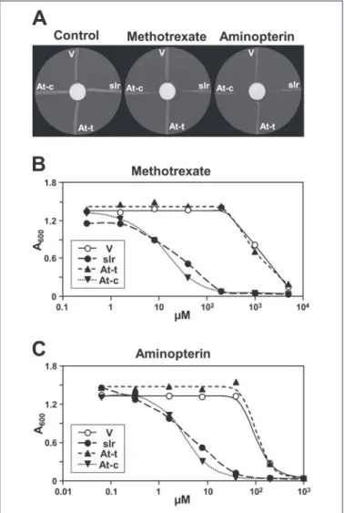
Antifolate Uptake—The antifolates aminopterin and methotrexate are close structural analogs of folic acid, differing only in one and two groups, respectively (Fig. 1A). E. coli normally has little capacity to take up these compounds, and they are inhibitory only at high concentra-tions (50, 51). Therefore, we tested antifolate transport activity by eval-uating growth of cells exposed to antifolate gradients on plates (Fig. 3A) or to various levels of antifolates in liquid medium (Fig. 3, B and C). Expression of slr0642 or the chimeric At2g32040 construct imparted large increases in sensitivity that were qualitatively obvious on plates and were quantified as 30 –100-fold in liquid culture. The truncated At2g32040 protein was again ineffective.
Subcellular Localization of At2g32040—This was investigated first by transient expression of an At2g32040::GFP fusion construct in
Arabi-dopsisprotoplasts. Consistent with the presence of a putative plastid targeting peptide, expression of the fusion protein resulted in green fluorescence that colocalized with the red autofluorescence of chloro-phyll; the green fluorescence was strongest at the chloroplast mar-gins, suggesting that the protein is concentrated in the envelope (Fig. 4A). Expression of GFP alone gave green fluorescence throughout the cytoplasm (Fig. 4A).
Import assays with isolated pea chloroplasts substantiated these results (Fig. 4B). After the import reaction with the full-length protein, chloroplasts contained a labeled product that was smaller than the full-length protein and was protected from attack by thermolysin, as expected for a translocated protein. In contrast, a truncated version lacking the predicted targeting peptide was not processed and was
ther-FIGURE 3. Antifolate uptake tests in E. coli expressing Synechocystis slr0642 or
mod-ified Arabidopsis At2g32040. Strain BN1163 harboring pACYC-RIL was transformed
with pLOI707HE with no insert (V) or containing slr0642 (slr), the truncated At2g32040 protein (At-t), or the chimeric At2g32040 protein (At-c). A, clones were streaked radially on minimal medium plates containing IPTG and 3.6MpABA, with a central 1-cm filter
paper disk impregnated with 10l of dimethyl sulfoxide alone (control) or containing 100 mMmethotrexate or 80 mMaminopterin. Plates were photographed after 2 days. B and C, growth (absorbance at 600 nm) after 9.5 h in liquid minimal medium containing IPTG, 3.6MpABA, and various concentrations of methotrexate (B) or aminopterin (C).
Data are the means of triplicate cultures; S.E. values wereⱕ9% of the means in all cases.
FIGURE 4. Evidence that the At2g23040 protein is targeted to chloroplasts. A, tran-sient expression in Arabidopsis protoplasts of GFP fused to the C terminus of At2g32040 (upper panels) or GFP alone (lower panels). GFP (green pseudo-color) and chlorophyll (red
pseudo-color) fluorescence were observed with an epifluorescence microscope. B,
pro-tein import into isolated pea chloroplasts. Full-length (FL) and truncated (T) versions of At2g32040 were translated in vitro in the presence of [3
H]leucine. The translation prod-ucts were incubated for 15 min in the light with Mg2⫹-ATP and chloroplasts (CP), which
were then purified, or first treated with thermolysin (TH) to remove adsorbed proteins. Proteins were separated by SDS-PAGE and visualized by fluorography. Samples were loaded on the basis of equal chlorophyll content next to aliquots of the respective trans-lation products. The molecular masses (kDa) shown in the figure were estimated from the positions of standards run on the same gel.
FBT Family Transporters
at INRA Institut National de la Recherche Agronomique, on November 8, 2010
www.jbc.org
molysin-sensitive, indicating that it had not been taken up. The latter protein (calculated molecular mass 51 kDa) behaved in SDS-PAGE as a 51-kDa species as expected, but the full-length protein (calcu-lated mass 61 kDa) migrated as a 72-kDa species (Fig. 4B)). Other FBT family proteins likewise migrate anomalously (52). Thus, although the migration of the imported protein as a 67-kDa band (Fig. 4B) implies that the point we chose to truncate At2g32040 is not identical to the normal processing site, electrophoretic anomalies preclude quantifying the discrepancy.
Expression of At2g32040 in Arabidopsis Organs—Quantitative RT-PCR analysis indicated that the At2g32040 gene is constitutively expressed throughout the plant at a low level (sequencing of the RT-PCR product obtained from root and young leaf RNA confirmed that it corresponded to At2g32040 mRNA). Thus, the abundance of At2g32040 transcripts varied rather little among major organs, from 5
to 14⫻ 104copies/250 ng of total RNA (Fig. 5A), which corresponds to
a frequency between 1 in 20,000 and 1 in 60,000 mRNAs (assuming that mRNA is 1% of total RNA and has an average size of 1.5 kb). Ubiquitous, low level expression is also reported for At2g32040 in the GENEVES-TIGATOR Arabidopsis microarray data base (53). The presence of At2g32040 transcripts in roots as well as aerial organs implies that the protein is expressed in non-green plastids as well as chloroplasts.
Identification and Analysis of a T-DNA Insertion Mutant—To explore the function of At2g32040 in vivo, an Arabidopsis line bearing a T-DNA insertion in the coding region of the gene was identified in the Salk collection (40). This mutant has a single T-DNA insertion in exon 1 at codon 24 (Fig. 5, B and C), as determined from the flanking DNA sequence and co-segregation of kanamycin resistance with the T-DNA. The insertion creates at least two in-frame stop codons and, therefore, is predicted to result in a complete knock-out.
Homozygous mutant plants did not differ noticeably from their wild type siblings in growth rate, morphology, leaf color, or fertility. Because At2g32040 is located in the plastid (Fig. 4), we compared the folate profiles of chloroplasts from wild type and mutant plants (Fig. 6). As in pea (15–17), total folate levels in wild type chloroplasts were low, about 50 pmol/mg of protein or 150 pmol/mg of chlorophyll, which is equiv-alent to a stromal concentration of⬃2M(taking stromal volume as 66 l/mg chlorophyll (54)). The most prominent folate forms were 5-methyl- and 5,10-methenyl-THF (the acidic HPLC mobile phase used in the analysis converts 10-formyl-THF to 5,10-methenyl-THF, so that the 5,10-methenyl-THF measured includes 10-formyl-THF plus any preexisting 5,10-methenyl-THF). Relative to wild type, mutant chloro-plasts contained significantly (p⬍ 0.05) more 5,10-methenyl-THF and
FIGURE 5. Expression of the At2g32040 mRNA in Arabidopsis organs and structure
of the T-DNA-mutated At2g32040 gene. A, levels of At2g32040 mRNA in Arabidopsis
organs, determined by real-time RT-PCR. Young leaves were harvested at 21 days, and mature leaves, stems, and siliques at 35 days. Roots were from hydroponically grown plants harvested at 35 days. Data are the means of three determinations for independent tissue samples and S.E. B, structure of the At2g32040 gene, with introns as solid lines and exons as boxes. The positions of the T-DNA insertion, of the start and stop codons, and of the primers used to identify wild type and mutant segregants are indicated. C, agarose gel analysis of the PCR products obtained from pooled genomic DNA from two wild type (WT) or three homozygous mutant plants using the primer pairs indicated. The approx-imate size of the products (bp) is indicated. kb, kilobases. KO, knock-out.
FIGURE 6. Chloroplast folate profiles of wild type and At2g32040 mutant
Arabidop-sis. Chloroplasts were purified by Percoll gradient centrifugation. Folates were analyzed
in deglutamylated samples by HPLC with electrochemical detection. Data are presented per unit protein in A and as a percent of total chloroplast folate in B. 5-CH3-THF,
5-methyl-THF; 5-CHO-THF, 5-formyl-5-methyl-THF; 5,10-CHATHF, 5,10-methenyl-5-methyl-THF; 10-CHO-DHF, 10-formyldihydrofolate (formed during isolation from 10-formyl-THF). Data are the means and S.E. from six wild type and five mutant chloroplast preparations. Asterisks indicate differences between mutant and wild type that are significant at p⬍ 0.05 by Student’s t test; percent data were arcsine transformed before the t test.
at INRA Institut National de la Recherche Agronomique, on November 8, 2010
www.jbc.org
total folate per unit protein (Fig. 6A) and, when data were expressed on a percent basis, a significantly lower proportion of 5-methyl-THF (Fig. 6B).
DISCUSSION
We report here the functional expression of an FBT family protein in
E. coli, an advance that should help studies of transport mechanisms. Expressing the Synechocystis slr0642 protein in E. coli was straightfor-ward, but success with Arabidopsis At2g32040 required the presence of a plasmid bearing rare AT-rich codons and modification of the protein. Deleting its N-terminal extension did not suffice; it was necessary also to replace the N terminus of the truncated protein with the corresponding sequence from slr0642, a region that may affect membrane insertion or translation in E. coli. This modification altered only the initial part (the first third) of the predicted transmembrane helix number 1, which part is not involved in substrate binding in other major facilitator superfam-ily proteins (55, 56).
As shown in Supplemental Fig. 1C, there is a remarkably high sequence conservation between the cyanobacterial carrier and At2g32040 that extends over the entire protein except the region between transmembrane helices 6 and 7, a large loop that separates the two halves of the molecule. The residues that are conserved both in these proteins and the Leishmania FT1, FT5, and BT1 carriers are like-wise spread throughout the sequence except for helices 6 and 7 and the region between them (see Supplemental Fig. 1C). In this interstitial region the Leishmania sequences are predicted to have two additional transmembrane␣-helices, an uncommon configuration in the major facilitator superfamily (57).
Folate transport was evaluated via bacterial growth, which is a sensi-tive way to detect transport activity but not to assess its rate or substrate specificity. For example, growth on 5-CHO-THF or folic acid lagged behind that on pABA, but this does not necessarily imply that the uptake rate of either folate limited growth. Both 5-CHO-THF and folic acid need enzymatic conversion to metabolically active forms (5,10-methe-nyl-THF and THF, respectively) before use (1, 58) so conversion rates could have been limiting. Moreover, 5-CHO-THF inhibits folate-de-pendent enzymes (58) and may have slowed growth on this account. Respecting substrate specificity, growth tests could not show whether or not slr0642 and At2g32040 act on dihydropteridines because E. coli cells took such compounds up unaided. These proteins, thus, remain uncon-firmed candidates for the pteridine transport activities observed or inferred in Cyanobacteria and chloroplasts (59 – 61).
Although E. coli is suspected to have a cryptic gene (abgT), not nor-mally expressed, that can allow transport of folate analogs and perhaps folates (51), three arguments exclude activation and selection of this or any other E. coli gene as an explanation for our data. First, curing port-proficient cells of the expression plasmid abolished folate trans-port capacity. Second, the antifolate uptake results of Fig. 3 could not have involved selection because transport-proficient cells were ipso facto unable to divide. Third, when cells harboring empty expression vector underwent prolonged incubation on folate media, the growth of colonies was far too infrequent to compromise the folate uptake exper-iments of Fig. 2, where all cells expressing an active FBT carrier grew.
The subcellular localization and expression pattern of At2g32040 imply that it has a housekeeping function in green and non-green plas-tids alike. That this function is folate transport is supported by the finding that ablating At2g32040 caused a 23% rise in chloroplast total folate and a 34% fall in the proportion of 5-methyl-THF. That these changes were not more marked implies that there are other chloroplast
folate transporters, and there is indeed recent evidence for a second one in Arabidopsis (62).
Because At2g32040 acts on 5-CHO-THF, folic acid, and antifolates, it is clearly not highly specific and so can presumably transport the mono-glutamyl form of any folate found in plastids. In this connection it is noteworthy that plastid folate transport fluxes include not only net import but also an exchange with the cytosol driven by methionine synthesis. Plastids synthesize homocysteine and contain an isoform of methionine synthase, which converts homocysteine to methionine using 5-methyl-THF as methyl donor (63). However, plastids cannot make 5-methyl-THF due to lack of the enzyme that produces it from 5,10-methylene-THF and so must import it from the cytosol (64) in exchange (not necessarily via the same carrier) for other folates in a shuttle reaction. Because stoichiometric, not catalytic, amounts of 5-methyl-THF are needed for methionine synthesis, this methyl group shuttle must greatly exceed the net import of folates into plastids.
Finally, this work establishes conservation of folate transport func-tion among FBT family proteins from species representing three king-doms of life, monera (Synechocystis), plants (Arabidopsis), and protists (Leishmania). Given the evidence for lateral transfer of genes from Cya-nobacteria to plants (47) and from CyaCya-nobacteria or plants to trypano-somatids and apicomplexans (45, 46), it seems likely that the FBT family arose in Cyanobacteria and passed to the other groups, the evolutionary driver being the selective advantage conferred by folate transport. Whatever the case, these folate carriers that function across kingdom boundaries may offer new options for engineering folate production in bacteria and plants (1, 65). Such carriers might be used, for example, to enhance folate export from bacteria or folate sequestration in storage compartments in plant cells.
Acknowledgments—We thank L. O. Ingram, V. de Cre´cy-Lagard, L. N. Csonka, F. Gerard, C. Dabney-Smith, and M. Ziemak for advice and help, W. Vermaas for Synechocystis DNA, B. Nichols for E. coli strain BN1163, N. Majdalani for strain NM1200, and Stratagene for pACYC-RIL.
REFERENCES
1. Scott, J., Re´beille´, F., and Fletcher, J. (2000) J. Sci. Food Agric. 80, 795– 824 2. Lucock, M. (2000) Mol. Genet. Metab. 71, 121–138
3. Cossins, E. A., and Chen, L. (1997) Phytochemistry 45, 437– 452
4. de Bree, A., van Dusseldorp, M., Brouwer, I. A., van het Hof, K. H., and Steegers-Theunissen, R. P. (1997) Eur. J. Clin. Nutr. 51, 643– 660
5. Re´beille´, F., and Douce, R. (1999) in Regulation of Primary Metabolic Pathways in
Plants(Kruger, N. J., Hill, S. A., and Ratcliffe, R. G., eds) pp. 53–99, Kluwer, Dordrecht, The Netherlands
6. Hanson, A. D., and Gregory, J. F., III (2002) Curr. Opin. Plant Biol. 5, 244 –249 7. Ravanel, S., Cherest, H., Jabrin, S., Grunwald, D., Surdin-Kerjan, Y., Douce, R., and
Re´beille´, F. (2001) Proc. Natl. Acad. Sci. U. S. A. 98, 15360 –15365
8. Basset, G., Quinlivan, E. P., Ziemak, M. J., Dı´az de la Garza, R., Fischer, M., Schiff-mann, S., Bacher, A., Gregory, J. F., III, and Hanson, A. D. (2002) Proc. Natl. Acad. Sci.
U. S. A. 99,12489 –12494
9. Basset, G. J., Quinlivan, E. P., Ravanel, S., Re´beille´, F., Nichols, B. P., Shinozaki, K., Seki, M., Adams-Phillips, L. C., Giovannoni, J. J., Gregory, J. F., III, and Hanson, A. D. (2004)
Proc. Natl. Acad. Sci. U. S. A. 101,1496 –1501
10. Goyer, A., Illarionova, V., Roje, S., Fischer, M., Bacher, A., and Hanson, A. D. (2004)
Plant Physiol. 135,103–111
11. Basset, G. J., Ravanel, S., Quinlivan, E. P., White, R., Giovannoni, J. J., Re´beille´, F., Nichols, B. P., Shinozaki, K., Seki, M., Gregory, J. F., III, and Hanson, A. D. (2004) Plant
J. 40,453– 461
12. Klaus, S. M., Wegkamp, A., Sybesma, W., Hugenholtz, J., Gregory, J. F., III, and Hanson, A. D. (2005) J. Biol. Chem. 280, 5274 –5280
13. Chen, L., Chan, S. Y., and Cossins, E. A. (1997) Plant Physiol. 115, 299 –309 14. Gambonnet, B., Jabrin, S., Ravanel, S., Karan, M., Douce, R., and Re´beille´, F. (2001) J.
Sci. Food Agric. 81,835– 841
15. Jabrin, S., Ravanel, S., Gambonnet, B., Douce, R., and Re´beille´, F. (2003) Plant Physiol.
131,1431–1439
16. Chan, S. Y., and Cossins, E. A. (2003) Pteridines 14, 67–76
FBT Family Transporters
at INRA Institut National de la Recherche Agronomique, on November 8, 2010
www.jbc.org
17. Orsomando, G., Dı´az de la Garza, R., Green, B. J., Peng, M., Rea, P. A., Ryan T. J., Gregory, J. F., III, and Hanson, A. D. (2005) J. Biol. Chem. 280, 28877–28884 18. Quinlivan, E. P., Roje, S., Basset, G., Shachar-Hill, Y., Gregory, J. F., III, and Hanson,
A. D. (2003) J. Biol. Chem. 278, 20731–20737
19. Brzezinska, A., Winska, P., and Balinska, M. (2000) Acta Biochim. Pol. 47, 735–749 20. Sirotnak, F. M., and Tolner, B. (1999) Annu. Rev. Nutr. 19, 91–122
21. Titus, S. A., and Moran, R. G. (2000) J. Biol. Chem. 275, 36811–36817
22. Ouellette, M., Drummelsmith, J., El-Fadili, A., Kundig, C., Richard, D., and Roy, G. (2002) Int. J. Parasitol. 32, 385–398
23. Shane, B., and Stokstad, E. L. (1975) J. Biol. Chem. 250, 2243–2253
24. Kumar, H. P., Tsuji, J. M., and Henderson, G. B. (1987) J. Biol. Chem. 262, 7171–7179 25. Tamura, T., Baggott, J. E., Johnston, K. E., Li, Q.J., and Antony, A. C. (1997)
Microbi-ology 143,2639 –2646
26. Beck, J. T., and Ullman, B. (1990) Mol. Biochem. Parasitol. 43, 221–230
27. Lemley, C., Yan, S., Dole, V. S., Madhubala, R., Cunningham, M. L., Beverley, S. M., Myler, P. J., and Stuart, K. D. (1999) Mol. Biochem. Parasitol. 104, 93–105 28. Richard, D., Kundig, C., and Ouellette, M. (2002) J. Biol. Chem. 277, 29460 –29467 29. Richard, D., Leprohon, P., Drummelsmith, J., and Ouellette, M. (2004) J. Biol. Chem.
279,54494 –54501
30. Chang, A. B., Lin, R., Studley, W. K., Tran, C. V., and Saier, M. H., Jr. (2004) Mol.
Memb. Biol. 21,171–181
31. Court, D. L., Swaminathan, S., Yu, D., Wilson, H., Baker, T., Bubunenko, M., Sawitzke, J., and Sharan, S. K. (2003) Gene (Amst.) 315, 63– 69
32. Yu, D., Ellis, H. M., Lee, E. C., Jenkins, N. A., Copeland, N. G., and Court, D. L. (2000)
Proc. Natl. Acad. Sci. U. S. A. 97,5978 –5983
33. Gibeaut, D. M., Hulett, J., Cramer, G. R., and Seemann, J. R. (1997) Plant Physiol. 115, 317–319
34. Arfman, N., Worrell, V., and Ingram, L. O. (1992) J. Bacteriol. 174, 7370 –7378 35. Sambrook, J., Fritsch, E. F., and Maniatis, T. (1989) Molecular Cloning: A Laboratory
Manual, 2nd Ed., p. A.3, Cold Spring Harbor Laboratory, Cold Spring Harbor, NY 36. Sherman, F. (1991) Methods Enzymol. 194, 3–21
37. Niwa, Y. (2003) Plant Biotechnol. 20, 1–11 38. Abel, S., and Theologis, A. (1994) Plant J. 5, 421– 427
39. Cline, K., Henry, R., Li, C., and Yuan, J. (1993) EMBO J. 12, 4105– 4114
40. Alonso, J. M., Stepanova, A. N., Leisse, T. J., Kim, C. J., Chen, H., Shinn, P., Stevenson, D. K., Zimmerman, J., Barajas, P., Cheuk, R., Gadrinab, C., Heller, C., Jeske, A., Koesema, E., Meyers, C. C., Parker, H., Prednis, L., Ansari, Y., Choy, N., Deen, H., Geralt, M., Hazari, N., Hom, E., Karnes, M., Mulholland, C., Ndubaku, R., Schmidt, I., Guzman, P., Aguilar-Henonin, L., Schmid, M., Weigel, D., Carter, D. E., Marchand, T., Risseeuw, E., Brogden, D., Zeko, A., Crosby, W. L., Berry, C. C., and Ecker, J. R. (2003)
Science 301,653– 657
41. Goyer, A., Collakova, E., Dı´az de la Garza, R., Quinlivan, E. P., Williamson, J., Gregory, J. F., III, Shachar-Hill, Y., and Hanson, A. D. (2005) J. Biol. Chem. 280, 26137–26142
42. Brock, I. W., Hazell, L., Michl, D., Nielsen, V. S., Moller, B. L., Herrmann, R. G., Klosgen, R. B., and Robinson, C. (1993) Plant Mol. Biol. 23, 717–725
43. Schulz, A., Knoetzel, J., Scheller, H. V., and Mant, A. (2004) J. Histochem. Cytochem.
52,701–704
44. Emanuelsson, O., Nielsen, H., and von Heijne, G. (1999) Protein Sci. 8, 978 –984 45. Hannaert, V., Saavedra, E., Duffieux, F., Szikora, J. P., Rigden, D. J., Michels, P. A., and
Opperdoes, F. R. (2003) Proc. Natl. Acad. Sci. U. S. A. 100, 1067–1071
46. Waller, R. F., McConville, M. J., and McFadden, G. I. (2004) Trends Parasitol. 20, 54 –57
47. Martin, W., Rujan, T., Richly, E., Hansen, A., Cornelsen, S., Lins, T., Leister, D., Stoebe, B., Hasegawa, M., and Penny, D. (2002) Proc. Natl. Acad. Sci. U. S. A. 99, 12246 –12251
48. Green, J. C., Nichols, B. P., and Matthews, R. G. (1996) in Escherichia coli and
Salmo-nella: Cellular and Molecular Biology, Vol. 1 (Neidhardt, F.C., ed) pp. 665– 673, American Society for Microbiology, Washington, D. C.
49. Facey, S. J., and Kuhn, A. (2004) Biochim. Biophys. Acta 1694, 55– 66
50. Hussein, M. J., Green, J. M., and Nichols, B. P. (1998) J. Bacteriol. 180, 6260 – 6268 51. Green, J. M., and Nichols, B. P. (2002) in Chemistry and Biology of Pteridines and
Folates(Milstien, S., Kapatos, G., Levine, R. A., and Shane, B., eds) pp. 631– 635, Kluwer Academic Publishers Group, Dordrecht, The Netherlands
52. Dole, V. S., Myler, P. J., Stuart, K. D., and Madhubala, R. (2002) FEMS Microbiol. Lett.
208,89 –91
53. Zimmermann, P., Hirsch-Hoffmann, M., Hennig, L., and Gruissem, W. (2004) Plant
Physiol. 136,2621–2632
54. Winter, H., Robinson, D. G., and Heldt, H. W. (1994) Planta 193, 530 –535 55. Abramson, J., Smirnova, I., Kasho, V., Verner, G., Kaback, H. R., and Iwata, S. (2003)
Science 301,610 – 615
56. Huang, Y., Lemieux, M. J., Song, J., Auer, M., and Wang, D. N. (2003) Science 301, 616 – 620
57. Pao, S. S., Paulsen, I. T., and Saier, M. H., Jr. (1998) Microbiol. Mol. Biol. Rev. 62, 1–34 58. Stover, P., and Schirch, V. (1993) Trends Biochem. Sci. 18, 102–106
59. Jansz, E. R., and Maclean, F. I. (1973) Can. J. Microbiol. 19, 381–387 60. Grodzinski, B., and Colman, B. (1973) J. Bacteriol. 115, 456 – 458
61. Iwai, K., Bunno, M., Kobashi, M., and Suzuki, T. (1976) Biochim. Biophys. Acta 444, 618 – 622
62. Bedhomme, M., Hoffmann, M., McCarthy, E. A., Gambonnet, B., Moran, R. G., Re´-beille´, F., Ravanel, S. (2005) J. Biol. Chem. 280, 34823–34831
63. Ravanel, S., Block, M. A., Rippert, P., Jabrin, S., Curien, G., Re´beille´, F., and Douce, R. (2004) J. Biol. Chem. 279, 22548 –22557
64. Hanson, A. D., Gage, D. A., and Shachar-Hill, Y. (2000) Trends Plant Sci. 5, 206 –213 65. Sybesma, W., Starrenburg, M., Kleerebezem, M., Mierau, I., de Vos W. M., and
Hu-genholtz, J. (2003) Appl. Environ. Microbiol. 69, 3069 –3076
at INRA Institut National de la Recherche Agronomique, on November 8, 2010
www.jbc.org