ENSEIGNEMENT SUPÉRIEUR EN RÉANIMATION MÉDECIN
Glucocorticoids in the treatment of acute respiratory distress syndrome*
Les glucocorticoïdes dans le traitement du syndrome de détresse respiratoire aiguë
F. Roche-Campo · H. Aguirre-Bermeo · J. Mancebo
Received: 22nd September 2011, Accepted: 3rd October 2011 © SRLF et Springer-Verlag France 2011
Abstract Acute respiratory distress syndrome (ARDS) is characterized by local inflammation and an intense systemic inflammatory reaction. Glucocorticoid administration has been suggested due to their anti-inflammatory properties. However, results from the initial studies of glucocorticoids in ARDS, which evaluated high-dose and short-term treat-ments, were negative. More recent studies have evaluated the effect of lower doses of glucocorticoids administered over longer periods, but the results thus far have been
incon-clusive.To cite this journal: Réanimation 21 (2012).
Keywords Glucocorticoids · Treatment · Acute respiratory distress syndrome
Résumé Le syndrome de détresse respiratoire aiguë (SDRA) est caractérisé par une inflammation locale et une réaction
inflammatoire systémique intense. L’administration de
glu-cocorticoïdes a été proposée en raison de leurs propriétés anti-inflammatoires. Cependant, les résultats des premières études concernant la prescription des glucocorticoïdes dans le SDRA, qui ont évalué de hautes doses avec des traite-ments de court terme, ont été négatifs. Des études plus récentes ont évalué des doses plus basses administrées sur des périodes plus longues, mais les résultats ont aussi été peu
concluants. Pour citer cette revue : Réanimation 21
(2012).
Mots clés Glucocorticoïdes · Traitement · Syndrome de détresse respiratoire aiguë
Introduction
Acute respiratory distress syndrome (ARDS) has been one of the greatest challenges faced by intensive care physicians since it was first described in 1967 [1]. Mortality rates remain high (approximately 50%) [2], and sequelae in survi-vors can be severe and long-lasting [3]. ARDS is character-ized by the sudden appearance of hypoxemia with radio-graphic evidence of bilateral lung infiltrates. ARDS is the physiological response of the lungs to numerous aggres-sions, both direct and indirect, which can lead to an increase in the permeability of the alveolocapillary membrane, sec-ondary lung edema, and persistent inflammation [4]. This increased alveolocapillary permeability is central to the pathophysiology of the syndrome and distinguishes ARDS from cardiogenic pulmonary edema that occurs due to an increase in the pulmonary capillary vascular pressure [5].
Various pharmacological and nonpharmacological strate-gies have been tried in an attempt to prevent or attenuate this inflammatory process [6]. Protective ventilation with low tidal volume has been shown to reduce the inflammatory response [7] and improve survival [8]. Encouraging results have also been obtained with prone position ventilation [9] and ventilating with a positive end-expiratory pressure to a
maximum plateau pressure of 30 cmH2O [10,11]. Results
from pharmacological interventions—including a variety of
anti-inflammatory and immunomodulating drugs—have
been disappointing [12]. Glucocorticoids (GCs) have shown some promising results in the past, so researchers have taken a renewed interest in this medication in recent years.
The use of GCs in intensive care dates back to the decade of the 1980s, when critical care clinicians began to better understand the role of inflammation in the pathogenesis of many diseases affecting critically ill patients. GCs were used to control or prevent inflammation in patients with ARDS or septic shock [13]. However, the high doses used proved to be more harmful than beneficial, and interest in these drugs
waned over time [14–17]. Now that we better understand
F. Roche-Campo (*) · H. Aguirre-Bermeo · J. Mancebo Servei de Medicina Intensiva, Hospital de Sant Pau, Barcelona, Espagne
e-mail : ferranroche@yahoo.es
* Cet article correspond à la conférence faite par l’auteur au congrès de la SRLF 2012 dans la session: SDRA au quotidien. DOI 10.1007/s13546-011-0316-1
why these mega doses failed, and as more recent studies have shown that the use of more appropriate physiological doses yields improved results, interest in the use of these drugs has increased.
The aim of this review is to evaluate the biological effects of GCs and to discuss the most relevant published studies of GCs in the treatment of ARDS. Although GCs are also used to treat septic shock and community-acquired pneumonia, both of which are known causes of ARDS [18], these spe-cific areas are not addressed directly in the present review.
Biological effects of glucocorticoids and basis
for its use in ARDS
Inflammation is the body’s reflex response to any type of
aggression [19]. The inflammatory response consists of a series of integrated reactions, whose objective is to destroy the harmful agent and repair damaged tissues. As long as the inflammatory process remains circumscribed and controlled, it is beneficial. However, uncontrolled inflammation is harmful. In ARDS, the intense inflammatory reaction is both local (the lungs) and systemic [20]. The evolutionary onset of the syndrome depends on the level and duration of this inflammatory process [21]. Persistently elevated levels of proinflammatory cytokines are associated with unresol-ving ARDS and a sign of worse prognosis [22].
The hypothalamic-pituitary-adrenal axis regulates the production of endogenous GCs, which, in addition to their metabolic and cardiovascular effects, play an essential role in modulating and limiting the inflammatory response [23]. Endogenous GCs are the natural drugs that protect us from stress and enhance survival [19]. Thus, while the adrenal cortex secretes 20 mg/day of cortisol in basal conditions, secretion may increase to between 200 and 400 mg/day in inflammatory processes, and basal levels of plasmatic corti-sol can increase by a multiple of 5 [24]. Elevated levels of cortisol are associated with more severe underlying disease and higher mortality [25,26].
More than 90% of circulating cortisol is bound to
corticosteroid-binding globulin. The remaining 10%—the
biologically active form—circulate freely [27]. During
acute illness, the levels of corticosteroid-binding globulin can decrease by up to 50%, leading to a corresponding large increase in free cortisol [28]. The free cortisol has a high affinity for a specific cell receptor called receptor alpha (GR-alpha), which is expressed in virtually all cells. The
cortisol–GR-alpha complex acts at the intracellular level
through genomic and nongenomic mechanisms, thereby inducing the production of anti-inflammatory proteins while inhibiting the production of proinflammatory proteins. This complex also inhibits fibroblast proliferation and colla-gen deposits [20]. However, endocolla-genous GCs are not always
able to modulate the inflammatory response, either because endogenous GC secretion is insufficient to overcome the inflammatory response (adrenal insufficiency) or because peripheral tissues become resistant (with or without adrenal insufficiency) [29]. This dynamic situation, defined as criti-cal illness-related corticosteroid insufficiency (CIRCI), may be reversed through the administration of exogenous GCs [30]. Unfortunately, the diagnostic criteria for CIRCI still remain to be well-defined [29].
Experimental and clinical evidence show that administra-tion of exogenous GCs regulates the inflammatory response, reduces markers of inflammation, and improves symptoms [30,31]. For this reason, GC therapy is considered biologi-cally plausible in ARDS.
Comments on the most relevant clinical studies
The first randomized controlled studies (RCTs) of GC administration date back to the 1980s. The hypothesis at
that time, based on experimental studies [32–35], was that
the use of high-dose, short-term GC treatment (e.g., 120 mg/kg per day of methylprednisolone for 48 hours) could pre-vent the emergence of ARDS in at-risk septic patients and
could also be used to treat early-stage ARDS [14–17].
How-ever, studies that evaluated this hypothesis in ARDS or in
septic shock patients found no benefits [36–38]. As we
dis-cuss in more detail below, it seems that harmful side effects caused by these high doses outweigh any beneficial effects of this therapy. In fact, recently published meta-analyses conclude that high doses of GCs not only fail to prevent ARDS in at-risk patients but may even hasten its onset [39]. Given the failure of this high-dose, short-term GC ther-apy, these drugs largely disappeared from the medical litera-ture until they regained popularity nearly a decade later, when promising results were reported for GC therapy in meningitis [40] and Pneumocystis carinii pneumonia [41]. An important conceptual change, which transformed our understanding of appropriate dosing levels, had taken place. In 1991, Schneider and Voerman [42] described for the first time how septic shock could be reversed using “only” physiological doses of GCs. These results established the basis for new dosing strategies in ARDS, which Meduri et al. adopted in their seminal study published just a few years afterwards [43]. As a result, researchers have carried out new studies that abandoned the original high-dose, short-term approach in favor of low to moderate doses delivered over prolonged periods (Table 1).
In 1998, Meduri et al. reported the results of a small ran-domized multicenter study that included 26 patients with ARDS of 7 days duration or more [44]. This study could
be considered the first study of GCs in ARDS of the
per day of methylprednisolone (16 patients) or placebo (8 patients) for 32 days. The main outcome measures were lung function and mortality, with patients in the treatment group showing significant improvement in both measures. Consequently, the authors concluded that prolonged admin-istration of methylprednisolone in patients with unresolving ARDS is associated with improved lung function and reduced mortality. The main limitations of that study were its small sample size and crossover design, which permitted patients in the placebo group who showed no improvement after 10 days of GC treatment (4 patients). Moreover, the study was stopped early after an intermediate analysis of efficacy; as a result, it is possible that the treatment effect was overestimated.
Nevertheless, these encouraging results prompted other investigators to undertake additional studies in the early 2000s, although no results were published until several years later.
The multicentric RCT published by Confalonieri et al. in 2005 [45] has been included in some meta-analyses, even though the primary focus was on patients with severe pneu-monia rather than ARDS. These authors evaluated 46 patients who had been admitted to the Intensive Care Unit (ICU) for severe community-acquired pneumonia. Patients were randomized to receive 240 mg/day of continu-ous hydrocortisone infusion or placebo for 7 days. The main objectives were to improve oxygenation, reduce the number of organ failures at day 8, and decrease the number of days in shock. Patients in the treatment group showed significant improvement in oxygenation, lung mechanics, and shock-free days; and they also showed a decrease in hospital mor-tality. The main limitation of the study (from the perspective of the present review) was the inclusion criteria: severe pneumonia rather than ARDS.
In 2006, the ARDS Network [46] published the largest study to date of GC therapy for ARDS. A total of 180 patients with a recent ARDS diagnosis (from 7 to 28 days following onset) were randomized to receive either 2 mg/kg per day of methylprednisolone or placebo for 25 days. The
GCs were withdrawn after 2–4 days. The primary endpoint
was mortality at day 60. Secondary endpoint included days free from mechanical ventilation, changes in biochemical markers, and the number of infectious complications. No differences in death rates at day 60 (29% in both groups) or day 180 (32% in both) were observed. However, the treat-ment group required fewer days of mechanical ventilation (7 vs. 11 days, p < 0.001) and fewer days in the ICU (6 vs. 9 days, p = 0.006) during the first 28 days, as well as fewer days of shock, better oxygenation, and better lung mechan-ics. Treatment was also associated with a reduction in noso-comial infections without an increase in muscular weakness. Post hoc analysis showed higher death rates at day 60 and 180 for the 48 patients in whom GC treatment was initiated
T able 1 Rand omized st udies foc used on the prolonged use of low –modera te doses of glucocorticoids (GCs) in acute lung lesions and ARDS St ud y Me th od ol og y P at ie n t p op ul at ion Nu mb er of pa ti en ts Tim in g of inc lu si on T rea tm en t Du ra ti on of tr ea tm en t, da ys Me du ri et al . (1 99 8) Pr os pe ct ive AR DS 24 L at e Me thy lp re dn is ol on e 2m g /k g Up to 32 Co nf al on ie ri et al . (2 00 5) Pr os pe ct ive S ev er e pn eu mo ni a 46 E ar ly Hy dr oc or tis on e 24 0 mg /d 7 St ei nb erg et al . (2 00 6) Pr os pe ct ive AR DS 18 0 L at e Me thy lp re dn is ol on e 2m g /k g Up to 25 An na ne et al . (2 00 6) Re tro sp ec tiv e S ep si s an d AR DS 17 7 E ar ly Hy dr oc or tis on e 20 0 mg /d 7 Me du ri et al . (2 00 7) Pr os pe ct ive AR DS 91 E ar ly Me thy lp re dn is ol on e 1m g /k g Up to 28 ARDS: acute resp iratory distress syndrom e; ICU: inten sive care unit; LIS: lung injury sco re; Early (<6 day s) ver sus late (>7 days).
13 or more days after the onset of ARDS. Another post hoc analysis evaluated the levels of type III procollagen peptide in bronchoalveolar lavage fluid taken from 91 patients dur-ing their admission. In these patients, the authors found that in the 46 patients with the lowest peptide values (<50th
per-centile)—a group that included most of the patients treated
after day 13—mortality at day 60 was higher for patients in
the treatment group (23 cases) compared to the placebo group (35% vs. 8%, p = 0.03). Conversely, in the 45 patients with the highest peptide values (>50th percentile), mortality at day 60 was lower in the treatment group (23 cases) than the placebo group (4% vs. 19%, p = 0.10). The authors con-cluded that the evidence did not support the use of methyl-prednisolone for persistent ARDS, despite the observed improvement in cardiopulmonary physiology. Moreover, they noted that mortality rates may actually increase when GC treatment is initiated more than 13 days after ARDS onset. Among the more notable limitations of the ARDS Network study is the early termination due to low enrollment (only 180 patients from 25 hospitals were included in a 6-year period). Likewise, a high percentage of patients in the treatment group required reintubation (20 vs. 6; p = 0.008). It has been hypothesized that this outcome could be explained by the abrupt discontinuation of the GC treatment, which may lead to renewed pulmonary inflamma-tion (the mechanisms by which abrupt discontinuainflamma-tion can be harmful are discussed in more detail below). Finally, there were several nonsignificant differences in the baseline characteristics of the small subgroup of patients who were randomized 13 or more days following onset, thus making comparison between these subgroups difficult.
In the same year (2006) that the ARDS Network study was published, Annane et al. [47] reported a post hoc analy-sis that evaluated a subgroup of patients from a larger clini-cal trial. The original study assessed low-dose GCs in 300 patients with septic shock. Of these original 300 cases, the authors analyzed the 177 patients who had a diagnosis of ARDS at the time of admission. Of these, 85 patients had received hydrocortisone (200 mg/d) for 7 days, while the remaining cases (92) received placebo. In their analysis, the authors differentiated between responders and non-responders by a rapid corticotrophin stimulation test. These researchers found that treatment with GCs was associated with lower mortality rates in patients with septic shock who did not respond to the corticotrophin test but not in the responder group nor in the group with septic shock with-out ARDS. Unfortunately, causal relations cannot be estab-lished due to the retrospective nature of the analysis.
In 2007, Meduri et al. [48] reported results of their second multicentric randomized study. This is the only RCT to date specifically focused on early ARDS (<7 days). A total of 91 patients were randomized within the first 72 hours of ARDS onset to receive 1 mg/kg per day of methylprednisolone
(n = 63) or placebo (n = 28) for 28 days. The main end points were lung injury scores and successful extubation at day 7. Patients in the treatment group showed a significant improvement in these measures in addition to a lower ICU mortality rate (21% vs. 43%; p = 0.03). This study had sev-eral limitations, including a low recruitment rate per center (five centers and a 10-year enrollment period) and the use of a crossover design (most patients in the placebo group who still required mechanical ventilation at day 9 were treated with GCs), a strategy that makes interpretation of results dif-ficult. In the placebo group, 45% of the patients required vasopressor support at the time of admission, so they did not receive GC therapy. Thus, this fact could be considered detrimental to the most severely ill patients [49]. In addition, the authors used the same sequential design as in their first study, in which measures of treatment effects influenced the decision to stop the study [50].
Comment on meta-analyses
Five meta-analyses or systematic reviews (but only few orig-inal, optimal papers) have been published since 2007. Despite variations among these five reviews in terms of methodology and study selection, the conclusions are gener-ally similar.
•
Agarwal et al. [51] evaluated two observational studiesand four RCTs and concluded that current evidence does not support the use of GCs in the management of early or late ARDS ;
•
Meduri et al. [52] evaluated five RCTs, including thestud-ies by Confalonieri et al. and Annane et al. They con-cluded that prolonged administration of GCs started before day 14 improved prognosis ;
•
Tang et al. [53] assessed five observational studies andfour RCTs of ARDS patients treated with low to moderate doses of GCs (including the Confalonieri study). The authors concluded that low doses are associated with less morbidity and mortality without an increase in side effects. They recommended that a new, rigorous trial be performed to confirm these findings in early ARDS ;
•
Peter et al. [39] used a rigorous statistical approach tocarry out a meta-analysis of a group of highly hetero-geneous studies. They evaluated nine RCTs, four of which were studies of ARDS prevention. The other five studies were RCTs for the treatment of persistent ARDS; of these, one evaluated high-dose treatment and another was the aforementioned study by Annane et al. This meta-analysis found that while GC treatment seems to yield positive results, no definitive conclusions can be made at present. The authors did, however, rule out the use of GC for preventive measures ;
•
Finally, Lamontagne et al. [54] evaluated 12 RCTs of patients with severe pneumonia, acute lung injury, or ARDS. These authors also found that prolonged GC treat-ment (at least 7 days) given before day 14 of onset seems to yield positive results. However, they too were unable to reach any firm conclusions.Factors that affect the response to treatment
with glucocorticoids
ARDS criteria and etiology
•
Most ARDS studies use diagnostic criteria described in1994 [55], a definition that, though straightforward, is not perfect: postmortem studies showed that 25% of lungs diagnosed with ARDS failed to meet the patho-logical criteria [56];
•
Many of these ARDS cases are secondary to septic shockand pneumonia [18]. The specific effects of GCs on both pathologies may be a confounding factor in the associa-tion between GCs and ARDS. The same can be said for etomidate, a drug which may induce transitory adrenal insufficiency and promote the overexpression of the inflammatory response [57];
•
Many of the studies of GCs for ARDS were designedbefore the clinical benefits of protective ventilation were described in the year 2000 [8]. Protective ventilation using low tidal volumes and limited airway pressures have proven to be less proinflammatory and less damag-ing than ventilation without volume limits [58]. If the volutrauma is reduced, the beneficial effects of GCs may be less than reported.
The individual variability of each patient
To believe that proinflammatory and anti-inflammatory pro-cesses occur at the same time and counterbalance each other in the same way in all patients is illusory. The course of these processes varies between individuals and depends both on the severity of the pathology as well as genetic factors and
individual susceptibility. Biomarkers of lung inflammation may be useful to individualize therapy [59].
Treatment strategies
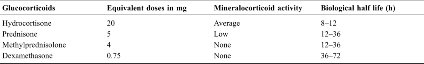
Administration of low doses of GCs, the usual strategy in septic shock, is quite different from the immunosuppressive doses that were used in the first studies of ARDS or in the more recent CRASH trial of patients with cranial trauma [60]. Much confusion persists in terms of the action of GCs with respect to the type of molecule used (either hydro-cortisone or methylprednisolone) and the dose (low or mod-erate). In septic shock, perhaps it is best to use molecules with a mineralocorticoid and GC effect at doses that replace the adrenal insufficiency, while in ARDS, perhaps it would be better to use molecules whose effect is purely GC at anti-inflammatory doses (Table 2). In addition, we must deter-mine when to initiate GC treatment with respect to ARDS onset (preventative, early, or late), the delivery method (intermittent bolus or continuous perfusion), treatment dura-tion (short or prolonged), and the appropriate discontinua-tion method (abrupt or progressive).
The future development of new, selective GCs that mimic the beneficial effects of natural GCs without their detrimen-tal side effects seems likely.
Adverse effects
The efficacy of GCs in alleviating inflammatory disorders is due to the different actions of the glucocorticoid receptor on multiple signaling pathways. Because there are multiple pathways involved, there is no selectivity, which in turn implies a high risk of adverse effects. Thus, it is essential that we employ strategies to prevent GC-related complica-tions in order to minimize the adverse effects of treatment while maximizing the beneficial effects. The following mea-sures have been proposed [61]:
•
Perform systematic microbiological infection surveillancefor early detection of nosocomial infections in patients who may have a blunted febrile response. Recent studies of low to moderate doses of GCs have not found a higher incidence of nosocomial infection [39,53]. At these doses,
Table 2 Equivalence between glucocorticoids (GCs)
Glucocorticoids Equivalent doses in mg Mineralocorticoid activity Biological half life (h)
Hydrocortisone 20 Average 8–12
Prednisone 5 Low 12–36
Methylprednisolone 4 None 12–36
GCs may reduce the incidence of ventilator-associated pneumonia (VAP) in selected populations [62]. However, retrospective studies in patients with ARDS secondary to the A/H1N1 influenza virus show an association between the use of GCs and VAP and an increase in mortality [63,64];
•
Avoid the concomitant use of muscle relaxants. When usedalone, GCs do not seem to increase muscle weakness [65]. However, combined use of GCs and muscle relaxants in asthmatic patients has been associated with a significant increase in muscle weakness [66]. Recently, the use of mus-cle relaxants during the first 48 hours after ARDS onset has been shown to reduce mortality without increasing muscle weakness [67]. In that study, 55% of patients received GCs for varying reasons at some time during treatment;
•
Avoid premature and abrupt discontinuation. As we havealready discussed, this is the main critique of the ARDS Network study. Administration of exogenous GCs may induce adrenal insufficiency through inhibition of cellular
receptors and/or negative feedback from the
hypothalamic-pituitary-adrenal axis. Once exogenous GCs are stopped, the receptors need time to recover (the precise time in ARDS patients is not well-known, but is estimated to range from 1 to 2 weeks) [68]. In experimen-tal acute lung injury, prolonged GCs administration decreased edema and lung collagen formation, whereas early withdrawal rapidly negated the positive effects of
therapy [33–35]. In unresolving ARDS, early
discontinu-ation of GC treatment was associated with physiological deterioration that improved following reinstitution of treatment [59,69]. Untreated adrenal insufficiency is asso-ciated with prolonged weaning [70]. In patients with sep-tic shock, premature discontinuation of GCs may be asso-ciated with a spike in markers of inflammation and a worsening of hemodynamic parameters [71];
•
Continuous perfusion rather than intermittent bolusadministration may reduce glycemia variability and improve its control [72]. Marked oscillations in glycemia levels increase oxidative stress [73] and may be a marker of poor prognosis in critically ill patients [74].
Conclusion
The various studies and meta-analyses on the prolonged use of low to moderate doses of GCs in ARDS suggest the pos-sibility that this medication may provide some benefits with a manageable risk profile (Table 3). Indeed, the use of GCs was actively recommended at a recent consensus conference of the American College of Critical Care Medicine: “Moderate-dose GC should be considered in the manage-ment strategy of patients with early severe ARDS and before day 14 in patients with unresolving ARDS. The role of GC
treatment in acute lung injury and less severe ARDS is less
clear” [29]. The first Surviving Sepsis campaign reached a
similar conclusion regarding the use of GCs for septic shock [75]. Nevertheless, given the difficulties of achieving consis-tently reproducible results thus far [76], the recommendation levels provided by recently published clinical guidelines have been lowered from previous guidelines [77]. Even so,
several authors [78–82] have expressed their disagreement
with these recommendations for two main reasons:
•
Most of the studies that the meta-analyses have evaluatedwere carried out by enthusiastic supporters of GC therapy, and these are the same people who have authored the American recommendations;
•
The primary endpoint of the ARDS Network trial, whichis considered to be the most rigorous study to date, was negative.
In order to resolve some of the uncertainties, a new study focused on low to moderate prolonged doses of methylpred-nisolone in early ARDS is being planned (the CARS trial, sponsored by the Clinical Trial Group of the European Soci-ety of Intensive Care Medicine).
Until new results become available, current evidence does not support the use of GCs for ARDS and, therefore, we do not recommend their use for the treatment of ARDS at this time.
Conflit d’intérêt : les auteurs déclarent ne pas avoir de
conflit d’intérêt.
Table 3 Reasons to administer or not (advantages vs. disad-vantages) glucocorticoids (GCs) in acute respiratory distress syndrome (ARDS)
Advantages
– Several studies show beneficial results for many secondary endpoints Mortality should not be the only argument to evaluate a therapeutic approach
– Few side effects are observed when GCs are properly administered at appropriate dosages
– Some beneficial results in related pathologies, such as community-acquired bacterial pneumonia
– Cheap and easily accessible Disadvantages
– Limitations in biological knowledge
– Serious methodological problems in several studies, a fact that also brings into question the validity of the meta-analyses – The largest and best-designed study to date had negative
results in its primary endpoint
– Early administration in patients with ARDS secondary to the A/H1N1 influenza virus could be dangerous – New studies, promoted by authors that currently defend
Références
1. Ashbaugh DG, Bigelow DB, Petty TL, Levine BE (1967) Acute respiratory distress in adults. Lancet 2:319–23
2. Rubenfeld GD, Herridge MS (2007) Epidemiology and outcomes of acute lung injury. Chest 131:554–62
3. Herridge MS, Tansey CM, Matte A, et al (2011) Functional dis-ability 5 years after acute respiratory distress syndrome. N Engl J Med 364:1293–304
4. Leaver SK, Evans TW (2007) Acute respiratory distress syn-drome. BMJ 335:389–94
5. Ware LB, Matthay MA (2005) Clinical practice. Acute pulmo-nary edema. N Engl J Med 353:2788–96
6. Diaz JV, Brower R, Calfee CS, Matthay MA (2010) Therapeutic strategies for severe acute lung injury. Crit Care Med 38:1644–50 7. Ranieri VM, Suter PM, Tortorella C, et al (1999) Effect of mechanical ventilation on inflammatory mediators in patients with acute respiratory distress syndrome: A randomized con-trolled trial. JAMA 282:54–61
8. Merci de fournir les auteurs]] (2000) Ventilation with lower tidal volumes as compared with traditional tidal volumes for acute lung injury and the acute respiratory distress syndrome. The acute respi-ratory distress syndrome network. N Engl J Med 342:1301–8 9. Abroug F, Ouanes-Besbes L, Dachraoui F, et al (2011) An
updated study-level meta-analysis of randomised controlled trials on proning in ARDS and acute lung injury. Crit Care 15:R6 10. Mercat A, Richard JC, Vielle B, et al (2008) Positive
end-expiratory pressure setting in adults with acute lung injury and acute respiratory distress syndrome: a randomized controlled trial. JAMA 299:646–55
11. Briel M, Meade M, Mercat A, et al (2010) Higher vs lower posi-tive end-expiratory pressure in patients with acute lung injury and acute respiratory distress syndrome: systematic review and meta-analysis. JAMA 303:865–73
12. Frank AJ, Thompson BT (2010) Pharmacological treatments for acute respiratory distress syndrome. Curr Opin Crit Care 16:62–8 13. Carlet J (1999) From mega to more reasonable doses of
corticos-teroids: a decade to recreate hope. Crit Care Med 27:672–4 14. Weigelt JA, Norcross JF, Borman KR, Snyder WH, 3rd (1985)
Early steroid therapy for respiratory failure. Arch Surg 120:536–40 15. Bernard GR, Luce JM, Sprung CL, et al (1987) High-dose corti-costeroids in patients with the adult respiratory distress syn-drome. N Engl J Med 317:1565–70
16. Bone RC, Fisher CJ, Jr, Clemmer TP, et al (1987) Early methyl-prednisolone treatment for septic syndrome and the adult respira-tory distress syndrome. Chest 92:1032–6
17. Luce JM, Montgomery AB, Marks JD, et al (1988) Ineffective-ness of high-dose methylprednisolone in preventing parenchymal lung injury and improving mortality in patients with septic shock. Am Rev Respir Dis 138:62–8
18. Rubenfeld GD, Caldwell E, Peabody E, et al (2005) Incidence and outcomes of acute lung injury. N Engl J Med 353:1685–93 19. Chrousos GP (2009) Stress and disorders of the stress system.
Nat Rev Endocrinol 5:374–81
20. Meduri GU, Annane D, Chrousos GP, et al (2009) Activation and regulation of systemic inflammation in ARDS: Rationale for pro-longed glucocorticoid therapy. Chest 136:1631–43
21. Baughman RP, Gunther KL, Rashkin MC, et al (1996) Changes in the inflammatory response of the lung during acute respiratory distress syndrome: Prognostic indicators. Am J Respir Crit Care Med 154:76–81
22. Meduri GU, Kohler G, Headley S, et al (1995) Inflammatory cytokines in the BAL of patients with ARDS. Persistent elevation over time predicts poor outcome. Chest 108:1303–14
23. Rhen T, Cidlowski JA (2005) Anti-inflammatory action of gluco-corticoids—new mechanisms for old drugs. N Engl J Med 353:1711–23
24. Cooper MS, Stewart PM (2007) Adrenal insufficiency in critical illness. J Intensive Care Med 22:348–62
25. Christ-Crain M, Stolz D, Jutla S, et al (2007) Free and total cor-tisol levels as predictors of severity and outcome in community-acquired pneumonia. Am J Respir Crit Care Med 176:913–20 26. Kellum JA, Kong L, Fink MP, et al (2007) Understanding the
inflammatory cytokine response in pneumonia and sepsis: results of the genetic and inflammatory markers of sepsis (GenIMS) study. Arch Intern Med 167:1655–63
27. Hamrahian AH, Oseni TS, Arafah BM (2004) Measurements of serum free cortisol in critically ill patients. N Engl J Med 350:1629–38
28. Ho JT, Al-Musalhi H, Chapman MJ, et al (2006) Septic shock and sepsis: a comparison of total and free plasma cortisol levels. J Clin Endocrinol Metab 91:105–114
29. Marik PE, Pastores SM, Annane D, et al (2008) Recommenda-tions for the diagnosis and management of corticosteroid insuffi-ciency in critically ill adult patients: Consensus statements from an international task force by the American College of Critical Care Medicine. Crit Care Med 36:1937–49
30. Meduri GU, Tolley EA, Chrousos GP, Stentz F (2002) Prolonged methylprednisolone treatment suppresses systemic inflammation in patients with unresolving acute respiratory distress syndrome: Evidence for inadequate endogenous glucocorticoid secretion and inflammation-induced immune cell resistance to glucocorticoids. Am J Respir Crit Care Med 165:983–91
31. Rocco PR, Souza AB, Faffe DS, et al (2003) Effect of corticoste-roid on lung parenchyma remodeling at an early phase of acute lung injury. Am J Respir Crit Care Med 168:677–84
32. Cheney FW, Jr, Huang TH, Gronka R (1979) Effects of methyl-prednisolone on experimental pulmonary injury. Ann Surg 190:236–42
33. Hesterberg TW, Last JA (1981) Ozone-induced acute pulmonary fibrosis in rats. Prevention of increased rates of collagen synthesis by methylprednisolone. Am Rev Respir Dis 123:47–52 34. Hakkinen PJ, Schmoyer RL, Witschi HP (1983) Potentiation of
butylated-hydroxytoluene-induced acute lung damage by oxygen. Effects of prednisolone and indomethacin. Am Rev Respir Dis 128:648–51
35. Kehrer JP, Klein-Szanto AJ, Sorensen EM, et al (1984) Enhanced acute lung damage following corticosteroid treatment. Am Rev Respir Dis 130:256–61
36. Sprung CL, Caralis PV, Marcial EH, et al (1984) The effects of high-dose corticosteroids in patients with septic shock. A pro-spective, controlled study. N Engl J Med 311:1137–43
37. Bone RC, Fisher CJ, Jr, Clemmer TP, et al (1987) A controlled clinical trial of high-dose methylprednisolone in the treatment of severe sepsis and septic shock. N Engl J Med 317:653–8 38. Merci de fournir les auteurs]] (1987) Effect of high-dose
gluco-corticoid therapy on mortality in patients with clinical signs of systemic sepsis. The Veterans Administration Systemic Sepsis Cooperative Study Group. N Engl J Med 317:659–65
39. Peter JV, John P, Graham PL, et al (2008) Corticosteroids in the prevention and treatment of acute respiratory distress syndrome (ARDS) in adults: meta-analysis. BMJ 336:1006–9
40. Lebel MH, Freij BJ, Syrogiannopoulos GA, et al (1988) Dexa-methasone therapy for bacterial meningitis. Results of two double-blind, placebo-controlled trials. N Engl J Med 319:964–71 41. Gagnon S, Boota AM, Fischl MA, et al (1990) Corticosteroids as adjunctive therapy for severe Pneumocystis carinii pneumonia in the acquired immunodeficiency syndrome. A double-blind, placebo-controlled trial. N Engl J Med 323:1444–50
42. Schneider AJ, Voerman HJ (1991) Abrupt hemodynamic improvement in late septic shock with physiological doses of glu-cocorticoids. Intensive Care Med 17:436–7
43. Meduri GU, Belenchia JM, Estes RJ, et al (1991) Fibroprolifera-tive phase of ARDS. Clinical findings and effects of corticoster-oids. Chest 100:943–52
44. Meduri GU, Headley AS, Golden E, et al (1998) Effect of pro-longed methylprednisolone therapy in unresolving acute respira-tory distress syndrome: a randomized controlled trial. JAMA 280:159–65
45. Confalonieri M, Urbino R, Potena A, et al (2005) Hydrocortisone infusion for severe community-acquired pneumonia: a prelimi-nary randomized study. Am J Respir Crit Care Med 171:242–8 46. Steinberg KP, Hudson LD, Goodman RB, et al (2006) Efficacy
and safety of corticosteroids for persistent acute respiratory dis-tress syndrome. N Engl J Med 354:1671–84
47. Annane D, Sebille V, Bellissant E (2006) Effect of low doses of corticosteroids in septic shock patients with or without early acute respiratory distress syndrome. Crit Care Med 34:22–30 48. Meduri GU, Golden E, Freire AX, et al (2007)
Methylpredniso-lone infusion in early severe ARDS: Results of a randomized controlled trial. Chest 131:954–63
49. Minneci PC, Deans KJ, Eichacker PQ, Natanson C (2009) The effects of steroids during sepsis depend on dose and severity of illness: An updated meta-analysis. Clin Microbiol Infect 15:308–18 50. Montori VM, Devereaux PJ, Adhikari NK, et al (2005) Random-ized trials stopped early for benefit: A systematic review. JAMA 294:2203–9
51. Agarwal R, Nath A, Aggarwal AN, Gupta D (2007) Do gluco-corticoids decrease mortality in acute respiratory distress syn-drome? A meta-analysis. Respirology 12:585–90
52. Meduri GU, Marik PE, Chrousos GP, et al (2008) Steroid treat-ment in ARDS: A critical appraisal of the ARDS network trial and the recent literature. Intensive Care Med 34:61–9
53. Tang BM, Craig JC, Eslick GD, et al (2009) Use of corticosteroids in acute lung injury and acute respiratory distress syndrome: a sys-tematic review and meta-analysis. Crit Care Med 37:1594–603 54. Lamontagne F, Briel M, Guyatt GH, et al (2010) Corticosteroid
therapy for acute lung injury, acute respiratory distress syndrome, and severe pneumonia: a meta-analysis of randomized controlled trials. J Crit Care 25:420–35
55. Bernard GR, Artigas A, Brigham KL, et al (1994) The American-European consensus conference on ARDS. Definitions, mechan-isms, relevant outcomes, and clinical trial coordination. Am J Respir Crit Care Med 149:818–24
56. Esteban A, Fernandez-Segoviano P, Frutos-Vivar F, et al (2004) Comparison of clinical criteria for the acute respiratory distress syndrome with autopsy findings. Ann Intern Med 141:440–5 57. Albert SG, Ariyan S, Rather A (2011) The effect of etomidate on
adrenal function in critical illness: A systematic review. Intensive Care Med 37:901–10
58. Parsons PE, Eisner MD, Thompson BT, et al (2005) Lower tidal volume ventilation and plasma cytokine markers of inflammation in patients with acute lung injury. Crit Care Med 33:1–6 discus-sion 230–2
59. Meduri GU, Tolley EA, Chinn A, et al (1998) Procollagen types I and III aminoterminal propeptide levels during acute respiratory distress syndrome and in response to methylprednisolone treat-ment. Am J Respir Crit Care Med 158:1432–41
60. Edwards P, Arango M, Balica L, et al (2005) Final results of MRC CRASH, a randomised placebo-controlled trial of intrave-nous corticosteroid in adults with head injury-outcomes at 6 months. Lancet 365:1957–9
61. Marik PE, Meduri GU, Rocco PR, Annane D (2011) Glucocorti-coid treatment in acute lung injury and acute respiratory distress syndrome. Crit Care Clin 27:589–607
62. Roquilly A, Mahe PJ, Seguin P, et al (2011) Hydrocortisone ther-apy for patients with multiple trauma: the randomized controlled HYPOLYTE study. JAMA 305:1201–9
63. Brun-Buisson C, Richard JC, Mercat A, et al (2011) Early corti-costeroids in severe influenza A/H1N1 pneumonia and acute respiratory distress syndrome. Am J Respir Crit Care Med 183:1200–6
64. Kim SH, Hong SB, Yun SC, et al (2011) Corticosteroid treatment in critically ill patients with pandemic influenza A/H1N1 2009 infection: analytic strategy using propensity scores. Am J Respir Crit Care Med 183:1207–14
65. Hough CL, Steinberg KP, Taylor Thompson B, et al (2009) Inten-sive care unit-acquired neuromyopathy and corticosteroids in sur-vivors of persistent ARDS. Intensive Care Med 35:63–8 66. Leatherman JW, Fluegel WL, David WS, et al (1996) Muscle
weakness in mechanically ventilated patients with severe asthma. Am J Respir Crit Care Med 153:1686–90
67. Papazian L, Forel JM, Gacouin A, et al (2010) Neuromuscular blockers in early acute respiratory distress syndrome. N Engl J Med 363:1107–16
68. Henzen C, Suter A, Lerch E, et al (2000) Suppression and recov-ery of adrenal response after short-term, high-dose glucocorticoid treatment. Lancet 355:542–5
69. Hooper RG, Kearl RA (1990) Established ARDS treated with a sustained course of adrenocortical steroids. Chest 97:138–43 70. Huang CJ, Lin HC (2006) Association between adrenal
insuffi-ciency and ventilator weaning. Am J Respir Crit Care Med 173:276–80
71. Keh D, Boehnke T, Weber-Cartens S, et al (2003) Immunologic and hemodynamic effects of“Low-dose” Hydrocortisone in sep-tic shock: a double-blind, randomized, placebo-controlled, cross-over study. Am J Respir Crit Care Med 167:512–20
72. Loisa P, Parviainen I, Tenhunen J, et al (2007) Effect of mode of hydrocortisone administration on glycemic control in patients with septic shock: a prospective randomized trial. Crit Care 11: R21
73. Ceriello A, Esposito K, Piconi L, et al (2008) Oscillating glucose is more deleterious to endothelial function and oxidative stress than mean glucose in normal and type 2 diabetic patients. Diabe-tes 57:1349–54
74. Egi M, Bellomo R, Stachowski E, et al (2006) Variability of blood glucose concentration and short-term mortality in critically ill patients. Anesthesiology 105:244–52
75. Dellinger RP, Carlet JM, Masur H, et al (2004) Surviving sepsis campaign guidelines for management of severe sepsis and septic shock. Crit Care Med 32:858–73
76. Sprung CL, Annane D, Keh D, et al (2008) Hydrocortisone ther-apy for patients with septic shock. N Engl J Med 358:111–24 77. Dellinger RP, Levy MM, Carlet JM, et al (2008) Surviving sepsis
campaign: International guidelines for management of severe sepsis and septic shock: 2008. Intensive Care Med 34:17–60 78. Thompson BT, Ancukiewicz M, Hudson LD, et al (2007) Steroid
treatment for persistent ARDS: a word of caution. Crit Care 11:425
79. Thompson BT (2010) Corticosteroids for ARDS. Minerva Anes-tesiol 76:441–7
80. Diaz JV, Calfee CS, Matthay MA (2011) Evidence-based support for prolonged glucocorticoid treatment in acute lung injury/acute respiratory distress syndrome. Crit Care Med 39:225–6 81. Matthay MA, Liu KD (2011) Con: Corticosteroids are not
indi-cated for treatment of acute lung injury from h1n1 viral pneumo-nia. Am J Respir Crit Care Med 183:1127–8
82. Sessler CN, Gay PC (2010) Are corticosteroids useful in late-stage acute respiratory distress syndrome? Respir Care 55:43–55