HAL Id: inserm-01193045
https://www.hal.inserm.fr/inserm-01193045
Submitted on 4 Sep 2015HAL is a multi-disciplinary open access archive for the deposit and dissemination of sci-entific research documents, whether they are pub-lished or not. The documents may come from teaching and research institutions in France or abroad, or from public or private research centers.
L’archive ouverte pluridisciplinaire HAL, est destinée au dépôt et à la diffusion de documents scientifiques de niveau recherche, publiés ou non, émanant des établissements d’enseignement et de recherche français ou étrangers, des laboratoires publics ou privés.
Incidence, risk factors and impact of age on retinal
detachment following cataract surgery in France: a
national population study
Vincent Daien, Annick Le Pape, Didier Hève, Isabelle Carriere, Max Villain
To cite this version:
Vincent Daien, Annick Le Pape, Didier Hève, Isabelle Carriere, Max Villain. Incidence, risk factors and impact of age on retinal detachment following cataract surgery in France: a national population study. Ophthalmology: Journal of The American Academy of Ophthalmology, Elsevier, 2015, pp.1. �10.1016/j.ophtha.2015.07.014�. �inserm-01193045�
Incidence, risk factors and impact of age on retinal detachment following cataract surgery in France: a national population study
V. DAIEN,1,2,3 A. LE PAPE,4 D. HEVE,4 I. CARRIERE,1 M. VILLAIN.2, 3
1
Inserm, U1061, Montpellier, F-34093 France
2
Univ Montpellier, Montpellier, F-34000 France
3
Department of Ophthalmology, Gui De Chauliac Hospital, Montpellier, F-34000, France
4
Regional Agency of Health, biostatistics department, F-34000 France.
Financial interest: No financial support was received for this submission
Conflict of interest: None of the authors has a conflict of interest with the submission Running head: Retinal detachment following cataract surgery
Correspondence to: Vincent DAIEN, Service d’ophtalmologie, Hôpital Gui de Chauliac, CHU de Montpellier, 80, avenue Augustin FLICHE 34295 MONTPELLIER cedex 5, FRANCE
Tel: 00 33 +6 73 05 58 77
Fax: 00 33 +4 67 33 75 57
ABSTRACT
Objective: To assess the incidence, risk factors and impact of age on retinal detachment (RD) following cataract surgery.
Design: Cohort study
Subjects: All patients > 40 years old who underwent a primary cataract surgery in France between January 2009 and December 2012.
Methods: A Cox proportional-hazard regression model was used to analyze risk factors of RD after cataract surgery.
Main Outcome Measures: Risk factors of RD after cataract surgery
Results: Over 4 years, 2,680,167 eyes in 1,787,021 patients (59.4% women, mean age 73.9 ±9.5 years) underwent cataract surgery. A total of 11,424 patients had RD after cataract surgery, with an estimated risk of 0.99% at 4 years after surgery. The odds ratio associated with increased risk of RD was 3.87 (95% confidence interval [95% CI] 3.79 to 3.95) for cataract surgery itself. The multi-adjusted hazard ratio (HR) associated with increased risk of RD was 5.22 (95% CI 5.05 to 5.39) for patients 40-54 years, 3.69 (95% CI 3.60 to 3.79) for those 55-64 years and 1.98 (95% CI 1.93 to 2.03) for those 65 to 74 as compared with those ≥ 75 years old. RD was associated with high myopia (HR 6.12; 95% CI 5.84 to 6.41), vitrectomy for peri-operative capsular rupture (HR 4.36; 95% CI 4.07 to 4.68), history of eye trauma (HR 3.98; 95% CI 3.69 to 4.30), extracapsular extraction (HR 3.11; 95% CI 2.94 to 3.30), male gender (HR 2.39; 95% CI 2.35 to 2.44) and history of diabetes (HR 1.18; 95% CI 1.15 to 1.21). In myopic patients, the multi-adjusted HR associated with increased risk of RD was 25.02 (95% CI 24.76 to 25.18) for patients 40-54 years old, 20.37 (95% CI 20.21 to 20.53) for those 55-64 years old and 17.05 (95% CI 16.85 to 17.25) for those 65 to 74 years old as compared with non-myopic patients ≥ 75 years old.
Conclusions: We provide a hierarchy of risk factors for RD onset: high myopia, young age, capsular rupture, history of eye trauma, extracapsular extraction technique, male gender and diabetes. Young age was an additional risk factor in myopic patients.
Introduction
Cataract surgery is the most commonly performed procedure in people older than 65 years in the developed world.1 Additionally, as the population ages in Western countries, the number of cataract operations is increasing.2
Retinal detachment (RD) after cataract extraction is a surgical vision-threatening event, with approximately half of patients not recovering better than 20/40 acuity.3 The risk of RD after cataract surgery has been estimated at 0.7% 4 and increases up to 20 years after surgery.5 As compared with the rate of 0.0065% to 0.079% for rhegmatogenous RD in the general population,4,6 cataract surgery increases the risk of RD at least 4-fold 4 and the absolute incidence of RD is substantial because of the large number of cataract surgeries performed.7
Cataract surgery continues to improve, with reduced incision size and multifocal lens technologies. The surgery is increasingly performed in relatively young patients,7 and clear lens extraction for correction of refractive errors or presbyopia is gaining in popularity.8,9 In previous studies, young age was suggested to be a risk factor of RD after cataract surgery.4 However, we need data on the accurate rate of RD by age group for complete patient information and consent before cataract surgery.
In the present study, we aimed to determine the incidence of RD after cataract surgery in France and potential risk factors. A secondary objective was to estimate the risk reduction of RD by year of delay in cataract surgery.
Methods
The study protocol was approved by the national health authority in France. Data source
The data for all patients > 40 years old who underwent cataract surgery in France between January 2009 and December 2012 were collected from the Programme de Médicalisation des Systèmes d'information (PMSI), a national database in France similar to the US Medicare system. Since 2004, each hospital’s budget depends on the medical activity described in the PMSI, which compiles discharge abstracts related to all admissions in the 1,546 French healthcare facilities, public or private, and each patient included in this study was followed until December 2012 by use of an identification number. Information in these abstracts covers both medical and administrative data,
including identification number, date of birth, and gender of patients. These data are rendered anonymous, and discharge abstracts related to a given patient can be linked. Routinely collected medical data include the principal diagnosis, secondary diagnoses, and procedures performed. Diagnoses identified during the hospital stay are coded according to the International Classification of Diseases, Tenth Revision (ICD-10). The PMSI database has provided accurate population-based data in cataract surgery in France between 2009 and 2012.10
Data extraction
For each patient, the cataract surgery was identified by the PMSI code BFGA004 corresponding to a cataract extraction, performed by phacoemulsification with intraocular lens implantation in a capsular bag, or by BFGA002, BFGA006, BFGA008 or BFGA009, corresponding to a manual extracapsular extraction. The peri-operative vitrectomy procedure used for posterior capsular rupture was recorded if the code BGFA008 was associated with the surgical care.
Socio-demographic variables including age and gender were recorded. Eye characteristics, including high myopia and history of eye trauma, were collected with ICD-10 diagnostic codes H44.2 or H52.1 and S05, respectively.
Post-operative RD was identified with the ICD-10 code H33 or H35.7 associated with medical care after cataract surgery. Patients with RD recorded before cataract extraction were not included because the aim of the study was to assess the incidence of pseudophakic RD.
We did not include subjects with a history of vitrectomy (done for all causes) to avoid an overestimation of the incidence of pseudophakic RD.
The cumulative risk of RD over the 4 year period in the French population was extracted from the database and compared to the risk of RD after cataract extraction.
Before extraction of the cohort, 0.26% of people were not included because of presumed coding errors. Excluded people were younger (p<0.01) and were more often men (p<0.01) than in the study population.
Statistical analysis
Data are described with meanSD and number (%). Kaplan-Meier analysis was used to assess the cumulative probability of RD after cataract surgery for the overall population and for patients
without any potential other risk factors’ of RD (patients who underwent phacoemulsification without a peri-operative complication [capsular lens rupture] or local condition that could increase the risk of RD [diabetes, history of eye trauma or high myopia]). The risk of cataract extraction itself was assessed by logistic regression. Cox proportional hazard regression was used to analyze the risk factors associated with RD, including age, gender, diabetes, high myopia, eye trauma, and anterior vitrectomy for peri-operative capsular rupture. A crude hazard ratio (HR) was calculated for each factor, and an adjusted HR was then used to detect the independent relationship between each factor and the occurrence of RD after cataract surgery. The effect of joint exposure to young age and myopia was assessed in the Cox model testing age*myopia interaction.
The duration of follow-up after cataract extraction was based on the patient’s last registration in the database, which was December 2012 for all patients. Differences between age groups were investigated by the log-rank test. Significance was set at P < 0.01. All analyses involved use of SAS 9.3 (SAS Inst., Cary, NC).
Results
Over the 4 years, 2,680,167 eyes in 1,787,021 patients > 40 years old (59.4% women, mean age 73.9 ±9.5 years) underwent cataract surgery; 11,424 patients had RD after cataract surgery, for an estimated risk of 0.99% at 4 years after cataract surgery. The cumulative probability of RD after cataract surgery increased in a near-linear manner over the study: 0.43%, 0.66%, 0.84%, 0.99%, at 1, 2, 3 and 4 years after cataract surgery, respectively (figure 1, total population). The median (interquartile range) number of days between cataract surgery and RD onset was 237 (75-738). In the same period, 2009-2012, among adults > 40 years old, 62,064 underwent RD in France (with or without a history of cataract surgery), giving a 4 year cumulative risk of RD of 0.19% as compared with 0.99% in pseudophakic patients (P<0.001). The odds ratio associated with increased risk of RD was 3.87 (95% confidence interval [95% CI] 3.79 to 3.95) for cataract surgery itself. Pseudophakic RD represented 18.4% of the total rate of RD in this 4-year study. The 4 year risk of RD in the French population was higher for older than younger adults (0.09%, 0.24%, 0.27% and 0.29% for patients 40-54, 55-64, 65-74 and ≥75 years old, respectively, p<0.001).
Risk factors of RD after cataract surgery
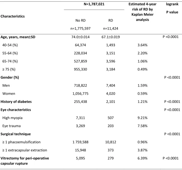
After cataract surgery, the mean age was lower for patients with than without RD (67.10.017 vs. 74.00.014 years, p<0.001; table 1). The risk of RD after cataract surgery was higher for younger than older adults (3.64%, 2.20%, 1.06%, and 0.49% for patients 40-54, 55-64, 65-74 and ≥75 years old, respectively, p<0.001), men than women (1.59% vs 0.59%, p<0.001) and patients with extracapsular extraction versus phacoemulsification (3.87% vs 0.96%, p<0.001). The frequency of RD onset was higher with posterior capsular rupture (6.39%), myopia (9.21%) and a history of eye trauma (7.58%) or diabetes (1.21%). The cumulative probabilities of each risk factor of RD during the 4 years are in figures 1 and 2.
The multi-adjusted HR associated with increased risk of RD was 5.22 (95% confidence interval [95% CI] 5.05 to 5.39) for patients aged 40-54 years, 3.69 (95% CI 3.60 to 3.79) for those 55-64 years and 1.98 (95% CI 1.93 to 2.03) for patients 65 to 74 years as compared with those ≥ 75 years old (P<0.001) (Table 2). Adjusted HR pairwise comparisons between age groups 40-54 versus 65-74, 55-64 versus 65-74 and 40-54 versus 55-55-64 were 2.65 (95% CI 2.48 to 2.81), 1.89 (95% CI 1.78 to 1.96) and 1.41 (95% CI 1.33 to 1.50), respectively.
RD was associated with high myopia (HR 6.12; 95% CI 5.84 to 6.41), vitrectomy for peri-operative capsular rupture (HR 4.36; 95% CI 4.07 to 4.68), history of eye trauma (HR 3.98; 95% CI 3.69 to 4.30), extracapsular extraction (HR 3.11; 95% CI 2.94 to 3.30), male gender (HR 2.39; 95% CI 2.35 to 2.44) and a history of diabetes (HR 1.18; 95% CI 1.15 to 1.21).
Interaction between each risk factors of RD was performed owing to statistical and clinical significance only between myopia and age groups (p<0.0001). We thus stratified the analysis by age. In myopic patients, the multi-adjusted HR associated with increased risk of RD was 25.02 (95% CI 24.76 to 25.18) for patients 40-54 years old, 20.37 (95% CI 20.21 to 20.53) for those 55-64 years old and 17.05 (95% CI 16.85 to 17.25) for those 65 to 74 years old as compared with non-myopic patients ≥ 75 years old. In non-myopic patients, the multi-adjusted HR associated with increased risk of RD was 5.46 (95% CI 5.40 to 5.52) for patients aged 40-54 years, 3.76 (95% CI 3.70 to 3.82) for those 55-64 years and 1.98 (95% CI 1.92 to 2.04) for those 65 to 74 years as compared with non-myopic patients ≥ 75 years old (P<0.001) (Table 3).
Age is a major marker of RD risk after cataract surgery
In the whole population, the younger the patient, the greater the risk of RD after cataract extraction performed with phacoemulsification (p logrank<.001; figure 1). We also assessed the effect of age on
RD onset in a subgroup of 1,504,637 patients who underwent surgery with phacoemulsification without any other risk factors of RD such as peri-operative capsular rupture, diabetes, high myopia, or eye trauma. Young age remained significantly associated with RD (p logrank <.001; figure 1). Relationship between age and risk of retinal detachment with a previous cataract surgery and in the French population is presented in figure 3.
Discussion
By using data from the PMSI program, an exhaustive national database from the 1,546 healthcare facilities in France, public or private, we determined an overall RD risk of 0.99% after cataract surgery during a recent 4-year period. In this work, including a wide range of risk factors, we provide a hierarchy of risk factors for RD onset: high myopia, young age, extracapsular extraction technique, eye trauma, peri-operative capsular rupture, male gender and diabetes.
Incidence of RD
Population-based studies of RD incidence after cataract surgery for patients who underwent surgery between 1980 and 2004 estimated RD based on sometimes a high proportion of extracapsular extraction.4 Phacoemulsification is now generally used for cataract extraction and continues to evolve, so recent data of the RD incidence after cataract surgery are of importance. A retrospective study in Western Australia of 129,982 cataract surgery patients from 46 health facilities over 21 years found an overall pseudophakic RD rate of 0.7% 11 and the rate of RD after cataract surgery decreased greatly from 1980 to 2001. A recent large retrospective case–control study in Singapore of 24,846 cataract operations performed between 2001 and 2003, with follow-up through 2008, found a pseudophakic RD rate of 0.16%,12 lower than rates previously found.4 The authors included only cases with RD surgery in the same hospital as the cataract surgery, so the rate could be underestimated. In this 4-year study among the general population in France, we found an estimated risk of RD of 0.99%.
In the present study, cataract surgery itself increased the risk of RD by 3-fold. This finding was close to the study by Bjerrum et al., who reported a 4-fold increase in risk associated with cataract surgery itself.13
Delay of RD onset
The study from Moorfields Eye Hospital found 75% of RD within the first 2 postoperative years, whereas Sheu et al.14 found that the mean time between cataract surgery to diagnosis was 40 months.15 In the present study, the median time for RD onset after cataract surgery was 237 (interquartile range 75-738) days and the risk of RD after cataract extraction increased in a nearly linear manner over time in the overall cohort. Regarding the shape of the slopes of RD after cataract surgery, in patients with vitrectomy for peri-operative capsular rupture a history of high myopia, or eye trauma, the RD incidence reached 4.5% to 5.2% during the first year.
Risk factors of RD after cataract surgery
Intraoperative posterior capsular rupture is one of the most significant risk factors for pseudophakic RD, leading to poor final visual acuity in most cases. The anterior movement of the vitreous due to posterior capsular rupture results in dynamic traction on the vitreous, with consequent retinal tear. In a case–control study performed in Sweden in 2003, among 324 patients with posterior capsular rupture, the risk of RD after cataract surgery was 10-fold increased in patients with capsule complications.16 In the present study, the risk of RD after cataract surgery was 4-fold increased with posterior capsular rupture. However, our HR was adjusted for multiple factors to take into account many other potential other risk factors of RD.
A study of patients who had undergone cataract surgery from 1980 through 2004 in Olmsted County, Minnesota, USA, found no difference in probability of RD with extracapsular extraction and phacoemulsification.5 We found extracapsular extraction as a major risk factor for RD after cataract extraction, with 3-fold increased risk of RD. This finding may be due to the progress in the technique of cataract surgery, which is now less traumatic than extracapsular extraction. Another hypothesis could be that patients undergoing extracapsular extraction had denser and harder crystalline lens.
A study from Moorefields Eye Hospital found a male predominance with pseudophakic RD: 67.5% males with RD versus 38.2% of controls.15 We found an estimated Cox proportion risk of 2.39 for men as compared with women. This increased risk of RD in men is not entirely understood. Sheu et al. hypothesized an increased and underreported history of trauma in men versus women.14 However,
this suggestion has not been verified and in our analysis, we included a history of eye trauma in the multivariate analysis.
Myopic eyes have a much higher risk of RD, whether pseudophakic or phakic. High myopia, defined as axial length ≥ 26 mm, is also an established risk factor of pseudophakic RD.4 The retrospective series by Sheu et al. 14 found an adjusted relative risk of 4.19 with high myopia as compared with axial length ≤ 23 mm. In the present study, the 4-year probability of RD after cataract surgery was 9.21% with high myopia, and the multi-adjusted HR was 6.12.
The retrospective study by Alio JL. et al. of patients with high myopia and a mean follow-up of 61.5 29.6 months found a higher risk of pseudohakic RD in myopic patients < 50 than > 50 years old (4.46% vs. 2.96%, respectively).17 Young age and high myopia were found as risk factors of RD. Thus, cataract surgery or refractive lens exchange in patients with high myopia appears to be associated with high risk of RD, and surgical indications may be carefully discussed.18 In the present analysis, young age was also a risk factor in myopic patients. Among myopic patients, the risk of RD increased by 25 in age group 40-54 years and by 20 in age group 55-64 years as compared with non-myopic ≥ 75 years old.
Young age is known to increase the risk of RD after cataract surgery. In a large retrospective case– control series of 63,298 cataracts between 1994 and 2003 at Moorfields Eye Hospital, young age was a significant risk factor for RD after surgery.15 The mean age with pseudophakic RD was 63.5 years and 71.9 years for controls. The odds ratio was 3.1 for patients ≤ 64 than > 64 years old 15 A series from Taiwan found 8-year RD rates of 6.65%, 2.57%, and 2.01% for patients ≤ 50, 50-60 and ≥ 60 years old, respectively.14 Our 4-year RD risk estimates were 3.6%, 2.2%, 1.1, and 0.5% for patients 40-54, 55-64, 65-74 and ≥75 years old, respectively. The incidence of RD was not significantly affected after adjusting for other risk factors (gender, history of diabetes, high myopia, eye trauma, and anterior vitrectomy for peri-operative capsular rupture and surgical technique) but was increased by 5-fold for patients 40-54 years old, 4-fold for those 55-64 and 2-fold for those 65-74 as compared with those ≥ 75. Of interest, the 4 year risk of RD (with or without a history of cataract extraction) in the French population was higher for older than younger adults.
The pathophysiology of increased risk of RD after cataract surgery in young patients remains speculative. In our study, the main causes of early cataract, such as diabetes, high myopia and a
history of eye trauma, were included in the multivariate analyses and the effect of age remained statistically significant. Another possible reason may be the status of the vitreous at the time of cataract extraction. Cataract extraction increases the risk of posterior vitreous detachment,19,20 and pseudophakic eyes have alterations in the vitreous humor structure that are not seen in phakic eyes.21 Changes in the vitreous induced by removal of the crystalline lens may underlie an increased risk of RD.22 The presence of a posterior vitreous detachment in older patients before cataract
extraction may protect against subsequent RD by limiting the force to the retina produced during cataract surgery. Consequently, cataract extraction in relatively young patients without previous posterior vitreous detachment may alter the microenvironment of the vitreous and retina, thereby increasing the risk of retinal break and RD.23 An alternative explanation is that eyes in some young patients are abnormal in their tendency to develop cataracts, which may predispose to pseudophakic RD.
Also people ≤ 65 years old who require cataract surgery may represent those with another pathology that could affect their risk of RD. Among these factors, we could include a history of diabetes, high myopia or ocular trauma. To assess the specific effect of age on the risk of pseudophakic RD, we performed a Kaplan-Meier analysis for a subgroup of patients without any history of diabetes, high myopia, or eye trauma (Figure 1). We also included these 3 covariates in the multivariate Cox model (Table 2). Young age remained significantly associated with RD in both analyses.
Optimal timing for cataract surgery
The optimal timing for cataract surgery is important for public health policies. In Western countries cataract surgery has achieved marked improvements in vision-related activity limitations 24 and may not be delayed beyond 20/40 or 20/50 of visual acuity so that older adults can maintain autonomy.25,26 Cataract surgery on eyes with poor preoperative visual acuity is related to surgical complications, and cataract surgery on eyes with excellent preoperative visual acuity is related to adverse visual results, including RD.27 As surgical techniques have improved, the demand for surgery at an early stage has increased. The observed strong association between age and pseudophakic RD may have implications when considering clear lens extraction as a refractive procedure in young patients. If refractive lens exchange becomes more commonly accepted, this procedure may add to the potential RD burden in younger eyes.
However, for young subjects with cataract associated with substantial vision loss, cataract surgery cannot be delayed. In both situations of refractive lens exchange or early cataract, we recommend
accurate screening for symptomatic flap tears and prophylactic laser treatment before the cataract extraction, and a close follow-up of the fundus in the weeks and months after surgery.28
Strengths and limitations
Strengths of the study are the population size, the national recruitment, the importance of its findings and potential impact on public health. Given the reliance on PMSI codes for the selection of patients and the ascertainment of outcomes, there was a potential for misclassification- or
underdetection-related biases. However, this situation may have only a minor impact on the findings. One possible reason for the increased risk of RD after cataract surgery in young patients may be the status of the posterior vitreous detachment at the time of cataract extraction. However, the administrative database did not include this information, so we could not assess its impact on the risk of pseudophakic RD. We could not exclude patients undergoing any other surgery that of vitrectomy (for example a trabeculectomy), which could have slightly increased the estimation of the risk of pseudophakic RD.
Conclusions
RD is one of the most serious complications following cataract surgery, with an overall risk of 0.99% in our 4-year study in the general population in France. We provide a hierarchy of risk factors for RD onset: high myopia, young age, vitrectomy for peri-operative capsular rupture, history of eye trauma, extracapsular extraction technique, male gender and diabetes. Screening and treatment for symptomatic flap tears before cataract surgery in young patients may help reduce the rate of RD after cataract surgery. Optimal timing of cataract surgery is an important issue for public health policies to obtain the best balance between maintaining autonomy in older adults and avoiding serious adverse events such as RD.
References
1. Bellan L. The Evolution of Cataract Surgery: The Most Common Eye Procedure in Older Adults. Geriatr Aging 2008;11:328–332.
2. Congdon N, Vingerling JR, Klein BEK, et al. Prevalence of cataract and pseudophakia/aphakia among adults in the United States. Arch Ophthalmol 2004;122:487–494.
3. Haddad WM, Monin C, Morel C, et al. Retinal detachment after phacoemulsification: a study of 114 cases. Am J Ophthalmol 2002;133:630–638.
4. Haug SJ, Bhisitkul RB. Risk factors for retinal detachment following cataract surgery. Curr Opin Ophthalmol 2012;23:7–11.
5. Erie JC, Raecker ME, Baratz KH, et al. Risk of retinal detachment after cataract extraction, 1980-2004: a population-based study. Trans Am Ophthalmol Soc 2006;104:167–175.
6. Algvere PV, Jahnberg P, Textorius O. The Swedish Retinal Detachment Register. I. A database for epidemiological and clinical studies. Graefes Arch Clin Exp Ophthalmol Albrecht Von Graefes Arch Für Klin Exp Ophthalmol 1999;237:137–144.
7. Baratz KH, Gray DT, Hodge DO, et al. Cataract extraction rates in Olmsted County, Minnesota, 1980 through 1994. Arch Ophthalmol 1997;115:1441–1446.
8. Colin J, Robinet A, Cochener B. Retinal detachment after clear lens extraction for high myopia: seven-year follow-up. Ophthalmology 1999;106:2281–2284; discussion 2285.
9. López Mato O. Retinal detachment after clear lens extraction for high myopia. Ophthalmology 2001;108:239.
10. Daien V, Le Pape A, Heve D, et al. Incidence and Characteristics of Cataract Surgery in France from 2009 to 2012: A National Population Study. Ophthalmology 2015.
11. Clark A, Morlet N, Ng JQ, et al. Whole population trends in complications of cataract surgery over 22 years in Western Australia. Ophthalmology 2011;118:1055–1061.
12. Quek DT-L, Lee SY, Htoon HM, Ang CL. Pseudophakic rhegmatogenous retinal detachment in a large Asian tertiary eye centre: a cohort study. Clin Experiment Ophthalmol 2012;40:e1–7. 13. Bjerrum SS, Mikkelsen KL, La Cour M. Risk of pseudophakic retinal detachment in 202,226 patients using the fellow nonoperated eye as reference. Ophthalmology 2013;120:2573–2579. 14. Sheu S-J, Ger L-P, Ho W-L. Late increased risk of retinal detachment after cataract extraction. Am J Ophthalmol 2010;149:113–119.
15. Tuft SJ, Gore DM, Bunce C, et al. Outcomes of pseudophakic retinal detachment. Acta Ophthalmol (Copenh) 2012;90:639–644.
16. Jakobsson G, Montan P, Zetterberg M, et al. Capsule complication during cataract surgery: Retinal detachment after cataract surgery with capsule complication: Swedish Capsule Rupture Study Group report 4. J Cataract Refract Surg 2009;35:1699–1705.
17. Alio JL, Ruiz-Moreno JM, Shabayek MH, et al. The risk of retinal detachment in high myopia after small incision coaxial phacoemulsification. Am J Ophthalmol 2007;144:93–98.
18. Alió JL. Lens surgery (cataract and refractive lens exchange) and retinal detachment risk in myopes: still an issue? Br J Ophthalmol 2011;95:301–303.
19. Sheard RM, Goodburn SF, Comer MB, et al. Posterior vitreous detachment after neodymium:YAG laser posterior capsulotomy. J Cataract Refract Surg 2003;29:930–934.
20. Hilford D, Hilford M, Mathew A, Polkinghorne PJ. Posterior vitreous detachment following cataract surgery. Eye Lond Engl 2009;23:1388–1392.
21. Neal RE, Bettelheim FA, Lin C, et al. Alterations in human vitreous humour following cataract extraction. Exp Eye Res 2005;80:337–347.
22. Mirshahi A, Hoehn F, Lorenz K, Hattenbach L-O. Incidence of posterior vitreous detachment after cataract surgery. J Cataract Refract Surg 2009;35:987–991.
23. Coppé AM, Lapucci G. Posterior vitreous detachment and retinal detachment following cataract extraction. Curr Opin Ophthalmol 2008;19:239–242.
24. Harrer A, Gerstmeyer K, Hirnschall N, et al. Impact of bilateral cataract surgery on vision-related activity limitations. J Cataract Refract Surg 2013;39:680–685.
25. Daien V, Pérès K, Villain M, et al. Visual impairment, optical correction, and their impact on activity limitations in elderly persons: the POLA study. Arch Intern Med 2011;171:1206–1207. 26. Daien V, Peres K, Villain M, et al. Visual acuity thresholds associated with activity limitations in the elderly. The Pathologies Oculaires Liées à l’Age study. Acta Ophthalmol (Copenh) 2014;92:e500– 506.
27. Lundström M, Goh P-P, Henry Y, et al. The Changing Pattern of Cataract Surgery Indications: A 5-Year Study of 2 Cataract Surgery Databases. Ophthalmology 2014.
28. Wilkinson CP. Evidence-based analysis of prophylactic treatment of asymptomatic retinal breaks and lattice degeneration. Ophthalmology 2000;107:12–15; discussion 15–18.
Figures legends
Figure 1. Effect of age on the cumulative probability of retinal detachment after cataract extraction for all patients in France and those undergoing only phacoemulsification (dashed lines) without any other risk factors (high myopia, diabetes, history of eye trauma, or peri-operative capsular rupture). P < .001, log-rank between each age group with or without other risk factors.
Figure 2. Cumulative probability of retinal detachment after cataract extraction in sub-groups of risk including high myopia, history of eye trauma, vitrectomy for peri-operative capsular rupture or history of diabetes.
Figure 3. Relationship between age and risk of retinal detachment with a previous cataract surgery and in the French population.
Table 1. Characteristics and estimated 4-year risk of retinal detachment (RD) among 1,787,021 patients who underwent cataract extraction in France between 2009 and 2012
Characteristics N=1,787,021 Estimated 4-year risk of RD by Kaplan Meier analysis logrank P value No RD n=1,775,597 RD n=11,424
Age, years, meanSD 74.00.014 67.10.019 P <0.0001
40-54 (%) 64,374 1,493 3.64% 55-64 (%) 228,034 3,151 2.20% 65-74 (%) 527,859 3,596 1.06% ≥ 75 (%) 955,330 3,184 0.49% Gender (%) P <0.0001 Men 718,822 7,404 1.59% Women 1,056,775 4,020 0.59% History of diabetes 255,438 2,101 1.21% P <0.0001 Eye characteristics P <0.0001 High myopia 7,311 507 9.21% Eye trauma 3,269 203 7.58% Surgical technique P <0.0001 ≥ 1 phacoemulsification 1 759,588 10,812 0.96% ≥ 1 extracapsular extraction 15,948 373 3.87%
Vitrectomy for peri-operative capsular rupture
Table 2. Crude and adjusted hazard ratio (HR) of retinal detachment after cataract extraction Crude HR 95% CI Adjusted HR* 95% CI Age, years ≥ 75 1.00 65-74 2.09 2.04, 2.14 1.98 1.93, 2.03 55-64 4.28 4.17, 4.39 3.69 3.60, 3.79 40-54 7.06 6.84, 7.28 5.22 5.05, 5.39 Gender Women 1.0 1.0 Men 2.73 2.68, 2.79 2.39 2.35, 2.44 History of diabetes 1.29 1.26, 1.32 1.18 1.15, 1.21 Eye characteristics High myopia 10.46 9.99, 10.95 6.12 5.84, 6.41 Eye trauma 9.65 8.98, 10.36 3.98 3.69, 4.30
Anterior vitrectomy for peri-operative capsular rupture
8.57 8.03, 9.13 4.36 4.07, 4.68
Surgical technique
≥ 1 extracapsular extraction
5.46 5.18, 5.76 3.11 2.94, 3.30
Abbreviations: 95% CI, 95% confidence interval
* Adjusted for age, gender, history of diabetes, high myopia, eye trauma, and anterior vitrectomy for peri-operative capsular rupture and surgical technique (extra capsular cataract extraction vs.
Table 3. Multi-adjusted hazard ratio (HR) of retinal detachment after cataract extraction among myopic and non-myopic patients.
Myopic patients Non-myopic patients
Adjusted HR* 95% CI Adjusted HR* 95% CI Age, years ≥ 75 1.00 65-74 1.19 0.87, 1.51 1.98 1.92, 2.04 55-64 1.37 1.07, 1.67 3.76 3.70, 3.82 40-54 1.72 1.42, 2.02 5.46 5.40, 5.52
Abbreviations: 95% CI, 95% confidence interval
* Adjusted for age, gender, history of diabetes, eye trauma, and anterior vitrectomy for peri-operative capsular rupture and surgical technique (extra capsular cataract extraction vs. phacoemulsification).