HAL Id: inserm-03223335
https://www.hal.inserm.fr/inserm-03223335v2
Submitted on 14 May 2021
HAL is a multi-disciplinary open access
archive for the deposit and dissemination of sci-entific research documents, whether they are pub-lished or not. The documents may come from teaching and research institutions in France or abroad, or from public or private research centers.
L’archive ouverte pluridisciplinaire HAL, est destinée au dépôt et à la diffusion de documents scientifiques de niveau recherche, publiés ou non, émanant des établissements d’enseignement et de recherche français ou étrangers, des laboratoires publics ou privés.
Spastic co-contraction is directly associated with altered
cortical beta oscillations after stroke
Alexandre Chalard, David Amarantini, Joseph Tisseyre, Philippe Marque, D.
Gasq
To cite this version:
Alexandre Chalard, David Amarantini, Joseph Tisseyre, Philippe Marque, D. Gasq. Spastic co-contraction is directly associated with altered cortical beta oscillations after stroke. Clinical Neu-rophysiology, Elsevier, 2020, 131 (6), pp.1345 - 1353. �10.1016/j.clinph.2020.02.023�. �inserm-03223335v2�
1 Original Research Article
Spastic co-contraction is directly associated with altered cortical beta oscillations after stroke
Alexandre Chalarda,b, David Amarantinia, Joseph Tisseyrea, Philippe Marquea,c, David
Gasqa,d,*
a ToNIC, Toulouse NeuroImaging Center, Université de Toulouse, Inserm, UPS, France b Ipsen Innovation, Les Ulis, France
c Department of Neurological Rehabilitation, University Hospital of Toulouse, Hôpital de
Rangueil, Toulouse, France
d Department of Functional Physiological Explorations, University Hospital of Toulouse,
Hôpital de Rangueil, Toulouse, France
*Correspondence to David Gasq, MD, PhD
Toulouse NeuroImaging Center, CHU Purpan, Pavillon Baudot, place du Dr Baylac 31024 Toulouse, France
Tél. +33 5 62 74 61 64 Fax. +33 5 62 74 61 63
Email: [email protected]
Word Count: 3850 words
2 Abstract (196 words)
Objective: Spastic co-contraction is a motor-disabling form of muscle overactivity occurring
after a stroke, contributing to a limitation in active movement and a certain level of motor impairment. The cortical mechanisms underlying spastic co-contraction remain to be more fully elucidated, the present study aimed to investigate the role of the cortical beta oscillations in spastic co-contraction after a stroke.
Method: We recruited fifteen post-stroke subjects and nine healthy controls. The subjects
were asked to perform active elbow extensions. In the study, multimodal analysis was performed to combine the evaluation of three-dimensional elbow kinematics, the elbow muscles electromyographic activations, and the cortical oscillatory activity.
Results: The movement-related beta desynchronization was significantly decreased in stroke
subjects compared to healthy participants. We found a significant correlation between the movement-related beta desynchronization and the elbow flexor activation during the active elbow extension in stroke subjects. When compared to healthy subjects, stroke subjects exhibited significant alterations in the elbow kinematics and greater muscle activation levels.
Conclusions: Cortical beta oscillation alterations may reflect an important neural mechanism
underlying spastic co-contraction after a stroke.
Significance: Measuring the cortical oscillatory activity could be useful to further characterize neuromuscular plasticity induced by recovery or therapeutic interventions.
Keywords: Neuronal Plasticity; Muscle Hypertonia; Brain Injuries; Movement; Upper Extremity
3 Highlights:
Spastic co-contraction is directly associated with alterations in movement kinematics and upper limb motor function.
Altered cortical oscillatory activity may reflect an important mechanism underlying spastic co-contraction.
Cortical beta oscillations may be a potential marker for motor recovery in stroke subjects.
4 1. Introduction
Spastic paresis is a disabling motor syndrome occurring after corticospinal pathway damage caused by, for example, a stroke. Considered as a neural disorder, spastic paresis associates motor paresis and antagonist overactivity (Baude et al., 2018; Gracies, 2005). One of the main features of antagonist overactivity is the spastic co-contraction which refers to an excessive degree of antagonist coactivation triggered by the volitional command of agonist muscles (Gracies, 2005). Spastic co-contraction is a disabling form of antagonist overactivity, contributing to the limitation of active movement with a deleterious impact on upper limb motor function (Chae et al., 2002; Chalard et al., 2019). Indeed, an extensive part of the literature has demonstrated the deleterious impact of spastic co-contraction on movement characteristics, including an increased duration of the movement, a lack of muscle
coordination and a limitation of the active range of motion (Arene and Hidler, 2009; Chalard et al., 2019; Gross et al., 2015; Sarcher et al., 2015). Spastic co-contraction has a mainly supraspinal origin, but could be also related to spinal mechanisms: while an abnormal pattern of the supraspinal descending drive leading to a loss of motor selectivity would be the
predominant mechanism underlying spastic co-contraction (Gracies et al., 1997; Schieber et al., 2009). Other mechanisms such as increased Renshaw inhibition and a reduction of reciprocal and presynaptic inhibition could contribute to the spastic co-contraction
(Bhagchandani and Schindler-Ivens, 2012; Katz and Pierrot-Deseilligny, 1982; Morita et al., 2001).
Electroencephalography (EEG) is a valuable non-invasive tool to assess and quantify the cortical activity during a motor task by recording the associated oscillatory activity of the brain (Ramos-Murguialday and Birbaumer, 2015) allowing, notably, to characterize the pathological patterns of cortical activation in post-stroke subjects (Kaiser et al., 2012; Park et al., 2016). Cortical oscillations in the beta-band frequency range (13-30 Hz) over the
5 sensorimotor cortex are heavily involved in motor control: they are present at rest and are decreased during movement, leading to a decrease in beta power which characterizes movement-related beta desynchronization (Pfurtscheller, 2001; Pfurtscheller and Lopes da Silva, 1999). The increase in the amplitude of the movement-related beta desynchronization corresponds to increased corticospinal excitability (Pfurtscheller and Lopes da Silva, 1999; Takemi et al., 2013), and can be interpreted as an electrophysiological correlate of cortical activations involved in the generation of movement. In post-stroke subjects, movement-related beta desynchronization has been used to investigate both the relationship between beta oscillations and motor impairments, and neuroplasticity induced by rehabilitation (Rossiter et al., 2015, 2014). The authors cited above have highlighted that abnormalities in cortical oscillatory activity could be an important mechanism in motor impairment, involved in
neuroplasticity following a therapeutic intervention. These studies have provided fundamental understanding of the adaptative and maladaptive neural mechanisms underlying motor
behavior in post-stroke subjects enabling the provision of patient-tailored rehabilitation intervention (Koch and Hummel, 2017).
Although alterations in various physiological neural mechanisms have been suggested to be involved in the emergence of spastic co-contraction (Baude et al., 2018), to date, much remains to be understood regarding the cortical mechanisms underlying the modulation of the spastic co-contraction after corticospinal pathway damage. To fill in the gaps on this issue, the aim of the present study has been to investigate the role of the cortical beta oscillations in spastic co-contraction after a stroke. We therefore assessed beta-band cortical oscillations using movement-related beta desynchronization during active elbow extension in post-stroke subjects and healthy controls. Since a decrease in movement-related beta desynchronization has been associated with altered motor function after a stroke (Rossiter et al., 2014), we hypothesized a decrease in movement-related beta desynchronization associated with an
6 increase in the antagonist co-contraction in post-stroke subjects. Such a change in cortical beta oscillatory activity during a motor task could reflect the alteration of the neural mechanisms underlying the spastic co-contraction after a stroke and thus provide key new elements related to the neural mechanisms behind motor impairment in chronic stroke subjects.
2. Methods
2.1. Participants
Twenty-four adults (≥ 18 years old) voluntarily participated in this study. They were allocated into two groups: the first was composed of fifteen post-stroke participants (STROKE) (4 females; mean (± SD) age: 55 ± 11 years, mean (± SD) Fugl-Meyer Upper Extremity score: 38 ± 9, mean (± SD) Erasmus modified Nottingham Sensory Assessment: 48 ± 14) and the second comprised nine healthy controls (CO) (3 females; mean (± SD) age: 43 ± 21 years) (see Table 1 for participant demographics).
Post-stroke participants were included if they were ≥ 6 months since stroke onset and were free of any antispastic treatment for ≥ 4 months. Exclusion criteria were comprehension disorders, neurodegenerative conditions, painful paretic upper limb during movement, active elbow extension ability ≤ 20° or elbow joint contracture. The presence of spasticity or the level of motor function was neither an inclusion nor an exclusion criterion applied for post-stroke participants. All participants gave and written informed consent prior to participation. This study was approved by local Research Ethics Board (No ID-RCB: 2017-A01616-47), and was conducted in accordance with the amended Declaration of Helsinki.
7 Recordings were made on both sides but subsequent analyzes concerned only the
non-dominant side in CO and the paretic side in STROKE.
2.2.1. Kinematics
Three-dimensional kinematic analysis was performed at 125 Hz using eight OptiTrack infrared cameras (model S250e, NaturalPoint, Corvallis, Oregon, USA). Six reflective markers were placed on both sides on the acromion, lateral epicondyle, and ulnar styloid.
2.2.2. Electromyography
Following appropriate skin preparation (Hermens et al., 2000), to reduce the impedance of skin-electrode interface below 5kΩ, surface electromyography (EMG) was acquired at 1000 Hz using Ag-AgCl bipolar electrodes in bipolar configuration with an inter-electrode distance of 20 mm using an MP150 system equipped with EMG100C amplifiers (Biopac Systems Inc., Goleta, CA, USA) with the ground electrode placed on the right mastoid. Following appropriate skin preparation (Hermens et al., 2000), the long head of the triceps brachii (TB) was taken to represent the elbow extensors, while the biceps brachii (BB), the brachioradialis (BR) and the brachialis (BA) were taken to represent elbow flexors
(Staudenmann and Taube, 2015).
2.2.3. Electroencephalography
EEG was recorded at 1024 Hz using a 64 channel ActiveTwo system (BioSemi
instrumentation, Amsterdam, The Netherlands), with scalp electrodes arranged according to the International 10-20 system.
8 Kinematics, EMG and EEG were synchronized using a common trigger controlled by the Biopac system.
2.3. Task
The task consisted of two sets of ten active elbow extension-flexion movements at a self-spontaneous speed. In a quiet room, participants were seated comfortably on a straight-backed chair with their shoulders fixed to the chair back by clavicular rings. The height of the table was adjusted to obtain an initial resting position corresponding to a shoulder flexed at 80° with internal rotation of 90°, the elbow flexed at 90° and the forearm pronated (forearm in front of the thorax). For each movement, an auditory signal indicated to the participants to start performing a full active elbow extension with the elbow lifted off the table. At the end of each elbow extension, participants had a random rest period of 8 to 15 s with their forearms resting on the table before returning to the initial position. To avoid fatigue, participants rested as needed between the two sets of ten movements.
2.4. Data analysis
Extension being a key phase for bringing the hand to a target, only elbow extension
movements, the deficit of which is a frequent post-stroke complaint, were considered in this study.
2.4.1. Preprocessing
Kinematic data were low-pass filtered at 6 Hz (Cahouët et al., 2002). Raw EMG signals were 10-400 Hz band-pass filtered, full wave rectified, and smoothed at 9 Hz to obtain the linear envelopes (Chalard et al., 2019). Raw EEG signals were 3-100 Hz band-pass filtered
9 derivations to reduce the effect of volume conduction using a spherical spline method (Perrin et al., 1989). The filters were applied on continuous signals; all were fourth-order, zero-lag Butterworth type. The onset and offset of each elbow extension were detected with a threshold of 0.01 °/s applied on the elbow angular velocity.
2.4.2. Data processing
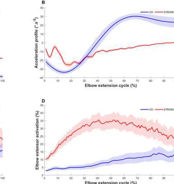
For each movement, the active elbow range of motion (Fig. 1.A) was computed in the horizontal plane from the Cartesian coordinates of the anatomical landmarks between the brachial and antebrachial segments. The movement duration was considered as the time in seconds required to extend the elbow at a self-spontaneous speed. The smoothness of the movement was quantified by using the normalized jerk metric defined as the number of peaks of the acceleration profile normalized by the mean angular velocity (Fig. 1.B): the greater the jerk, the less smooth was the movement (de los Reyes-Guzmán et al., 2014).
For both elbow flexors and extensors, muscle activation was computed as the ratio (expressed as a percentage) of the root mean-square value of the EMG envelope obtained during the elbow extension movement and that obtained during a maximal isometric voluntary contraction of the same muscle (Fig. 1.C & 1.D). For elbow flexor activation, the average ratio of the three corresponding muscles was used.
EEG data were segmented into epochs from -3s prior to the start and +3s after the end of the movement. A visual inspection was performed to reject epochs contaminated with artifacts. The EEG power spectrum was obtained using Morlet wavelet with the MATLAB package developed by Grinsted et al. and adapted by Bigot et al (Bigot et al., 2011; Grinsted et al., 2004). The scale resolution of the wavelet (parameter ‘nvoice’), the number of scales used in the wavelet analysis (parameter ‘J1’) and the Morlet mother wavelet parameter (parameter ‘wavenumber’) were set respectively to 7, 25 and 10 to provide a satisfactory compromise
10 between time and resolution for the identification of oscillatory activity on the [0.31·10 -2 : 0.22 : 39.98] Hz frequency range. To cope with the inter-trial duration variability that can
lead to power spectrum cancelation, a normalization procedure was used to obtain a time-frequency EEG power spectrum with time expressed as a percentage of elbow extension movement time (Fauvet et al., 2019).
The event-related EEG power were obtained by the method described by Pfurtscheller (Pfurtscheller, 2001), where a decrease / increase in power corresponds to an event-related desynchronization / synchronization respectively.
Event-related EEG power = 10×log10 ([𝐴 𝐵⁄ ]) (1) where A is the absolute power during the elbow movement (period between the movement onset and the movement offset), and B is the median of the absolute power during a baseline period ranging from -2.9 s to -0.9 s before the start of the movement.
In the current work, our analysis was focused on the movement-related beta
desynchronization corresponding to the event-related EEG power on the EEG-channels over each sensorimotor cortex (Cz, FCz, CPz, C1, C2, C3, C4, C5, C6, FC1, FC2, FC3, FC4, FC5, FC6, CP1, CP2, CP3, CP4, CP5, CP6) during elbow extension in the beta-band frequency (i.e., [13-30 Hz]). To compare the subjects between them independently of the side of the brain injury (STROKE) or manual laterality (CO), we flipped EEG-data of subjects with right cerebral lesion (STROKE) or left manual laterality (CO).
2.5. Statistical analysis
To assess the potential significant differences between groups in the movement-related beta desynchronization, we identified sensorimotor EEG electrodes showing significant inter-group differences in movement-related beta desynchronization using a data-driven non-parametric cluster-based randomization test (10,000 permutations) at the topographical level
11 (Maris and Oostenveld, 2007; Oostenveld et al., 2011). This procedure obviates questions of multiple comparisons at multiple electrodes (type I risk of error inflation) while having a greater sensitivity than a conservative Bonferroni procedure to correct alpha-level (Maris and Oostenveld, 2007). For comparison, movement-related beta desynchronization was then considered as the mean of all the significant cluster values.
All variables showed normal distribution (Shapiro-Wilk test; P > 0.05) and homogeneity of variance (Levene’s test; P > 0.05). Independent t-tests were then used to compare kinematic data (active elbow range of motion, movement duration, and smoothness), and EEG data (movement-related beta desynchronization) between STROKE and CO. A mixed analysis of variance (ANOVA) was performed with Muscle (elbow flexors vs elbow extensor) as within-subject factor and Group (STROKE vs CO) as a between-within-subject factor. For all analyses, the significance was set at P < 0.05. We expressed effect sizes of the independent t-tests in standardized terms with the Hedge’s g (Lakens, 2013). The magnitude of the effect size was interpreted as small (g > 0.2), moderate (g > 0.5) or large (g > 0.8) (Cohen, 1977). The underlying relationship between kinematic, EMG and EEG data in STROKE was explored with multiple partial correlations allowing control of cofounding factors. We applied a threshold of 0.4 to isolate the remarkable correlations highlighting the organization of the links between variables, and generated a correlation iconography diagram (Lesty et al., 2004).
3. Results
3.1. Elbow extension kinematics are altered in STROKE
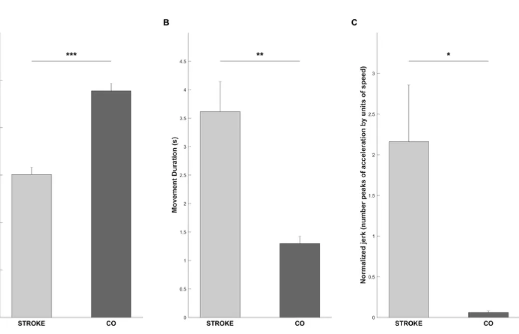
Statistical analysis revealed a significant difference with a large effect size between STROKE and CO on the active elbow range of motion (t22 = 7.62, p < 0.001, g = 3.07) (Fig 2.A), the
12 movement duration (t22 = 3.38, p = 0.003, g = 1.36) (Fig. 2.B), and the jerk
(t22 = 2.31, p = 0.03, g = 0.92) (Fig. 2 C). In STROKE, the active elbow range of motion was
decreased by 35.2 ± 2.3°, the movement duration of the elbow extension was increased by 2.8 ± 1.6 s, and the normalized jerk was increased by 2.1 ± 2.63.
3.2. EMG patterns reveal overactivity of elbow muscles in STROKE
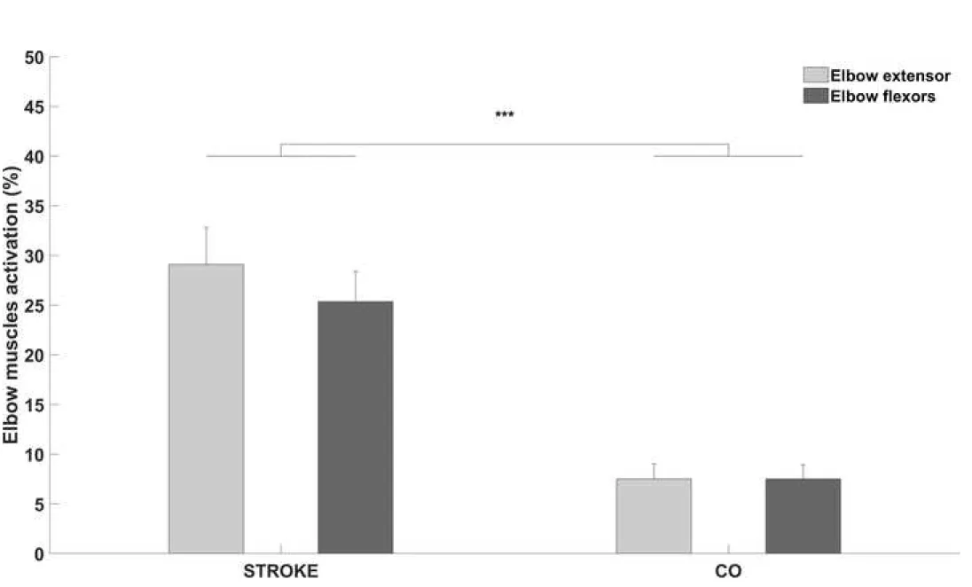
The mixed ANOVA revealed a significant Group effect with a large effect size between two groups on elbow extensor activation and elbow flexors activation
(F1,22 = 21.8, p < 0.001, ή = 0.95) (Fig. 3), but did not reveal nor a Muscle effect (F1,22 = 0.98,
p = 0.33, ή = 0.05) nor a significant interaction between Group and Muscle (F1,22 = 0.98, p =
0.33). In STROKE, the elbow extensor activation was increased by 21.4 ± 8%, and the elbow flexor activation was increased from 17.8 ± 7.1%.
3.3. Cortical oscillatory beta activity is altered during elbow extension in STROKE
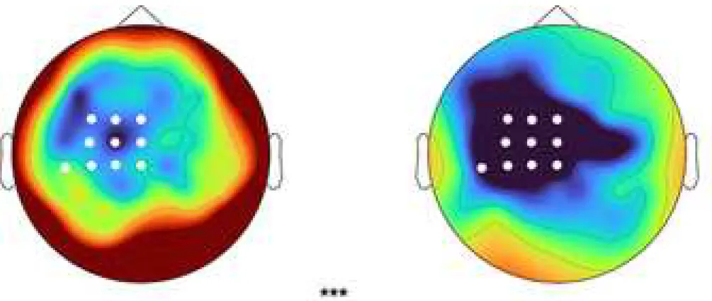
A preliminary analysis did not reveal any differences between STROKE and CO in the absolute power during the baseline period, allowing to interpret the movement-related beta desynchronization related to the elbow extension. The time-frequency topographic plot (Fig. 4) represent the response pattern of movement-related beta desynchronization during an elbow extension for both groups. The non-parametric cluster-based permutation analysis revealed a significant difference (p < 0.01) between STROKE and CO in the movement-related beta desynchronization (Fig. 4). The topographical distribution of the cluster
corresponded to the ipsilesional sensorimotor electrodes for STROKE (e.g., Cz, C1, C3, CPz, CP1, CP3, CP5, FCz, FC1, FC3). The movement-related beta desynchronization was
decreased by 0.9 ± 0.3 db in STROKE compared to CO during the elbow extension
13 covariance on movement-related beta desynchronization with Group (STROKE vs CO) as between factor and Age as a covariable revealed a Group effect on the movement-related beta desynchronization while controlling for age (F1,22 = 5.02, p < 0.05). The non-parametric cluster-based permutation was also performed on the movement-related alpha
desynchronization and did not reveal any differences between STROKE and CO (see supplemental material).
3.4. Remarkable correlations between EEG, EMG and kinematic data in STROKE
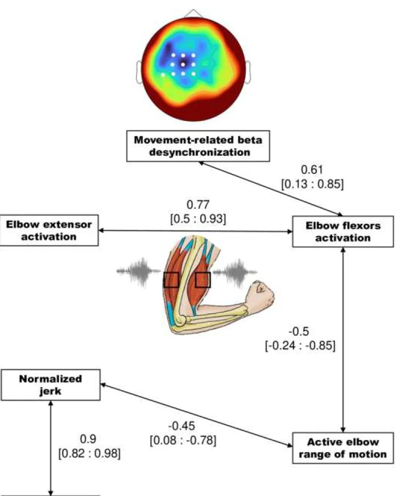
The correlation iconography diagram representing significant partial correlations is presented in Fig. 5. Movement-related beta desynchronization correlated positively with elbow flexor activation, meaning that greater elbow flexor activation was associated with less movement-related beta desynchronization. Elbow flexor activation cormovement-related positively with elbow extensor activation, meaning that an increase in the elbow flexor activation was associated with an increase in the elbow extensor activation. Elbow flexor activation correlated negatively with the active elbow range of motion, meaning that an increase in elbow flexor activity during the movement was associated with a reduced active elbow range of motion. Active elbow range of motion was correlated negatively with the normalized jerk, meaning that a reduced active elbow range of motion was associated with greater acceleration peaks. Finally, the normalized jerk was positively correlated with the movement duration - the more the movement was jerky the longer it took to extend the elbow.
4. Discussion
We aimed to investigate the role of the cortical beta oscillations in the excessive increase of antagonist co-contraction in post-stroke subjects during active elbow extension. To the best of
14 our knowledge, this study is the first to measure cortical activity directly during an active elbow extension and to examine the cortical mechanisms underlying the spastic co-contraction in post-stroke subjects. We reported a new key finding - a strong association between movement-related beta desynchronization and the elbow flexor activation during elbow extension, providing new insights into neural mechanisms underlying contributing to the motor impairment after stroke.
In line with our hypotheses, we showed significant differences in kinematic variables between post-stroke and healthy participants. In post-stroke subjects, the active range of motion was limited, slower and less smooth. These results are consistent with the literature that has reported the impairment of the motor behavior through kinematic analyses after a stroke (Murphy et al., 2011; Murphy and Häger, 2015). Our results showed an association between the jerk and the active elbow range of motion where post-stroke subjects with a jerky
movement will tend to have a limited active elbow range of motion, reflecting the impairment of various characteristics of the movement. Our results showed an association between
movement duration and movement smoothness, which supports the idea of using movement duration as an easy-to-use clinical marker relating the movement characteristics (Murphy et al., 2011).
Our findings also highlighted a pathological pattern of muscle overactivity on both elbow flexors and extensors, suggesting that a motor task as simple as extending the elbow
represents a considerable effort for post-stroke subjects. As previously shown (Chalard et al., 2019), we found a negative association between the elbow flexor activation and the active elbow range of motion confirming that excessive antagonist co-contraction is one of the mechanisms contributing to motor impairment (Baude et al., 2018; Chalard et al., 2019; Sarcher et al., 2015). Excessive antagonist co-contraction produces an active resistance by generating an opposite force at the elbow, which limits the active extension movement
15 (Gracies, 2015). Furthermore, we showed a link between elbow flexor and extensor
activations in post-stroke subjects, highlighting that an excessive antagonist co-contraction could contribute to the concomitant increase of elbow extensor activation, in order to
counteract the active resistance generated by the elbow flexors. Taken together, these results strongly support and confirm that spastic co-contraction is a disabling form of muscle overactivity generating abnormal patterns of motor behavior leading to alterations in
movement characteristics and upper limb motor function (Baude et al., 2018; Chalard et al., 2019; Gracies, 2015).
After eliminating the cofounding factors in the partial correlations, our data do not directly link the modulation of the jerk or the movement duration with elbow muscles or cortical activations. It is likely that the soft tissue plastic rearrangements, muscle contracture or other modalities of muscle overactivity (i.e. spasticity and spastic dystonia) occurring in the spastic paresis syndrome, could have a detrimental impact on the different components of the elbow kinematics (Gracies, 2005). Moreover, beta oscillations in the ipsilesional sensorimotor cortex of post-stroke subjects emerged as the strongest feature of our data, reflecting significant differences between STROKE and CO in cortical beta oscillatory activity spread across the sensorimotor cortex. In line with other studies (Gerloff, 2006; Rossiter et al., 2014), our results indicated a decrease in movement-related beta desynchronization in post-stroke subjects during the movement. As suggested by Rossiter et al. (2014), post-stroke subjects may lack the ability to modulate their motor cortex during the movement, reflecting an impaired ability to generate volitional descending motor signals. Cortical oscillatory activity provides a direct measure of neuronal activity that can help us to understand the link between neuroplasticity and post-stroke motor impairment. Although the role of the movement-related beta desynchronization in post-stroke and healthy subjects is documented, the cortical neural signature underlying the control of the antagonist co-contraction in post-stroke subjects
16 remains unexplored. We found an association between the decrease of the movement-related beta desynchronization and the increase of elbow flexor activation (i.e. the antagonist co-contraction). In healthy subjects, some studies have suggested a specific encoding for the antagonist muscles through the cortical oscillations in the beta frequency band which could play a critical role in the modulation of antagonist co-activation (Dal Maso et al., 2017, 2012; Takemi et al., 2018). Even if correlations do not provide proof of causal relationship, our results highlight that an alteration of beta cortical oscillations represents a key mechanism associated to spastic co-contraction in post-stroke situations. Alterations in various
physiological mechanisms, such as a reduction in inhibitory mechanisms (reciprocal
inhibition, reduction of presynaptic inhibition), have been suggested as being involved in the emergence of spastic co-contraction (Baude et al., 2018). We can hypothesize that a decrease in the movement-related beta desynchronization could reflect the reduced cortical influences on spinal mechanisms regulating the antagonist co-activation. Considering that the
modulation of cortical oscillations can reflect the balance between inhibitory and excitatory processes (Hall et al., 2011; Muthukumaraswamy et al., 2013), we can suppose that the observed decrease in cortical oscillatory activity can be due to a disturbance between
inhibitory and excitatory processes. The movement-related desynchronization may also reflect the conjunction of multiple factors associated with sensory aspects of motor control, notably the potential deficit in sensory feedback during movement (Müller et al., 2003). In chronic stroke situations, it has been shown that abnormal inter-hemispheric interactions occur during movement generation and influence functional recovery (Murase et al., 2004). Although we have not investigated the potential interaction between excessive inter-hemispheric inhibition and spastic co-contraction, an inter-hemispheric inhibition may be partly involved in the decrease in the cortical oscillatory activity in the ipsilesional hemisphere (Takechi et al., 2014) and, thus, contribute to the emergence of spastic co-contraction.
17
5. Limitations
In this study we focused on the cortical mechanisms underlying spastic co-contraction. One limitation of this study is the variability in the subject groups with regard to the time since the stroke and localizing the lesion and that little is known about the influence of lesion location on spastic co-contraction and on the cortical oscillatory activity. Taken together, and due to the small sample of the study and the absence of a matched control group, any generalization of these results should be treated with caution.
6. Conclusion
These results suggest that cortical beta oscillation alterations may reflect an important neural mechanism underlying with spastic co-contraction of elbow muscles during movement following a stroke. The present findings extend the current concept of cortical oscillations toward an underlying neural mechanism associated with the motor impairment after a stroke. Movement-related beta desynchronization is increasingly being used to investigate the functional role of cortical oscillations in motor function, making cortical beta oscillations a potential marker for motor recovery in stroke subjects. Our study pleads for an EEG-based assessment of cortical oscillatory activity as a target to understand and characterize
neuromuscular plasticity induced by therapeutic interventions.
Conflict of Interest Statement
Alexandre Chalard is employee of Ipsen Innovation within the framework of a CIFRE PhD fellowship. The other authors in this study declare that there is no conflict of interest.
18 Funding
This research did not receive any specific grant from funding agencies in the public, commercial, or not-for-profit sectors.
Acknowledgement
We thank Marie Belle, Lisa Corbière, Emmeline Montané, Célia Pinto, Maxime Fauvet for their help during the experimental protocol. We thank Dr Camille Cormier and Dr Yann Le Déan for their help in the patients’ clinical assessments.
References
Arene, N., Hidler, J., 2009. Understanding Motor Impairment in the Paretic Lower Limb After a Stroke: A Review of the Literature. Top. Stroke Rehabil. 16, 346–356. https://doi.org/10.1310/tsr1605-346
Baude, M., Nielsen, J.B., Gracies, J.-M., 2018. The neurophysiology of deforming spastic paresis: A revised taxonomy. Ann. Phys. Rehabil. Med.
https://doi.org/10.1016/j.rehab.2018.10.004
Bhagchandani, N., Schindler-Ivens, S., 2012. Reciprocal inhibition post-stroke is related to reflex excitability and movement ability. Clin. Neurophysiol. 123, 2239–2246. https://doi.org/10.1016/j.clinph.2012.04.023
Bigot, J., Longcamp, M., Dal Maso, F., Amarantini, D., 2011. A new statistical test based on the wavelet cross-spectrum to detect time-frequency dependence between non-stationary signals: application to the analysis of cortico-muscular interactions. Neuroimage 55, 1504–18. https://doi.org/10.1016/j.neuroimage.2011.01.033
Cahouët, V., Martin, L., Amarantini, D., 2002. Static optimal estimation of joint accelerations for inverse dynamics problem solution. J. Biomech. 35, 1507–1513.
Chae, J., Yang, G., Park, B.K., Labatia, I., 2002. Muscle weakness and cocontraction in upper limb hemiparesis: relationship to motor impairment and physical disability.
Neurorehabil. Neural Repair 16, 241–248.
Chalard, A., Amarantini, D., Tisseyre, J., Marque, P., Tallet, J., Gasq, D., 2019. Spastic co-contraction, rather that spasticity, is associated with impaired active function in adults with acquired brain injury: A pilot study. J. Rehabil. Med. 51, 307–311.
https://doi.org/10.2340/16501977-2528
Cohen, J., 1977. CHAPTER 2 - The t Test for Means, in: Statistical Power Analysis for the Behavioral Sciences (Revised Edition). Academic Press, pp. 19–74.
19 Dal Maso, F., Longcamp, M., Amarantini, D., 2012. Training-related decrease in antagonist
muscles activation is associated with increased motor cortex activation: evidence of central mechanisms for control of antagonist muscles. Exp. Brain Res. 220, 287–295. https://doi.org/10.1007/s00221-012-3137-1
Dal Maso, F., Longcamp, M., Cremoux, S., Amarantini, D., 2017. Effect of training status on beta-range corticomuscular coherence in agonist vs. antagonist muscles during
isometric knee contractions. Exp. Brain Res. 235, 3023–3031. https://doi.org/10.1007/s00221-017-5035-z
de los Reyes-Guzmán, A., Dimbwadyo-Terrer, I., Trincado-Alonso, F., Monasterio-Huelin, F., Torricelli, D., Gil-Agudo, A., 2014. Quantitative assessment based on kinematic measures of functional impairments during upper extremity movements: A review. Clin. Biomech. 719–727. https://doi.org/10.1016/j.clinbiomech.2014.06.013
Fauvet, M., Cremoux, S., Chalard, A., Tisseyre, J., Gasq, D., Amarantini, D., 2019. A novel method to generalize time-frequency coherence analysis between EEG or EMG signals during repetitive trials with high intra-subject variability in duration, in: 2019 9th International IEEE/EMBS Conference on Neural Engineering (NER). Presented at the 2019 9th International IEEE/EMBS Conference on Neural Engineering (NER), pp. 437–440. https://doi.org/10.1109/NER.2019.8716973
Gerloff, C., 2006. Multimodal imaging of brain reorganization in motor areas of the
contralesional hemisphere of well recovered patients after capsular stroke. Brain 129, 791–808. https://doi.org/10.1093/brain/awh713
Gracies, J.-M., 2015. Coefficients of impairment in deforming spastic paresis. Ann Phys Rehabil Med 58, 173–8. https://doi.org/10.1016/j.rehab.2015.04.004
Gracies, J.-M., 2005. Pathophysiology of spastic paresis. II: Emergence of muscle overactivity. Muscle Nerve 31, 552–571.
Gracies, J.-M., Wilson, L., Gandevia, S.C., 1997. Stretched position of spastic muscles aggravates their co-contraction in hemiplegic patients. Ann Neurol 42, 438–9.
Grinsted, A., Moore, J.C., Jevrejeva, S., 2004. Application of the cross wavelet transform and wavelet coherence to geophysical time series. Nonlinear Process. Geophys. 11, 561– 566. https://doi.org/10.5194/npg-11-561-2004
Gross, R., Leboeuf, F., Hardouin, J.B., Perrouin-Verbe, B., Brochard, S., Rémy-Néris, O., 2015. Does muscle coactivation influence joint excursions during gait in children with and without hemiplegic cerebral palsy? Relationship between muscle coactivation and joint kinematics. Clin. Biomech. 1088–1093.
https://doi.org/10.1016/j.clinbiomech.2015.09.001
Hall, S.D., Stanford, I.M., Yamawaki, N., McAllister, C.J., Rönnqvist, K.C., Woodhall, G.L., Furlong, P.L., 2011. The role of GABAergic modulation in motor function related neuronal network activity. NeuroImage 56, 1506–1510.
https://doi.org/10.1016/j.neuroimage.2011.02.025
Hermens, H.J., Freriks, B., Disselhorst-Klug, C., Rau, G., 2000. Development of recommendations for SEMG sensors and sensor placement procedures. J. Electromyogr. Kinesiol. 10, 361–374.
Kaiser, V., Daly, I., Pichiorri, F., Mattia, D., Müller-Putz, G.R., Neuper, C., 2012. Relationship between electrical brain responses to motor imagery and motor
20 impairment in stroke. Stroke J. Cereb. Circ. 43, 2735–2740.
https://doi.org/10.1161/STROKEAHA.112.665489
Katz, R., Pierrot-Deseilligny, E., 1982. Recurrent inhibition of alpha-motoneurons in patients with upper motor neuron lesions. Brain J. Neurol. 105, 103–124.
https://doi.org/10.1093/brain/105.1.103
Koch, P.J., Hummel, F.C., 2017. Toward precision medicine: tailoring interventional strategies based on noninvasive brain stimulation for motor recovery after stroke. Curr. Opin. Neurol. 30, 388–397. https://doi.org/10.1097/WCO.0000000000000462 Lakens, D., 2013. Calculating and reporting effect sizes to facilitate cumulative science: a
practical primer for t-tests and ANOVAs. Front. Psychol. 4. https://doi.org/10.3389/fpsyg.2013.00863
Lesty, C., Pleau-Varet, J., Kujas, M., 2004. Geometric Method and Generalized Linear Models. Two Opposite Multiparametric Approaches Illustrated on a Sample of Pituitary Adenomas. J. Appl. Stat. 31, 191–213.
https://doi.org/10.1080/0266476032000148867
Maris, E., Oostenveld, R., 2007. Nonparametric statistical testing of EEG- and MEG-data. J. Neurosci. Methods 164, 177–190. https://doi.org/10.1016/j.jneumeth.2007.03.024 Morita, H., Crone, C., Christenhuis, D., Petersen, N.T., Nielsen, J.B., 2001. Modulation of
presynaptic inhibition and disynaptic reciprocal Ia inhibition during voluntary movement in spasticity. Brain J. Neurol. 124, 826–837.
Müller, G.R., Neuper, C., Rupp, R., Keinrath, C., Gerner, H.J., Pfurtscheller, G., 2003. Event-related beta EEG changes during wrist movements induced by functional electrical stimulation of forearm muscles in man. Neurosci. Lett. 340, 143–147.
https://doi.org/10.1016/S0304-3940(03)00019-3
Murase, N., Duque, J., Mazzocchio, R., Cohen, L.G., 2004. Influence of interhemispheric interactions on motor function in chronic stroke. Ann. Neurol. 55, 400–409. https://doi.org/10.1002/ana.10848
Murphy, M.A., Häger, C.K., 2015. Kinematic analysis of the upper extremity after stroke – how far have we reached and what have we grasped? Phys. Ther. Rev. 20, 137–155. https://doi.org/10.1179/1743288X15Y.0000000002
Murphy, M.A., Willén, C., Sunnerhagen, K.S., 2011. Kinematic Variables Quantifying Upper-Extremity Performance After Stroke During Reaching and Drinking From a Glass. Neurorehabil. Neural Repair 25, 71–80.
https://doi.org/10.1177/1545968310370748
Muthukumaraswamy, S.D., Myers, J.F.M., Wilson, S.J., Nutt, D.J., Lingford-Hughes, A., Singh, K.D., Hamandi, K., 2013. The effects of elevated endogenous GABA levels on movement-related network oscillations. NeuroImage 66, 36–41.
https://doi.org/10.1016/j.neuroimage.2012.10.054
Oostenveld, R., Fries, P., Maris, E., Schoffelen, J.-M., 2011. FieldTrip: Open source software for advanced analysis of MEG, EEG, and invasive electrophysiological data. Comput. Intell. Neurosci. 2011, 156869. https://doi.org/10.1155/2011/156869
Park, W., Kwon, G.H., Kim, Y.-H., Lee, J.-H., Kim, L., 2016. EEG response varies with lesion location in patients with chronic stroke. J. Neuroengineering Rehabil. 13, 21. https://doi.org/10.1186/s12984-016-0120-2
21 Perrin, F., Pernier, J., Bertrand, O., Echallier, J.F., 1989. Spherical splines for scalp potential
and current density mapping. Electroencephalogr. Clin. Neurophysiol. 72, 184–187. https://doi.org/10.1016/0013-4694(89)90180-6
Pfurtscheller, G., 2001. Functional brain imaging based on ERD/ERS. Vision Res. 41, 1257– 1260. https://doi.org/10.1016/S0042-6989(00)00235-2
Pfurtscheller, G., Lopes da Silva, F.H., 1999. Event-related EEG/MEG synchronization and desynchronization: basic principles. Clin. Neurophysiol. 110, 1842–1857.
https://doi.org/10.1016/S1388-2457(99)00141-8
Ramos-Murguialday, A., Birbaumer, N., 2015. Brain oscillatory signatures of motor tasks. J. Neurophysiol. 113, 3663–3682. https://doi.org/10.1152/jn.00467.2013
Rossiter, H.E., Borrelli, M.R., Borchert, R.J., Bradbury, D., Ward, N.S., 2015. Cortical mechanisms of mirror therapy after stroke. Neurorehabil. Neural Repair 29, 444–452. https://doi.org/10.1177/1545968314554622
Rossiter, H.E., Boudrias, M.-H., Ward, N.S., 2014. Do movement-related beta oscillations change after stroke? J. Neurophysiol. 112, 2053–2058.
https://doi.org/10.1152/jn.00345.2014
Sarcher, A., Raison, M., Ballaz, L., Lemay, M., Leboeuf, F., Trudel, K., Mathieu, P.A., 2015. Impact of muscle activation on ranges of motion during active elbow movement in children with spastic hemiplegic cerebral palsy. Clin. Biomech. 30, 86–94.
Schieber, M.H., Lang, C.E., Reilly, K.T., McNulty, P., Sirigu, A., 2009. Selective activation of human finger muscles after stroke or amputation. Adv. Exp. Med. Biol. 629, 559– 575. https://doi.org/10.1007/978-0-387-77064-2_30
Staudenmann, D., Taube, W., 2015. Brachialis muscle activity can be assessed with surface electromyography. J. Electromyogr. Kinesiol. 25, 199–204.
https://doi.org/10.1016/j.jelekin.2014.11.003
Takechi, U., Matsunaga, K., Nakanishi, R., Yamanaga, H., Murayama, N., Mafune, K., Tsuji, S., 2014. Longitudinal changes of motor cortical excitability and transcallosal
inhibition after subcortical stroke. Clin. Neurophysiol. 125, 2055–2069. https://doi.org/10.1016/j.clinph.2014.01.034
Takemi, M., Maeda, T., Masakado, Y., Siebner, H.R., Ushiba, J., 2018. Muscle-selective disinhibition of corticomotor representations using a motor imagery-based brain-computer interface. NeuroImage 183, 597–605.
https://doi.org/10.1016/j.neuroimage.2018.08.070
Takemi, M., Masakado, Y., Liu, M., Ushiba, J., 2013. Is event-related desynchronization a biomarker representing corticospinal excitability? Conf. Proc. Annu. Int. Conf. IEEE Eng. Med. Biol. Soc. IEEE Eng. Med. Biol. Soc. Annu. Conf. 2013, 281–284. https://doi.org/10.1109/EMBC.2013.6609492
Tisseyre, J., Marquet-Doléac, J., Barral, J., Amarantini, D., Tallet, J., 2019. Lateralized inhibition of symmetric contractions is associated with motor, attentional and executive processes. Behav. Brain Res. 361, 65–73.
22 Figure captions
Figure 1. Time series representation of (A.) the active elbow extension, (B.) the acceleration profile, (C.) the elbow flexors activation, and (D.) extensor activation for a typical subject in post-stroke (STROKE) (red) and in healthy controls CO (blue). The shaded area corresponds to the standard error.
Figure 2. Bar charts representing (A.) the active elbow range of motion, (B.) the movement duration, and (C.) the normalized jerk for post-stroke (STROKE) (light gray) and healthy controls CO (dark gray). *, **, *** indicates a significant difference between groups (p < 0.05; p < 0.01; p < 0.001, respectively); vertical bars represent standard error of the mean.
Figure 3. Bar charts representing the elbow extensor activation (light gray) and elbow flexor activation (dark gray) for post-stroke (STROKE) and healthy controls (CO). *** indicates significant difference between groups (p < 0.001); vertical bars represent standard error of the mean.
Figure 4. Bar chart and scalp topography of movement-related beta desynchronization during the elbow extension for post-stroke (STROKE) and healthy controls (CO). White dots on the scalp topography represent the significant cluster of difference between groups obtained by the cluster-based permutation analysis. *** indicates a significant difference between groups (p < 0.001); vertical bars represent standard error of the mean.
Figure 5. Correlation iconography diagram representing only the significant partial
23 activations and elbow extension parameters for post-stroke (STROKE). A link between two nodes represents a significant correlation (p < 0.05); brackets represent the 95th confidence interval for the correlation coefficient.
Participants Age Sex Time Since
Stroke (months) Stroke type, sub-type Stroke side, location
Fugl-Meyer Upper Extremity (/66) Erasmus modified Nottingham Sensory Assessment (/64) Active elbow extension range of motion (°) CONTROL (n = 9) 43 ± 21 6 M / 3 F - - - - - 96 ± 8 STROKE 1 61 M 50 Hemorrhagic, microangiopathy Right, basal ganglia and corona radiata 38 34 56 STROKE 2 59 M 17 Ischemic, MCA (atheroma) Right, cortical and subcortical territories
of MCA 46 27 52
STROKE 3 69 F 19 Ischemic, vertebral artery (atheroma) Right, Pons 44 60 74 STROKE 4 65 M 74 Ischemic, carotid artery dissection Right, cortical and subcortical territories
of MCA 32 50 58
STROKE 5 50 M 29 Hemorrhagic, microangiopathy Left basal ganglia and internal capsule 42 60 75 STROKE 6 51 F 12
Ischemic (with hemorrhagic transformation), MCA
(cardio-embolic)
Left, subcortical territory of MCA 24 61 74
STROKE 7 57 M 14 Ischemic, microangiopathy Left, posterior limb of the internal
capsule 45 56 40
STROKE 8 75 M 26
Ischemic (with hemorrhagic transformation), MCA
(cardio-embolic)
Left, cortical and subcortical territories
of MCA 26 33 61
STROKE 9 46 M 8
Ischemic (with hemorrhagic transformation), carotid artery
dissection
Left, cortical and subcortical territories
of MCA 53 54 56
STROKE 10 65 M 116 Ischemic, MCA (atheroma) Right, cortical and subcortical territories
of MCA 30 62 52
STROKE 11 49 M 13 Ischemic, microangiopathy Predominant right, Pons (paramedian) 53 63 60 STROKE 12 33 F 12 Ischemic, vertebral artery dissection Predominant left, Pons and middle
cerebellar peduncles 45 48 80 STROKE 13 33 F 12 Ischemic, carotid artery dissection Left, subcortical territories of MCA 25 62 56 STROKE 14 57 M 18 Ischemic, MCA (atheroma) Right, cortical and subcortical territories
of MCA 41 26 66
STROKE 15 56 M 22 Ischemic, MCA (atheroma) Right, cortical and subcortical territories
of MCA 29 30 40
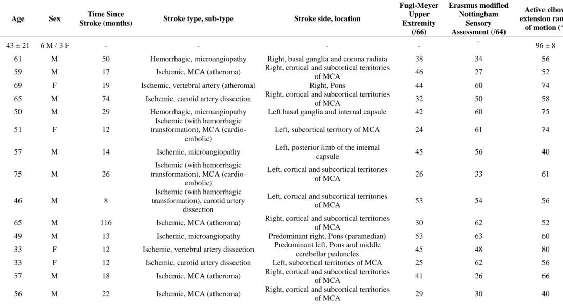
Table I. Participants' demographics (mean ± SD). Table
Abbreviations: MCA, middle cerebral artery. Cortical territory of MCA: lateral surface of the hemisphere, except for the medial part of the frontal and the parietal lobe (anterior cerebral artery), and the inferior part of the temporal lobe (posterior cerebral artery). Subcortical territory of MCA: Basal ganglia (globus pallidus, caudate, putamen), internal capsule and corona radiata.
Supplemental material: Movement-related alpha desynchronization.
Supplemental Figure 1: Boxplot and topographic representation of movement-related alpha and beta desynchronization over the ipsilesional cortex for STROKE (gray) and CO (white). White circles represent electrodes of interest (Cz, C1, C3, CPz, CP1, CP3, CP5, FCz, FC1, FC3). We performed a two-way mixed ANOVA with Frequency (Alpha vs Beta) as a within-subject factor and Group (STROKE vs CO) as a between-within-subject factor, on the movement-related desynchronization. The ANOVA revealed a Frequency effect (F1,22 = 46.4, p < 0.01)
and a Group x Frequency interaction (F1,22 = 6.2, p < 0.05), post-hoc analyses showed only a
significant difference for STROKE vs CO for movement-related beta desynchronization (t22 =
2.9, p < 0.01). The analysis did not reveal nor a Group effect (F1,22 = 3.1, p = 0.1) nor a
significant difference for STROKE vs CO for movement-related alpha desynchronization (t22
= 0.53, p = 0.6). Optional e-only supplementary files