Publisher’s version / Version de l'éditeur:
Vous avez des questions? Nous pouvons vous aider. Pour communiquer directement avec un auteur, consultez la
première page de la revue dans laquelle son article a été publié afin de trouver ses coordonnées. Si vous n’arrivez pas à les repérer, communiquez avec nous à PublicationsArchive-ArchivesPublications@nrc-cnrc.gc.ca.
Questions? Contact the NRC Publications Archive team at
PublicationsArchive-ArchivesPublications@nrc-cnrc.gc.ca. If you wish to email the authors directly, please see the first page of the publication for their contact information.
https://publications-cnrc.canada.ca/fra/droits
L’accès à ce site Web et l’utilisation de son contenu sont assujettis aux conditions présentées dans le site LISEZ CES CONDITIONS ATTENTIVEMENT AVANT D’UTILISER CE SITE WEB.
Journal of Natural Products, 2019-07-08
READ THESE TERMS AND CONDITIONS CAREFULLY BEFORE USING THIS WEBSITE.
https://nrc-publications.canada.ca/eng/copyright
NRC Publications Archive Record / Notice des Archives des publications du CNRC :
https://nrc-publications.canada.ca/eng/view/object/?id=5226b968-f88d-4c79-9a94-256602ec4b20
https://publications-cnrc.canada.ca/fra/voir/objet/?id=5226b968-f88d-4c79-9a94-256602ec4b20
NRC Publications Archive
Archives des publications du CNRC
This publication could be one of several versions: author’s original, accepted manuscript or the publisher’s version. / La version de cette publication peut être l’une des suivantes : la version prépublication de l’auteur, la version acceptée du manuscrit ou la version de l’éditeur.
For the publisher’s version, please access the DOI link below./ Pour consulter la version de l’éditeur, utilisez le lien DOI ci-dessous.
https://doi.org/10.1021/acs.jnatprod.9b00318
Access and use of this website and the material on it are subject to the Terms and Conditions set forth at
Structure elucidation and relative toxicity of
(24R)-24-hydroxyyessotoxin from a Namibian isolate of Gonyaulax spinifera
Rajotte, Isabelle; Rafuse, Cheryl; Wright, Elliott J.; Achenbach, John C.;
Ellis, Lee D.; McCarron, Pearse
†
Biotoxin Metrology, National Research Council Canada, 1411 Oxford Street, Halifax, Nova Scotia B3H 3Z1, Canada
‡
Aquatic and Crop Resource Development, National Research Council Canada, 1411 Oxford Street, Halifax, Nova Scotia B3H 3Z1,
Canada
*
S Supporting InformationABSTRACT:
Liquid chromatography−high-resolution mass
spectrometry (LC-HRMS) analysis of a Namibian strain of
Gonyaulax spinifera showed the presence of a number of
yessotoxins (YTXs). Principal among these were YTX (1),
homoYTX (2), and a tentative hydroxylated analogue that did not
correspond to any previously confirmed YTX structures.
Culturing the G. spinifera strain afforded sufficient biomass for
purification of the new analogue through a series of solvent
partitioning and chromatographic steps, yielding ∼0.9 mg as a
solid. NMR spectroscopy, ion-trap mass spectrometry, and HRMS
identified the new analogue as hydroxyYTX (7). Purified
24-hydroxyYTX was quantitated by NMR, and its relative toxicity
evaluated using two embryonic zebrafish toxicity assays. 24-HydroxyYTX demonstrated reduced toxicity compared to YTX.
Y
essotoxins (YTXs) are a class of sulfated polyether toxins
(
Figure 1
) produced by some marine dinoflagellates that
can accumulate in filter-feeding shellfish.
1YTX (1) was first
isolated from shellfish,
2and since then, various dinoflagellates
have been reported to produce YTXs including Protoceratium
reticulatum (Clapare
̀de & Lachmann) Bütschli, 1885,
3Lingulodinium polyedrum (F. Stein) J. D. Dodge, 1989,
4Gonyaulax spinifera (Clapare
̀de & Lachmann) Diesing,
1866,
5and Gonyaulax taylorii M.C.Carbonell-Moore, 1996.
6A number of YTX structures including 1a-homoYTX (2),
45-hydroxyYTX (3),
745-hydroxyhomoYTX (4),
844-carboxyYTX
(5),
9and 41a-homoYTX (6),
10among others,
1have been
elucidated by NMR, while numerous other analogues have
been observed but only tentatively identified by LC-MS.
11Many YTX analogues are biosynthetic products of algae;
however some variants such as 3 and 5 are metabolic products
of shellfish. 45-hydroxyYTX (3) was first isolated and
identified from Japanese scallops in 1996
12and was
subsequently detected in shellfish from a number of other
locations.
7,8YTXs were originally classified as diarrhetic poisoning toxins
due to their positive response when administered
intra-peritoneally in the traditional mouse bioassay for lipophilic
toxins.
13However, this toxicity is greatly diminished when
YTXs are administered orally.
14Recent studies have suggested
cardiotoxicity
15and immunotoxicity
16in rodents via
intra-peritoneal administration. Due to evidence of reduced toxicity
upon oral consumption, levels of YTXs regulated in seafood by
the European Union were changed from a limit of 1 mg/kg to
3.75 mg/kg.
17Research on YTXs remains of interest despite further
recommendations for less stringent regulatory limits for YTXs
in seafood.
18Several groups have studied possible synergistic
effects with other lipophilic toxins. Aasen et al. reported no
enhanced activity of YTX in a mouse bioassay when
administered orally in combination with azaspiracid-1.
19However, Ferron et al. reported a synergistic toxic effect on
human intestinal cell models when YTX was combined with
azaspiracid-1.
20This indicates further work may be merited on
the study of YTX toxicity. Continued development of
improved analytical methods is necessary for detection of
marine biotoxins in complex matrices, and profiling of
toxin-producing algae for new analogues is also an important
activity.
21,22Methods using high-resolution mass spectrometry
(HRMS) have been developed for profiling and quantitation of
marine toxins including YTXs.
23,24Furthermore, there is also
interest in YTX as a synthetic target.
25,26In 2011 a bloom of dinoflagellates was reported in Walvis
Bay, Namibia, from which a strain of G. spinifera, a known
producer of YTXs,
27was isolated.
28Here we report on the
purification, structure elucidation, and relative toxicity
assess-ment of a new hydroxyYTX analogue from the Namibian G.
spinifera strain.
Received: April 5, 2019 Published XXXX by the American Chemical
Society A DOI:10.1021/acs.jnatprod.9b00318
J. Nat. Prod. XXXX, XXX, XXX−XXX
Downloaded via NATL RESEARCH COUNCIL CANADA on July 11, 2019 at 19:30:05 (UTC).
■
RESULTS AND DISCUSSION
LC-MS Profiling. LC-HRMS analysis of the Namibian G.
spinifera strain revealed the presence of various YTX analogues.
The most prominent were 1a-homoYTX and YTX, both
confirmed by accurate mass and retention time matching with
certified reference materials (CRMs), and an earlier eluting
oxygenated YTX, tentatively identified as hydroxyYTX (7) due
to its retention time (
Figure 2
). Tentative identification was
supported by the measured accurate of m/z 1157.4662 [M −
H]
−, corresponding to an oxygenated YTX (C
55
H
81O
22S
2−) (Δ
−
0.4 ppm). A series of minor YTX analogues were also
detected with masses of m/z 1047.3939 [M − H]
−(tentatively
identified as 41-ketoYTX (8) by MS/MS), m/z 1061.4094 [M
−
H]
−(unidentified), m/z 1089.4934 [M − H]
−(tentatively
identified as 44,55-dihydroxy-41a-homoYTX (9)), m/z
1101.4403 [M − H]
−(tentatively identified as trinorYTX
(10) by MS/MS), and m/z 1117.4350 [M − H]
−(unidentified),
11,29the most significant of which was
44,55-dihydroxy-41a-homoYTX.
11Assuming equimolar response in
the full-scan LC-HRMS measurements, the new hydroxyYTX
analogue was ∼30% of the major component (1a-homoYTX),
making it a viable target for isolation and structure elucidation.
The potential presence of a hydroxylated YTX was of interest
as 45-hydroxyYTX has been tentatively identified by LC-MS in
an Italian strain of P. reticulatum
30and in a strain of G. spinifera
from South Africa.
31However, 45-hydroxyYTX has been
confirmed as a metabolite of YTX in shellfish.
32The putative
hydroxyYTX identified in the current work had an earlier LC
retention time than the previously reported 45-hydroxyYTX, as
determined through comparison with a matrix reference
material containing YTXs (
Figure S1
).
33,34Structure Elucidation.
Culturing of G. spinifera,
meth-anolic extraction of the biomass, and multiple chromatographic
steps yielded 7 as a colorless solid (∼0.9 mg). A series of
LC-Figure 1.Structure of YTX analogues mentioned in the text and calculated m/z values in negative ionization mode.
Journal of Natural Products
ArticleDOI:10.1021/acs.jnatprod.9b00318
J. Nat. Prod. XXXX, XXX, XXX−XXX
MS experiments comparing YTX (1) and the new analogue
(7) helped localize the position of the extra oxygen (
Figure 3
).
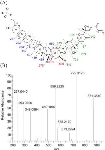
In negative ion mode the first major fragment observed for
YTXs typically corresponds to a loss of SO
3[M − SO
3]
−,
which corresponds to m/z 1061 and 1077 for 1 and 7,
respectively. The fragmentation of the [M − SO
3]
−ion follows
with a retro-ene reaction,
35forming the fragment m/z 924 for
1
11and m/z 940 for 7. Compared to 1, fragments 16 Da higher
were observed for 7, m/z 871, 815, 759, 729, 673, and 647
corresponding to cleavages in rings K down to H (
Figure 3
a).
Fragment m/z 647, corresponding to the first cleavage of ring
G, marks the last +16 Da fragment. Below this point, the
fragmentation diverges between 1 and 7, indicating
mod-ification on a carbon below C-27. On ring G, the fragments
observed for 7 were not consistent with those found in 1.
However, fragments m/z 559 and lower were present in both 1
and 7, indicating the presence of an additional oxygen between
C-24 and 26 in the form of a hydroxylated analogue (
Figure 3
).
The structure of 7 was established by comparing it with 1 in
a series of NMR experiments (
1H,
13C, COSY, TOCSY,
ROESY, HSQC, and HMBC) on the purified compounds. In
accord with the chemical formula indicated by HRMS, the
13C
and HSQC NMR spectra revealed the presence of 55 carbon
signals for 7 (6 methyl, 17 methylene, 25 methine, and 7
nonprotonated carbons). Comparison to the signals for 1 (6
methyl, 18 methylene, 24 methine, and 7 nonprotonated
carbons) indicated that a methylene had been converted to a
methine, suggesting hydroxylation at C-24 or C-25.
Comparison of the
1H spectra revealed a distinct shift of
26-Me (deshielded by 0.05 ppm), but no change in multiplicity,
and minor shifts for 23-Me (deshielded by 0.01 ppm) and
19-Me (shielded 0.01 ppm). All the other methyls were equivalent
for 1 and 7 (
Figure 4
a). Hydroxylation at C-26 was excluded
due to the doublet splitting of 26-Me that remained unchanged
(
Figure 4
a). The
1H NMR spectra also showed major
differences in the signals at 3.3−3.6 ppm (
Figure 4
b).
Compound 1 showed a doublet of doublets at 3.49 ppm
corresponding to H-22;
36however, for 7 the signal at 3.48 ppm
is a doublet. 2D NMR spectra (COSY and TOCSY) revealed
that the signal at 3.48 ppm in 7 did not correspond to H-22
(
Figure S2
). The COSY NMR spectrum of YTX revealed that
the signal at 3.49 ppm (H-22; dd; J = 4.5, 11.9 Hz) correlated
with signals at 1.94 and 1.73 ppm (H-21 α and H-21 β,
respectively), confirming its correlation to H-22. However, the
COSY NMR spectrum of 7 showed that the signal at 3.48 ppm
(H-24; d; J = 10.2 Hz) correlated with signals 1.98 (H-25 β),
1.39 (H-25 α), and 1.14 ppm (23-Me). Moreover, in the
TOCSY NMR spectrum of 1 the signal at 3.49 ppm (H-22; dd;
J = 4.5, 11.9 Hz) correlated with signals at 3.43 (H-20), 1.94
(H-21 α), and 1.73 (H-21 β). However, the TOCSY NMR
spectrum of 7 revealed that the signal at 3.48 ppm (H-24; d; J
= 10.2 Hz) correlated with signals at 3.23 (H-28), 2.76 (H-27),
1.98 (H-25 β), 1.82 (H-26), 1.39 (H-25 α), and 1.08 (26-Me)
ppm. Therefore, even though the signals are superimposed,
they belong to two different spin systems. The peak at 3.48
ppm belongs to H-24 in 7. C-24 in 1 is a methylene, and the
protons appear as two distinct signals (1.48 ppm, 1.75 ppm,
m), while C-24 in 7 is an oxygenated methine revealing a
change in multiplicity and a major shift (deshielded >1.73
ppm).
Figure 2. LC-HRMS chromatogram showing YTX analogues in a Namibian strain of G. spinifera (LC-MS method A).
Figure 3.(a) LC-MS/MS (LTQ) negative ion fragmentation of YTX and 24-hydroxyYTX (method B). Blue fragments are consistent between YTX and 24-hydroxyYTX, green corresponds to YTX-specific fragments M+16 (= M+O), and red fragments are unique to 24-hydroxyYTX. (b) HRMS/MS fragmentation of 24-hydroxyYTX (method A, collision energy 150 eV).
DOI:10.1021/acs.jnatprod.9b00318
J. Nat. Prod. XXXX, XXX, XXX−XXX
The HSQC NMR spectrum of 1 showed correlations
between the
13C signal at 47.0 ppm (CH
2
; C-24) and
1H
signals at 1.48 (m; H-24 β) and 1.75 (m; H-24 α). In 7 the
13C
signal for C-24 was present as a methine at 84.2 ppm and
correlated only to the proton at 3.48 ppm (d; H-24),
confirming that the hydroxy group is positioned on C-24.
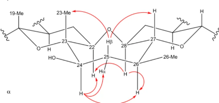
The configuration at C-24 was determined by examination
of the coupling constants and correlations observed in the
ROESY NMR experiment (
Figure S3
). The ROESY spectrum
revealed that H-24 correlated with signals at 3.39 (H-22 α),
1.39 (H-25 α), and 1.82 (H-26 α) ppm (
Figure 5
). There were
no correlations to either 23-Me(β) or 26-Me(β). Furthermore,
on the alpha face, the signal at 3.23 ppm (H-28 α) correlated
with signals at 3.39 (H-22 α) and 1.82 (H-26 α) ppm. On the
beta face, the signal at 1.98 ppm (H-25 β) correlated with
signals 1.14 (23-Me β) and 2.76 (H-27 β). As a result, the
hydroxy group must have a β orientation, thus assigning the
configuration of this new analogue as (24R)-24-hydroxyYTX
based on the known absolute configuration as YTX (1). The
complete assignment of 24-hydroxyYTX is reported in
Table 1
.
Comparison of YTX and 24-hydroxyYTX signals in both
1H
and
13C experiments showed major ppm shifts in C-22 to C-26
as well as 23-Me.
Although there have been previous observations of
hydroxylated YTXs in marine microalgae
11,30by LC-MS, this
is the first specific report defining the identity of a hydroxyYTX
from algae. 45-hydroxyYTX (3) is a known metabolite of
shellfish;
32therefore it would be unexpected for it to also be a
biosynthetic product of algae. A sample of the P. reticulatum
extract analyzed in an earlier study
30was provided for
LC-HRMS comparison against the 24-hydroxyYTX (7) identified
in this work. No clear peak for either 3 or 7 was observed in
the sample provided; however 1 and 2 were confirmed as
previously reported (
Figure S4
). Trace levels of tentative
hydroxylated YTXs were observed at retention times different
from 1 and 7; however, it was not possible to identify these
due to the lower concentrations present in the sample.
Recently, a strain of G. spinifera from Walker Bay, South Africa,
was reported
31to produce 3 as part of a YTX toxin profile
similar to the Namibian strain of G. spinifera (2 most
significant, with lower amounts of a hydroxyYTX and 1).
The identity of the hydroxyYTX reported was determined
using the EU harmonized standard operating procedure for
LC-MS determination of lipophilic marine biotoxins,
37which
includes a transition for 3. Due to relative proximity of the
locations where the respective strains of G. spinifera were
isolated, similarities in the profiles reported, and the ease of
misidentification of hydroxyYTX analogues using conventional
LC-MS methods in the absence of individual toxin standards, it
should be considered whether the hydroxylated compound
reported
31is actually 7.
The toxin profile of the G. spinifera strain studied in this
work is of interest in terms of the biosynthesis of these
compounds. Considering the reported biosynthesis of YTX
38and that the most abundant analogue in this culture is
homoYTX, the presence of an equivalent homo analogue of
24-hydroxy-YTX might reasonably be expected. However, no
additional homoYTX analogues were detected by LC-HRMS.
The presence of a mixed algal culture is unlikely, as the strain
analyzed was cultured from a single-cell isolate,
39and an
equivalent toxin profile was reported by Pitcher et al.
31in the
G. spinifera from South Africa. Future work should consider the
biosynthesis of YTXs by these particular strains of G. spinifera.
Relative Toxicity.
To assess the toxicity of 24-hydroxyYTX
(7), a stock solution was accurately quantitated by qNMR
40and a reference material was prepared by making accurate
dilutions in MeOH. Toxicity was compared to accurately
quantitated 1 in vivo by means of embryonic zebrafish (Danio
rerio) toxicity testing. Embryonic zebrafish toxicity assays have
been used as an alternative model for testing of algal
toxins.
41,42As YTX toxicity has already been described using
Figure 4.1H spectra (700 MHz, CD
3OD) for YTX (1) (blue) and 24-hydroxyYTX (7) (red) overlaid showing ranges for (a) 1.40 to 0.95 ppm and (b) 3.6 to 3.4 ppm.
Figure 5.3D representation of 24-hydroxyYTX and observed ROESY correlations (red arrows).
Journal of Natural Products
ArticleDOI:10.1021/acs.jnatprod.9b00318
J. Nat. Prod. XXXX, XXX, XXX−XXX
this model
41and shown to affect cardiovascular function in
other animal models,
15both a zebrafish embryonic
devel-opmental toxicity assay and a zebrafish embryonic
cardiotox-icity model were used to assess both 1 and the new 7. Exposure
of developing zebrafish embryos to both 1 and 7 caused an
edema phenotype (
Figure S5
), as reported in the previous
study.
41However, 7 was slightly but significantly less toxic in
this assay, with a resultant EC
50of 51 nM (95% confidence
interval, 42−62) as compared to an EC
50of 7.0 nM (95%
confidence interval, 3.5−14) for 1. Toxicity curves are shown
(
Figure 6
a). Both compounds exhibited bradycardia in the
zebrafish model, consistent with what has been observed with
YTX in the rat model.
15Compounds 1 and 7 both reduced
heart rate relative to carrier controls (
Figure 6
b). However, as
observed in the zebrafish embryo toxicity model, 7 was not as
toxic.
■
CONCLUSIONS
(24R)-24-HydroxyYTX was isolated from a Namibian strain of
G. spinifera and structurally elucidated through a combination
of NMR and LC-MS experiments. Embryonic zebrafish toxicity
assays demonstrated that this new YTX analogue was less toxic
than YTX. The production of hydroxylated analogues of YTXs,
both as biosynthetic products of microalgae and as metabolic
products of shellfish feeding on YTX-producing algae, requires
consideration when assessing YTX uptake and transformation
in seafood, and when performing regulatory monitoring for
hydroxylated YTXs in seafood. This also highlights the
challenge when assigning tentative identities to new toxin
analogues on the basis of LC and MS experiments alone, a
situation that is equally applicable to other toxin classes.
This work expands knowledge on the occurrence of
toxin-producing harmful algal species in Western Africa, which is of
importance considering the increased international exploitation
of seafood as a commodity for human consumption.
■
EXPERIMENTAL SECTION
General Experimental Procedures. All NMR spectra were obtained with a Bruker Avance III 700 MHz instrument. Experiments for structure elucidation were performed in CD3OD at 20 °C with a 1.7 mm TXI cryoprobe. Chemical shifts were reported relative to the methyl of CHD2OD (1H 3.31, 13C 49.00). Standard Bruker pulse sequences were used for1H,13C, COSY, TOCSY (120 ms), HSQC, HMBC, and ROESY. 24-HydroxyYTX (7) was dissolved in ∼30 μL of CD3OD. A sample of YTX (1) (∼0.2 mg)36was dissolved in ∼30 μL of CD3OD. Quantitative NMR experiments were performed by dissolving the purified 7 in 700 μL of CD3OD and running at 20 °C with a 5 mm TXI cryoprobe using benzoic acid as an external standard.40LC-MS (method A): The YTX profile of the Namibian G.
spinefera culture was assessed by LC-HRMS using a Q Exactive HF Orbitrap mass spectrometer (ThermoFisher Scientific) equipped with a heated electrospray ionization interface (HESI) connected to an Agilent 1200 G1312B binary pump, G1367C autosampler, and G1316B column oven (Agilent). Separations were performed on a Phenomenex Luna C18column (50 × 2 mm, 1.7 μm) eluting with 25−75% B over 25 min, hold (5 min), and re-equilibration (back to 25% over 10 min) (A: 5 mM NH4Ac in deionized H2O; B: 5 mM NH4Ac in 95% CH3CN) at 150 μL min
−1. The MS was operated in negative ion mode and calibrated in the range m/z 150−2000. The spray voltage was 2.70 kV, the capillary temperature was 350 °C, and the sheath and auxiliary gas flow rates were 40 and 15 (arbitrary units), respectively. The MS was operated in negative full scan (FS) mode: scan range m/z 500−1500, resolution setting at 60 000, AGC target 3 × 10⁶, max IT 200 ms; all ion fragmentation: scan range m/z 100−1500, resolution setting at 60 000, AGC target 3 × 10⁶,
8 36.5, CH2 1.39, 2.17, m 33 76.8, C 9 78.3, CH 3.13, m 33-Me 15.3, CH3 1.20, s 10 78.3, CH 3.13, m 34 73.3, CH 3.76, dd (12.6, 4.0) 11 36.2, CH2 1.39, 2.25, m 35 31.7, CH2 1.48, m, 2.10, dt (10.9, 4.2) 12 77.6, CH 3.03, m 36 73.2, CH 4.05, ddd (11.5, 9.6, 4.6) 13 78.1, CH 3.06, m 37 73.0, CH 3.38, m 14 38.0, CH2 1.43, 2.29, m 38 39.0, CH2 2.43, dd (12.5, 4.5), 2.71, td (12.2, 6.2) 15 81.1, CH 3.33, m 39 143.2, C 16 82.4, CH 3.21, m b 115.7, CH2 4.77, s, 5.00, s 17 30.2, CH2 1.79, 1.95, m 40 85.1, CH 3.86, s 18 40.8, CH2 1.83, 1.90, m 41 78.4, C 19 78.5, C 41-Me 26.2, CH3 1.38, s 19-Me 23.4, CH3 1.25, s 42 136.7, CH 5.81, d (16.0) 20 82.2, CH 3.40, m 43 130.6, CH 6.28, d (16.0) 21 32.5, CH2 1.78, 1.96, m 44 145.5, C 22 84.0, CH 3.39, m 44-methylene 116.6, CH2 4.96, s, 5.04, s 23 79.6, C 45 37.8, CH2 2.96, dd (6.6, 1.6) 23-Me 15.1, CH3 1.14, s 46 137.6, CH 5.87, ddt (16.8, 10.1, 6.5) 24 84.2, CH 3.48, d (10.2) 47 116.6, CH2 5.06, m, 5.08, dd (1.7, 1.6) 25 41.5, CH2 1.39, 1.98, m DOI:10.1021/acs.jnatprod.9b00318 J. Nat. Prod. XXXX, XXX, XXX−XXX E
maximum IT 200 ms, normalized collision energy (35 eV); and parallel reaction monitoring mode for MS/MS spectra: m/z 1141.5, 1155.5, 1157.5, with resolution setting at 30 000, AGC target 2 × 10⁵, max IT 100 ms, isolation window 4.0 m/z, and collision energy of 150 eV. LC-MS (method B): Fragmentation patterns were assessed using linear ion trap mass spectrometry (Thermo Scientific LTQ-XL) with an HESI source in negative mode. Instrument calibration was performed according to the manufacturer’s recommendations, and instrument default settings were used unless otherwise stated. Test solutions dissolved in methanol were introduced by infusion at 5 μL min−1
.The instrument was tuned using CRM-YTX. The HESI source voltage was 3 kV, temperature at 40 °C, sheath gas at 15 arbitrary units, aux and sweep gases off. Data for 1 and 7 were acquired using normal scan rate in full scan from m/z 150 to 2000, from which target ions were isolated. Once isolated, CID normalized collision energy was increased until sufficient fragmentation was achieved, and additional ions were isolated for further fragmentation as required. Method C: Analysis of fractions during the purification process was performed by selected reaction monitoring (SRM) on an Agilent 1200 HPLC connected to a Sciex API 4000 Qtrap. Chromatography conditions were as described for method A. Mass spectrometry conditions: source temperature 350 °C, DP −60 V, and CE −65 eV for a range of SRM transitions corresponding to known YTX analogues: m/z 1155 > 1075; 1047 > 967; 991 > 911; 1203 > 1123; 1189 > 1109; 1175 > 1095; 1169 > 1089; 1191 > 111.; 1187 > 1107; 1173 > 1093; 1290 > 1210; 1304 > 1224; 1085 > 1005; 1171 > 1091; 1157 > 1077; 1101 > 1021; 1141 > 1061.
Toxins and Other Materials.Distilled H2O was further purified using a UV purification system (Thermo Scientific) or a Milli-Q water purification system (Millipore Ltd.). MeOH and CH3CN (Optima LC-MS grade) were from Fisher Scientific. Hexanes, chloroform, and butanol were from Caledon. CD3OD (99.8%) was from Cambridge Isotope Laboratories. Ammonium hydroxide, calcium chloride dihydrate, HEPES buffer, magnesium sulfate heptahydrate, potassium chloride, and sodium chloride were from Sigma−Aldrich. Sephadex LH-20 was from Amersham Biosciences, and C18 silica (40 μm) was from Bakerbond. YTX (CRM-YTX-c (lot # 20151125)) and in-house purified YTX), homoYTX (CRM-hYTX (lot # 20111102)), and a freeze-dried mussel tissue (CRM-FDMT1)34were from the National Research Council Canada (Biotoxin Metrology). YTX used for NMR and LTQ experiments was purified in-house from material provided for developing the original CRM-YTX.
Culturing of Gonyaulax spinifera.A clonal isolate of G. spinifera obtained from Namibia, Africa,28was grown in L1 medium in 250 mL
flasks at 18 °C under a 14:10 h light:dark photoperiod. An approximate photon flux density of 80−100 μmol quanta m−2 s−1 cool white light was maintained. Light was measured outside the flask using a Li-Cor model LI-185B quantum/photometer. Cultures were scaled-up from 100 mL stock cultures to 300 L in a photobioreactor under the same environmental conditions using full-strength L1 medium and harvested in late exponential growth phase by gravity filtration through a 10 μm Nitex mesh sieve. Cells were concentrated by centrifugation at 2100g for 15 min at 4 °C in 200 mL polypropylene centrifuge tubes to yield 85 g of biomass.
Isolation of YTXs. Biomass (85 g wet) was extracted by sonication with MeOH (3 × 250 mL) on ice and centrifuged. The methanolic supernatants were combined and evaporated in vacuo. The residue (5.4 g) was dissolved in 70% MeOH/H2O (100 mL) and partitioned with hexanes (3 × 100 mL). The MeOH/H2O fraction was evaporated in vacuo and dissolved in H2O (150 mL) for partitioning with CHCl3(3 × 100 mL) and BuOH (3 × 100 mL). The CHCl3and BuOH fractions were combined, and the solvent was removed in vacuo. The residue (2.2 g) was dissolved in a minimum volume of MeOH and subjected to two successive Sephadex LH-20 columns (3 × 57 cm) eluting with MeOH (no pressure, ∼2 mL min−1). Fractions of 3.5 min were collected and YTXs eluted in fractions 15−49. Following analysis by LC-MS (method C), selected fractions were combined and evaporated to dryness (337 mg). The fraction containing YTXs was subjected to a C18flash chromatography column (1.2 × 13 cm) eluting with 100 mL volumes in a step gradient (10, 20, 30, 40, 50, and 60% MeOH in H2O; N2pressure: 15 psi). Fractions of 1.3 min were collected. Hydroxylated YTXs eluted in fractions 71−84, and hYTX (2) and YTX (1) coeluted in fractions 85−168. Selected fractions were combined and evaporated to dryness. The 24-hydroxyYTX fraction was subjected to HPLC purification (Luna C18; 3 μm; 250 × 4.6 mm, Phenomenex) eluting with A [H2O (5 mM NH4Ac)]/B (CH3CN) in a step gradient (37% B for 15 min; 47% B for 10 min; 70% B for 10 min; and equilibration) at 0.85 mL min−1. UV was monitored at 210, 230, and 254 nm. 24-HydroxyYTX eluted at 20.5 min to afford 0.87 mg (determined by quantitative1H NMR) of pure 7.
24-HydroxyYTX (7): 1H (700 MHz) and 13C (175 MHz) NMR data (CD3OD),Table 1; HRMS m/z 1157.4662 [M − H]−(calcd for C55H81O22S2, 1157.4666).
Larval Zebrafish Toxicity Assays. All larvae were derived from breeding AB/Tub hybrid wild-type zebrafish (Danio rerio) housed following Canadian Council for Animal Care (CCAC) guidelines in a ZebTec recirculating water aquaria system (Tecniplast USA, Exton, PA, USA). The aquaria were kept at a constant 28.5 °C under a 14 h light:10 h dark lighting cycle. In the zebrafish embryo toxicity assay zebrafish embryos at 6 h postfertilization (hpf) were placed in 150 μL of HE3 media (5 mM NaCl, 0.17 mM KCl, 0.33 mM CaCl2·2H2O, 0.33 mM MgSO4·7H2O, 10 mM HEPES, pH 7.2) in a well of a flat-bottom 96-well polystyrene plate with one embryo per well. The exposure was initiated by adding 150 μL of a 2× solution of the desired concentration of either 1 or the purified 7 in HE3 media with 1% MeOH (v/v). Experimental replicates consisted of 12 embryos Figure 6. (A) Dose−response curve for YTX (●) and
24-hydroxyYTX (□) in the zebrafish embryo toxicity test. SEM values for each concentrated tested are plotted (n ≥ 4). Linear regression analysis was used to fit dose−response curves to the data sets and determine EC50values. (B) Heart rate toxicity analysis. SEM values normalized to the heart beats within 15 s observed in carrier control are shown (n ≥ 3). Replicate means were analyzed by two-way ANOVA followed by a Dunnett’s multiple comparison test (values significantly different than carrier controls indicated by ****p < 0.0001, ***p < 0.0002).
Journal of Natural Products
ArticleDOI:10.1021/acs.jnatprod.9b00318
J. Nat. Prod. XXXX, XXX, XXX−XXX
manually dechorionated using Dumont #3 forceps (Fisher Scientific) at 24−26 hpf and allowed to recover in HE3 media at 28.5 °C for at least 2 h. At 29 hpf the dechorionated larvae were incubated in toxin in a 96-well flat-bottomed polystyrene plate as above and incubated at 28.5 °C on a 14:10 h light:dark photoperiod. At 52 hpf the plates containing larvae and toxin were removed from the incubator, the film removed, and the plate allowed to sit upon the lit stage of a dissecting stereomicroscope for 1 h to come to room temperature. Heart beats within a 15 s interval were manually counted using the stereo-microscope for at least three larvae per concentration tested. A percentage of control heart rate for each experimental replicate was calculated by dividing the mean of the heart rates observed for each concentration of toxin by the mean of the heart rate observed for the carrier control [HE3 media w/0.5% methanol (v/v)] for each plate. At least three replicates were performed for every concentration of YTX and 24-hydroxyYTX tested. A two-way ANOVA with Dunnett’s multiple comparison test was used to test significance between the values obtained for every toxin concentration and the carrier control.
■
ASSOCIATED CONTENT
*
S Supporting InformationThe Supporting Information is available free of charge on the
ACS Publications website
at DOI:
10.1021/acs.jnat-prod.9b00318
.
LC-HRMS/MS chromatogram showing YTX profile in
Namibian G. spinifera; comparison of COSY and
TOCSY spectra for 1 and 7; ROESY spectrum between
3.6 and 3.4 ppm; LC-MS/MS chromatograms showing
extracted traces of hydroxyYTX and YTX from reference
materials and an Italian P. reticulatum extract;
representative phenotypes of zebrafish larvae exposed
to dilution of either 1 or 7; NMR spectra for 7 (
1H,
13C,
COSY, TOCSY, HSQC, HMBC, and ROESY); NMR
spectra for 1 (
1H and
13C); LTQ MS fragmentation
pathways of 1 and 7; NMR spectroscopic data for 1 and
7
(
)
■
AUTHOR INFORMATION
Corresponding Author*
Tel: +1 9024266182. E-mail:
pearse.mccarron@nrc-cnrc.gc.
ca
.
ORCID
Pearse McCarron:
0000-0002-7746-8432 NotesThe authors declare no competing financial interest.
■
ACKNOWLEDGMENTS
C. Chikwililwa of the Ministry of Fisheries and Marine
Resources, Namibia, is acknowledged for providing the G.
spinifera strain used in this study. J. Waniek and D. Schulz-Bull
of the Namibian Ministry of Fisheries are also thanked. Various
colleagues at the NRCC contributed to this work including N.
Lett. 1987, 28, 5869−5872.
(3) Samdal, I. A.; Naustvoll, L. J.; Olseng, C. D.; Briggs, L. R.; Miles, C. O. Toxicon 2004, 44, 75−82.
(4) Paz, B.; Riobó, P.; Luisa Fernández, M.; Fraga, S.; Franco, J. M. Toxicon 2004, 44, 251−258.
(5) Rhodes, L.; McNabb, P.; de Salas, M.; Briggs, L.; Beuzenberg, V.; Gladstone, M. Harmful Algae 2006, 5, 148−155.
(6) Álvarez, G.; Uribe, E.; Regueiro, J.; Blanco, J.; Fraga, S. Harmful Algae 2016, 58, 8−15.
(7) Ciminiello, P.; Fattorusso, E.; Forino, M.; Magno, S.; Poletti, R.; Viviani, R. Toxicon 1999, 37, 689−693.
(8) Satake, M.; Tubaro, A.; Lee, J.-S.; Yasumoto, T. Nat. Toxins 1997, 5, 107−110.
(9) Ciminiello, P.; Fattorusso, E.; Forino, M.; Poletti, R.; Viviani, R. Eur. J. Org. Chem. 2000, 2000, 291−295.
(10) Miles, C. O.; Wilkins, A. L.; Jensen, D. J.; Cooney, J. M.; Quilliam, M. A.; Aasen, J.; MacKenzie, A. L. Chem. Res. Toxicol. 2004, 17, 1414−1422.
(11) Miles, C. O.; Samdal, I. A.; Aasen, J. A. G.; Jensen, D. J.; Quilliam, M. A.; Petersen, D.; Briggs, L. M.; Wilkins, A. L.; Rise, F.; Cooney, J. M.; Lincoln MacKenzie, A. Harmful Algae 2005, 4, 1075− 1091.
(12) Satake, M.; Terasawa, K.; Kadowaki, Y.; Yasumoto, T. Tetrahedron Lett. 1996, 37, 5955−5958.
(13) Terao, K.; Ito, E.; Oarada, M.; Murata, M.; Yasumoto, T. Toxicon 1990, 28, 1095−1104.
(14) Aune, T.; Sørby, R.; Yasumoto, T.; Ramstad, H.; Landsverk, T. Toxicon 2002, 40, 77−82.
(15) Ferreiro, S. F.; Vilariño, N.; Carrera, C.; Louzao, M. C.; Cantalapiedra, A. G.; Santamarina, G.; Cifuentes, J. M.; Vieira, A. C.; Botana, L. M. Chem. Res. Toxicol. 2016, 29, 981−990.
(16) Ferreiro, S. F.; Vilariño, N.; Carrera, C.; Louzao, M. C.; Santamarina, G.; Cantalapiedra, A. G.; Cifuentes, J. M.; Vieira, A. C.; Botana, L. M. Toxicon 2017, 129, 74−80.
(17) Commission, E. Commission regulation No. 786/2013, 16 August 2013, amending Annex III to regulation No 853/2004. Off. J. Eur. Union 2013.
(18) Alexander, J.; Benford, D.; Cockburn, A.; Cravedi, J.-P.; Dogliotti, E.; Di Domenico, A.; Fernández-Cruz, M. L.; Fink-Gremmels, J.; Fürst, P.; Galli, C.; Grandjean, P.; Gzyl, J.; Heinemeyer, G.; Johansson, N.; Mutti, A.; Schlatter, J.; van Leeuwen, R.; Van Peteghem, C.; Verger, P. Marine biotoxins in shellfish − Yessotoxin group: Scientific opinion of the panel on contaminants in the food chain; European Food Safety Authority: The EFSA Journal 2008, 2 December 2008, pp 1−62.
(19) Aasen, J. A. B.; Espenes, A.; Miles, C. O.; Samdal, I. A.; Hess, P.; Aune, T. Toxicon 2011, 57, 909−917.
(20) Ferron, P.-J.; Dumazeau, K.; Beaulieu, J.-F.; Le Hégarat, L.; Fessard, V. Toxins 2016, 8, 50−63.
(21) Peter, C.; Krock, B.; Cembella, A. Harmful Algae 2018, 78, 9− 17.
(22) Qiu, J.; Rafuse, C.; Lewis, N. I.; Li, A.; Meng, F.; Beach, D. G.; McCarron, P. Harmful Algae 2018, 77, 108−118.
(23) Orellana, G.; Van Meulebroek, L.; Van Vooren, S.; De Rijcke, M.; Vandegehuchte, M.; Janssen, C. R.; Vanhaecke, L. Anal. Bioanal. Chem. 2015, 407, 6345−6356.
(24) Zendong, Z.; McCarron, P.; Herrenknecht, C.; Sibat, M.; Amzil, Z.; Cole, R.; Hess, P. J. Chrom. A 2015, 1416, 10−21.
DOI:10.1021/acs.jnatprod.9b00318
J. Nat. Prod. XXXX, XXX, XXX−XXX
(25) Yokoyama, H.; Nishida, K.; Togawa, T.; Yamagami, M.; Miyazawa, M.; Hirai, Y. Tetrahedron Lett. 2016, 57, 4379−4381.
(26) Zhang, Y.; Rainier, J. D. J. Antibiot. 2016, 69, 259−272. (27) Riccardi, E.; Riccardi, M.; Guerrini, F.; Roncarati, F.; Milandri, A.; Cangini, M.; Pigozzi, S.; Ceredi, A.; Ciminiello, P.; Dell’Aversano, C.; Fattorusso, E.; Forino, M.; Tartaglione, L.; Pistocchi, R. Harmful Algae 2009, 8, 279−290.
(28) Chikwililwa, C., Neumann, C., Schulz-Bull, D. E., Waniek, J. J., Wasmund, N.; Currie, B. 9th International Conference on Molluscan Shellfish Safety; McLeod, Ed.; Sydney, Australia, 2013; p 213.
(29) Krock, B., Alpermann, T., Tillmann, U., Grant, P. C., Cembella, A. D. 12th International Conference on Harmful Algae; Copenhagen, Moestrup, O.; Doucette, G.; Enevoldsen, H.; Godhe, A.; Hallegraeff, G.; Luckas, B.; Lundholm, N.; Lewis, J.; Rengefors, K.; Sellner, K.; Steidinger, K.; Tester, P.; Zingone, A., Eds.; 2008.
(30) Ciminiello, P.; Dell’Aversano, C.; Fattorusso, E.; Forino, M.; Magno, S.; Guerrini, F.; Pistocchi, R.; Boni, L. Toxicon 2003, 42, 7− 14.
(31) Pitcher, G. C.; Foord, C. J.; Macey, B. M.; Mansfield, L.; Mouton, A.; Smith, M. E.; Osmond, S. J.; van der Molen, L. Harmful Algae 2019, 81, 30−41.
(32) Aasen, J.; Samdal, I. A.; Miles, C. O.; Dahl, E.; Briggs, L. R.; Aune, T. Toxicon 2005, 45, 265−272.
(33) McCarron, P.; Emteborg, H.; Giddings, S. D.; Wright, E.; Quilliam, M. A. Anal. Bioanal. Chem. 2011, 400, 847−858.
(34) McCarron, P.; Wright, E.; Emteborg, H.; Quilliam, M. A. Anal. Bioanal. Chem. 2017, 409, 95−106.
(35) Demarque, D. P.; Crotti, A. E. M.; Vessecchi, R.; Lopes, J. L. C.; Lopes, N. P. Nat. Prod. Rep. 2016, 33, 432−455.
(36) Loader, J. I.; Hawkes, A. D.; Beuzenberg, V.; Jensen, D. J.; Cooney, J. M.; Wilkins, A. L.; Fitzgerald, J. M.; Briggs, L. R.; Miles, C. O. J. Agric. Food Chem. 2007, 55, 11093−11100.
(37) EU-RL. EU-Harmonised Standard Operating Procedure for Determination of Lipophilic Marine Biotoxins in Molluscs by LC-MS/MS. Version 5; European Union Reference Laboratory for Marine Biotoxins, 2015.
(38) Yamazaki, M.; Tachibana, K.; Satake, M. Tetrahedron 2011, 67, 877−880.
(39) Chikwililwa, C. Marine phycotoxins in the northern Benguela region: biological and chemical parameters promoting the production of harmful algal blooms. Ph.D. Thesis, University of Rostock, Rostock, Germany, 2014.
(40) Burton, I.; Quilliam, M. A.; Walter, J. A. Anal. Chem. 2005, 77, 3123−3131.
(41) Lewis, N. I.; Wolny, J. L.; Achenbach, J. C.; Ellis, L.; Pitula, J. S.; Rafuse, C.; Rosales, D. S.; McCarron, P. Harmful Algae 2018, 75, 45−56.
(42) Argyle, P. A.; Harwood, D. T.; Rhodes, L. L.; Champeau, O.; Tremblay, L. A. N. Z. J. Mar. Freshwater Res. 2016, 50, 444−456.
Journal of Natural Products
ArticleDOI:10.1021/acs.jnatprod.9b00318
J. Nat. Prod. XXXX, XXX, XXX−XXX